HAL Id: hal-01898492
https://hal.univ-lorraine.fr/hal-01898492
Submitted on 18 Oct 2018
HAL is a multi-disciplinary open access archive for the deposit and dissemination of sci-entific research documents, whether they are pub-lished or not. The documents may come from teaching and research institutions in France or abroad, or from public or private research centers.
L’archive ouverte pluridisciplinaire HAL, est destinée au dépôt et à la diffusion de documents scientifiques de niveau recherche, publiés ou non, émanant des établissements d’enseignement et de recherche français ou étrangers, des laboratoires publics ou privés.
Roudayna Diab
To cite this version:
Roudayna Diab. Apport de la nanotechnologie dans le domaine de l’antibiothérapie. Sciences phar-maceutiques. 2015. �hal-01898492�
AVERTISSEMENT
Ce document est le fruit d'un long travail approuvé par le jury de
soutenance et mis à disposition de l'ensemble de la
communauté universitaire élargie.
Il est soumis à la propriété intellectuelle de l'auteur. Ceci
implique une obligation de citation et de référencement lors de
l’utilisation de ce document.
D'autre part, toute contrefaçon, plagiat, reproduction illicite
encourt une poursuite pénale.
Contact : ddoc-thesesexercice-contact@univ-lorraine.fr
LIENS
Code de la Propriété Intellectuelle. articles L 122. 4
Code de la Propriété Intellectuelle. articles L 335.2- L 335.10
http://www.cfcopies.com/V2/leg/leg_droi.php
2015
_______________________________________________________________________________
FACULTE DE PHARMACIE
T H E S E
Présentée et soutenue publiquement Le 02 octobre 2015, sur un sujet dédié à :
Apport de la nanotechnologie dans le domaine de
l'antibiothérapie
pour obtenir
le Diplôme d'Etat de Docteur en Pharmacie
par
Roudayna DIAB
Née le 18 janvier 1977 au LIBAN
Membres du Jury
Président : Monsieur Bertrand RIHN, Professeur des Universités, Faculté de Pharmacie Directeur de thèse : Monsieur Raphaël DUVAL, Professeur des Universités, Faculté de Pharmacie Juges : Madame Andreea PASC, Maître de Conférences, Faculté des Sciences et Technologies
Madame Béatrice DEMORE, Maître de Conférences, Faculté de Pharmacie et Praticien Hospitalier, Centre Hospitalier Régional et Universitaire de Nancy
DOYEN
Francine PAULUS Vice-Doyen Béatrice FAIVRE Directeur des Etudes
Virginie PICHON Conseil de la Pédagogie Président, Brigitte LEININGER-MULLER Collège d'Enseignement Pharmaceutique Hospitalier
Président, Béatrice DEMORE Commission Prospective Facultaire
Président, Christophe GANTZER Vice-Président, Jean-Louis MERLIN
Commission de la Recherche Président, Raphaël DUVAL
Responsable de la filière Officine Béatrice FAIVRE Responsables de la filière Industrie Isabelle LARTAUD,
Jean-Bernard REGNOUF de VAINS
Responsable de la filière Hôpital Béatrice DEMORE
Responsable Pharma Plus ENSIC Jean-Bernard REGNOUF de VAINS Responsable Pharma Plus ENSAIA Raphaël DUVAL
Responsable de la Communication Marie-Paule SAUDER Responsable de la Cellule de Formation Continue Béatrice FAIVRE
et individuelle
Responsable de la Commission d'agrément Béatrice FAIVRE
des maîtres de stage
Responsables des échanges internationaux Bertrand RIHN Responsable ERASMUS Mihayl VARBANOV
DOYENS HONORAIRES Chantal FINANCE Claude VIGNERON PROFESSEURS EMERITES Jeffrey ATKINSON Max HENRY Gérard SIEST Claude VIGNERON
PROFESSEURS HONORAIRES MAITRES DE CONFERENCES HONORAIRES
Roger BONALY Monique ALBERT
Pierre DIXNEUF Mariette BEAUD
Marie-Madeleine GALTEAU Gérald CATAU
Thérèse GIRARD Jean-Claude CHEVIN
Michel JACQUE Jocelyne COLLOMB
Pierre LABRUDE Bernard DANGIEN
Lucien LALLOZ Marie-Claude FUZELLIER
Pierre LECTARD Françoise HINZELIN
Vincent LOPPINET Marie-Hélène LIVERTOUX
Marcel MIRJOLET Bernard MIGNOT
Maurice PIERFITTE Jean-Louis MONAL Janine SCHWARTZBROD Blandine MOREAU Louis SCHWARTZBROD Dominique NOTTER
ASSISTANTS HONORAIRES
Anne ROVEL
Marie-Catherine BERTHE Maria WELLMAN-ROUSSEAU
Annie PAVIS
ENSEIGNANTS Section CNU* Discipline d'enseignement
PROFESSEURS DES UNIVERSITES - PRATICIENS HOSPITALIERS
Danièle BENSOUSSAN-LEJZEROWICZ 82 Thérapie cellulaire
Chantal FINANCE 82 Virologie, Immunologie
Jean-Louis MERLIN 82 Biologie cellulaire
Alain NICOLAS 80 Chimie analytique et Bromatologie
Jean-Michel SIMON 81 Economie de la santé, Législation pharmaceutique
PROFESSEURS DES UNIVERSITES
Jean-Claude BLOCK 87 Santé publique
Christine CAPDEVILLE-ATKINSON 86 Pharmacologie
Raphaël DUVAL 87 Microbiologie clinique
Béatrice FAIVRE 87 Biologie cellulaire, Hématologie
Luc FERRARI 86 Toxicologie
Pascale FRIANT-MICHEL 85 Mathématiques, Physique
Christophe GANTZER 87 Microbiologie
Frédéric JORAND 87 Eau, Santé, Environnement
Isabelle LARTAUD 86 Pharmacologie
Dominique LAURAIN-MATTAR 86 Pharmacognosie
Brigitte LEININGER-MULLER 87 Biochimie
Pierre LEROY 85 Chimie physique
Philippe MAINCENT 85 Pharmacie galénique
Alain MARSURA 32 Chimie organique
Patrick MENU 86 Physiologie
Jean-Bernard REGNOUF de VAINS 86 Chimie thérapeutique
Bertrand RIHN 87 Biochimie, Biologie moléculaire
MAITRES DE CONFÉRENCES DES UNIVERSITÉS - PRATICIENS HOSPITALIERS Béatrice DEMORE 81 Pharmacie clinique
Julien PERRIN 82 Hématologie biologique
Marie SOCHA 81 Pharmacie clinique, thérapeutique et biotechnique
Nathalie THILLY 81 Santé publique
MAITRES DE CONFÉRENCES
Sandrine BANAS 87 Parasitologie
Xavier BELLANGER 87 Parasitologie, Mycologie médicale
Emmanuelle BENOIT 86 Communication et Santé
Isabelle BERTRAND 87 Microbiologie
Michel BOISBRUN 86 Chimie thérapeutique
François BONNEAUX 86 Chimie thérapeutique
Ariane BOUDIER 85 Chimie Physique
Cédric BOURA 86 Physiologie
Igor CLAROT 85 Chimie analytique
Joël COULON 87 Biochimie
Sébastien DADE 85 Bio-informatique
Dominique DECOLIN 85 Chimie analytique
ENSEIGNANTS (suite) Section CNU* Discipline d'enseignement
Florence DUMARCAY 86 Chimie thérapeutique
François DUPUIS 86 Pharmacologie
Adil FAIZ 85 Biophysique, Acoustique
Anthony GANDIN 87 Mycologie, Botanique
Caroline GAUCHER 85/86 Chimie physique, Pharmacologie
Stéphane GIBAUD 86 Pharmacie clinique
Thierry HUMBERT 86 Chimie organique
Olivier JOUBERT 86 Toxicologie, Sécurité sanitaire
Francine KEDZIEREWICZ 85 Pharmacie galénique
Alexandrine LAMBERT 85 Informatique, Biostatistiques
Julie LEONHARD 86 Droit en Santé
Faten MERHI-SOUSSI 87 Hématologie
Christophe MERLIN 87 Microbiologie environnementale
Maxime MOURER 86 Chimie organique
Coumba NDIAYE 86 Epidémiologie et Santé publique
Francine PAULUS 85 Informatique
Caroline PERRIN-SARRADO 86 Pharmacologie
Virginie PICHON 85 Biophysique
Sophie PINEL 85 Informatique en Santé (e-santé)
Anne SAPIN-MINET 85 Pharmacie galénique
Marie-Paule SAUDER 87 Mycologie, Botanique
Rosella SPINA 86 Pharmacognosie
Gabriel TROCKLE 86 Pharmacologie
Mihayl VARBANOV 87 Immuno-Virologie
Marie-Noëlle VAULTIER 87 Mycologie, Botanique
Emilie VELOT 86 Physiologie-Physiopathologie humaines
Mohamed ZAIOU 87 Biochimie et Biologie moléculaire
Colette ZINUTTI 85 Pharmacie galénique
PROFESSEUR ASSOCIE
Anne MAHEUT-BOSSER 86 Sémiologie
PROFESSEUR AGREGE
Christophe COCHAUD 11 Anglais
*Disciplines du Conseil National des Universités :
80 : Personnels enseignants et hospitaliers de pharmacie en sciences physico-chimiques et ingénierie appliquée à la santé
81 : Personnels enseignants et hospitaliers de pharmacie en sciences du médicament et des autres produits de santé
82 : Personnels enseignants et hospitaliers de pharmacie en sciences biologiques, fondamentales et cliniques 85 ; Personnels enseignants-chercheurs de pharmacie en sciences physico-chimiques et ingénierie appliquée à la santé 86 : Personnels enseignants-chercheurs de pharmacie en sciences du médicament et des autres produits de santé 87 : Personnels enseignants-chercheurs de pharmacie en sciences biologiques, fondamentales et cliniques 32 : Personnel enseignant-chercheur de sciences en chimie organique, minérale, industrielle
e jure, en présence des maîtres de la Faculté, des conseillers de l’ordre des pharmaciens et de mes condisciples :
’ honorer ceux qui m’ont instruit dans les préceptes de mon art et de leur témoigner ma reconnaissance en restant fidèle à leur enseignement.
’exercer, dans l’intérêt de la santé publique, ma profession avec conscience et de respecter non seulement la législation en vigueur, mais aussi les règles de l’honneur, de la probité et du désintéressement.
e ne jamais oublier ma responsabilité et mes devoirs envers le malade et sa dignité humaine ; en aucun cas, je ne consentirai à utiliser mes connaissances et mon état pour corrompre les mœurs et favoriser des actes criminels.
Q
ue les hommes m’accordent leur estime si je suis fidèle à mes promesses.Q
ue je sois couvert d’opprobre et méprisé de mes confrères si j’y manque.« LA FACULTE N’ENTEND DONNER AUCUNE APPROBATION, NI IMPROBATION AUX OPINIO NS EMISES DANS LES THESES, CES OPINIONS DOIVENT ETRE CONSIDEREES COMME PROPRES A LEUR AUTEUR ».
Remerciements
À Monsieur Bertrand Rihn,
Je vous suis reconnaissante pour votre soutien, vos conseils et votre accueil.Veuillez trouver dans cet ouvrage le témoignage de mon respect et de ma gratitude.
À Monsieur Raphaël Duval,
Merci d’avoir accepté de diriger cette thèse et pour tous les échanges scientifiques que nous avons eus à travers les projets multidisciplinaires. Tu trouveras dans cet écrit toute ma gratitude et mon amitié.
À Madame Béatrice Démoré,
Merci d’avoir accepté de juger cette thèse, pour tes conseils pour le stage hospitalier et pour ta disponibilité. Tu trouveras dans ce manuscrit toute ma reconnaissance.
À Madame Andreea Pasc,
Merci d’avoir accepté de juger cette thèse malgré ton emploi du temps très chargé. Merci pour ton accueil et ta bonne humeur.
Tu trouveras dans ce manuscrit toute mes plus sincères remerciements et mon amitié. À Madame Véronique Meyer,
Merci d’avoir accepté de juger cette thèse. Soyez assurée de mon respect et ma gratitude. À tous mes collègues enseignants-chercheurs, personnels administratifs et étudiants à la faculté de pharmacie,
Je vous adresse mes remerciements les plus sincères. Sans votre gentillesse et disponibilité, je n’aurais pas pu réussir cette expérience unique !
1
Table des matières
LISTE DES ABREVIATIONS ... 3
INTRODUCTION GENERALE ... 5
CHAPITRE 1 ... 7
INTERACTIONS NANOPARTICULE-BACTERIE: DE NOUVEAUX HORIZONS POUR COMBATTRE LA RESISTANCE AUX ANTIBIOTIQUES ... 7
“INSIGHTS IN NANOPARTICLE- BACTERIUM INTERACTIONS: NEW FRONTIERS TO BYPASS BACTERIAL RESISTANCE TO ANTIBIOTICS” ... 7
ABSTRACT ... 11
1.INTRODUCTION ... 11
2.HOW CAN NANOPARTICLES HELP TO BYPASS BACTERIAL DRUG RESISTANCE? ... 13
2.1. Alteration of bacteria’s efflux pump activity ... 13
2.2. Antibiofilm activity ... 14
2.3. Enhanced penetration through biofilms ... 14
2.4. Protection against enzymatic degradation and inactivation by polyanionic compounds... 15
2.5. Intracellular bacterial killing ... 17
2.6. Specific targeting and sustained-release ... 17
2.7. Down-regulation of bacteria’ oxidative-stress resistance genes ... 18
3.MECHANISMS OF NANOPARTICLE- BACTERIUM INTERACTIONS ... 18
3.1. Internalization mechanism of liposomes ... 19
3.2. Internalization mechanisms of polymeric and inorganic nanoparticles ... 21
4.FACTORS AFFECTING NANOPARTICLE- BACTERIUM INTERACTION ... 22
4.1. Nanoparticle-related factors ... 22
4.2. Antibiotic-related factors ... 24
4.3. Bacterium-related factors ... 27
REFERENCES... 28
CHAPITRE 2 ... 35
DEVELOPPEMENT DES LIPOSOMES STABILISES STERIQUEMENT COMME VECTEURS DU S-NITROSOGLUTATHION POUR CIBLER LES MACROPHAGES ... 35
“ELABORATION OF STERICALLY STABILIZED LIPOSOMES FOR S-NITROSOGLUTATHIONE TARGETING TO MACROPHAGE” ... 35
ABSTRACT ... 39
1.INTRODUCTION ... 39
2.MATERIALS AND METHODS ... 41
2.1. Materials ... 41 2.2. Encapsulation screening ... 42 2.3. Preparation of liposomes... 42 2.4. Characterization of liposomes ... 42 2.5. In vitro release ... 43 2.6. Cytotoxicity studies ... 44
2.7. Cell uptake studies ... 45
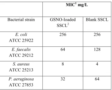
2.8. Liposomal GSNO antibacterial activity assessments... 46
3.RESULTS AND DISCUSSION ... 47
3.1. Screening the liposome manufacturing process ... 47
3.2. Physico-chemical characterization of liposomes ... 48
3.3. In vitro release kinetics ... 49
3.4. Cytotoxicity studies ... 50
2
3.6. Liposomal GSNO antibacterial activity studies ... 54
4.SUMMARY ... 55
REFERENCES... 56
CHAPITRE 3 ... 62
MICROENCAPSULATION DE LA RIFAMPICINE EN UTILISANT LE PALMITATE DE SACCHAROSE COMME TENSIOACTIF ALTERNATIF A L’ALCOOL POLYVINYLIQUE POUR CIBLER LES MACROPHAGES ALVEOLAIRES ... 62
“FORMULATION AND IN VITRO CHARACTERIZATION OF INHALABLE POLYVINYL ALCOHOL-FREE RIFAMPICIN-LOADED PLGA MICROSPHERES PREPARED WITH SUCROSE PALMITATE AS STABILIZER: EFFICIENCY FOR EX VIVO ALVEOLAR MACROPHAGE TARGETING” ... 62
ABSTRACT ... 66
1.INTRODUCTION ... 66
2.MATERIALS AND METHODS ... 67
2.1. Materials ... 67
2.2. Microsphere preparation ... 67
2.3. Microsphere characterization ... 68
2.4. Morphology analysis ... 68
2.5. In vitro RIF release studies ... 68
2.6. Aerodynamic evaluations ... 70
2.7. Alveolar macrophage cells ... 70
2.8. RIF uptake by alveolar macrophage ... 70
2.9. Toxicity assay ... 71
3.RESULTS AND DISCUSSION ... 71
3.1. Preparation and characterization of RIF-loaded microspheres ... 71
3.2. Morphology analysis ... 73
3.3. In vitro RIF release studies ... 74
3.4. Aerodynamic behavior assessment of RIF-loaded microspheres ... 75
3.5. Rifampicin uptake by alveolar macrophage ... 75
3.6. Cell toxicity ... 75
4.CONCLUSION ... 77
REFERENCES... 77
CONCLUSION GENERALE ... 80
3
Liste des abréviations
Alginate lyase AlgL
Aerodynamic diameter AED Alveolar macrophages AM Colony-forming units CFU Confocal laser scanning microscopy CLSM
Cystic fibrosis CF
Dichloromethane DCM
Dimethyl sulfoxide DMSO
Dimyristoylphosphatidylglycerol DMPG Dipalmitoyphosphatidylcholine DPPC Dipalmitoyphosphatidylglycerol DPPG Dioleylphosphatidylethanolamine DOPE Distearylphosphatidylethanolamine DSPE Encapsulation efficiency EE
Ethidium bromide EtBr
Ethylene diamine tetra-acetate EDTA Gram-negative bacteria GNB Gram-positive bacteria GPB Hexagonal II phase HII Hydrogenated soybean phosphatidylcholine HSPC Isosorbide mononitrate ISMN Lipopolysaccharides LPS
Lipoteichoic acids LTA
Mass median aerodynamic diameter MMAD Mesoporous silica nanoparticles MSN
Methicillin-resistant Staphylococcus aureus MRSA
Microspheres MS
Minimum bactericidal concentration MBC Minimum inhibitory concentration MIC
Multi-drug resistance MDR
Multilamellar vesicles MLV Nanoparticles (nanoparticules) NP
Nile Red NR
Oil/water O/W
Oleoyl chitosan nanoparticles OCNP
Phosphatidic acid PA
Phosphatidylcholine PC Phosphatidylethanolamine PE Phosphatidylglycerol PG Phosphatidylinositol PI
4
Phosphatidylserine PS Polydispersity index PDI Polyethylene glycol PEG
Polylactic acid PLA
Poly(lactide-co-glycolide) acid PLGA
Polyvinyl alcohol PVA
Nitric oxide NO
Quaternary ammonium compounds QAC Reactive nitrogen species RNS Reactive oxygen species ROS
Rifampicin RIF
Stearylamine SA
Sterically stabilized anionic liposomes SSAL Sterically stabilized cationic liposomes SSCL Sterically-stabilized liposomes SSL Solid lipid nanoparticles SLN
S-nitrosoglutathione GSNO
Teichoic acids TA
Transmission electron microscopy TEM
Vancomycin Van
5
Introduction générale
La chimiothérapie des maladies infectieuses a révolutionné le 20ème siècle. Des millions de
vies humaines ont été épargnées grâce aux antibiotiques. Toutefois, ces derniers semblent être les victimes de leur propre succès. L’utilisation massive des antibiotiques dans le monde a engendré une pression de sélection, ce qui a favorisé l’apparition des souches résistantes au traitement.
Le besoin de renouveler l’arsenal thérapeutique croît sans cesse. Grâce à la chimie organique, de nouvelles molécules actives contre les bactéries multi-résistantes voient le jour, avec la crainte d’apparition prochaine de souches résistantes.
Les cliniciens tentent de trouver une issue à cette impasse en proposant des associations non conventionnelles d’antibiotiques. Ces nouvelles associations visent à élargir le spectre thérapeutique et/ou de contourner la résistance bactérienne. La réintroduction d’anciennes molécules, jadis abandonnées en raison de leur toxicité sévère, telles que la colistine et le chloramphénicol, constitue parfois l’option clinique de dernier recours pour traiter les infections récalcitrantes.
Le développement de vecteurs non viraux capables de piloter l’antibiotique et de franchir les barrières biologiques bactériennes ou celles de l’hôte, est une approche qui séduit de plus en plus de chercheurs dans le monde. Le premier avantage est que cette approche peut intégrer toutes les autres stratégies proposées jusque-là pour combattre la résistance bactérienne, comme nous le schématisons dans la figure 1.
Figure 1. Les principales stratégies connues à ce jour visant à combattre la résistance bactérienne aux antibiotiques.
Les nouvelles molécules issues de la synthèse organique peuvent être nanoencapsulées dans le but de corriger les caractéristiques physico-chimiques non favorables à une administration
6
optimale, telles que la faible solubilité aqueuse et/ou la faible stabilité, qui constituent des obstacles majeurs à leur développement comme futurs médicaments.
La nanotechnologie offre aussi la possibilité de co-formuler deux ou plusieurs molécules dans le même vecteur et les dresser contre une cible commune. De plus, les anciennes molécules retirées du marché en raison de leur toxicité sévère, peuvent être vectorisées dans le but de limiter leur action hors cible. Autrement dit, la nanotechnologie permet de diminuer considérablement la toxicité d’antibiotiques en ciblant principalement l’organe, le tissue ou les cellules infectées.
Le second grand avantage de la vectorisation d’antibiotiques est que les vecteurs peuvent être conçus de façon à produire une cinétique de libération qui s’accorde avec la pharmacodynamie de l’antibiotique encapsulé. Ainsi, les antibiotiques dont l’activité est temps-dépendante, peuvent être formulés dans des particules à profil de libération prolongée ; tandis que les nanoparticules permettant une libération contrôlée et, si possible, sélective de la cible seront mieux adaptées aux antibiotiques dont l’activité est dose-dépendante. Ce point est traité en détails dans le chapitre 1, section 4.2.
Le troisième avantage se traduit par l’interaction spécifique nanoparticule-bactérie. Une fois maîtrisée, cette interaction apporterait une solution radicale à la résistance bactérienne au traitement. C’est pourquoi, le premier chapitre de ce manuscrit est dédié à l’analyse de ces interactions.
Dans cette thèse, nous avons associé recherche bibliographique et travail expérimental pour aborder quelques aspects de l’apport important de la nanotechnologie dans le domaine de l’antibiothérapie. Ainsi, le premier chapitre constitue une synthèse de 98 articles scientifiques traitant l’encapsulation d’antibiotiques dans des vecteurs non viraux, leurs mécanismes d’interactions avec les bactéries et leurs modes d’action pour contourner la résistance bactérienne.
Les deuxième et troisième chapitres présentent des travaux originaux de vectorisation d’antibiotiques. Le deuxième chapitre présente, donc, l’élaboration des nanoliposomes chargés en une nouvelle molécule avec potentiel anti-infectieux, le S-nitrosoglutathion (GSNO), un donneur endogène de l’oxyde nitrique, comme une nouvelle thérapeutique antibactérienne. L’objectif principal de l’encapsulation est de réaliser une libération sélective du GSNO dans les macrophages afin de renforcer ses capacités de défense, surtout dans le cas des infections causées par des bactéries multi-résistantes. Gardons à l’esprit que la libération non contrôlée de l’oxyde nitrique peut être néfaste, vu son fort potentiel pro-inflammatoire. Le troisième et dernier chapitre de ce manuscrit traite la microencapsulation d’un antituberculeux commercialisé depuis plusieurs années, la rifampicine. Cette fois-ci, l’objectif est de cibler les macrophages, siège de l’infection intracellulaire causée par les mycobactéries, mais aussi de formuler une poudre pour inhalation permettant une administration pulmonaire de la rifampicine ; une forme galénique de la rifampicine qui n’existe pas sur le marché et qui n’est pourtant pas sans intérêts directs.
Chapitre 1
Interactions nanoparticule-bactérie: de nouveaux
horizons pour combattre la résistance aux
antibiotiques
“Insights in Nanoparticle- Bacterium interactions:
New Frontiers to Bypass Bacterial Resistance to
8
Ce chapitre est rédigé sous forme d’une revue de la littérature scientifique, actuellement sous presse dans le journal « Current Pharmaceutical Design ».
Cette revue est une tentative de répondre à la première question posée dans cette thèse : comment la nanotechnologie peut-elle améliorer l’activité antibactérienne des antibiotiques voire contourner la résistance bactérienne ?
Les différentes facettes des antibiotiques nano-encapsulés sont, donc, présentées à travers de récents travaux de recherche, à savoir : le pouvoir pénétrant à travers les biofilms bactériens (Meers et al.,2008) ; (Alipour et al.,2008) ; la stabilité face aux attaques enzymatiques (Alipour et al.,2009) ; (Wright,2005) ; la capacité d’agir sur les bactéries intracellulaires confinées dans les macrophages (Abed &Couvreur,2014) ; le ciblage d’organes voire de tissus qui a permis d’améliorer considérablement la tolérance de l’hôte aux antibiotiques (Xiong et
al.,2014). De plus, certains types de nanoparticules, comme les nanoparticules inorganiques
(Pati et al.,2014), ont une action bactéricide intrinsèque pour laquelle aucune forme de résistance n’est connue à ce jour.
En outre, la nanoencapsulation permet d’associer les antibiotiques dans le même vecteur dans le but d’obtenir une action synergique. Cette stratégie a montré une capacité à déjouer la résistance bactérienne en inhibant la pompe d’efflux de la paroi bactérienne, comme il a été démontré pour les nanoliposomes contenant la pipérine (Khameneh et al.,2015) ; ou alors en inhibant la formation des biofilms et/ou en éradiquant les biofilms déjà formés, comme c’est le cas des nanoparticules libérant l’oxyde nitrique (Jardeleza et al.,2011).
Malheureusement, la nano- et la micro-encapsulation des antibiotiques sont réalisées via des procédés qui s’avèrent plus ou moins efficaces en fonction du type d’antibiotique et du type du vecteur visé. Il est évident que l’efficacité d’encapsulation affecte directement l’activité bactérienne du vecteur ainsi conçu.
Il est important de noter que l’encapsulation s’est montrée, parfois, néfaste quant à l’activité de l’antibiotique encapsulé, surtout pour ceux qui agissent sur la membrane bactérienne. A titre d’exemple, le méropénème encapsulé dans des Fluidosomes® a montré une diminution de
9
son activité antibactérienne de 4 à 16 fois, sur les différentes souches bactériennes testées (Drulis-Kawa et al.,2006).
Toutes ces remarques conduisent à la deuxième grande question posée dans cette thèse : quelles sont donc les mécanismes d’interactions entre les trois acteurs, à savoir, l’antibiotique, le vecteur et la bactérie ? Et, quelles sont les facteurs qui régissent ces interactions ?
Afin de répondre à ces questions, une analyse des données de la littérature a été réalisée et constitue la deuxième partie de la revue. La figure 2 présente un aperçu général des facteurs régissant les interactions nanoparticule-bactérie. La compréhension des interactions qui s’opèrent entre la bactérie, la nanoparticule et l’antibiotique encapsulée permettrait, sans doute, de guider le choix de vecteurs et de formulation en fonction de l’antibiotique et du type de la bactérie visée.
Figure 2. Les principaux facteurs régissant les interactions nanoparticule-bactérie, présentés sous forme d'un diagramme d'Ishikawa.
10
Insights in nanoparticle- bacterium interactions: new frontiers to bypass
bacterial resistance to antibiotics
R. Diab
a,b†, B. Khameneh
c, O. Joubert
dand R.E. Duval
a,b,ea CNRS, UMR 7565, SRSMC, Vandœuvre-lès-Nancy, F-54506, France
b Université de Lorraine, UMR 7565, SRSMC, Nancy, F-54001, France
c Department of Food and Drug Control, Students Research Committee, Mashhad University of
Medical Sciences, Mashhad, Iran
d Université de Lorraine, CITHEFOR, EA 3452,Faculté de Pharmacie, Nancy, France e ABC Platform®, Nancy, F-54001, France
† To whom correspondence should be directed
Dr. Roudayna Diab
SRSMC, UMR 7565, CNRS-Université de Lorraine, Faculté de Pharmacie, Université de Lorraine 5, rue Albert Lebrun, BP 80403
54001 Nancy Cedex France
Tel +33 3 83 68 22 74 Fax +33 3 83 68 23 01
11 A b s t r a c t
Nanotechnology has been revealed as a fundamental approach for antibiotics delivery. In this paper, recent findings demonstrating the superiority of nanocarried-antibiotics over “naked” ones and the ways by which nanoparticles can help to overwhelm bacterial drug resistance are reviewed. The second part of this paper sheds light on nanoparticle-bacterium interaction patterns. Finally, key factors affecting the effectiveness of nanoparticles interactions with bacteria are discussed.
Key words: antibiotic, controlled-release, liposome, nanoparticle, resistance, targeting.
1 . I n t r o d u c t i o n
Antibiotics, drugs that saved millions of lives in the twentieth century, seem to be of a decreasing efficacy nowadays. Recalcitrant infections threaten people lives, inflicting heavy burdens on the society. Common solutions are: developing new molecules [1]; [2]; re-introducing some abundant molecules [3]; using plant polyphenolic compounds [4], etc. Unfortunately, in the long run, all of these solutions could have lesser or even null efficacy because of the emergence of resistant strains. Today, there is a growing need of a radical approach enabling to short-circuit the bacterial resistance. This approach is supposed to help antibiotics to bypass the multiples bacterial barriers in order to reach their therapeutic targets. Barriers could mainly be summarized by bacterial biofilms, cell walls and destructive enzymes (Fig. 1). The viscous mucus surrounding the bacterial foci could also be considered as an additional barrier.
Generally speaking, antibiotics are ineffective against biofilms due to their inability to cross such a complex matrix. Biofilms are composed of a wide variety of extracellular biopolymers such as polysaccharides, proteins, glycoproteins and glycolipids and in some cases they contain amounts of extracellular DNA [5]. Besides, biofilms shelter bacterial cells called “persisters” that represent the most resistant phenotype [6]. Bacteria embedded in biofilms are characterized by a slow metabolism which reinforces their resistance against antibiotics, especially those acting by inhibiting cell wall or protein synthesis [6]. In addition, in biofilms the hypoxic and acidic environment may deactivate pH-sensitive antibiotics [7].
Bacterial cell wall is another obstacle to the effective delivery of antibiotics, owing to its electrical charge and special architecture. Both Gram-negative (GNB) and Gram-positive bacteria (GPB) are negatively-charged. Their wall contains several anionic components, such as teichoic (TA), lipoteichoic acids (LTA) (in GPB), lipopolysaccharides (LPS) (in GNB), peptidoglycan layers and phospholipids (in both types). Therefore, permeation of anionic antibiotics, e.g. β-lactams, across the bacterial wall in both types would be restricted [8]. Moreover, with regard to the wall architecture, GPB’s wall is simply composed of an outer hydrophilic thick layer of peptidoglycan covered by TA and LTA, and a cytoplasmic membrane [9]. On the other hand, GNB have a complex wall organized in outer lipophilic layer mainly composed of LPS and proteins, followed by an aqueous periplasmic space and then by an internal peptidoglycan wall that directly covers the cytoplasmic membrane [9]. The GNB wall’s outer membrane is crossed by tiny aqueous channels called porins enabling small hydrophilic molecules to permeate. Consistently, porins characteristics such as size, structure and expression level considerably affect the antibacterial spectrum of hydrophilic
12
antibiotics [10]. This can explain, in part, why GNB are intrinsically resistant to hydrophilic antibiotics of high molecular weight, e.g. glycopeptides, which are only effective against GPB [10]; [11].
Furthermore, the enzymatic barrier represented by a number of virulence factors produced by opportunistic bacteria jeopardizes antibiotic effectiveness. Pseudomonas aeruginosa,
Staphylococcus aureus and Escherichia coli have all the enzymatic weapons causing damage
to both administered antibiotics and host cells [12]. For instance, P. aeruginosa produces a myriad of enzymes, such as elastase, chitinase, lipase, and proteases, that are destructive for host tissues; in addition to metallo-β-lactamases and aminoglycoside acetyltransferase that can deactivate the most effective antibiotics [13]; [14]; [15].
In some contexts, the situation could be more complex. For instance in cystic fibrosis (CF), the, abundantly secreted viscous mucus creates an additional physico-chemical barrier to antibiotics. This mucus contains abnormally high concentrations of neutrophil-derived DNA and filamentous actin, which are produced as a result of the inflammatory response to bacterial virulence factors. These former interact with glycoproteins, e.g. mucin, resulting in viscous sputa covering the epithelial surface, and thus favoring the bacterial adherence and subsequently biofilm production [16].
Fig. 1. The main potential barriers to antibiotic delivery.
Nanotechnology has been revealed as a fundamental approach for antibiotics delivery allowing the above mentioned barriers to be overcome. In this paper, recent findings pointing out the superiority of nanocarried-antibiotics over “naked” ones and the ways by which nanoparticles (NP) can help to overwhelm bacterial drug resistance are reviewed. The second part of this paper sheds light on NP-bacterium interaction patterns. Finally, key factors affecting the effectiveness of NP interaction with bacteria are discussed.
13
2 . H o w c a n n a n o p a r t i c l e s h e l p t o b y p a s s b a c t e r i a l d r u g r e s i s t a n c e ?
All over the world, researchers developed different nanotechnology- based approaches with the aim to overcome the currently-known bacterial resistance mechanisms to antibiotics. Some revealed promising findings and led to clinical trials. Today, several “nano-antibiotics”
are clinically-approved for human use. For instance, AX-TobraTM is an inhalable liposomal
tobramycin based on Fluidosomes® technology, claimed for the treatment of Pseudomonas aeruginosa pulmonary infections in cystic fibrosis. It is developed and commercialized by Axentis pharma (Zurich, Swizerland). PulmaquinTM and LipoquinTM are two inhalable liposomal dosage forms of ciprofloxacin for the treatment of serious infectious diseases encountered in cystic fibrosis or in non-cystic fibrosis bronchiectasis. They are developed and commercialized by Grifols, S.A. and Aradigm Corporation (Hayward, CA, USA). These novel formulations were recently reviewed and discussed in details [17]; [18]. Arikace® is an inhaled liposomal dosage form of amikacin developed for the treatment of cystic
fibrosis-associated pulmonary infections caused by P. aeruginosa [19]. Now, it is undergoing phase
III clinical trials.
Numerous approaches are still under investigation. They are reviewed hereafter.
2.1. Alteration of bacteria’s efflux pump activity
In this regard, recently-reported advances could be mentioned. Khameneh et al. developed piperine-containing nanoliposomes as a vector for gentamicin. The liposomal formulation was specifically developed to fight methicillin-resistant Staphylococcus aureus (MRSA), an antibiotic-resistant bacteria which is widely recognized as a nosocomial pathogen [20].
The encapsulation of gentamicin in classical nanoliposomes or piperine-containing nanoliposomes resulted in a dramatic decrease of minimum inhibitory concentration (MIC) values of 16- and 32 folds, respectively. Similarly, minimum bactericidal concentration (MBC) values were also reduced 4- and 8- folds for encapsulated gentamicin in classical nanoliposomes or piperine-containing nanoliposomes, respectively. These hopeful results were attributed to the piperine inhibiting effect on the bacterial efflux pump. This argument was confirmed using ethidium bromide (EtBr) fluorescence assay. The fluorescence of this compound occurs only when it is bound to nucleic acid. Accordingly, bacterial suspension was incubated with EtBr for 30 min in the presence of: i) bare nanoliposomes (without piperine), ii) piperine-containing nanoliposomes or iii) piperine in its free form. After centrifugation and washing of bacteria, the loss of fluorescence was checked in order to investigate the efflux of EtBr outside bacterial cells. Consistently, a gradual decrease of fluorescence during the assay period was observed in the first case, i.e. in the absence of piperine. However, in the presence of piperine the fluorescence was significantly enhanced indicating a significant inhibition of the efflux pump [20]. Therefore, the enhanced antibacterial activity of gentamicin encapsulated in piperine-containing nanoliposomes is likely to be the consequence of an increase in its intracellular concentration. It is of note that piperine in its free form was less effective in inhibiting the efflux pump than the liposomal one, as demonstrated by the EtBr fluorescence assay.
14 2.2. Antibiofilm activity
Nitric oxide (NO)-releasing NP were found to prevent the formation of bacterial biofilms and to eradicate already formed biofilms. Some examples of recent breakthroughs in this domain are presented hereafter.
Jardeleza et al. encapsulated isosorbide mononitrate (ISMN), as NO donor into different liposomal formulations with the purpose to enhance the antibiofilm activity against
Staphylococcus aureus’s biofilms [21]. NO-releasing multilamellar vesicles (MLV)
efficiently eliminated S. aureus’s biofilms in vitro. A five min-exposure to 60 mg/mL ISMN-loaded MLV induced an almost complete eradication of the biofilms. Paradoxically, the authors observed that at low concentrations NO-releasing MLV enhanced the formation of biofilms, which is in accordance with previously obtained results [22].
Duong et al. developed nanoparticulate NO-core cross-linked star polymers as new therapeutics able to combating biofilms that are frequently formed during long exposure of the body to medical devices and catheters [23]. These systems were found to release NO in a controlled and slowed-down manner in bacterial cultures and showed great efficacy in preventing both cell attachment and biofilm formation in P. aeruginosa over time. This study unveiled, in part, the inherent mechanisms of NO’s antibiofilm activity. Accordingly, NO-releasing NP inhibit the switch of planktonic cells in contact with a surface to the biofilm form by continuously stimulating phosphodiesterase activity. Thus, NO-releasing NP maintained low intracellular concentrations of cyclic di-guanosine monophosphate (c-di-GMP) in the growing bacterial population, thereby confining growth to an unattached free-swimming mode [23].
The dual delivery of two antibiotics via their co-encapsulation in nanoliposomes is another proposed strategy to bypass resistance mediated by biofilm formation. For instance, Moghadas-Sharif proposed vancomycin/rifampin-co-loaded nanoliposomes as a new therapeutic against Staphylococcus epidermidis [24]. This strategy was based on two points. First, combination therapy of vancomycin and rifampicin helps avoid the emergence of rifampin-resistant strains. Indeed, numerous studies have already reported the antibiofilm activities of rifampin in combinations with other antibiotics [25]; [26]; [27]. Second, rifampicin fails alone to eradicate bacterial biofilm [28]. Nevertheless, the developed liposomal combination was ineffective to eradicate S. epidermidis‘s biofilm. The authors attributed this result to the lack of liposomal adsorption or low penetration into the bacterial biofilm [24]. A more adjusted formulation with enhanced penetration behavior into the biofilm may lead to the initially expected effect.
2.3. Enhanced penetration through biofilms
Several research papers reported the improved penetration across bacterial biofilms as a plausible reason behind the enhanced antibacterial activity of encapsulated antibiotics against resistant bacteria. For instance, liposomal encapsulation of polymyxin B was first described by Alipour and co-authors as a strategy to enhance its antibacterial activity against P.
aeruginosa resistant strains [29]. As they expected, lower MIC values were observed for
liposomal formulations with respect to that of the free drug. In an attempt to elucidate the involved mechanisms, the researchers focused on the drug uptake and more precisely on its
15
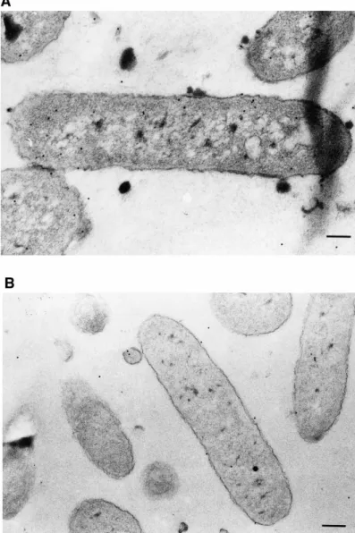
penetration across the biofilm formed by the polymyxin B-resistant P. aeruginosa strain. They used a coupled immunocytochemistry-transmission electron microscopy (TEM) imaging technique. Accordingly, a clinical strain of P. aeruginosa resistant to polymyxin B was incubated either with free or liposomal polymyxin B at sub-MIC concentrations (i.e. 64 and 16 µg/mL, respectively). Untreated bacteria were used as control. Penetration efficiency into biofilms was checked at predetermined intervals of 0, 4, 8 and 16 h at 37°C. TEM studies showed that the uptake of polymyxin B-loaded liposomes by the resistant strain was higher than that of the free drug [29]. It is important to mention that the treatment with both free drug and empty liposomes did not display a superior effectiveness with regard to the free drug indicating that the enhanced activity can only be attributed to the entrapped form.
Furthermore, the superiority of liposomal aminoglycosides was demonstrated on in vivo chronic Pseudomonas infection model [30]. Consistently, mucoid P. aeruginosa-containing agar beads were instilled intratracheally to Sprague-Dawley female rats. After the establishment of infection, animals were treated by inhalation over 14 days. Two treatment regimens were used; tri-weekly dosing schedule with free or liposomal amikacin at 6 mg/kg per dose and compared with the classical aminoglycoside regimen, i.e. a twice daily dosing of free tobramycin at the same dose (6 mg/kg/day). Finally, animals were killed and lungs were homogenized. Homogenates were subsequently cultured on agar plates. Then, colony-forming units (CFU) were counted in order to assess the effectiveness of the treatment. The researchers found that “free amikacin was relatively ineffective in the reduction of CFU under these conditions, while bacteria were undetectable in a large proportion of the group treated with liposomal amikacin” [30]. Interestingly, the thrice-weekly treatment with the liposomal amikacin was as effective as the twice-daily treatment with free tobramycin. Although, tobramycin showed a lower MIC value than amikacin against the planktonic form of P.
aeruginosa [30]. The authors explained the observed enhanced effectiveness of liposomal
amikacin by the enhanced penetration through biofilm and by the drug sustained-release pattern. The researchers have demonstrated the drug sustained-release profile from liposomes in CF-patients ‘sputa [30]. They also checked biofilm penetration on in vitro 4 days- grown biofilms produced by a mucoid form of PA01, prepared using rat lung models with chronic infections. For this aim, fluorescently-labeled liposomal amikacin was used and biofilm penetration was imaged by confocal laser scanning microscopy (CLSM) [30].
2.4. Protection against enzymatic degradation and inactivation by polyanionic compounds
Nanoparticulate delivery systems provide a physical barrier shielding the entrapped antibiotic from aggregation and inactivation with polyanionic compounds, such as bacterial endotoxins
e.g. LPS and LTA. Additionally, encapsulation may protect antibiotics against enzymatic
degradation by β-lactamases, macrolide esterases and other bacterial enzymes [31].
Two decades ago, Lagacé et al. demonstrated that liposomal encapsulation of ticarcillin or tobramycin reverse the resistance of P. aeruginosa strains towards these both antibiotics [32]. Growth inhibition of ticarcillin- and tobramycin- resistant strains was achieved using ticarcillin and tobramycin liposomal formulations at 2 % and 20 % of their respective MIC. Liposomal formulations were as effective against the lactamase -producing strains as β-lactamase -non producing ones.
16
Recently, Alipour et al. demonstrated the versatility of liposomal encapsulation in protecting tobramycin or polymyxin B from inhibition by LPS, LTA, neutrophil-derived DNA, actin filaments (F-actin) and glycoproteins e.g. mucin, common components in the CF-patients ‘sputa [33]. Being polycationic, tobramycin and polymyxin B can bind to these polyanionic compounds and thereby have their bioactivity reduced. The authors postulated that “liposomes are able to reduce the antibiotic contact with polyanionic factors in the sputum and to enhance bacteria-antibiotic interactions” [33]. In vitro stability studies revealed that liposomal formulations were stable after an 18 h-incubation at 37°C with i) a supernatant of biofilm-forming P. aeruginosa, ii) a combination of DNA, F-actin, LPS and LTA or iii) an intact or an autoclaved patient’s sputum. No significant differences with respect to control (before incubation) were observed. Furthermore, the antibacterial potency of liposomal antibiotics were checked after both short (3 h) and prolonged (18 h) exposure to a combination of DNA/F-actin or LPS/LTA at different concentrations. It was found that for both free and liposomal drugs the antibioactivity was reduced in a concentration-dependent manner. However, much higher concentrations (100 to 1000 mg/L) and (500 to 100 mg/L) of LPS/LTA and DNA/F-actin, respectively, were needed to inhibit liposomal forms in comparison to free drugs. The authors explained this finding by the increased viscoelasticity induced by the high concentrations of polyanionic elements that may hinder the interaction of liposomes with bacteria. Indeed, the early leakage of antibiotics from liposomes cannot be used as a plausible cause of the inactivation of liposomal antibiotic because in vitro stability studies showed that liposomal vesicles were not disrupted [33].
To further confirm the superiority of liposomal forms, the authors studied the bactericidal activity of liposomal formulations versus free forms against P. aeruginosa found in CF-patients’ sputa. The antibacterial activities of liposomal formulations were 4-fold higher when compared to the free drugs, despite the presence of different bacterial strains in the patient’s sputum. It is of note that liposomal tobramycin reduced growth at a high concentration (128 mg/L), whereas liposomal polymyxin B did it at a markedly lower concentration (8 mg/L). The dissimilar activities of tobramycin and polymyxin B was attributed to their different sites of action. The different behaviors of liposomal formulations as a function of the encapsulated drug will be discussed thoroughly in the following sections of this review.
The same research group conducted a meticulously detailed study confirming the inhibiting effect of the polyanionic compounds in CF-patients ‘sputa, i.e. neutrophil-derived DNA, mucoid P. aeruginosa-produced alginates and mucins, on the antibacterial activities of free and liposomal aminoglycosides [34]. It was found that bactericidal concentrations of aminoglycosides were increased by 8- to 256-folds against biofilm-forming strain, while the treatment with alginate lyase (AlgL) improved the eradication of this latter. The activity of the tested aminoglycosides, i.e. tobramycin, gentamicin and amikacin, was significantly increased by the concomitant use of recombinant human DNase or AlgL. However, liposomal antibiotic formulations did not display an additional effectiveness with respect to the free drugs, unless used in combination with AlgL.
These non-conclusive results could be explained by the fact that the authors compared free and liposomal antibiotics at high concentration (512 mg/L). Very often, the superiority of the liposomal formulation over the free form was easier to be demonstrated at low tested
17
concentrations, i.e. 1 mg/L for liposomal amikacin [30], 8 mg/L for liposomal tobramycin [35], 1.7 mg/L for liposomal mupirocin [36].
2.5. Intracellular bacterial killing
Obviously, the intracellular location reinforces bacterial resistance as it shields them from both humoral and cellular host defenses and also from the action of therapeutic agents. Indeed, intracellular bacteria, such as Mycobacterium tuberculosis and Listeria
monocytogenes, use cells of the innate immune system, not only as reservoirs to launch
recurrent infections but even more as vectors enabling them to invade other sites of the body [37]. On the other hand, most of antibiotics, e.g. aminoglycosides, β-lactams and glycopeptides, have restricted cellular penetration while others can readily diffuse, e.g. fluoroquinolones and macrolides. Unfortunately, these latter suffer from low intracellular retention [38]. Accordingly, a small number of available antibiotics are effective against intracellular infections. To fight intracellular infections, NP are promising vectors allowing antibiotics to target macrophages and to reach bacteria located in intracellular compartments. In this field, a recent review article has already highlighted the role of NP for targeting intracellular infections [39].
Furthermore, engineered NP enable to keep their loaded antibiotics intact. We recently demonstrated that sterically-stabilized liposomes (SSL) loaded with S-nitrosoglutathione could be good candidates for macrophage targeting (unpublished data). We found that SSL are predominantly internalized by caveolae-dependent endocytosis which is the preferred pathway for drug delivery systems as it avoids the fusion with lysosomes and the subsequent drug degradation in its highly acidic environments.
2.6. Specific targeting and sustained-release
Inherent toxicity of antibiotics is a crucial drawback that led to limit or even to stop the use of some of them, such as aminoglycosides and lipopeptides known for their neuro- and nephrotoxicity [40]. Therefore, specific targeting to bacteria would counteract drug toxicity, since it enables to avoid non-selective and uncontrolled delivery to host cells.
To date, few works reported the design of NP with a specific targeting to bacteria for therapeutic purposes. Some examples are presented hereafter. Qi et al. elaborated mesoporous silica NP (MSN) as nanocarriers of vancomycin (Van) in order to specifically target GPB over macrophage-like cells [41]. The specific recognition was based on hydrogen bonding interactions of Van with the terminal D-alanyl-D-alanine moieties of GPB. Cell viability assay showed a good biocompatibility of Van-MSN with human embryonic kidney and human hepatocytes.
Tang et al. have recently described the design of a nanoparticulate carrier loaded with a fluorescent dye, and called it “nanoprobe” for diagnostic purposes [42]. The surface of the nanoprobe was grafted with a bacterial ligand, i.e. concanavalin A, and therefore displayed a high affinity to bacteria. The developed nanoprobe was shown to rapidly detect and quantify the extent of bacterial colonization on wounds and catheters in real time.
Prolonged or sustained release of the loaded antibiotic is of great importance for antibiotics with time-dependent action, such as lipoproteins, β-lactams, glycopeptides and some fluoroquinolones. The importance of the sustained-release profile was highlighted by Meers et
18
al. [30]. Thanks to the prolonged release of amikacin from liposomes, this latter was as
effective, when administered tri-weekly, as free tobramycin administered twice-daily and despite the fact that MIC of tobramycin is lower than that of amikacin. Additional examples of antibiotic-loaded polymeric NP were recently reviewed [43].
2.7. Down-regulation of bacteria’ oxidative-stress resistance genes
Bacterial adaptation to oxidative and nitrosative stress could be considered as a resistance mechanism to host defenses [44]. Indeed, innate immune cells generate reactive oxygen species (ROS) and reactive nitrogen species (RNS) such as superoxide and peroxynitrite, respectively, in order to kill phagocyted bacteria [45]. Consistently, pathogenic bacteria resist to host-mediated oxidative stress by up-regulating the expression of their antioxidant enzymes [46]. Importantly, it was claimed that many antibiotics exert their bactericidal effects via the production of hydroxyl radicals, regardless of their molecular targets [47].
Recently, it was found that metal NP, namely zinc oxide-NP (ZnO-NP), exert by themselves bactericidal effects on GPB and GNB [48]. A synergistic killing effect on acid fast bacteria (i.e. Mycobacterium bovis-BCG) was also observed for ZnO-NP when used in combination with rifampicin [48]. Moreover, ZnO-NP effectively killed MRSA clinical strains [48].
Several mechanisms were found to be involved in ZnO-NP antibacterial activities. Most importantly, ZnO-NP were found to down-regulate the transcription of oxidative stress resistance genes in S. aureus. Strictly speaking, the treatment with 300 µg/mL of ZnO-NP decreased the transcription of peroxide stress regulon katA and perR genes by 10- and 3.1- folds, respectively, when compared to untreated bacteria [48]. These results highlight the importance of ZnO-NP in fighting drug-resistant bacteria.
It is of note that ZnO-NP induced oxidative stress response on macrophages, as ROS and NO production was markedly increased, thus reinforcing their bacterial killing capacity [48]. Generally speaking, metal NP, such gold or silver NP, are known to induce oxidative stress in host cells, which is considered as one of mechanisms involved in their toxic effects [49]; [50]. However, to the best of our knowledge, Pati et al. were the first to demonstrate the opposite effect on bacterial cells [48].
3 . M e c h a n i s m s o f n a n o p a r t i c l e - b a c t e r i u m i n t e r a c t i o n s
NP interactions with bacterial cells or biofilms are ruled by two types of driving forces, i.e. electrostatic and hydrophobic. Evidences of electrostatic NP interactions with bacteria are multiple. For instance, positively-charged NP, especially those with zeta potential above +40 mV, are known to alter the bacterial cell membrane permeability by acting as detergents, causing an osmotic damage finally leading to cell death. This effect is weakened or nonexistent for NP with low positive potential or negatively-charged ones, respectively [8]. It is noteworthy that such interactions are unlikely to be overcome by bacterial adaptive resistance based on a single gene mutation, since bacterial membrane is highly evolutionarily conserved [51].
Hydrophobic interactions were reported in numerous research papers dealing with the antibacterial efficacy of quaternary ammonium compounds (QAC) micelles or QAC-containing micelles [52]; [53]; [54]. It was found that the antibacterial efficacy of these latter
19
vary as a function of the QAC’s alkyl chain length. The optimal activity was observed for the more hydrophobic ones, i.e. containing C12 to C16 alkyl chain, when compared to those with short carbon chain (< C12) [52]. This was attributed to a stronger adherence to bacterial biofilms of QAC with longer alkyl chains [53].
Moreover, Cottenye et al. reported that hydrophobic interactions play an important role in liposome adherence to biofilms [54]. This is on the basis of their observation that only the fluorescent dye encapsulated in liposomes was tracked while the bare one was readily washed out of the biofilms. Accordingly, they concluded that the bound liposomes remained intact in the biofilms and no drastic reorganization in the liposomal wall occurred due to hydrophobic interactions with the biofilms [54].
During the last decades, researchers attempted to gain deep insights in NP interactions with bacterial cell-wall. The involved mechanisms are likely to be directly dependent on nanoparticle type and structural composition. The main interaction patterns are discussed hereafter.
3.1. Internalization mechanism of liposomes 3.1.1. Passive fusion “stalk mechanism”
Taking into account the barriers and the interaction driving forces, NP were designed in order to give rise to different types of interaction with bacteria or their biofilms. Passive fusion is the commonly described interaction mechanism with the bacterial cell-wall for NP in general but also particularly for fusogenic liposomes. The formulation of fusogenic liposomes is based on the combined use of lipids forming hexagonal II (HII) phase and lipids forming lamellar phase [55]. The lipids forming HII phase, such as phosphatidylethanolamine (PE), phosphatidylserine (PS) or phosphatidic acid (PA), have a small polar head showing a cone shaped-molecule. When used in formulation, the negative curvature stress leads to the spontaneous formation of inverted micelles (HII phase) [56]. In contrast, lipids with similar packing ratios for the polar head and the hydrophobic queue, such as phosphatidylcholine (PC), phosphatidylglycerol (PG) or phosphatidylinositol (PI), have a cylindric shape and produce a small or no curvature stress. They form lamellar phases [57].
The principal fusion mechanism as identified by Markin et al. was called the stalk mechanism [58]. The approaching membranes form an hourglass-shaped structure, called stalk, generating a local stress and spontaneous curvature in these membranes followed by bilayer reorganization. The stalk formation is promoted by an HII forming lipids since they stabilize the hemi-fusion intermediate structures. This hypothesized mechanism was subsequently confirmed thanks to TEM. The intermediate structure consisting in lamellar/HII transition phase was observed in a mixture of PC/PE after dehydration [59].
It is of note that there is a category of lipids called lysolipids, characterized by a bully polar head, molecularly shaped as inverted cone and so form hexagonal I (HI) phase. They produce a positive curvature stress in the membranes and inhibit the stalk formation of approaching membranes and thereby their fusion [60]. Accordingly, the design of fusogenic liposomes requires a careful qualitative and quantitative choice of phospholipids as it directly influences the stability of lamellar/HII transition phase [55].
20 3.1.2. Chemically triggered fusion
Passive fusion of liposomes with biological membranes could be considered as a drawback preventing a specific delivery of their loaded drugs. Thus, another generation of liposomes has been developed by grafting a hydrophilic polymer, i.e. polyethylene glycol (PEG), that confers a higher stability to liposomal wall and inhibiting its spontaneous fusion with biological membranes. Hence, liposomes containing PEG-lipid conjugates are called sterically-stabilized liposomes (SSL). Nonetheless, in order to restore the fusiogenecity in the vicinity of the site of action, e.g. biofilms or bacterial cell membrane, Kirpotin et al. elaborated liposomes containing pH-sensitive fusogenic phospholipid, i.e.
dioleylphosphatidylethanolamine (DOPE), and a small percentage of a disulfide-linked PEG conjugate with distearylphosphatidylethanolamine (mPEG-DTP-DSPE) [61]. The authors demonstrated that the thiolytic cleavage of the disulfide bridge resulted in the detachment of PEG from DOPE-containing liposomes, and subsequently liposomal fusion occurred at pH 5.5 accompanied with the leakage of the entrapped dye.
3.1.3. Fusion triggered by specific “ligand-receptor” recognition
In an attempt to target a specific type of bacteria, researchers focused on the outer membrane composition of the considered bacterium. This is with the aim to find a membrane component that specifically interacts with a ligand grafted on NP surface. Accordingly, Bardonnet et al. took benefit of the fact that some strains of Helicobacter pylori has an outer membrane protein (BabA2 adhesin) that binds with the fucosylated Lewis b (Leb) histo-blood group antigen expressed by human gastric epithelial cells [62]. Based on this phenomenon,
Bardonnet et al. elaborated liposomes containing a synthesized glycolipid (Fuc-E4-Chol) composed of cholesterol as an anchor part, four ethylene glycol residues as a linker, and fucose as an exposed part at the surface of liposomes [63]. These liposomes were used as delivery systems for ampicillin and metronidazole and were found to be effective against both the spiral and the coccoid bacterial forms.
In this study, the authors attributed the interactions H. pylori-liposomes to four events [63]. The first one is the incorporation cholesterol in the formulation of liposomes which enhances their interaction with H. pylori, because of the specific affinity of this latter to this steroid. The second phenomenon is the electrostatic interaction as H. pylori is negatively-charged. The authors found that liposomes exhibiting a lower negative zeta-potential (between -2.9 and -4.3 mV) were more efficient than those with a higher one (between -12.2 and -20 mV). The third phenomenon is the specific interaction of fucosylated liposomes-BabA2 adhesin, since better results were obtained with liposomes grafted with the synthesized glycolipid Fuc-E4-Chol. Finally, the fourth important point was the age of the bacterial culture or, in other words, the bacterium phenotype. During aging, the morphology of H. pylori evolves from the spiral to the coccoid resistant form. Fucosylated liposomes were found to interact with both phenotypes, whereas analogous ones without Fuc-E4-Chol were only able to interact with H.
pylori spiral form. Consistently, the authors supposed that both phenotypes express BabA2
21
3.2. Internalization mechanisms of polymeric and inorganic nanoparticles
Internalization of inorganic/polymeric NP by viable bacteria was previously demonstrated. Kumar et al. evidenced the uptake of inorganic NP, sizing from 30 to 50 nm, by viable bacteria using flow cytometry [64]. The researchers exposed E. coli to high concentrations of ZnO NP and TiO2 NP, up to 80 µg/ml, for periods ranging from 30 to 90 min. it was shown
that NP were taken up in a concentration-dependent manner. It is of note that over the longest incubation period, cell death did not exceed 13.3%.
Recently, Curia et al. reported the internalization of polyurethane NP by S. aureus by a “Trojan horse” mechanism [65]. Polyurethane NP were naturally generated from the bacterial action on plastic materials. Generated NP was covered by a protein corona and were shown to be taken up by a dynamic movement of the bacterial membrane. The authors tried to explain this phenomenon, usually observed in Eukaryote and not in Prokaryote, by the fact that NP covering with a protein corona modified the electromagnetic parameters of engineered NP-bacterium interactions.
Little is known about the exact mechanisms of the interaction of inorganic or polymeric NP with bacterial walls and membranes. LPS, phospholipids seem to be the target sites for inorganic NP action; whereas phospholipids and outer membrane proteins could be the sites of action for polymeric NP. Jiang et al. studied the interaction of Al2O3 NP with micelles and
vesicles formed of LPS/PE extracted from E. coli, as bacterial membrane models [66]. A strong attachment of NP with LPS/PE vesicles and micelles was evidenced by atomic force microsocopy. After an exposure of 24 h, it was observed that LPS vesicles were coated with NP that formed a layer of tens of nanometer; while the exposure to NP disturbed the stability of PE vesicles that became larger with thicker walls.
These observations are consistent with previous findings, reported by Fortunellai and Monti [67]. The researchers studied the interaction of three phospholipids (DOPC, DOPS, DMTAP) with TiO2 surfaces. They showed that interactions influenced the conformation properties of
phospholipids and reduced the mobility of lipid bilayers. Phosphate or carbonyl oxygens of the lipids constituted the target sites of interactions [67].
Xi et al. studied the interaction of OCNP, polymeric NP that are formed of oleoyl-chitosan, with S. aureus and E. coli as models of GPB and GNB, respectively [68]. This work focused on the role of membrane proteins and phospholipids in NP-bacterium interactions. The change in membrane protein conformation was investigated by measuring the fluorescence of a protein residue, tyrosine (Tyr). Tyr is normally located in both sides of the bacterial membrane. If the NP interacted with membrane proteins, the conformation of these latter would be altered, and Tyr residues located inside the membrane would be exposed to the surface and thus increasing the fluorescence intensity. Accordingly, it was found that for both
S. aureus and E. coli the fluorescence intensity of Tyr residues was increased after a 1
h-incubation with OCNP, in a concentration-dependent manner. The authors deduced that “OCNP influenced the structure of cell membranes by interacting with proteins on the cell
membrane of the bacteria” [68].
Phospholipids were also evaluated as a potential target for OCNP. Toward this aim, OCNP were treated with yolk lecithin in order to simulate the effect of membrane phospholipids. The treated on non-treated OCNP were incubated with S. aureus or with E. coli for 24 h. The role of lecithin in NP-bacterium interaction was appreciated by comparing the growth inhibition