DOI 10.1007/s00421-014-2889-7 OrIgInAl ArtIclE
Locomotor and diaphragm muscle fatigue in endurance athletes
performing time‑trials of different durations
Thomas U. Wüthrich · Elisabeth C. Eberle · Christina M. Spengler
received: 10 December 2013 / Accepted: 6 April 2014 / Published online: 29 April 2014 © Springer-Verlag Berlin Heidelberg 2014
magnitude of inspiratory muscle fatigue were also not dif-ferent (both p > 0.05).
Conclusion Different levels of leg muscle fatigue in run-ners and cyclists could in part be related to the specific muscle activation patterns including concentric contrac-tions in both modalities but eccentric contraccontrac-tions in run-ners only. Diaphragm fatigue likely resulted from the large ventilatory load which is characteristic for both exercise modalities and which was higher in 15tts than in 30tts (+27 %, p < 0.01) while postural demand appears to be of less importance.
Keywords Self-paced exercise · Fatigue · locomotor muscles · respiratory muscles · Hyperpnoea
Abbreviations
cV coefficient of variation MVV Maximal voluntary ventilation M-wave compound muscle action potential Pdi transdiaphragmatic pressure
Pdi,tw transdiaphragmatic twitch pressure
Pes Esophageal pressure
Pes,tw Esophageal twitch pressure
Pga gastric pressure
Pga,tw gastric twitch pressure
Pm Mouth pressure
Pm,tw Mouth twitch pressure
PtPdi,in Inspiratory transdiaphragmatic pressure–time product
PtPga,in Inspiratory transdiaphragmatic pressure–time
product
PtPes,in Inspiratory esophageal pressure–time product PtPes,ex Expiratory esophageal pressure–time product PtPga,ex Expiratory gastric pressure–time product
Qtw Quadriceps twitch force
Abstract
Purpose Fatigue in leg muscles might differ between run-ning and cycling due to inherent differences in muscle acti-vation patterns. Moreover, postural demand placed upon the diaphragm during running could augment the develop-ment of diaphragm fatigue.
Methods We investigated quadriceps and diaphragm fatigue in 11 runners and 11 cyclists (age: 29 ± 5 years; ˙
VO
2,peak: 66.9 ± 5.5 ml min−1 kg−1) by assessing
quadri-ceps twitch force (Qtw) and transdiaphragmatic twitch
pres-sure (Pdi,tw) before and after 15- and 30-min time-trials (15tt, 30tt). Inspiratory muscle fatigue was also obtained after volitional normocapnic hyperpnoea (nH) where pos-tural demand is negligible. We hypothesized that running and cycling would induce different patterns of fatigue and that runners would develop less respiratory muscle fatigue when performing nH.
Results the reduction in Qtw was greater in cyclists
(32 ± 6 %) compared to runners (13 ± 8 %, p < 0.01), but not different for 15tts (23 ± 13 %) and 30tts (21 ± 11 %, p = 0.34). Overall Pdi,tw was more reduced
after 15tts (24 ± 8 %) than after 30tts (20 ± 9 %, p = 0.04) while being similar for runners and cyclists (p = 0.78). Meanwhile, breathing duration in nH and the
communicated by nicolas Place.
t. U. Wüthrich · E. c. Eberle · c. M. Spengler (*) Exercise Physiology lab, Institute of Human Movement Sciences and Sport, EtH Zurich, Winterthurerstrasse 190, 8057 Zurich, Switzerland
e-mail: christina.spengler@hest.ethz.ch c. M. Spengler
Zurich center for Integrative Human Physiology (ZIHP), University of Zurich, Zurich, Switzerland
˙ VO
2 Oxygen consumption
˙
VO2,peak Oxygen consumption at peak workload
WOB Work of breathing
ΔPdi,tw reduction in transdiaphragmatic twitch pressure
ΔQtw reduction in quadriceps twitch force 15tt 15 min time-trial
30tt 30 min time-trial Introduction
neuromuscular fatigue develops when one or several processes which allow the muscle to contract and thus to generate force are impaired, independent of whether a given task can be sustained or not (Bigland-ritchie and Woods 1984). these impairments can occur at differ-ent sites between brain and muscle and the location and extent strongly depend on the task that is performed (Allen et al. 2008; gandevia 2001). How different components of fatigue contribute to exercise limitation has been a matter of extensive debate and several different models were intro-duced (Abbiss and laursen 2005). Experimental data sug-gest that the level of peripheral locomotor muscle fatigue associated with unconscious and/or consciously perceived afferent feedback might contribute to these exercise limit-ing factors (Amann 2011). Amann and co-workers (2009) showed that attenuation of type 3 and 4 afferent feedback can substantially alter central motor output to working limbs in cycling exercise resulting in unusually high levels of peripheral fatigue. It is thus believed that type 3 and 4 afferent feedback from working muscles likely contributes to the impaired willingness and/or ability to drive a muscle in order to avoid the development of fatigue beyond a cer-tain functional limit (Amann and Secher 2010).
to date, several studies have described the occurrence of locomotor muscle fatigue in knee extensors and plantar flexors during self-paced running or cycling exercise with test durations and intensities ranging from brief sprints up to events of several hours and days (Martin et al. 2010; Millet et al. 2011; ross et al. 2010; Saugy et al. 2013; Skof and Strojnik 2006a, b). A broad range of reductions (i.e. 0–30 %) in muscle twitch force of these muscles in response to nerve stimulation have been reported. Yet, no study has systematically investigated the extent of leg mus-cle fatigue in self-paced exercise of different durations and intensities performed by the same individuals. Similar end-exercise levels of leg muscle fatigue in such trials would potentially provide further support to the importance of afferent feedback in the regulation of exercise intensity as described above. However, fatigue could also be affected by the type of muscle contraction depending on the exercise modality being performed. running and cycling—the two most common types of locomotion—are very dissimilar in
terms of the activation pattern (i.e. concentric-eccentric vs. mainly concentric) which suggests different fatigue mecha-nisms to be present (Bijker et al. 2002).
During intense whole-body exercise not only leg mus-cles are active and develop fatigue, but also respiratory muscles are under severe strain, the level mainly depend-ing on exercise intensity and the related level of ventilation [e.g. (Johnson et al. 1993; Verges et al. 2006b)]. respira-tory muscle work has been shown to require up to 15 % of whole-body oxygen consumption ( ˙VO2) at peak workload
( ˙VO2,peak) in endurance-trained athletes highlighting the substantial work performed by these muscles (Aaron et al.
1992). In contrast to locomotor muscles, however, fatigue of respiratory muscles has not yet been described for competition like self-paced exercise although fatigue was repeatedly found to develop during intense constant-load exercise [e.g. (Johnson et al. 1993; Mador et al. 1993)]. Moreover, no data are available using objective measure-ments to obtain potential differences in the level of stress and thus fatigue of respiratory muscles between running and cycling. the fact that the diaphragm appears to be substantially involved in the maintenance of posture sug-gests that the load placed on this muscle could be larger in running compared to cycling [e.g. (Hodges and gandevia
2000)].
therefore, the aim of the present study was (i) to objec-tively quantify both locomotor and inspiratory muscle fatigue after competition-like running and cycling time-trials of different durations/intensities (i.e. 15 and 30 min; 15tt and 30tt) in a group of well-trained runners and cyclists, (ii) to investigate potential differences between running and cycling in the level of quadriceps and inspira-tory muscle fatigue in these trials, and (iii) to test whether runners and cyclists differ in terms of fatigability during volitional normocapnic hyperpnoea where no additional postural demand from the gait is expected. We hypoth-esized that both the quadriceps and inspiratory muscles would yield a very specific fatigue profile for running and cycling and that runners would sustain normocapnic hyper-pnoea at the same relative intensity for a longer duration than cyclists.
Methods Subjects
twenty-two male endurance athletes (runners n = 11, cyclists n = 11) with normal lung function and respira-tory muscle strength (table 1) gave their written informed consent to participate in this study. Subjects were free of acute or chronic disease and were non-smokers. A minimal
˙ VO
frequent participation in endurance competitions (in the respective sports activity) were mandatory to be included in this study. Subjects were requested to keep their per-sonal training constant throughout the course of the study. they were asked to refrain from strenuous physical activity for the 2 days prior to the test, to completely refrain from exercising for 24 h before testing, to sleep at least 7 h the night before the test, to abstain from caffeinated beverages on test days and to have their last meal at least 2 h prior to reporting to the laboratory. the study was approved by the local ethics committee and was performed according to the Declaration of Helsinki.
Experimental overview
Subjects reported to the laboratory on four different days separated by at least 48 h and scheduled at the same time of day to control for confounding circadian influences (Scheer et al. 2010). All testing had to be completed within a maximum of 3 weeks. runners performed all tests on a treadmill while cyclists performed tests either on a station-ary cycling ergometer or a road bike mounted on an indoor cycle trainer (for details see below). On the first day, sub-jects performed an incremental running or cycling test to exhaustion. they were then introduced to pulmonary func-tion and respiratory strength testing and were extensively
familiarized with the technique of magnetic stimulation. On the second day, baseline pulmonary function and respir-atory strength measurements were obtained and inspirrespir-atory muscle contractility was assessed before and after a bout of exhaustive normocapnic hyperpnoea. On the third and fourth days, diaphragmatic and quadriceps muscle contrac-tility were assessed before and after self-paced running or cycling time-trials of 15 (15tt) and 30 min (30tt) dura-tion, performed in a randomized and balanced order. lung function and respiratory muscle strength
lung function including maximal voluntary ventilation in 12 s (MVV) was assessed according to current AtS/ErS guidelines (Miller et al. 2005) using a metabolic cart with a calibrated volume sensor (Oxycon pro, Jaeger, Höchberg, germany). Maximal inspiratory pressure (at residual vol-ume) and maximal expiratory pressure (at total lung capac-ity) were measured using a handheld device (MicrorPM, MicroMedical, Kent, UK). Variables of pulmonary func-tion and respiratory muscle strength are reported both in absolute values and in percent of predicted values (Quanjer et al. 1993; Wilson et al. 1984).
Perception of respiratory and leg exertion
respiratory exertion and leg exertion were assessed by means of a linear scale ranging from 0 to 10, where 0 corre-sponded to no and 10 to maximal perception of respiratory or leg exertion. to ensure a proper understanding of the term respiratory exertion, subjects were extensively ques-tioned about their prior experience with respiratory sen-sations before the first exercise test (lansing et al. 2000). thereafter, a definition was given for respiratory exertion (how difficult it is to breathe) which is distinguished from breathlessness (the sensation of “not getting enough air”). Incremental exercise
An incremental running or cycling test to exhaustion was performed to determine ˙VO2,peak as well as maximal run-ning velocity or maximal cycling power output.
For the running incremental test, subjects stood for 5 min on a treadmill (Quasar, h/p/cosmos, traunstein, ger-many) with a mouthpiece and nose clip in place before starting to run with a velocity of 10 km h−1. thereafter,
treadmill speed was increased by 1.5 km h−1 every 2 min
to the point of exhaustion. At the end of each stage and at the point of exhaustion subjects were asked to rate their perception of respiratory and leg exertion. Ventilation and gas exchange were measured breath by breath via the meta-bolic cart with calibrated volume, cO2 (infrared absorption principle) and O2 sensors (paramagnetic principle). Heart
Table 1 Subject characteristics for runners and cyclists
Values are mean ± SD. ˙VO2,peak peak oxygen consumption, MIP maximal inspiratory pressure, MEP maximal expiratory pressure, FVC forced vital capacity, FEV1 forced expiratory volume in the first second, PEF peak expiratory flow, MVV maximal voluntary ventila-tion
* Significantly different between runners and cyclists, p < 0.05
runners cyclists Age (years) 31 ± 4 27 ± 6* Height (m) 1.81 ± 0.08 1.81 ± 0.04 Body mass (kg) 72.1 ± 7.3 75.5 ± 8.2 ˙ VO2,peak (l min−1 kg−1) 66.7 ± 5.5 67.2 ± 6.6 MIP (cmH2O) 133 ± 17 138 ± 28 MIP (%pred) 121 ± 16 121 ± 27 MEP (cmH2O) 207 ± 45 208 ± 36 MEP (%pred) 137 ± 31 134 ± 26 FVc (l) 6.20 ± 0.55 6.17 ± 0.81 FVc (%pred) 118 ± 9 114 ± 10 FEV1 (l) 4.76 ± 0.34 4.85 ± 0.62 FEV1 (%pred) 109 ± 10 107 ± 10 PEF (l s−1) 10.5 ± 0.8 10.1 ± 0.4 PEF (%pred) 106 ± 8 103 ± 10 MVV (l min−1) 204 ± 19 197 ± 23 MVV (%pred) 134 ± 13 126 ± 15
rate was obtained beat by beat using a Suunto t6 heart rate monitor (Suunto Oy, Vantaa, Finland). At rest and at termi-nation of exercise, 20 µl of capillary blood was drawn from an ear lobe to assess blood lactate concentration enzymati-cally (BIOSEn c_line Sport®, EKF-diagnostic, Barleben,
germany).
For cyclists, the incremental test was performed on a cycling ergometer (Ergoline 900, Ergoline, Blitz, ger-many). cyclists were monitored in the same way as run-ners. the protocol consisted of a resting period (5 min) before cycling at 100 W for 2 min, followed by increments of 30 W every 2 min until the point of exhaustion. cycling cadence was chosen by the subject during the first stage and held constant for the remainder of the test. the test was either terminated when subjects voluntarily stopped or when cadence dropped below 70 rpm. In addition to the above-described measurements, samples of capillary blood were drawn at the end of each stage.
normocapnic hyperpnoea
First, subjects performed 2 min of normocapnic hyper-pnoea at 40 % and 2 min at 60 % of their individual MVV to warm up before they were required to breathe at the tar-get ventilation of 80 % MVV to exhaustion. the mouth-piece was connected to a two-way valve (Hans rudolph, Shawnee, KS, USA) via the volume-sensor of the meta-bolic cart. normocapnia was maintained by adding cO2 to the inspirate. Subjects received both visual and verbal feedback to keep minute ventilation at the target level. Vis-ual feedback was provided via a computer screen showing minute ventilation online while verbal feedback consisted of telling the subjects to breathe “more” or “less” (without referring to tidal volume or breathing frequency) when they deviated from the target by ≥5 % for several breaths. the test was terminated if target ventilation could no longer be achieved. Before and immediately after normocapnic hyperpnoea, mouth twitch pressure (Pm,tw) was assessed
(for details see below). If Pm,tw was reduced by <20 % from baseline, subjects were required to continue breathing at the same target ventilation until task failure was reached again (one subject only).
time-trial exercise
For running and cycling time-trial exercise, subjects were asked to cover as much distance as possible once in 15 min and once in 30 min.
After 2 min at rest subjects were requested to warm up for 5 min at an intensity corresponding to 50 % of their individual maximal running velocity (for runners) and 1.5 W kg−1 body mass (for cyclists) followed by a “running
start”, meaning that treadmill speed was steadily increased
during the final minute of the warm up period until sub-jects signaled that their initial running velocity for the time-trial was reached (within 30–45 s). cyclists performed on a road bike equipped with a calibrated SrM system (Power MtB, Schoberer rad Messtechnik SrM, Jülich, germany) stationed on an indoor trainer (tacx Basic cycle-force, tacx, Wassenaar, nederland) and they increased power output to their starting time-trial load approximately 10 s before the start. thereafter, subjects were allowed to freely adjust their running velocity or cycling power out-put. All subjects were encouraged verbally throughout the entire test and received feedback of the elapsed time from a screen in front of the treadmill or cycle but were naïve to velocity/power output and distance covered. Ventilation, gas exchange, esophageal (Pes) and gastric (Pga) pressures, and heart rate were measured continuously and stored simultaneously in a computer (Maclab Software, ADIn-struments, castle Hill, Australia). At rest, every 3 min dur-ing the time-trial as well as at the end of the test, subjects were asked to rate their perception of respiratory and leg exertion. Before and immediately after the time-trial, Qtw and Pdi,tw were assessed (for details see below). At rest and
at termination of the time-trial, 20 µl of capillary blood was drawn from an earlobe to assess blood lactate concentra-tion. In cycling time-trials, additional samples of capillary blood were drawn every 3 min during the test.
Magnetic stimulation of femoral and phrenic nerve roots Quadriceps muscle contractility was objectively determined by assessing quadriceps twitch torque (Qtw) in response to magnetic femoral nerve stimulation before and 10 min after completion of the time-trial (Fig. 1). Subjects were studied semi-supine on a custom-made chair. care was taken that the knee was flexed at exactly 90° and the leg was passively stabilized to prevent lateral motion. the ankle was fixed to a force transducer (strain gage lc4102-K060, A&D cO, tokyo, Japan) by a non-elastic ankle strap. Force signals were recorded on the computer. the femoral nerve was stimulated with a 70-mm figure-of-eight coil powered by a MagStim 200 stimulator (MagStim, Whitland, UK). the center of the coil was placed in the femoral triangle just lateral to the femoral artery and was repositioned sys-tematically to determine the position that resulted in the largest Qtw. this position was marked and used for the remainder of the study. Sarcolemmal membrane excitabil-ity for the quadriceps was determined by means of peak-to-peak amplitudes of compound muscle action potentials (M-wave). M-waves were recorded using bipolar surface silver electrodes on the muscle belly of the vastus lateralis with a recording diameter of 1 cm and an inter-electrode distance of 2 cm. the skin was carefully shaved, treated with an abrasive paste, and cleaned using alcohol prior to
electrode placement. EMg signals were recorded at a sam-pling rate of 4,000 Hz, pre-amplified (gain = 1,000), band-pass filtered (20–1,000 Hz; nihon Kohden, Bad Homburg, germany; common mode rejection ratio ≥94 db), A/D converted (Maclab interface, ADInstruments, castle Hill, Australia), and stored in the computer.
Inspiratory muscle contractility was assessed by means of phrenic nerve stimulation using a circular 90 mm coil powered by the magnetic stimulator before and 2 min after completion of the time-trial (Fig. 1). For assessment of Pm,tw, a differential pressure transducer (DP45‐34, Vali-dyne, northridge, cA, USA) was connected to a mouth-piece. For assessment of transdiaphragmatic twitch pres-sure (Pdi,tw), conventional balloon-catheters (esophageal
and gastric balloons containing 1 and 2 ml of air, respec-tively) were inserted through the nose and positioned in the esophagus and in the stomach to measure Pes and Pga according to current guidelines (AtS/ErS 2002). Balloon-catheters were separately connected to differential pressure transducers (DP45‐34, Validyne, northridge, cA, USA). Pressure signals were amplified (cD 19A, Validyne, north-ridge, cA, USA), A/D converted and recorded in the com-puter. Pdi was calculated by online subtraction of Pes from Pga. cervical magnetic stimulation of the phrenic nerves was applied while subjects were comfortably seated on a
chair with a nose-clip in place and the center of the coil was positioned at the 7th cervical vertebra. the subject’s position on the chair and the coil position on the neck were marked and continuously monitored throughout the experi-ment. Stimulations for the assessment of Pm,tw during the 2nd visit were performed at the start of a gentle expiratory effort from functional residual capacity through a small leak while stimulations for the assessment of Pdi,tw were applied at the end of a passive expiration corresponding to functional residual capacity with subjects’ mouth closed. Pes was continuously monitored on an oscilloscope (tek-tronix, Beaverton, Or, USA) by a second experimenter to ensure that identical lung volumes were achieved imme-diately prior to stimulations. In addition, pre-twitch Pes
-recordings showing a deviation from baseline Pes were rejected post hoc. For one subject, Pdi could not be calcu-lated due to a defective gastric balloon.
the experimental protocol for the assessment of both inspiratory and quadriceps muscle strength consisted of 9 potentiated twitches at 100 % of the stimulator output. Qtw force was measured in a fully relaxed state. to investigate fatigue-induced changes in muscle contractility, potenti-ated twitches were used since they are known to be more sensitive to changes occurring within a fatigued muscle (Kufel et al. 2002). Special care was taken to assure that
Fig. 1 response to femoral
(left panels) and cervical (right panel) magnetic stimulation before and after a time-trial in a representative participant. Qtw, quadriceps twitch force; M-wave, compound muscle action potential of the quadri-ceps; Pes, esophageal pres-sure; Pga, gastric pressure; Pdi, transdiaphragmatic pressure (Pdi = Pga − Pes)
investigated muscles were indeed in a fully potentiated state. Potentiated twitches were assessed after 3–4 submax-imal warm-up contractions and 3 maxsubmax-imal efforts lasting 5 s. After the third and sixth stimulation, another maximal effort followed to maintain the potentiated state. to ensure supramaximal stimulation before and after exercise, 3 addi-tional twitches were performed at 70, 80, 90, 94 and 98 % (for phrenic nerve stimulation) or 60, 70, 80, and 90 % (for femoral nerve stimulation) of the stimulator output either on the third or fourth visit in a randomized and balanced order. A plateau for Qtw and Pdi,tw was observed in most subjects. Moreover, no statistically relevant difference was found between the highest and the second highest stimu-lator output before and after exercise indicating maximal depolarization of the femoral and phrenic nerves at all times.
Data analysis and statistics
lung function variables were determined according to AtS/ErS guidelines (Miller et al. 2005). Maximal inspira-tory and expirainspira-tory pressures were selected as the highest of 3 values with no more than 5 % deviation. ˙VO2,peak was calculated as the highest 15-s average while maximal run-ning velocity and maximal cycling power output (last com-pleted stage + time on the final stage × increment). For twitch pressures and force, the average amplitude (baseline to peak) of 10 unpotentiated and 6–9 potentiated twitches was calculated. A twitch was rejected post hoc if Pes prior to twitch stimulation deviated from baseline. Average peak-to-peak amplitude of M-waves was calculated from valid Qtw responses only. For maximal voluntary diaphragm con-tractions and maximal voluntary quadriceps contraction the average of the two highest volitional values obtained dur-ing the potentiation maneuvers was calculated. Voluntary activation ratio for the diaphragm and the quadriceps was calculated with the following formula:
A correction term A was included to account for super-imposed stimulations which were not delivered at the high-est volitionally produced pressure/force.
Ventilation and gas exchange variables were aver-aged over 1 min. Work of breathing (WOB) was calcu-lated breath-by-breath as the area within the Pes-volume loop, multiplied by breathing frequency, converted into joules and averaged over 3 min. Every 3 min inspiratory Voluntary activation ratio (%)
=
1− A × superimposed twitch amplitude Amplitude of potentiated twitch
× 100
A = Volitional pressure/force just before superimposed twitch Highest volitional pressure/force during maneuver
diaphragmatic, esophageal and gastric pressure–time prod-ucts (PtP; PtPdi,in, PtPes,in and PtPga,in) as well as
expira-tory esophageal and gastric PtPs (PtPes,ex and PtPga,ex) were averaged over 10 consecutive breaths.
Unpaired t tests were used to assess differences in sub-jects’ characteristics between runners and cyclists. For the comparison of twitch parameters and exercise response data between time-trials a linear mixed model was calcu-lated with test (15tt and 30tt) and exercise modality (running and cycling) as main factors. to further account for differences among subjects a random intercept per sub-ject was included. to test for supramaximal twitch stimu-lation a second linear mixed model was calculated with stimulator intensity as main factor. Post hoc pairwise com-parisons were only made with 100 % stimulator output and Bonferroni correction was applied to correct for mul-tiple comparisons. End-exercise parameters were compared between time-trials by use of the Wilcoxon signed rank test due to non-parametric distribution. twitch reductions and exercise responses in normocapnic hyperpnoea were com-pared between runners and cyclists using unpaired t tests. Statistical analyses were performed with SPSS Statistics 19 (IBM company, new York, nY, USA). All data are shown as mean ± SD. the level of significance was set at p < 0.05 for all statistical comparisons.
Results
runners covered a distance of 4.0 ± 0.2 and 7.7 ± 0.5 km (15tt and 30tt, respectively) at an average speed of 16.1 ± 0.8 and 15.3 ± 0.96 km h−1 while cyclists covered
a distance of 10.9 ± 0.9 and 20.2 ± 2.1 km (15tt and 30tt, respectively) at an average power output of 303 ± 31 and 279 ± 28 W.
Quadriceps muscle fatigue after running and cycling time-trials
Volitional quadriceps muscle strength, i.e. maximal volun-tary quadriceps contraction, was significantly reduced after both 15tt and 30tt (main effect of time point, p < 0.01; Fig. 2). the reduction was not different between both time-trials (p = 0.66) and independent of the exercise modality (interaction test × exercise modality, p = 0.99).
Quadriceps muscle contractility, i.e. potentiated Qtw,
was significantly reduced from baseline to post-exercise in both time-trials (both p < 0.01, Fig. 2). the reduction in Qtw (ΔQtw) was significantly more pronounced in cyclists compared to runners (exercise modality, p < 0.01) while no significant difference between 15tt and 30tt was present (effect of test, p = 0.34) and no interaction was observed (p = 0.06). M-wave peak-to-peak amplitude was
significantly reduced after both time-trials with no effect of test (p = 0.35), exercise modality (p = 0.93) or their inter-action (p = 0.47). Quadriceps voluntary activation ratio was similarly reduced after both time-trials irrespective of exercise modality (Fig. 2).
cV for potentiated Qtw were similar at rest (15tt: 1.9 ± 1.3 %; 30tt: 2.4 ± 1.5 %) and after the time-trial (15tt: 4.2 ± 1.6 %; 30tt: 4.2 ± 1.8 %). the between-day cV of potentiated Qtw was 4.4 ± 2.3.
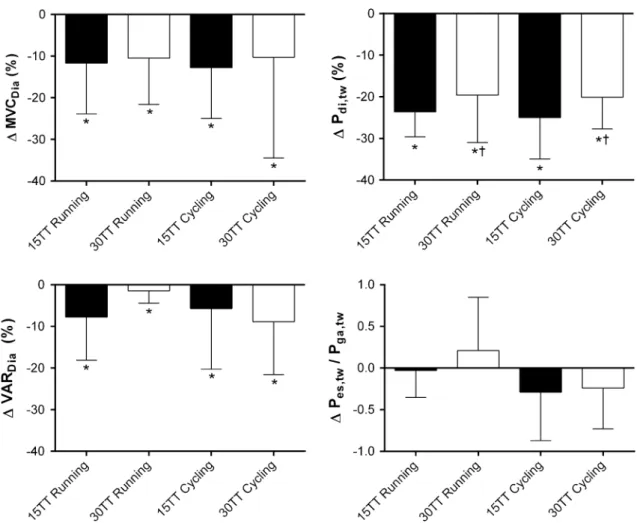
Diaphragm fatigue after running and cycling time-trials Volitional diaphragm muscle strength, i.e. maximal vol-untary diaphragm contractions, was significantly reduced after both time-trials indicated by a significant main effect of time point (p < 0.01, Fig. 3) without being different between 15tt and 30tt (main effect of test, p = 0.62) and exercise modality (interaction test × exercise modality, p = 0.86).
Diaphragm muscle contractility, i.e. potentiated Pdi,tw, was significantly impaired after both time-trials compared to resting values (both p < 0.01, Fig. 3). the decrease in Pdi,tw (ΔPdi,tw) resulted from a significant reduction in both
Pes,tw and Pga,tw (all p < 0.01) and exceeded 10 % in all sub-jects in the 15tt and in 19/22 subsub-jects in the 30tt. the level of ΔPdi,tw was significantly larger in the 15tt com-pared to the 30tt (effect of test, p = 0.04). the type of exercise (running or cycling) had no effect on the decrease in twitch pressures (no effect of exercise modality, p = 0.78 and interaction of test × exercise modality, p = 0.82). the change in the ratio of Pes,tw/Pga,tw was not different between
tests (p = 0.29) but tended to be different for running and cycling (p = 0.06, Fig. 3). the average Pes baseline immediately preceding each twitch was constant for all measurements, suggesting identical lung volumes before stimulations (15tt: before −4.6 ± 1.5 cmH2O vs. after
−4.8 ± 1.8 cmH2O, p = 0.52; 30tt: before −4.8 ± 1.8 vs.
after −5.1 ± 2.3 cmH2O, p = 0.37). Diaphragm voluntary Fig. 2 reduction in quadriceps muscle strength after 15 and
30 min time-trials (15tt, 30tt) in runners and cyclists. Values are mean ± SD (n = 12). MVcQuad, voluntary maximal contraction of the quadriceps; Qtw, quadriceps twitch force; PtP M-Wave,
peak-to-peak amplitude of compound muscle action potentials; VArQuad, vol-untary activation ratio of the quadriceps. *Significantly reduced com-pared to baseline, p < 0.05; #Significantly different between runners and cyclists, p < 0.05
activation ratio was similarly reduced after both time-trials irrespective of exercise modality (Fig. 3).
Within-day coefficients of variation (cV) for potenti-ated Pdi,tw were similar at rest (15tt: 5.4 ± 2.9 %; 30tt:
5.6 ± 3.1 %) and after the time-trial (15tt: 6.8 ± 3.2 %; 30tt: 5.2 ± 2.1 %). the between-day cV of potentiated Pdi,tw was 4.0 ± 3.2 %.
Exercise response and respiratory muscle work during time-trial exercise
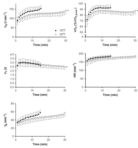
Average exercise responses for runners and cyclists during the 15tt and 30tt are given in table 2 as well as in Fig. 4. ˙
VO
2 was not different between runners and cyclists (main
effect of exercise modality, p = 0.25) and significantly higher in the 15tt compared to the 30tt (+4.4 ± 4.5 %, main effect of test, p < 0.01) with no interaction test × exercise modality (p = 0.38). In addition, min-ute ventilation (+6.4 ± 7.3 %) and inspiratory as well as
expiratory muscle work were higher in the 15tt indicated by a significant main effect of test (all p < 0.001) while no effect was observed for exercise modality and the interac-tion test × exercise modality (table 2). ˙VO2 (runners and
cyclists) rose to 93 ± 5 % ˙VO2,peak in the final 30 s of the
15tt and 88 ± 6 % ˙VO2,peak in the 30tt. Heart rate of the
final 30 s reached 98 ± 4 % (15tt) and 99 ± 3 % (30tt) of the maximum heart rate obtained in the incremental test. Perception of respiratory and leg exertion reached maximal or near maximal levels (i.e. ≥9) at the end of the 15tt (in 18/22 subjects for respiratory exertion and in 19/22 for leg exertion) and 30tt (in 16/22 and 21/22 for respiratory and leg exertion, respectively).
Inspiratory muscle fatigue after isolated normocapnic hyperpnoea
normocapnic hyperpnoea lasted for 7.8 ± 3.8 min in runners and 11.0 ± 9.0 min in cyclists (p = 0.30)
Fig. 3 reduction in diaphragm muscle strength after 15 and
30 min time-trials (15tt, 30tt) in runners and cyclists. Values are mean ± SD (n = 12). Pdi,tw, transdiaphragmatic twitch pres-sure; Pes,tw, esophageal twitch pressure; Pga,tw, gastric twitch
pres-sure; MVcDia, diaphragm voluntary maximal contraction; VArDia, voluntary activation ratio of the diaphragm. *Significantly reduced compared to baseline, p < 0.05; †Significantly different from 15tt, p < 0.05
at an average minute ventilation of 157 ± 12 and 149 ± 9 l min−1 (p = 0.08) corresponding to 77 ± 4 %
MVV in both groups (p = 0.97). tidal volume of both groups averaged at 2.164 ± 0.385 l (corresponding to ~35 % of vital capacity) and breathing frequency was 74 ± 12 breaths min−1 without being different between
runners and cyclists. During the course of normocap-nic hyperpnoea, tidal volume decreased significantly while breathing frequency increased (tidal volume: from 2.588 ± 0.518 to 1.796 ± 0.415 l; breathing frequency: from 63 ± 12, to 85 ± 16 breaths min−1) independent of
exercise modality. normocapnia was achieved in all sub-jects throughout normocapnic hyperpnoea with an average end-tidal cO2 partial pressure of 39.2 ± 1.2 mmHg.
After normocapnic hyperpnoea, Pm,tw was significantly reduced by 35.8 ± 11.0 %. Data of one subject had to be excluded due to the inability to produce reliable Pm,tw measurements after normocapnic hyperpnoea. the aver-age reduction in Pm,tw for runners (37.1 ± 8.4 %, n = 11)
and cyclists (34.4 ± 13.5 %, n = 10) were not signifi-cantly different (p = 0.60). Within-day cVs for Pm,tw were
6.1 ± 2.5 % before and 7.8 ± 3.9 % after normocapnic hyperpnoea.
Discussion
the present study sought to investigate differences in the level of fatigue in locomotor and diaphragm muscles during self-paced exercise of different durations (15 vs. 30 min) and between different exercise modalities (run-ning and cycling). Present findings obtained in endurance trained runners and cyclists (in their respective disci-pline) suggest that both quadriceps and diaphragm muscle fatigue is present after time-trials but quadriceps muscle fatigue is substantially more pronounced in cyclists com-pared to runners (independent of exercise duration/inten-sity) while diaphragm fatigue is more pronounced in the
Table 2 Exercise response to time-trials
PEtcO2, end tidal cO2 partial pressure for 15 and 30 min time-trials (15tt, 30tt). End-exercise values are given for perception of respiratory exertion, leg exertion and blood lactate concentration
Values are mean ± SD. HR heart rate, ˙VE minute ventilation, VT tidal volume, fB breathing frequency, PTP pressure–time product, PTPdi,in inspir-atory diaphragmatic PtP, PTPes,in inspiratory esophageal PtP, PTPes,ex expiratory esophageal PtP, PTPga,ex expiratory gastric PtP, WOB work of breathing, ˙VO2 oxygen consumption; ˙VcO2 carbon dioxide elimination
* Significantly different between 15tt vs. 30tt, p < 0.05 # Significantly different between runners and cyclists, p > 0.05 ‡ Interaction test × exercise modality, p < 0.05
15tt 30tt
running cycling running cycling
Hr (beats min−1) 174 ± 7 178 ± 6 169 ± 13 177 ± 8
Exercise intensity (% max) 84.1 ± 1.4 77.3 ± 4.1 79.9 ± 2.1 71.1 ± 4.0*, #
˙ VE (l min−1) 125 ± 15 138 ± 20 120 ± 13 126 ± 16* Vt (l) 2.77 ± 0.34 3.03 ± 0.40 2.64 ± 0.30 2.88 ± 0.35* fB (breaths min−1) 46.1 ± 7.0 46.1 ± 7.2 46.1 ± 6.2 44.6 ± 7.7 PtPdi,in (cmH2O s min−1) 737 ± 133 666 ± 229 658 ± 93 549 ± 164* PtPes,in (cmH2O s min−1) 444 ± 81 551 ± 142 404 ± 85 457 ± 89* PtPes,ex (cmH2O s min−1) 385 ± 135 373 ± 154 339 ± 109 309 ± 100* PtPga,ex (cmH2O s min−1) 461 ± 133 416 ± 141 402 ± 90 293 ± 92*
WOB (joule min−1) 410 ± 137 516 ± 158 343 ± 109 384 ± 112*,‡
˙ VO2 (l min−1) 4.27 ± 0.30 4.51 ± 0.46 4.12 ± 0.38 4.27 ± 430* ˙ VcO2 (l min−1) 4.29 ± 0.28 4.66 ± 0.55 4.01 ± 0.31 4.20 ± 0.50*,‡ ˙ VO2 (% ˙VO2,peak) 89.5 ± 3.5 89.5 ± 4.4 86.0 ± 3.8 84.6 ± 3.6* ˙ VE/ ˙VO2 29.3 ± 2.7 30.3 ± 1.6 29.1 ± 2.8 29.3 ± 1.7 ˙ VE/ ˙VcO2 29.1 ± 2.8 29.5 ± 1.9 29.7 ± 2.4 30.0 ± 1.8 PEtcO2 (mmHg) 37.7 ± 3.2 37.6 ± 2.2 36.9 ± 2.9 37.2 ± 2.3 respiratory exertion 9.0 ± 0.78 8.8 ± 1.2 8.7 ± 1.0 9.0 ± 0.8 leg exertion 9.0 ± 1.0 9.3 ± 0.5 9.4 ± 0.7 9.1 ± 0.5
shorter, more intense time-trial (independent of exercise modality). In addition, no difference in time to exhaus-tion and in the rate of fatigue development was evident between runners and cyclists performing isolated nor-mocapnic hyperpnoea.
Quadriceps muscle fatigue after running and cycling time-trials of different durations
Both time-trials (15 and 30 min) induced significant reduc-tions in volitional maximal quadriceps muscle force. this reduction is likely to be the result of both a reduction in muscle contractility and the ability to voluntarily drive the muscle as both Qtw amplitude and quadriceps voluntary activation ratio were significantly decreased after time-trials (Fig. 2). the fact that M-Wave amplitude was also reduced in both time-trials (Fig. 2), indicates that periph-eral fatigue (ΔQtw) may have included not only contractile failure but possibly also alterations in the electrical propa-gation of the action-potential across the neuromuscular
junction and along the sarcolemma and t-tubules (i.e. reduced membrane excitability).
the reductions in maximal voluntary quadriceps con-traction (7 %) and Qtw (13 %) after running time-trials are in line with other studies investigating quadriceps muscle fatigue in tests of similar duration/intensity (nummela et al. 2008). Similarly, reductions in maximal voluntary quadriceps contraction after cycling time-trials are in the range of those found in other studies using cycling tasks of approximately the same duration (Bentley et al. 2000; lep-ers et al. 2002). reductions in Qtw in cyclists were, how-ever, slightly larger than those in the latter studies and are more consistent with those seen in somewhat shorter time-trials [e.g. (Amann et al. 2006)]. to the best of our knowl-edge, this is the first time that locomotor muscle fatigue was systematically investigated after self-paced exercise of different durations/intensities in the same group of indi-viduals. Interestingly, peripheral quadriceps muscle fatigue was not different between the 15 and 30tts. these results may be viewed in support of the idea of afferent feedback
Fig. 4 Development of
ventila-tory variables, relative oxygen consumption and heart rate during 15 and 30 min time-trials (15tt, 30tt). Values are mean ± SD (n = 22); ˙VE minute ventilation; VT tidal volume, fB breathing frequency, ˙VO2 oxygen consumption, HR heart rate (n = 21)
interacting at the spinal and/or supraspinal level possi-bly to avoid the development of fatigue beyond a certain functional limit (Amann and Secher 2010; racinais et al.
2007). Alternatively, fatigue was suggested to be the conse-quence of a metabolic challenge mainly to type 2 fibers and to develop rather early during exercise which would then explain similar levels of contractile fatigue (Decorte et al.
2012; Sargeant 2007).
However, Qtw was substantially more impaired in cyclists than in runners after time-trials in the present study. Both methodological and physiological mechanisms may explain these findings. First, one has to consider whether the quadriceps is the ‘right’ muscle to compare the occur-rence of fatigue in locomotor muscles between running and cycling. In fact, there is evidence showing that EMg activity of the quadriceps muscle does not increase linearly with the increasing mechanical power output for running but does so during cycling (Bijker et al. 2002). For the calf muscles on the other hand, a linear relation between EMg activity and mechanical power was detected in both running and cycling exercise which could imply that this muscle group would have been better comparable. How-ever, when Martin and co-workers (2010) measured muscle contractility in both the knee extensors and plantar flexors at the same time, no difference in the amount of reduction in twitch force was present. Furthermore, when looking at running exercise of comparable duration to our study, reductions in twitch force of the calf muscle [i.e. ~16 % (girard et al. 2012)] were of similar magnitude as those found for the quadriceps muscle here (~13 %). Hence, we believe that the quadriceps muscle represents a valid model for comparing the occurrence of locomotor muscle fatigue in running and cycling. Second, since runners and cyclists only performed in their respective sports, one has to con-sider that two different populations were investigated which per se could have affected the level of contractile fatigue. the present study design was introduced to minimize a potential bias of running/cycling experience on time-trial performance but clearly limits what can be concluded in terms of the mechanism responsible for the finding of aug-mented levels of contractile fatigue in cyclists. Despite of these limitations, it is important also to consider the type of muscle contraction specific to running and cycling. the quadriceps muscle is exposed to repeated stretch–shorten-ing cycles (i.e. eccentric and concentric contractions) dur-ing runndur-ing while cycldur-ing predominantly involves concen-tric contractions (Bijker et al. 2002). Owing to eccentric contractions in running, one would expect both metabolic and mechanical stress to contribute to the loss in Qtw while mechanical stress is likely less prominent and metabolic stress more pronounced when cycling. there is indeed some indication that metabolic load on the quadriceps was higher in cyclists than in runners as relative whole-body
˙ VO
2 was not different between exercise modalities but
blood lactate concentrations at the end of exercise was higher in cyclists (table 2). In fact, both metabolic and mechanical stress can impair contractile function and thus exercise performance (Amann and Dempsey 2008; Mar-cora et al. 2008). contractile impairment, however, was reported to differ between concentric and eccentric con-tractions both in terms of magnitude and timing (Smith and newham 2007). the latter authors reported that contrac-tile fatigue was less pronounced after eccentric compared to concentric contractions and that peak reduction after eccentric contractions occurs ~60 min after task cessa-tion while in concentric exercise peak reduccessa-tion occurred ~20 min into recovery. thus, the time point when Qtw was
reassessed (i.e. 10 min after completion of time-trials) after time-trials might explain some of the observed differences between runners and cyclists in the present study. Interest-ingly, despite smaller levels of peripheral muscle fatigue in runners, similar reductions in maximal voluntary quadri-ceps contractions were detected in presence of a similar drop in voluntary activation ratio (Fig. 2). Unfortunately, the present study does not allow for a conclusive discus-sion. Apart from methodological concerns regarding the sensitivity of the superimposed twitch technique (gande-via 2001), one could also hypothesize that this apparent discrepancy points towards effects of muscle damage (due to eccentric contractions in running) which act beyond the excitation–contraction coupling system and are thus not detected by the assessment of Qtw (Iguchi and Shields
2010). this clearly needs further investigations. Diaphragm muscle fatigue after running and cycling time-trials of different durations
the average intensities of both self-paced exercise tests were 90 % (15tt) and 85 % ˙VO2,peak (30tt), i.e. they were performed at and above the intensity (i.e. ≥85 % ˙VO2,peak)
reported to be required for diaphragm fatigue to occur (Johnson et al. 1993). the degree of diaphragm fatigue that subjects developed in the two time-trials compared well to that observed after exhaustive constant-load exercise (Babcock et al. 1995a, b, 2002; Johnson et al. 1993; Mador et al. 1993; taylor et al. 2006; Verges et al. 2007, 2006a,
b; Walker et al. 2011; Wuthrich et al. 2013). the one study that assessed diaphragm fatigue after constant-load exercise at 90–95 % and 80–85 % ˙VO2,peak unfortunately included a different number of subjects with a large varia-tion in fitness level and the actual exercise intensity ranged from 60 % to over 110 % ˙VO2,peak in the two trials such that
a direct comparison is not possible (Johnson et al. 1993). thus, the hypothesis that more intense cycling would cause larger degrees of diaphragm fatigue was proposed but has not been investigated systematically. Present findings
suggest substantially more pronounced levels of diaphragm fatigue after the shorter more intense 15tt (ΔPdi,tw 24 %) than after the longer 30tt (ΔPdi,tw 20 %)—a finding which was anticipated given the relationship between exer-cise intensity and the level of diaphragm fatigue observed by Johnson and co-workers (1993). Several factors may have contributed to the observed difference in diaphragm fatigue while total respiratory muscle work that accumu-lated during the entire exercise duration can be excluded, i.e. the sum of WOB was approximately 1.5 times larger in the 30tt where fatigue was smaller. However, when considering respiratory muscle work on a per minute basis, we found significantly higher levels for the shorter, more intense 15tt (table 2), i.e. the higher exercise intensity of the 15tt not only induced a significantly higher average ventilation (+6.4 %) but also an even more pronounced increase in respiratory muscle work per minute (+16 % in PtPdi,in, +15 % in PtPes,in and +27 % in WOB; Fig. 5).
Of note is that higher exercise intensity not only increases respiratory muscle work but may also affect blood flow distribution to all working muscles. While increased respiratory muscle work was shown to compro-mise leg blood flow (Harms et al. 1997), two recent studies reported that blood flow to intercostal muscles may actually
be compromised at high ventilatory levels during whole-body exercise (Henderson et al. 2012; Vogiatzis et al.
2009). the latter studies showed that intercostal blood flow reaches a peak around 80 % maximal cycling power out-put in an incremental test. In the 15tt of the present study, average relative running velocity (84 % maximal running velocity) and cycling power output (80 % maximal cycling power output) were in fact at or above this threshold sug-gesting the potential for blood flow restrictions to respira-tory muscles in contrast to the 30tt, where relative run-ning velocity (80 % maximal runrun-ning velocity) and cycling power output (71 % maximal cycling power output) were at or clearly below this threshold suggesting no compro-mise of the respiratory muscle blood flow. the potentially larger mismatch between oxygen demand and supply in the inspiratory muscles along with higher respiratory mus-cle work during the more intense 15tt may have led to an increased accumulation of metabolites known to impair the function of the contractile apparatus thus explaining in part why diaphragm fatigue was more pronounced in the shorter 15tts (Allen et al. 2008).
contrary to our original hypothesis, similar levels of inspiratory muscle fatigue were observed for runners and cyclists. We hypothesized that diaphragm fatigue would be
Fig. 5 Variables of respiratory muscle work during 15 and 30 min
time-trials (15tt, 30tt). Values are mean ± SD (n = 22). R rest, WU warm up, PTPdi,in, inspiratory transdiaphragmatic pressure–time
product, PTPes,in inspiratory esophageal pressure–time product, WOB work of breathing
more pronounced in runners due to the postural demand associated with running. We based our hypothesis mainly on findings by Hodges and gandevia (2000) who reported tonic and phasic diaphragm activity during postural challenge superimposed on breathing. We suggested that this postural demand in concert with severe ventilatory work might place a greater load on the diaphragm in runners compared to cyclists. this is supported by an immediate increase in Pga at the start of exercise which was consistently seen in run-ners but not in cyclists (data not shown). this increase in intra-abdominal pressure is thought to help improving the stiffness and stability of the spine when the maintenance of posture is challenged but may also increase the load on the working diaphragm (Henke et al. 1988; Hodges et al. 2005). this increase in Pga also increased PtPga,in, thus explain-ing why the ratio of PtPes,in/PtPdi,in was higher in runners than cyclists (data not shown). these observations suggest a somewhat higher postural demand in runners which does, however, not seem to exert a substantial effect on the devel-opment of inspiratory muscle fatigue.
Although inspiratory muscle fatigue was not differ-ent between exercise modalities, the contribution of the diaphragm and of rib cage muscles might have differed between running and cycling since in runners, the ratio of Pes,tw/Pga,tw (Similowski et al. 1998) remained unchanged (15tt) or even slightly increased (30tt) while in cyclists it was reduced in both time-trials with the effect of exer-cise modality showing a tendency to statistical significance (p = 0.06). together with significantly reduced Pdi,tw and
Pga,tw in both exercise modalities, this may be interpreted
to mean that running time-trials induced predominantly diaphragm fatigue while cycling induced more global inspiratory muscle fatigue (Similowski et al. 1998). the significance of this potential difference was then tested in volitional normocapnic hyperpnoea where no difference in postural demand was present for subjects trained in the dif-ferent exercise modalities.
In contrast to our hypothesis task failure in volitional normocapnic hyperpnoea occurred slightly but not sig-nificantly earlier in runners than in cyclists. runners per-formed at somewhat higher absolute ventilation but at the same relative ventilation as cyclists (i.e. 77 % MVV) and inspiratory muscles were similarly fatigued in runners and cyclists indicated by a 36 % reduction in Pm,tw. Also, run-ners and cyclists showed similar rates of fatigue develop-ment (i.e. the reduction in Pm,tw divided by the duration of
normocapnic hyperpnoea, data not shown) which further highlights the finding of similar fatigue resistance for both groups.
collectively, results from normocapnic hyperpnoea tests confirm findings during whole-body exercise where the magnitude of inspiratory muscle fatigue was similar in runners and cyclists. together, one could postulate that
ventilatory demand during whole-body exercise substan-tially exceeds that of posture with the latter consequently not affecting the development of fatigue to a significant degree—thereby also not inducing specific training adap-tations. this, however, requires further investigation but is supported by the finding that posture-related diaphragm activity decreases when ventilatory demand increases (Hodges et al. 2001).
Methodological considerations and limitations
Influence of end‑expiratory lung volume on the assessment of diaphragm fatigue
constant lung volumes before application of magnetic stimulations are crucial to achieve reproducible twitch measurements since an inverse linear relationship exists between Pdi,tw and lung volume (Hamnegard et al. 1995;
Walker et al. 2011). thus, lung volumes were closely monitored in the present study by measurement of Pes before each stimulation. Baseline Pes was similar within a sequence of twitches and also before and after exercise. thus, altered lung volumes are unlikely to have biased our outcomes but lung volumes could not be monitored during Pm,tw measurements. Also, similar cVs of Pm,tw and Pes,tw suggest that lung volumes were constant also during Pm,tw
assessments.
M‑Wave during assessment of diaphragm fatigue
EMg responses to magnetic stimulation were not assessed for inspiratory muscles. this opens the possibility of reduced excitability of the muscular membrane resulting from electrolytic disturbances after exercise which would lead to an overestimation of contractile fatigue (Fowles et al. 2002). However, such changes were not commonly seen in studies assessing diaphragm fatigue and EMg concomitantly in our laboratory or those of others (guen-ette et al. 2010; Johnson et al. 1993; Verges et al. 2006a). Hence, an overestimation of contractile diaphragm fatigue in the present study is unlikely, although it cannot be com-pletely ruled out.
Selection of investigated population
In the present study, only experienced endurance athletes were included performing in their respective exercise modalities in order to guarantee replication of a maximal effort as well as optimal pacing in both the 15tt and 30tt. this is a prerequisite when investigating potentially limit-ing processes within the human body and was likely to be achieved here as indicated by several findings. Percep-tion of respiratory and leg exerPercep-tion increased to ≥9 (out
of a maximum of 10) by the end of the test in most of the subjects and heart rate of the final 30 s of both time-trials was at 98–99 % of the maximal heart rate achieved in the incremental test. Also, average ˙VO2 of the final 30 s rose to approximately 93 % (15tt) and 88 % of ˙VO2,peak (30tt) which is in line with other studies where maximal time-trial exercise or exhaustive constant-load tests were performed (Amann et al. 2009; Johnson et al. 1993). these findings strongly suggest that subjects achieved a truly maximal effort in both time-trials. Yet, it remains speculative how these results may be transferred to other populations such as healthy subjects of different sex and age or patients. Existing studies on the development of quadriceps mus-cle fatigue at the limit of tolerance suggest that levels of fatigue are remarkably similar between young and elderly individuals as well as patients suffering from chronic obstructive pulmonary disease (Amann 2011; Mador et al.
2000a, b, 2001; Saey et al. 2005) while findings for dia-phragm fatigue seem to be more variable when considering healthy subjects of different fitness and age or patients suf-fering from chronic obstructive pulmonary disease (Bab-cock et al. 1996; Mador et al. 2000a; Polkey et al. 1995). Conclusion
the present study provides evidence that alterations in con-tractility of the quadriceps muscle after intense self-paced whole-body exercise are distinctly different for running and cycling while these alterations are very similar in the two exercise intensities of a single exercise modality. the level of inspiratory muscle fatigue is, however, independ-ent of the exercise modality and related more to exercise intensity and the associated respiratory muscle work. Pos-sibly, the postural demand placed upon the diaphragm dur-ing runndur-ing resulted in a larger contribution of diaphrag-matic fatigue to total inspiratory muscle fatigue compared to cycling which did, however, not lead to greater fatigue resistance during volitional hyperpnoea in runners com-pared to cyclists.
Acknowledgments We thank all the subjects for their time and
maximal efforts put into this study and Dr. ruth Briggs for English editing. this research is financially supported by the Swiss Office of Sports (grant no. 11-11).
Conflict of interest the authors declare that no conflict of interest
exists.
References
Aaron EA, Seow Kc, Johnson BD, Dempsey JA (1992) Oxygen cost of exercise hyperpnea: implications for performance. J Appl Physiol 72:1818–1825
Abbiss cr, laursen PB (2005) Models to explain fatigue during pro-longed endurance cycling. Sports Med 35:865–898
Allen Dg, lamb gD, Westerblad H (2008) Skeletal muscle fatigue: cellular mechanisms. Physiol rev 88:287–332
Amann M (2011) central and peripheral fatigue: interaction during cycling exercise in humans. Med Sci Sports Exerc 43:2039–2045 Amann M, Dempsey JA (2008) locomotor muscle fatigue modifies
central motor drive in healthy humans and imposes a limitation to exercise performance. J Physiol 586:161–173
Amann M, Secher nH (2010) Point: counterpoint: Afferent feed-back from fatigued locomotor muscles is/is not an important determinant of endurance exercise performance. J Appl Physiol 108:452–454
Amann M, Eldridge MW, lovering At, Stickland MK, Pegelow DF, Dempsey JA (2006) Arterial oxygenation influences central motor output and exercise performance via effects on peripheral locomotor muscle fatigue in humans. J Physiol 575:937–952 Amann M, Proctor lt, Sebranek JJ, Pegelow DF, Dempsey JA (2009)
Opioid-mediated muscle afferents inhibit central motor drive and limit peripheral muscle fatigue development in humans. J Physiol 587:271–283
AtS/ErS (2002) Statement on respiratory muscle testing. Am J respir crit care Med 166:518–624
Babcock MA, Johnson BD, Pegelow DF, Suman OE, griffin D, Demp-sey JA (1995a) Hypoxic effects on exercise-induced diaphrag-matic fatigue in normal healthy humans. J Appl Physiol 78:82–92 Babcock MA, Pegelow DF, Mcclaran Sr, Suman OE, Dempsey JA
(1995b) contribution of diaphragmatic power output to exercise-induced diaphragm fatigue. J Appl Physiol 78:1710–1719 Babcock MA, Pegelow DF, Johnson BD, Dempsey JA (1996)
Aero-bic fitness effects on exercise-induced low-frequency diaphragm fatigue. J Appl Physiol 81:2156–2164
Babcock MA, Pegelow DF, Harms cA, Dempsey JA (2002) Effects of respiratory muscle unloading on exercise-induced diaphragm fatigue. J Appl Physiol 93:201–206
Bentley DJ, Smith PA, Davie AJ, Zhou S (2000) Muscle activation of the knee extensors following high intensity endurance exercise in cyclists. Eur J Appl Physiol 81:297–302
Bigland-ritchie B, Woods JJ (1984) changes in muscle contractile properties and neural control during human muscular fatigue. Muscle nerve 7:691–699
Bijker KE, de groot g, Hollander AP (2002) Differences in leg mus-cle activity during running and cycling in humans. Eur J Appl Physiol 87:556–561
Decorte n, lafaix PA, Millet gY, Wuyam B, Verges S (2012) central and peripheral fatigue kinetics during exhaustive constant-load cycling. Scand J Med Sci Sports 22:381–391
Fowles Jr, green HJ, tupling r, O’Brien S, roy BD (2002) Human neuromuscular fatigue is associated with altered na+-K+-AtPase activity following isometric exercise. J Appl Physiol 92:1585–1593 gandevia Sc (2001) Spinal and supraspinal factors in human muscle
fatigue. Physiol rev 81:1725–1789
girard O, Millet gP, Micallef JP, racinais S (2012) Alteration in neu-romuscular function after a 5 km running time trial. Eur J Appl Physiol 112:2323–2330
guenette JA, romer lM, Querido JS, chua r, Eves nD, road JD, McKenzie Dc, Sheel AW (2010) Sex differences in exercise-induced diaphragmatic fatigue in endurance-trained athletes. J Appl Physiol 109:35–46
Hamnegard cH, Wragg S, Mills g, Kyroussis D, road J, Daskos g, Bake B, Moxham J, green M (1995) the effect of lung volume on transdiaphragmatic pressure. Eur respir J 8:1532–1536 Harms cA, Babcock MA, Mcclaran Sr, Pegelow DF, nickele gA,
nelson WB, Dempsey JA (1997) respiratory muscle work com-promises leg blood flow during maximal exercise. J Appl Physiol 82:1573–1583
Henderson Wr, guenette JA, Dominelli PB, griesdale DE, Querido JS, Boushel r, Sheel AW (2012) limitations of respiratory mus-cle and vastus lateralis blood flow during continuous exercise. respir Physiol neurobiol 181:302–307
Henke Kg, Sharratt M, Pegelow D, Dempsey JA (1988) regulation of end-expiratory lung volume during exercise. J Appl Physiol 64:135–146
Hodges PW, gandevia Sc (2000) Activation of the human diaphragm during a repetitive postural task. J Physiol 522(Pt 1):165–175 Hodges PW, Heijnen I, gandevia Sc (2001) Postural activity of
the diaphragm is reduced in humans when respiratory demand increases. J Physiol 537:999–1008
Hodges PW, Eriksson AE, Shirley D, gandevia Sc (2005) Intra-abdominal pressure increases stiffness of the lumbar spine. J Bio-mech 38:1873–1880
Iguchi M, Shields rK (2010) Quadriceps low-frequency fatigue and muscle pain are contraction-type-dependent. Muscle nerve 42:230–238
Johnson BD, Babcock MA, Suman OE, Dempsey JA (1993) Exer-cise-induced diaphragmatic fatigue in healthy humans. J Physiol 460:385–405
Kufel tJ, Pineda lA, Mador MJ (2002) comparison of potentiated and unpotentiated twitches as an index of muscle fatigue. Muscle nerve 25:438–444
lansing rW, Im BS, thwing JI, legedza At, Banzett rB (2000) the perception of respiratory work and effort can be independ-ent of the perception of air hunger. Am J respir crit care Med 162:1690–1696
lepers r, Maffiuletti nA, rochette l, Brugniaux J, Millet gY (2002) neuromuscular fatigue during a long-duration cycling exercise. J Appl Physiol 92:1487–1493
Mador MJ, Magalang UJ, rodis A, Kufel tJ (1993) Diaphragmatic fatigue after exercise in healthy human subjects. Am rev respir Dis 148:1571–1575
Mador M, Kufel tJ, Pineda lA (2000a) Quadriceps and diaphrag-matic function after exhaustive cycle exercise in the healthy elderly. Am J respir crit care Med 162:1760–1766
Mador MJ, Kufel tJ, Pineda lA, Sharma gK (2000b) Diaphragmatic fatigue and high-intensity exercise in patients with chronic obstruc-tive pulmonary disease. Am J respir crit care Med 161:118–123 Mador MJ, Kufel tJ, Pineda lA, Steinwald A, Aggarwal A,
Upad-hyay AM, Khan MA (2001) Effect of pulmonary rehabilitation on quadriceps fatiguability during exercise. Am J respir crit care Med 163:930–935
Marcora SM, Bosio A, de Morree HM (2008) locomotor muscle fatigue increases cardiorespiratory responses and reduces perfor-mance during intense cycling exercise independently from meta-bolic stress. Am J Physiol-reg 294:r874–r883
Martin V, Kerherve H, Messonnier lA, Banfi Jc, geyssant A, Bonne-foy r, Feasson l, Millet gY (2010) central and peripheral con-tributions to neuromuscular fatigue induced by a 24-h treadmill run. J Appl Physiol 108:1224–1233
Miller Mr, crapo r, Hankinson J, Brusasco V, Burgos F, casaburi r, coates A, Enright P, van der grinten cP, gustafsson P, Jensen r, Johnson Dc, MacIntyre n, McKay r, navajas D, Pedersen OF, Pellegrino r, Viegi g, Wanger J (2005) general considerations for lung function testing. Eur respir J 26:153–161
Millet gY, tomazin K, Verges S, Vincent c, Bonnefoy r, Boisson rc, gergele l, Feasson l, Martin V (2011) neuromuscular con-sequences of an extreme mountain ultra-marathon. PloS One 6:e17059
nummela At, Heath KA, Paavolainen lM, lambert MI, St clair gib-son A, rusko HK, noakes tD (2008) Fatigue during a 5-km run-ning time trial. Int J Sports Med 29:738–745
Polkey MI, Kyroussis D, Keilty SE, Hamnegard cH, Mills gH, green M, Moxham J (1995) Exhaustive treadmill exercise does
not reduce twitch transdiaphragmatic pressure in patients with cOPD. Am J respir crit care Med 152:959–964
Quanjer PH, tammeling gJ, cotes JE, Pedersen OF, Peslin r, Yer-nault Jc (1993) lung volumes and forced ventilatory flows. report Working Party Standardization of lung Function tests, European community for Steel and coal. Official Statement of the European respiratory Society. Eur respir J Suppl 16:5–40 racinais S, girard O, Micallef JP, Perrey S (2007) Failed excitability
of spinal motoneurons induced by prolonged running exercise. J neurophysiol 97:596–603
ross EZ, goodall S, Stevens A, Harris I (2010) time course of neu-romuscular changes during running in well-trained subjects. Med Sci Sports Exerc 42:1184–1190
Saey D, Michaud A, couillard A, cote cH, Mador MJ, leBlanc P, Jobin J, Maltais F (2005) contractile fatigue, muscle morphom-etry, and blood lactate in chronic obstructive pulmonary disease. Am J respir crit care Med 171:1109–1115
Sargeant AJ (2007) Structural and functional determinants of human muscle power. Exp Physiol 92:323–331
Saugy J, Place n, Millet gY, Degache F, Schena F, Millet gP (2013) Alterations of neuromuscular function after the World’s most challenging mountain Ultra-Marathon. PloS One 8:e65596 Scheer FA, Hu K, Evoniuk H, Kelly EE, Malhotra A, Hilton MF, Shea
SA (2010) Impact of the human circadian system, exercise, and their interaction on cardiovascular function. Proc natl Acad Sci USA 107:20541–20546
Similowski t, Straus c, Attali V, Duguet A, Derenne JP (1998) cervical magnetic stimulation as a method to discriminate between diaphragm and rib cage muscle fatigue. J Appl Physiol 84:1692–1700
Skof B, Strojnik V (2006a) neuro-muscular fatigue and recovery dynamics following anaerobic interval workload. Int J Sports Med 27:220–225
Skof B, Strojnik V (2006b) neuromuscular fatigue and recovery dynamics following prolonged continuous run at anaerobic threshold. Br J Sports Med 40:219–222
Smith Ic, newham DJ (2007) Fatigue and functional performance of human biceps muscle following concentric or eccentric contrac-tions. J Appl Physiol 102:207–213
taylor BJ, How Sc, romer lM (2006) Exercise-induced abdominal muscle fatigue in healthy humans. J Appl Physiol 100:1554–1562 Verges S, notter D, Spengler cM (2006a) Influence of diaphragm and rib cage muscle fatigue on breathing during endurance exercise. respir Physiol neurobiol 154:431–442
Verges S, Schulz c, Perret c, Spengler cM (2006b) Impaired abdom-inal muscle contractility after high-intensity exhaustive exercise assessed by magnetic stimulation. Muscle nerve 34:423–430 Verges S, lenherr O, Haner Ac, Schulz c, Spengler cM (2007)
Increased fatigue resistance of respiratory muscles during exer-cise after respiratory muscle endurance training. Am J Physiol regul Integr comp Physiol 292:r1246–r1253
Vogiatzis I, Athanasopoulos D, Habazettl H, Kuebler WM, Wagner H, roussos c, Wagner PD, Zakynthinos S (2009) Intercostal mus-cle blood flow limitation in athletes during maximal exercise. J Physiol 587:3665–3677
Walker DJ, Walterspacher S, Schlager D, Ertl t, roecker K, Windisch W, Kabitz HJ (2011) characteristics of diaphragmatic fatigue during exhaustive exercise until task failure. respir Physiol neu-robiol 176:14–20
Wilson SH, cooke nt, Edwards rH, Spiro Sg (1984) Predicted nor-mal values for maxinor-mal respiratory pressures in caucasian adults and children. thorax 39:535–538
Wuthrich tU, notter DA, Spengler cM (2013) Effect of inspira-tory muscle fatigue on exercise performance taking into account the fatigue-induced excess respiratory drive. Exp Physiol 98:1705–1717