Copyright © 2020 Contestabile et al.
This is an open-access article distributed under the terms of the Creative Commons Attribution 4.0 International license, which permits unrestricted use, distribution and reproduction in any medium provided that the original work is properly attributed.
Variable interhemispheric asymmetry in layer
V of the supplementary motor area following
cervical hemisection in adult macaque
monkeys
https://doi.org/10.1523/ENEURO.0280-20.2020
Cite as: eNeuro 2020; 10.1523/ENEURO.0280-20.2020 Received: 29 June 2020
Revised: 24 August 2020 Accepted: 3 September 2020
This Early Release article has been peer-reviewed and accepted, but has not been through the composition and copyediting processes. The final version may differ slightly in style or formatting and will contain links to any extended data.
Alerts: Sign up at www.eneuro.org/alerts to receive customized email alerts when the fully formatted version of this article is published.
1
Variable interhemispheric asymmetry in layer V of the
1
supplementary motor area following cervical hemisection in adult
2
macaque monkeys.
3 4
A. Contestabile1,2, R. Colangiulo2, M. Lucchini 1,2, E. M. Rouiller2 and E. Schmidlin2
5 6
(1) Department of Basic Neuroscience, University of Geneva; 1, Rue Michel-Servet, CH-1205
7
Genève, Switzerland. 8
(2) Department of Neurosciences and Movement Sciences, Section of Medicine, Faculty of
9
Sciences and Medicine, Fribourg Center of Cognition, University of Fribourg, Chemin du 10
Musée 5, CH-1700 Fribourg, Switzerland. 11
12
Running title: SMA asymmetry in SCI monkeys 13
14
Keywords: spinal cord injury, supplementary motor area, layer V pyramidal neurons, 15
corticospinal projections, interhemispheric asymmetry, SMI-32, non-human primate. 16
17
Text pages: 28 18
Words: 6364 total words (without abstract, significance statements, acknowledgments, list of 19
references and figure legends). Abstract. 146 words. Introduction: 981 words. Material and 20
Methods: 2021 words. Results: 1506 words. Discussion: 1856 words. Tables: 1. Figures: 5. 21
22
Post-revision corrections after revision in eNeuro are labelled in red. 23
24
Address for correspondence: Dr. Eric Schmidlin, Department of Neurosciences, Faculty of 25
Sciences and Medicine, University of Fribourg, Chemin du Musée 5, CH-1700 Fribourg, 26
2 Switzerland. Phone: +41 26 300 86 09. Fax: +41 26 300 96 75. E-mail: 27
Eric.Schmidlin@unifr.ch 28
3
ABSTRACT
29
Motor cortical areas from both hemispheres play a role during functional recovery 30
after a unilateral spinal cord injury (SCI). However, little is known about the morphological 31
and phenotypical differences that a SCI could trigger in corticospinal neurons of the 32
ipsilesional and contralesional hemisphere. Using an SMI-32 antibody which specifically 33
labeled pyramidal neurons in cortical layers V, we investigated the impact of a unilateral 34
cervical cord lesion on the rostral part (F6) and caudal part (F3) of the supplementary motor 35
area (SMA) in both hemispheres of eight adult macaque monkeys compared with four intact 36
control monkeys. We observed in F3 (but not in F6) interindividual variable and adaptive 37
interhemispheric asymmetries of SMI-32 positive layer V neuronal density and dendritic 38
arborization, which are strongly correlated with the extent of the SCI as well as the duration 39
of functional recovery, but not with the extent (percentage) of functional recovery. 40
41 42
Significance statement
43
1. This study consists in a precise quantification on two different levels of the histological 44
consequences on the long term of a traumatic and sudden unilateral interruption of the 45
corticospinal tract at cervical level in 8 non-human primates (adult macaque monkeys). 46
2. The lesion affected the density and the morphology of layer v pyramidal neurons in the 47
supplementary motor area (SMA), in the form of an interhemispheric adaptive asymmetry, 48
correlated to the lesion size and duration of functional recovery. 49
3. These changes are reminiscent of those observed in SMA after unilateral lesion of the 50
primary motor cortex, suggesting to some extent comparable mechanism of functional 51
motor recovery from unilateral cortical or spinal lesion. 52
4. The dendritic arborization in the basal dendrites of the SMI-32 positive neurons in layer 53
V showed a more prominent interhemispheric effect of the lesion than the apical dendrites. 54
4
Introduction
56
In non-human primates, the hand area of the primary motor cortex (M1 or F1) is 57
subdivided in an old M1 and a new M1 (Rathelot and Strick, 2009). The new M1 is at the 58
origin of the corticomotoneuronal (CM) projection, representing the anatomical support of 59
manual dexterity, a prerogative of primates (Lawrence and Kuypers, 1968; Courtine et al., 60
2007; Lemon, 2008; Yoshida and Isa, 2018). Although M1 is the main contributor to the 61
corticospinal (CS) projection (including the CM projection), non-primary motor areas such as 62
the premotor cortex (PM), the supplementary motor area (SMA-proper or F3) and the 63
cingulate motor areas (CMA) are also at the origin of CS projections (Luppino et al., 1994; 64
Rouiller et al., 1994; Dum and Strick, 1996; Rouiller, 1996). In particular, SMA projects to the 65
cervical spinal cord, where the motoneurons controlling hand (fingers) motor function are 66
located (Jenny, Inukai and Strick, 1983). There is evidence that part of the CS projection 67
from SMA may be CM (Rouiller, 1996), but the influence of SMA on hand motoneurons is 68
functionally less strong than the one of M1 (Maier et al., 2002; Boudrias et al., 2010). The 69
multiple representations of the hand in several motor cortical areas (M1, PM, SMA, CMA) of 70
primates is the basis for a vicarious scenario of functional redistribution of hand function 71
control in case of selective and focal lesion affecting a motor structure. For instance, after 72
unilateral lesion of the hand area in M1 functional recovery, though often incomplete, 73
depends on plasticity of intact non-primary motor areas, such as PM (Liu and Rouiller, 1999; 74
Dancause et al., 2005; Hoogewoud et al., 2013; Orczykowski et al., 2018) and/or SMA 75
(McNeal et al., 2010; Morecraft et al., 2015). Although such rearrangement of the cortical 76
motor circuits is believed to occur mostly in the ipsilesional hemisphere, there is still 77
controversy about the role played by the contralesional hemisphere (see e.g. (Morecraft et 78
al., 2016; Savidan et al., 2017) for the non-human primate). In a recent article from this 79
laboratory, we reported that a unilateral lesion of the M1 hand area led to a variable 80
interhemispheric asymmetry in the detection of layer V pyramidal neurons in SMA, identified 81
with the marker SMI-32 (Contestabile et al., 2018). This anatomical variable interhemispheric 82
5 imbalance possibly reflects an adaptive interhemispheric contribution of the bilateral SMA to 83
recovery, depending on the lesion size as well as on the duration of functional recovery 84
(Contestabile et al., 2018). It was argued that these observations may represent a putative 85
anatomical support of diaschisis, originally defined as a “loss of function and electrical activity 86
in an area of the brain due to a lesion in a remote area that is neuronally connected with it” 87
(Finger, Koehler and Jagella, 2004 and see von Monakow, 1914). In other words, due to a 88
unilateral M1 lesion, the function of the SMA is affected at distance differently on the 89
ipsilesional versus contralesional hemispheres, because the reciprocal connections between 90
M1 and SMA are stronger ipsilaterally than contralaterally, in the intact state. More recently, 91
the concept of diaschisis has been extended to a structural dimension, in the form of 92
“connectional diaschisis”, focused on post-lesion changes of connectivity in networks/circuits 93
directly or indirectly related to the focal lesion site (Carrera and Tononi, 2014). 94
In the current study, the main goal was to transpose this concept of interhemispheric 95
adaptable SMA reorganization related to functional recovery from a lesion of M1 96
(Contestabile et al., 2018) to a cervical spinal cord hemisection. Does a unilateral spinal cord 97
injury (SCI) also affect differently the contralesional versus the ipsilesional SMA, because the 98
CS projection from SMA is predominantly crossed (Fig. 1A and B)? Following a hemisection 99
of the cervical cord in macaque monkeys, it was observed that the CS axotomy did not lead 100
to a retrograde death of CS neurons in M1 (Wannier et al., 2005), but instead to a somatic 101
shrinkage of CS neurons in layer V of the contralesional M1 (Beaud et al., 2008). As far as 102
the functional recovery from CS tract injury or cervical cord lesion is concerned, the 103
underlying mechanisms are multiple and complex, involving plasticity at spinal, subcortical 104
and cortical levels, depending also on the time course of rehabilitative training (Galea and 105
Darian-Smith, 1997; Freund et al., 2007; Nishimura et al., 2007; Nishimura and Isa, 2009, 106
2012; Zaaimi et al., 2012; Sugiyama et al., 2013; Isa and Nishimura, 2014; Chao et al., 107
2019). At cortical level in particular, following unilateral cervical lesion, the bilateral M1 areas 108
are involved in the functional recovery at early stage whereas, at later stage, the 109
contralesional M1 area plays a major role, together with the bilateral PM (Freund et al., 2007; 110
6 Nishimura et al., 2007; Nishimura and Isa, 2012; Isa and Nishimura, 2014). Moreover, a 111
recent study identified two distinct cortical network dynamics that are implicated in the 112
recovery of a unilateral SCI: the grasping-related intrahemispheric interactions from the 113
contralesional PM to the contralesional M1, and motor-preparation-related interhemispheric 114
interactions from the contralesional to ipsilesional PM (Chao et al., 2019). 115
What happens in SMA layer V bilaterally after cervical hemisection and functional 116
recovery? One may hypothesize that CS neurons in SMA also survive (as those in M1: see 117
above), but their phenotype is likely to be modified (connectional diaschisis), in a variable 118
manner depending on the hemisphere (because of the asymmetric strength of its CS 119
projection), as well as on lesion parameters, such as lesion extent, as well as on functional 120
recovery parameters (duration of recovery, extent of recovery,). To test these different 121
hypotheses, SMI-32 labelled neurons in the bilateral SMAs were analyzed histologically in 122
eight macaque monkeys subjected to cervical hemisection and compared to intact monkeys 123
(n = 4), based on the assumption that graded changes in the phenotype of CS neurons in 124
SMA impacts on the detection of those neurons using the marker SMI-32 (see (Contestabile 125
et al., 2018); see also (Geyer et al., 2000)). 126
7
Material and methods
127
The methods to analyze the histological sections in SMA following SCI were similar to 128
those reported in detail in a recently published open access article (Contestabile et al., 129
2018), also focused on SMA but following a lesion of the primary motor cortex (M1). 130
Nevertheless, for the sake of convenience, the methods are reminded here, though in a 131
somewhat shorter version. 132
133
Macaque monkeys 134
The present histological analysis in SMA was conducted on 12 adult macaque 135
monkeys (10 Macaca fascicularis, 2 Macaca mulatta; Table 1). All procedures were 136
conducted in accordance with guidelines of the federal and local (cantonal) veterinary 137
authorities (veterinary authorizations: 157e/04, 175/04, 187/05, 188/06, 193/07). All 12 138
monkeys included in the current study were already reported in previous articles addressing 139
distinct issues related to C7 spinal cord lesion (Freund et al., 2006, 2007, 2009; Beaud et al., 140
2008, 2012; Hoogewoud et al., 2013) or as intact control animals (Contestabile et al., 2018). 141
Eight animals (Mk-CG, Mk-CGa, Mk-CH, Mk-CP, Mk-CS, Mk-AG, Mk-AM and Mk-AP) were 142
subjected to a unilateral cervical cord lesion at C7 level, for whom detailed and 143
comprehensive experimental data were reported previously, such as lesion characteristics, 144
behavior, plasticity of CS projection system in M1 and at spinal cord level, effects of the anti-145
Nogo-A antibody therapy (Freund et al., 2006, 2007, 2009; Beaud et al., 2008, 2012; 146
Hoogewoud et al., 2013). Four other monkeys (Mk-IC, Mk-IE, Mk-IR, and Mk-IZ) had no 147
lesion (intact monkeys) and were used as controls; they already appeared as such in a 148
recent study (Contestabile et al., 2018). Three out of eight cervical cord lesioned monkeys 149
were treated with anti-Nogo-A antibody (Mk-AG, Mk-AM and Mk-AP). Comprehensive 150
descriptions of the anti-Nogo-A antibody treatment procedure were published earlier (Freund 151
et al., 2006, 2007, 2009; Beaud et al., 2008, 2012; Hoogewoud et al., 2013). In short, the 152
anti-Nogo-A antibody treatment (14.8 mg) was applied during for 4 weeks immediately after 153
8 the SCI. The anti-Nogo-A antibody was delivered from an osmotic pump, placed in the back 154
of the animal. The other five monkeys subjected to SCI received a control antibody (Freund 155
et al., 2006, 2007, 2009). At the end of the behavioral assessments (see below), the animals 156
were euthanized under deep anesthesia obtained with an intraperitoneal overdose of 157
pentobarbital sodium (90 mg/kg body weight), as previously reported (Freund et al., 2006, 158
2007, 2009; Beaud et al., 2008, 2012; Hoogewoud et al., 2013). 159
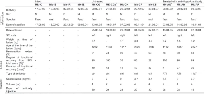
Table 1. Summary of the individual properties of each monkey
Intact Untreated Treated with anti-Nogo-A antibody
Mk-IC Mk-IE Mk-IR Mk-IZ Mk-CG MK-CGa3
Mk-CH Mk-CP Mk-CS Mk-AG3 Mk-AM Mk-AP General informa tion Birthday 17.07.99 15.06.96 02.02.04 12.05.96 20.02.01 21.05.03 20.02.01 22.12.97 30.04.97 28.03.02 20.02.01 09.03.98 Sex M M F M M M M F M M M F
Species Fasc mul Fasc Fasc fasc fasc fasc fasc mul fasc fasc fasc
Date of sacrifice 17.08.09 15.02.02 22.12.09 09.02.04 13.01.05 19.01.07 07.02.05 09.11.04 21.09.01 03.08.05 14.02.05 16.11.04
Lesion
Date of lesion - - - - 25.08.04 16.08.06 29.09.04 04.05.04 07.03.01 13.04.05 29.09.04 02.06.04
SCI side - - - - left right right left left left right left
Weight at time of
lesion (kg) - - - - 5.1 - 4.1 3.8 4.0 3.7 4.5 4.2
Age at the time of the
lesion (days) - - - - 1282 1183 1317 2325 1407 1112 1317 2277
Hemisection extent
(%) - - - - 51 73 90 45 63 78 80 58
Degree of functional recovery from SCI,
total score (%)1 - - - - 90 100 53 83 22 100 96 99
Duration of functional
recovery (days)2 - - - - 49 43 41 49 47 7 27 36
Treatment
Type of antibody - - - - ctrl ctrl ctrl ctrl ctrl ATI ATI 11c7
Cocentration (mg/ml) - - - - 9 7 9 3.7 3.7 3.6 9 3.7
Volume (ml) - - - - 4 2 4 4 4 2 4 4
Days of antibody
injection - - - - 30 29 28 29 32 28 28 15
M = Male; F = female; fasc = M. fascicularis, mul = M. mulatta and rhe = Rhesus. 1
Expressed in percentages of postlesion total score at plateau divided by pre-lesion total score in the modified Brinkman board task: all slots. 2 Time interval from the day of lesion to the beginning of postlesion plateau, as defined by Kaeser et al. 2011 (Kaeser et al., 2011). 3
Mk-CGa and Mk-AG suffered from a more caudal lesion than C7/C8. These monkeys were taken into acount in this study but with some reserves.
160
Behavior 161
As recently reported (Contestabile et al., 2018), manual dexterity was quantified in the 162
eight monkeys subjected to unilateral SCI, based on the “modified Brinkman board” task, 163
testing the precision grip (opposition of thumb and index finger), needed to grasp small food 164
pellets in 25 vertically oriented slots and 25 horizontally oriented slots, randomly positioned 165
over a Perspex board (Schmidlin et al., 2011). After habituation to the housing facility and 166
transfer to a primate chair using positive reinforcement (see 167
http://www.unifr.ch/neuro/rouiller/home/nhp), the macaques monkeys were progressively 168
9 trained daily to perform the “modified Brinkman board” task, separately for each hand, until 169
they reached a pre-lesion plateau of performance. The duration of the training period and the 170
plateau level of performance was variable across monkeys, as reported earlier based on a 171
large cohort of animals (Kaeser et al., 2014). Then, at plateau, the monkeys were tested 172
daily, once for each hand, during a pre-lesion phase of several weeks in order to establish a 173
score of manual dexterity of reference in the intact monkey. The same behavioral test 174
procedure was pursued also daily after the SCI, without additional training besides the test 175
itself. Based on the post-lesion tests, it was possible to assess the progressive functional 176
recovery from the SCI and the performance level of the post-lesion plateau. 177
The dramatic drop of score (usually to zero) immediately after the lesion and the 178
deficits of precision grip following such cervical lesion have been reported in detail earlier 179
(Freund et al., 2006, 2007, 2009; Hoogewoud et al., 2013) and are summarized in Figure 1D. 180
After unilateral C7 injury, a progressive though incomplete functional recovery was observed 181
until reaching a plateau of motor performance a few weeks after the SCI (Freund et al., 2006, 182
2007, 2009; Hoogewoud et al., 2013) (Fig. 1D). The behavioral parameter of interest here 183
was the percentage of functional recovery for the ipsilesional hand, given by the average 184
total score (number of pellets retrieved from both vertical and horizontal wells in 30 s) at 185
post-lesion plateau divided by the average total score at pre-lesion plateau (Freund et al., 186
2006, 2007, 2009; Hoogewoud et al., 2013) (Fig. 1D). Moreover, the plots of manual 187
dexterity scores as a function of time were used to define the time duration until the post-188
lesion plateau was reached (duration of functional recovery) (Fig. 1D). 189
190
Unilateral cervical cord lesion 191
The surgical procedure to perform the unilateral lesion was described in detail in 192
previous reports (Freund et al., 2006, 2007, 2009; Beaud et al., 2008, 2012; Hoogewoud et 193
al., 2013), an experimental procedure summarized below. Anesthesia was induced by an 194
intramuscular injection of ketamine (Ketalar; Parke-Davis; 5 mg/kg, i.m.) and atropine was 195
injected i.m. (0.05 mg/kg) to reduce bronchial secretions. Before surgery, the analgesic 196
10 Carprofen was delivered (Rymadil, 4 mg/kg, s.c.). Then, deep and stable anesthesia was 197
obtained via a continuous perfusion (0.1 ml/minute/kg) through an intravenous catheter 198
placed in the femoral vein of a mixture of 1% propofol (Fresenius) and a 4% glucose solution. 199
The monkey was positioned in a stereotaxic headholder in a ventral decubitus position, 200
allowing the spinal processes from C2 to Th1 to be exposed, preceding a complete C6 201
laminectomy and an upper C7 hemi-laminectomy. The dorsal root entry zone at the C7/C8 202
border was then identified, providing a medial landmark for placing a surgical blade (No. 11; 203
Paragon), which was inserted vertically 4 mm in depth to generate an incomplete section of 204
the cervical cord at C7 level. In most cases, such a section completely interrupted the CS 205
tract in the dorsolateral funiculus unilaterally. Following the SCI, the monkey was kept alone 206
in a separate cage for a couple of days in order to monitor its health condition and provide 207
specific post-operative care (antibiotics, analgesics,). 208
209
Histology and neuroanatomical material for analysis 210
The general histological and analysis methods are similar to those recently reported 211
(Contestabile et al., 2018) and repeated in a shorter version below. After euthanasia, the 212
spinal cord segment (C3-T4) comprising the SCI was cut parasagittally in either three or five 213
series of respectively 50 or 30 Pm sections and processed to visualize either BDA, Nissl or 214
SMI- 32 staining (Fig. 1A-C). The extent of the cervical cord lesion was measured in 215
consecutive longitudinal sections (scar tissue and absence of neuronal staining) and 216
transposed in a transversal reconstruction to assess the percentage of lesion with respect to 217
the entire corresponding hemi-cervical cord (white and grey matter). 218
In all monkeys (SCI and intact), the brain was cut in the frontal plane into 50 Pm-thick 219
frozen sections with both hemispheres facing each other on the same slide (Fig. 2A). 220
Sections were collected in 5 or 8 consecutive series, one of them immunoreacted to visualize 221
the marker SMI-32 (Sternberger and Sternberger, 1983), as previously reported (Liu et al., 222
2002; Wannier et al., 2005; Beaud et al., 2008). Frontal brain sections were examined under 223
bright-field illumination (at a total magnification of 200x). Brain regions of interest (mostly 224
11 SMA) were vectorised using Neurolucida 9 (MBF Bioscience) with a computer-interfaced 225
Olympus BX40 microscope (Olympus Schweiz AG), a computer-controlled motorized stage 226
(Märzhäuser Wetzlar GmbH, type EK 32 75 50) and a digital camera (Olympus U-PMTVC). 227
The Photomicrographs captured with a digital camera were processed using the CorelDraw 228
software (color, brightness, and contrast were not modified) and then quantification was 229
conducted using the software Neuroexplorer (MBF Bioscience) (example in Fig. 2B-D). At 230
that step, the investigator of the histological material was blinded against information on 231
animal group (intact versus SCI) and side of the cervical cord hemisection. The size of the 232
SCI expressed as a percentage of hemisection extent were reported previously (Freund et 233
al., 2006, 2007, 2009; Beaud et al., 2008, 2012; Hoogewoud et al., 2013) and are reminded 234
in Table 1. SMA and its two subdivisions (F3 and F6, respectively corresponding to SMA-235
proper and pre-SMA) in both hemispheres was delineated from M1 or PM laterally and from 236
cingulate motor area (CMA) ventrally, based on cytoarchitectural landmarks (Liu et al., 2002). 237
SMI-32-positive neurons were counted in SMA layers V and the surface of the delineated F3 238
or F6 was calculated in relation with the defined limits. Based on an exhaustive plotting 239
method (Fregosi and Rouiller, 2017; Fregosi et al., 2017, 2018), the cellular density in layer V 240
of F3, respectively F6, was computed on individual sections. It is the number of identified 241
SMI-32-positive cells in that layer in one hemisphere divided by the corresponding volume of 242
SMA (given by the thickness of the section multiplied by the area-of-interest). The criteria 243
used to include a neuron were the same proposed in our previous article (Contestabile et al., 244
2018): (1) SMI-32-positive; (2) the soma, the nucleus or the nucleolus and at least four 245
proximal dendrites of the neuron have to be identifiable; and (3) the neuron is located in the 246
cortical layer V. The criteria to set the rostrocaudal limit between F3 and F6, as well as the 247
precise territories of F3 and F6 analyzed were the same as in Contestabile et al. 2018. 248
249
Interhemispheric difference of cell density (IDCD) 250
An interhemispheric difference of cell density (IDCD) in SMA layer V was computed in 251
each histological section by subtracting the SMI-32-positive neuron density in the ipsilesional 252
12 SMA (relatively intact side) from the SMI-32-positive neuron density in the contralesional 253
SMA (strongly connected with the SCI) (Fig. 3 B and D). Positive IDCD means that more 254
SMI-32 neurons were found in SMA strongly connected to the side where the spinal cord 255
injury was performed, whereas negative IDCD corresponds to a larger number of SMI-32 256
neurons in layer V of SMA homolateral to the SCI. In intact monkeys, IDCD was expected to 257
be close to zero whereas, in monkeys subjected to SCI, the IDCD was hypothesized to 258
diverge from zero (see Fig. 5A, e.g. gray line). As for our previous work (Contestabile et al., 259
2018), the interhemispheric comparison was made on each individual section, so that the 260
mirrored SMA territories analyzed on each side had comparable area and position. 261
262
Single SMI-32 labelled neuron analysis 263
To investigate whether the unilateral SCI impacted on the microstructure of single 264
SMI-32 positive neurons in SMA layer V, in particular on their dendritic trees, a so-called 265
Sholl analysis for performed, identical to the one reported recently (Contestabile et al., 2018). 266
The sampling and inclusion criteria were similar: three SMI-32- positive pyramidal neurons of 267
layer V per hemisphere per section were analyzed in four different sections (12 neurons per 268
hemisphere in total) in each of five representative monkeys: two intact monkeys and three 269
monkeys with SCI. The following criteria were applied to specifically select SMI-32-positive 270
neurons: (1) to be representative of the entire region-of-interest, one neuron was picked in 271
each of the dorsal, middle, and ventral parts of the medial wall in F3; and (2) at least intact 272
primary and secondary dendrites and an apical dendrite reaching the layer III without 273
interruption had to be clearly identified. Two distinct regions of the dendritic tree of each SMI-274
32 positive neurons were analyzed: the basal dendrites excluding the axon and the apical 275 dendrite. 276 277 Statistics 278
Statistical analysis was conducted with GraphPad Prism 7 (San Diego, CA, USA). 279
The normality of sample distributions was assessed with the Shapiro–Wilk criterion. In each 280
13 monkey, the statistical significance of IDCDs of SMI-32-positive neurons between the 281
ipsilesional and contralesional hemispheres was assessed using a paired t-test or a 282
Wilcoxon test (according to the data distribution) as the neuronal density was directly 283
compared across the two hemispheres on the same section. In order to compare the 284
interhemispheric morphological data and the results of the Sholl analysis, a two-way RM 285
ANOVA with Bonferroni’s posttest correction were used (*: p 0.05, **: p 0.01, ***: p 0.001, 286
****: p 0.0001). Data are represented as the mean ± s.e.m. and the significance was set at 287
95% of confidence. 288
14
Results
289
Based on a previously available case ((Rouiller, 1996); intact monkey 93-81), 290
subjected to a unilateral injection of the anterograde tracer BDA in the SMA (F3, focused to 291
the hand area), the bilateral distribution of CS axons was established at cervical level. The 292
majority of CS projection originating from SMA crossed the midline (79.7%) whereas only 293
20.3% of the BDA positive CS axons were located on the ipsilateral side of the cervical cord 294
(11.7% in the dorsolateral part and 8.6% in the ventromedial part) (Fig. 1A and B). These 295
results suggest that a unilateral cervical cord injury (SCI, Fig. 1C) retrogradely impacts more 296
on the contralesional SMA than on the ipsilesional one (connectional diaschisis). 297
As previously reported (Freund et al., 2006, 2007, 2009; Beaud et al., 2008, 2012; 298
Hoogewoud et al., 2013), eight macaques monkeys (see Table 1) were unilaterally injured at 299
C7 level (SCI animals: Mk-CG, Mk-CGa, Mk-CH, Mk-CP and Mk-CS; SCI + Anti-Nogo-A 300
animals: Mk-AG, Mk-AM and Mk-AP) and the manual dexterity was quantified using the 301
“modified Brinkman board” task. After unilateral C7 injury, the ipsilesional manual dexterity 302
was severely affected, followed by a progressive though incomplete functional recovery, 303
reaching a plateau of motor performance a few weeks after the SCI (Fig. 1D). Thanks to this 304
longitudinal quantification of the manual dexterity, it was possible to calculate the extent of 305
functional recovery (in %) of the ipsilesional hand and the time duration needed to reach the 306
post-lesional plateau (duration of functional recovery) (see Table 1 and Fig. 1D). 307
308
Neuronal density in the layer V of SMA after a SCI 309
In order to quantify the effect of a unilateral SCI (Fig. 1C) on SMA layer V pyramidal 310
neurons, SMI-32 labelled neurons in the bilateral F3 (SMA-proper) and F6 (pre-SMA) were 311
analyzed histologically in eight macaque monkeys subjected to cervical hemi-section and 312
compared to intact monkeys (n = 4). First, we quantified the density of SMI-32 positive layer 313
V neurons in the delimited F3 and F6 areas in both hemispheres along the rostro-caudal axis 314
in coronal sections (Fig. 2A–D). In F6 and F3 layer V, the SMI-32 positive neurons’ densities 315
15 ranged approximately from 50 cells/mm3 to 250 cells/mm3 across
316
sections/hemispheres/monkeys in the intact group, whereas in the lesioned monkeys, SMI-317
32-positive neurons’ densities in SMA layer V ranged approximately from 100 to 700 318
cells/mm3 (Fig. 2E). With respect to those ranges of cellular densities, in the SCI group, there
319
is no obvious difference between the anti-Nogo-A antibody treated monkeys (n = 3) and the 320
control antibody treated animals (n = 5). In addition, the range of SMI-32-positive neurons’ 321
densities in the SMA of SCI animals seemed to reach higher values than the intact group (in 322
fact higher than 3 intact monkeys with low cellular densities in F3, whereas the fourth intact 323
monkey exhibits a cellular density comparable to 5 monkeys of the SCI group). However, the 324
very large interindividual variability of the histological staining quality precludes interindividual 325
comparison between the 2 groups of monkeys and strongly affects the absolute 326
quantification of neuronal density. The quality of SMI-32 staining in 3 of the 4 intact monkeys 327
may have been different than in the SCI monkeys, leading to the differences in the absolute 328
numbers of SMI-32 positive neurons between the two groups. On the other hand, the quality 329
of staining does not interfere when the comparison is intraindividual and furthermore 330
restricted to a single histological section. For this reason, we performed a direct comparison 331
between the hemispheres (left hemisphere compared to the corresponding right hemisphere 332
on the same section). 333
334
Interhemispheric difference of cell density (IDCD) 335
As expected, intact monkeys presented no significant difference in their IDCDs for the 336
32 positive neurons in layer V of F3 (Fig. 3A-B). On the other hand, the IDCDs of SMI-337
32 positive neurons were significantly different in six out of eight monkeys subjected to SCI 338
(Fig. 3B). However, the IDCD was not systematically biased toward the same hemisphere 339
(ipsilesional versus contralesional; Fig. 3B). Four lesioned monkeys (CG, CGa, Mk-340
CP and Mk-AP) exhibited a significantly higher contralesional density of SMI-32 positive 341
neurons in F3 layer V, corresponding to a positive IDCD, while two SCI monkeys (Mk-AG 342
16 and Mk-AM) had a significantly higher ipsilesional density of SMI-32 positive neurons in F3 343
layer V, corresponding to a negative IDCD (Fig. 3A-B). 344
In order to have an internal control, the cellular density of SMI-32 positive neurons 345
was also quantified in the layer V of F6 (pre-SMA), a motor cortical area lacking corticospinal 346
neurons (Luppino et al., 1994). The assessment of cellular density in F6 layer V showed no 347
significant IDCD, both in intact animals and in SCI animals (Fig. 3C-D). This observation 348
indicates that SMI-32 positive neurons in F6 layer V were not affected by the SCI, as 349
expected. 350
Finally, we did not observe significant difference between the calculated IDCD 351
between intact and injured animals neither in F3 nor in F6 (Fig. 3B’ and D’). 352
353
Arborization of layer V SMI-32 positive neurons in F3 after SCI 354
Microstructural changes of the basal (Fig. 4A) and apical dendritic (Fig. 4G) 355
arborization of SMI-32 positive neurons located in layer V of F3 was assessed based on a 356
Sholl analysis conducted in two control animals (Mk-IR and Mk-IE) and in three 357
representative lesioned monkeys (Mk-CGa, Mk-CP and Mk-AG). For all five animals, the 358
Sholl analysis yielded a classical inverted U-shape curve with a tail on the right, 359
corresponding to an increase of dendritic intersection numbers going away from the soma up 360
to a peak, followed by a comparable progressive decrease ending with slower fading at 361
larger distances from the soma. In most analyzed monkeys, the number of dendritic 362
intersections peaked at a distance of about 50 Pm from the soma, both for basal and apical 363
dendrites (Fig. 4). As excepted, no interhemispheric difference was observed in the 2 control 364
monkeys for both the basal (Fig. 4B-C) and apical dendrites (Fig. 4H-I). After SCI, 2 out of 3 365
monkeys exhibited in F3 interhemispheric differences in the basal dendritic arborization (Fig. 366
4D-F). In fact, for Mk-CP and Mk-AG we observed the same interhemispheric bias consistent 367
with the IDCD bias already observed for layer V in F3 (Fig. 3B, 4E-F) for the basal dendrites. 368
A comparable tendency (not statistically significant) was observed for Mk-CGa, the third 369
representative lesioned animal. This significant interhemispheric difference of basal dendritic 370
17 arborizations was verified within a limited distance range from the soma, going from 40 to 80 371
Pm. (Fig. 4E-F). Interestingly, and in contrast to the basal dendrites, the apical dendrites 372
exhibited clearly less pronounced interhemispheric difference in their numbers of dendritic 373
intersections (Fig. 4J-L). 374
375
Relationship of IDCDs in F3 with percentage of hemi-section extent, duration and 376
extent of functional recovery after SCI 377
Are the morphological interhemispheric changes on SMI-32 positive neurons in F3 378
layer V induced by SCI (Figs. 2-3) related to the characteristics of the lesion (size) and/or the 379
properties of functional recovery? To this aim, IDCDs of SMI-32 neurons in F3 were plotted 380
as a function of lesion extent (percentage of hemi-section), durations of functional recovery 381
and extent in percent of functional recovery (Fig. 5A-C). Interestingly, IDCDs in F3 layer V 382
were significantly inversely correlated with the size of the SCI, expressed as the percentage 383
of hemi-section (Fig. 5A; R = - 0.857, p = 0.006), independently of the presence/absence of 384
anti-Nogo-A antibody treatment. A similar inverse correlation was found between IDCDs and 385
lesion size in F3 after unilateral M1 lesion (Contestabile et al., 2018). In both cases, these 386
correlation data indicate that a small lesion (ipsilateral in the case of cortical lesion or 387
contralateral in case of SCI) is associated with a largely positive IDCD whereas large lesions 388
are associated to negative IDCDs. 389
The relationship between the median IDCD values and the duration of functional 390
recovery from SCI (Fig. 5B) was then investigated, based on behavioral data derived from 391
the modified Brinkman board task: the IDCDs in F3 layer V were correlated with the duration 392
of functional recovery (Fig. 5B; R = 0.734, p = 0.038), again independently of the 393
presence/absence of treatment. Interestingly, there was no statistically significant correlation 394
between the duration of functional recovery from SCI and the hemi-section extent of the 395
spinal cord (Fig. 5C; R = - 0.525, p = 0.181). However, the 3 SCI monkeys which received 396
the anti-Nogo-A antibody treatment recovered faster (shorter time to reach the post-lesional 397
plateau) than the control antibody treated monkeys, a difference already visible in panel B of 398
18 Figure 5. In contrast to the parameters lesion extent and duration of functional recovery, 399
IDCDs in F3 layer V were not correlated with the parameter extent of functional recovery 400
(expressed in %) (Fig. 5D; R = 0.054, p = 0.889), assessed with the total score of pellets 401
retrieval in the modified Brinkman board task. Finally, as observed in Contestabile et al. 402
2018, no correlation was observed between the percentage of functional recovery in the 403
modified Brinkman board task and duration of functional recovery (Fig. 5E; R = 0.176, p = 404
0.301) suggesting that a longer duration of functional recovery did not mean a better 405
functional recovery. 406
19
Discussion
407
The major conclusion of the present study is that a unilateral cervical cord hemi-408
section retrogradely induced an interhemispheric asymmetry of SMI-32 staining in F3 layer V 409
in non-human primates, which is correlated with the extent of the SCI as well as the duration 410
of functional recovery, but not with the extent (percentage) of the functional recovery. These 411
conclusions are coherent with the observations described recently by our laboratory showing 412
similar results in F3 layer V in SMA after a unilateral M1 lesion (Contestabile et al., 2018). A 413
coherent correlation between IDCDs and lesion size as well as duration of functional 414
recovery (but not extent of recovery) were observed in this study. In other words, the 415
interhemispheric effects on SMI-32 staining in F3 layer V observed after M1 lesion 416
(Contestabile et al., 2018) can be generalized to another type of unilateral lesion, namely a 417
cervical cord hemi-section. If the morphological interhemispheric changes reflect variable 418
balanced contributions of the two hemispheres in the process of functional recovery, then 419
comparable mechanisms of functional recovery at cortical level may take place, irrespective 420
of the site of lesion (cortical or spinal), at least for injury restricted to one side of the brain or 421
of the spinal cord. 422
The lateralization of the unilateral lesion, either in M1 or at cervical cord level, is of 423
importance. The computation formula to establish the IDCD was chosen to be reversed in 424
the present study (cervical cord lesion) as compared to our recent report on unilateral M1 425
lesion (Contestabile et al., 2018). The reason for such reversal is that the unilateral M1 lesion 426
was on the same hemisphere as the F3 area, which exhibits (before the lesion) the 427
quantitatively predominant corticocortical connection with the injured M1, whereas the 428
transcortical connection from the opposite F3 is less strong (see (Contestabile et al., 2018); 429
their Fig. 1). In case of cervical cord hemi-section, the predominant corticospinal projection 430
from F3 is crossed (originating mainly from the opposite hemisphere), reason why the IDCD 431
formula was reversed (see methods above). Due to this reversal, then the correlation plots in 432
the M1 lesion model ((Contestabile et al., 2018); their Fig. 5) and in the present study (Fig. 5) 433
20 should appear similar, if the same correlations are verified. This is indeed the case for the 434
correlation between IDCDs and the lesion extent (compare Fig. 5B of (Contestabile et al., 435
2018) and the present Fig. 5A). In contrast, although there is a correlation in both lesion 436
models between IDCDs and duration of functional recovery, the correlation was inverse in 437
the M1 lesion model ((Contestabile et al., 2018); their Fig. 5F) and positive in the present SCI 438
model (Fig. 5B). As argued above, due to the reversal of the IDCD formula, we would have 439
intuitively expected a similar correlation direction. This is not the case, suggesting that the 440
mechanisms of recovery reflected by the correlation between IDCD and duration of functional 441
recovery may be independent of the lateralization of the unilateral lesion, either cortical or 442 cervical. 443 444 Connectional diaschisis 445
The results obtained in Contestabile et al. (2018), and in this study could be linked 446
with the old concept of diaschisis (von Monakow, 1914), defined as a “loss of function and 447
electrical activity in an area of the brain because of a lesion in a remote area that is 448
neuronally connected with it”, although it corresponds more to a “connectional diaschisis” 449
(Carrera and Tononi, 2014; see introduction above). The unilateral SCI may have affected 450
the morphology, phenotype and cellular expression of layer V neurons in the contralesional 451
hemisphere in a different manner with respect to the neurons localized in the ipsilesional 452
hemisphere. This interhemispheric difference in phenotype could have influenced the affinity 453
for the SMI-32 antibody and explain the observed interhemispheric asymmetry of density of 454
SMI-32-positive layer V neurons. This interpretation is moreover supported by the neuronal 455
reconstruction. In fact, the directions of interhemispheric differences in dendritic arborization 456
complexity (Fig. 4) were consistent with the direction of the IDCD especially in the 457
arborization of the basal dendrites which are more implicated in the integration of the 458
neuronal response and have a strong effect on action potential output due to their direct 459
attachment to the cell body and the proximity to the axon (Nevian et al., 2007; Zhou et al., 460
2008). The changes of dendritic arborization following spinal cord injury can be linked to the 461
21 recently published observation depicting that axotomy of peripheral motor projections induce 462
changes in the dendritic arborization of M1 pyramidal neurons in the rodent model submitted 463
to a permanent lesion of the facial nerve (Urrego, Troncoso and Múnera, 2015). In another 464
recent study aiming at measuring the structural changes in the CNS of human patients 465
suffering from degenerative pathology affecting interaction between cortical motoneurons 466
and spinal motoneurons such as Amyotrophic Lateral Sclerosis (ALS) or dementia, the 467
authors observed a degeneration of the apical dendrites of pyramidal neurons located in 468
layer V of M1 (Betz’s cells as described in the paper), to a larger extent in patients suffering 469
from ALS (Genç et al., 2017). These results are consistent with a lower dendritic arborisation 470
observed in the contralesional SMA (fig. 4 G, L) in animals having a larger extent of SCI, as 471
ALS is a major degenerative disease affecting a majority of motoneurons in the ventral horn 472
of the spinal cord. 473
Interestingly, it seems that anti-Nogo-A antibody treatment does not have an effect on 474
the observed interhemispheric difference in phenotype. The three treated animals showed 475
results that are comparable to only lesioned animals. Neutralization of Nogo-A was found to 476
promote sprouting of corticospinal axons in macaque monkeys and to lead to a complete 477
recovery of the manual dexterity after a unilateral hemisection lesion irrespectively on the 478
lesion extent (Freund et al., 2006, 2007, 2009). However, it is possible that anti-Nogo-A 479
antibody treatment could not preserve the phenotype of F3 layer V pyramidal neurons since 480
the SCI immediately affect the neuronal connection and altered the phenotype of the 481
corticospinal neurons in an irreversible manner. 482
483
A possible role of the ipsilesional hemisphere during the functional recovery from SCI 484
The correlation between IDCD asymmetry and the duration of functional recovery is 485
also reminiscent of the controversy related to the respective contributions of the ipsilesional 486
versus contralesional hemispheres in the functional recovery following a unilateral SCI. 487
Previous studies in rodents (Ghosh et al., 2009) and humans (Lundell et al., 2011) 488
demonstrated that the ipsilesional hemisphere underwent a profound reorganization of the 489
22 motor areas after cervical SCI. Moreover, these previous findings suggest a correlation 490
between brain activity, duration and extent of functional recovery and are consistent with our 491
results. In fact, they revealed that monkeys with larger lesion tend to show shorter duration of 492
functional recovery and negative IDCD suggesting a more prominent reorganization and a 493
predominant contribution of the ipsilesional hemisphere (as compared to the contralesional 494
one), immediately after severe SCI. On the other hand, the data also show that a more 495
moderate lesion leads to a higher SMI-32 positive neurons density in the contralesional 496
hemisphere suggesting a more important reorganization of the hemisphere predominantly 497
affected by the spinal hemisection. Interestingly, previous studies reported an upregulation of 498
the expression of Sema3A and NRP-1 in motoneurons located in the contralesional 499
hemisphere after SCI in rats (De Winter et al., 2002; Hashimoto et al., 2004). It is likely that 500
neurons whose descending fibres were transected upregulated the production of proteins 501
involved in the regulation of axonal re-growth and maintenance. This phenomenon may 502
possibly be correlated with the higher density of neurons in the contralesional hemisphere 503
that express SMI-32, a major component of the neuronal cytoskeleton providing structural 504
support to the axon. However, in a recent study based on meta-analysis of fMRI data 505
assessing the possible reorganisation of different regions of the cerebral cortex after spinal 506
cord injury in human patients, the authors showed changes in bilateral SMA, but were unable 507
to specify the type of change or potential role in the functional recovery (Wang et al., 2019). 508
509
Limitations 510
The present study involves a limited number of monkeys subjected to SCI (n=8), 5 of 511
them received a control antibody, while 3 monkeys were subjected to anti-Nogo-A antibody 512
treatment, as one may reasonably expect from a non-human primate study, mostly for ethical 513
reasons. However, in spite of the limited number of monkeys, the data are internally coherent 514
(e.g. F3 versus F6) and statistically significant correlations of IDCDs with lesion extent and 515
duration of functional recovery emerged (Fig. 5). Furthermore, these correlation data are fully 516
23 consistent with previous data derived from another pool of monkeys (n=9) subjected to 517
another type of unilateral lesion (M1) affecting the CS projection (Contestabile et al., 2018). 518
An important limitation of this study is due to the impossibility to compare the absolute 519
values of SMI-32-positive neurons’ densities across animals. Although the raw data seems to 520
show a higher density of SMI-32 neurons in both hemispheres of SCI monkeys as compared 521
to intact animals, such interindividual comparison between the 2 groups of monkeys is 522
problematic due to a very large interindividual variability of the histological staining quality, 523
strongly affecting the absolute quantification of neuronal density. The quality of SMI-32 524
staining in 3 of the 4 intact monkeys may have been different (lower) than in the SCI 525
monkeys, leading to the differences in the absolute numbers of SMI-32 positive neurons 526
between the two groups. In one of the intact monkeys (Mk-IC), the absolute cellular density is 527
comparable to that of 5 of the SCI monkeys, corresponding thus to a partial overlap between 528
the 2 groups (which are not statistically different with respect to their IDCDs in F3 and F6 529
(Fig. 3B and D). On the other hand, the quality of staining does not interfere when the 530
comparison is intraindividual and furthermore restricted to a single histological section (left 531
hemisphere compared to the corresponding right hemisphere on the same section, both 532
hemispheres processed histologically in the very same way and at the same time point). 533
Further limitations were related to the impossibility in some monkeys to perform the 534
cell counting and consecutive IDCD analysis in F6 because of the lack of brain tissue. 535
Finally, the histological condition of some SMI-32 sections was not optimal to perform the 536
neuronal dendritic reconstruction and for this reason the morphological sholl analysis was 537
limited to 5 animals (Fig. 4). 538
539
Conclusion 540
Altogether, the current study on SCI and the recent investigation on M1 lesion (Contestabile 541
et al., 2018) both show that a unilateral lesion affecting the CS projection system, either at its 542
origin (in M1) or along its trajectory (at cervical level) results in a variable and adaptable 543
interhemispheric balance of F3 layer V pyramidal neurons, detected with the marker SMI-32, 544
24 in a manner which is systematically correlated with the extent of the lesion as well as the 545
duration of the functional recovery of manual dexterity. These observations suggest that the 546
controversy on which hemisphere (ipsilesional versus contralesional) is involved in the 547
functional recovery may be resolved by considering a contribution of both, however in an 548
adaptable and finely tuned balance depending on the lesion properties (e.g. extent) and 549
evolving with time during the recovery, the duration of the latter being a crucial factor in this 550
process. 551
25
Acknowledgements
552
The authors thank the excellent technical assistance of Mrs Véronique Moret, 553
Christine Roulin, and Christiane Marti (histology), of Mr. Laurent Bossy, Joseph Corpataux 554
and Jacques Maillard (animal care taking), André Gaillard (mechanics), Bernard Aebischer 555
and Andrea Francovich (electronics), and Laurent Monney (informatics). 556
This work was supported by: Swiss National Science Foundation, Grants No 31-557
61857.00, 310000-110005, 31003A-132465, 310030B-149643 (EMR), No 320030-160229 558
(ES), the National Centre of Competence in Research (NCCR) on “Neural plasticity and 559
repair”; Novartis Foundation; The Christopher Reeves Foundation (Springfield, NJ, USA); 560
The Swiss Primate Competence Centre for Research (SPCCR: ). 561
Conflict of interest: the anti-Nogo-A antibody was provided by Novartis Pharma AG. 562
563
Authors’ contribution
564
A.C. and R.C. performed most of the analysis of the histological data. M.L. and A.C. 565
contributed to the Sholl analysis. A.C., E.M.R. and E.S. prepared the figures and wrote the 566
manuscript. E.M.R. and E.S. coordinated all aspects of the work. E.S. designed and 567
supervised the morphological analysis; EMR designed and coordinated the overall 568
behavioural and lesional study. 569
26 Captions to figures
571
Figure 1- Corticospinal projection of SMA. 572
(A) Upper panel: schematic representation of a macaque brain showing the location of pre-573
SMA (area F6), SMA-proper (area F3) and M1. Lower panel: schematic representation of the 574
corticospinal projections of F6 (bilateral). (B) Lower panel: upper cervical spinal cord section 575
showing the localization of CS axons labelled by a unilateral BDA injection in the SMA. 576
Upper panel: Pie chart reporting the percentage of BDA positive CS axons present in the 577
ipsilateral ventromedial, ipsilateral dorsolateral and contralateral dorsomedial part of the 578
spinal cord. The majority of BDA positive CS axons are located in the contralateral 579
dorsolateral part of the spinal cord. (C) Photomicrographs of sagittal histological sections of 580
the spinal cord of a lesioned animal (Mk-CS) showing the induced permanent lesion at the 581
C7/C8 level. The histological sections derived from two different series processed to 582
visualize SMI-32 staining (left) or Nissl staining (right). Scale bar: 500 μm. (D) Graphical 583
representation of behavioral performance in the modified Brinkman board task of the 584
macaque monkeys included in this study (SCI in green and SCI + anti-Nogo-A treatment in 585
dark grey). The manual performance of the ipsilesional hand is given by the score (number of 586
pellets retrieved in the first 30 s of the task from the randomly distributed wells) as a function 587
of time (days) before and after a SCI. Day 0 corresponds to the day of the lesion (vertical red 588
line). Pre and post-lesional average of the scores are indicated with horizontal grey lines. 589
The total duration of functional recovery of manual dexterity after lesion is given by the time 590
interval between the lesion (red vertical line) and the onset of the postlesion plateau (brown 591
area). Figure Contributions: EMR and ES performed and supervised the experiments on the 592
monkeys and analyzed the data. 593
594
Figure 2 - Neuronal density in the layer V of SMA in intact and SCI monkeys 595
(A) Photomicrograph of coronal brain histological section of an intact macaque monkey (Mk-596
IR) stained with SMI-32 and magnified in F3 (scale bar: 40 μm). Layer III and V are visible 597
and 32 positive pyramidal neurons are indicated with arrows. (B-D). Localization of SMI-598
27 32 positive layer V neurons in bilateral F3 and F6 areas of three representative macaque 599
monkeys (intact: Mk-IR, SCI: Mk-CG and SCI + anti-Nogo-A treatment: Mk-AM). 4 sections 600
per macaque monkeys are shown and the relative rostro-caudal position of the sections from 601
the F3-F6 border is reported in millimeters. Negative distance values belong to F6 and 602
positive distance values belong to F3. (E) Graphs representing the rostro-caudal gradient 603
(from F6 to F3) of SMI-32 positive cell density in layer V of all monkeys (intact: Mk-IC, Mk-IE, 604
Mk-IR and Mk-IZ; SCI: Mk-CG, Mk-CGa, Mk-CH, Mk-CP and Mk-CS; SCI + anti-Nogo-A 605
treatment: Mk-AG, Mk-AM and Mk-AP). The cell density for each hemisphere is plotted as a 606
function of the distance from the F3-F6 border, which has been set to 3 mm rostrally to the 607
genu of the arcuate sulcus. Negative distance values belong to F6 and positive distance 608
values belong to F3. The symbol # was used to indicate that the analyzed cortex region was 609
not complete (sections lacking for the analysis). Figure Contributions: EMR and ES 610
performed the experiments on the monkeys and generated the histological sections; AC and 611
RA performed the microscopic analysis of the histological sections; AC analyzed the data. 612
613
Figure 3 - Interhemispheric difference of cell density (IDCD) after SCI in F3 and F6 614
(A) Histograms reporting the cell density of layer V SMI-32 positive neurons in F3 for intact 615
(white background), SCI (light grey background) and SCI treated monkeys (dark grey 616
background). For each couple of histograms, are reported the side (L = left and R = right) 617
and the location in function of the SCI (Ipsi = ipsilesional and Contra = contralesional). As 618
statistical test, a paired t-test or Wilcoxon test was performed (p – values are reported) 619
comparing the cell density in the two hemispheres in each consecutive histological section. 620
(B) Histograms showing the IDCD of layer V SMI-32 positive neurons of F3 for intact (white 621
background), SCI (light grey background) and SCI treated monkeys (dark grey background). 622
A positive IDCD corresponds to an ipsilesional bias in pyramidal cell density while a negative 623
IDCD corresponds to a contralesional bias in pyramidal cell density. (B’ upper left inset) 624
Comparison of calculated IDCD in F3 between groups. No statistical difference is observed 625
(unpaired t-test: t(10) = 0.8276, p = 0.4272). (C) Histograms reporting the cell density of layer
28 V SMI-32 positive neurons in F6 for intact (white background), SCI (light grey background) 627
and SCI treated monkeys (dark grey background). For each couple of histograms, are 628
reported the side (L = left and R = right) and the location in function of the SCI (Ipsi = 629
ipsilesional and Contra = contralesional). As statistical test, a paired t-test or Wilcoxon test 630
was performed (p – values are reported) comparing the cell density in the two hemispheres 631
in each consecutive histological section. (D) Histograms showing the IDCD of layer V SMI-32 632
positive neurons of F6 for intact (white background), SCI (light grey background) and SCI 633
treated monkeys (dark grey background). (D’ upper left inset) Comparison of calculated 634
IDCD in F6 between groups. No statistical difference is observed (unpaired t-test: t(7) = 1.084,
635
p = 0.3142). For A and C, the median and the interquartile range are indicated. For B and D, 636
the mean ± SD are reported. Abbreviations: L = left hemisphere, R = right hemisphere, *:p 637
⩽ 0.05, **: p ⩽ 0.01). Figure Contributions: EMR and ES performed the experiments on the 638
monkeys and generated the histological sections; AC and RA performed the microscopic 639
analysis of the histological sections; AC analyzed the data. 640
641
Figure 4 - Arborization of layer V SMI-32 positive neurons in F3 after a SCI 642
(A) Neuronal reconstruction example of basal dendrites of a layer V SMI-32 positive neuron. 643
(B - F) Sholl profiles of basal dendrites of layer V SMI-32-positive neurons in each 644
hemisphere in two intact monkeys (A-B) and in three SCI monkeys (D-F). For each monkey, 645
the IDCD are reported in the upper right angle. (G) Neuronal reconstruction example of 646
apical dendrite of a layer V SMI-32 positive neuron. (H - L) Sholl profiles of apical dendrites 647
of layer V SMI-32-positive neurons in each hemisphere in two intact monkeys (H-I) and in 648
three SCI monkeys (J-L). The curves represent the mean intersection values ± SD. As 649
statistical test, a two-way ANOVA wit Bonferroni’s multiple comparison post test was 650
performed (*: p ⩽ 0.05, **: p ⩽ 0.01, ***: p ⩽ 0.001). The p - values for the distance to soma 651
main effect (up) and hemisphere main effect (right) are report for each monkey. # in the 652
upper right angle means a significative p – value for the interaction between the distance to 653
29 soma and the hemisphere main effect. Figure Contributions: AC and ML performed the 654
microscopic analysis of the histological sections; AC analyzed the data. 655
656
Figure 5 - Relationship of IDCD, proportion of hemisection extent, duration of recovery 657
and percentage of functional recovery 658
(A) The interhemispheric difference in cell density (IDCD) in F3 was plotted in function of the 659
proportion of hemisection extent. The gray line represents the hypothesized interhemispheric 660
cell density asymmetry (see introduction). (B) The interhemispheric difference in cell density 661
(IDCD) in F3 was plotted in function of the duration of functional recovery. (C) The duration of 662
functional recovery was plotted in function of the proportion of hemisection extent. (D) The 663
interhemispheric difference in cell density (IDCD) in F3 was plotted in function of the 664
functional recovery. (E) The functional recovery of the animals was plotted in function of the 665
duration of functional recovery. Intact (in white), SCI (light gray) and SCI treated (dark gray) 666
animals are divided by color-code. In each graph, the R and the p – value of the linear 667
regression are reported. Figure Contributions: AC, RA, EMR and ES performed the 668
experiments and collected the data. AC analyzed the data. 669
670
30 References
671
Beaud, M. L. et al. (2008) ‘Anti-Nogo-A antibody treatment does not prevent cell body 672
shrinkage in the motor cortex in adult monkeys subjected to unilateral cervical cord lesion’, 673
BMC Neuroscience. doi: 10.1186/1471-2202-9-5. 674
Beaud, M. L. et al. (2012) ‘Invasion of lesion territory by regenerating fibers after spinal cord 675
injury in adult macaque monkeys’, Neuroscience. doi: 10.1016/j.neuroscience.2012.09.052. 676
Boudrias, M. H. et al. (2010) ‘Forelimb muscle representations and output properties of motor 677
areas in the mesial wall of rhesus macaques’, Cerebral Cortex. doi: 10.1093/cercor/bhp136. 678
Carrera, E. and Tononi, G. (2014) ‘Diaschisis: Past, present, future’, Brain. doi: 679
10.1093/brain/awu101. 680
Chao, Z. C. et al. (2019) ‘Dynamic Reorganization of Motor Networks During Recovery from 681
Partial Spinal Cord Injury in Monkeys’, Cerebral Cortex. doi: 10.1093/cercor/bhy172. 682
Contestabile, A. et al. (2018) ‘Asymmetric and Distant Effects of a Unilateral Lesion of the 683
Primary Motor Cortex on the Bilateral Supplementary Motor Areas in Adult Macaque 684
Monkeys’, The Journal of Neuroscience. doi: 10.1523/jneurosci.0904-18.2018. 685
Courtine, G. et al. (2007) ‘Stance- and Locomotion-Dependent Processing of Vibration-686
Induced Proprioceptive Inflow From Multiple Muscles in Humans’, Journal of 687
Neurophysiology. doi: 10.1152/jn.00764.2006. 688
Dancause, N. et al. (2005) ‘Extensive cortical rewiring after brain injury.’, The Journal of 689
neuroscience : the official journal of the Society for Neuroscience. doi: 690
10.1523/JNEUROSCI.3256-05.2005. 691
Dum, R. P. and Strick, P. L. (1996) ‘Spinal cord terminations of the medial wall motor areas 692
in macaque monkeys.’, The Journal of neuroscience : the official journal of the Society for 693
Neuroscience. 694
Finger, S., Koehler, P. J. and Jagella, C. (2004) ‘The Monakow Concept of Diaschisis: 695
Origins and Perspectives’, Archives of Neurology. doi: 10.1001/archneur.61.2.283. 696
Fregosi, M. et al. (2017) ‘Corticobulbar projections from distinct motor cortical areas to the 697
reticular formation in macaque monkeys’, European Journal of Neuroscience. doi: 698
31 10.1111/ejn.13576.
699
Fregosi, M. et al. (2018) ‘Changes of motor corticobulbar projections following different lesion 700
types affecting the central nervous system in adult macaque monkeys’, European Journal of 701
Neuroscience. doi: 10.1111/ejn.14074. 702
Fregosi, M. and Rouiller, E. M. (2017) ‘Ipsilateral corticotectal projections from the primary, 703
premotor and supplementary motor cortical areas in adult macaque monkeys: a quantitative 704
anterograde tracing study’, European Journal of Neuroscience. doi: 10.1111/ejn.13709. 705
Freund, P. et al. (2006) ‘Nogo-A-specific antibody treatment enhances sprouting and 706
functional recovery after cervical lesion in adult primates’, Nature Medicine. doi: 707
10.1038/nm1436. 708
Freund, P. et al. (2007) ‘Anti-Nogo-A antibody treatment enhances sprouting of corticospinal 709
axons rostral to a unilateral cervical spinal cord lesion in adult macaque monkey’, Journal of 710
Comparative Neurology. doi: 10.1002/cne.21321. 711
Freund, P. et al. (2009) ‘Anti-Nogo-A antibody treatment promotes recovery of manual 712
dexterity after unilateral cervical lesion in adult primates - Re-examination and extension of 713
behavioral data’, European Journal of Neuroscience. doi: 10.1111/j.1460-714
9568.2009.06642.x. 715
Galea, M. P. and Darian-Smith, I. (1997) ‘Manual dexterity and corticospinal connectivity 716
following unilateral section of the cervical spinal cord in the macaque monkey’, Journal of 717
Comparative Neurology. doi: 10.1002/(SICI)1096-9861(19970512)381:3<307::AID-718
CNE4>3.0.CO;2-6. 719
Genç, B. et al. (2017) ‘Apical dendrite degeneration, a novel cellular pathology for Betz cells 720
in ALS’, Scientific Reports. doi: 10.1038/srep41765. 721
Geyer, S. et al. (2000) ‘Neurofilament protein distribution in the macaque monkey 722
dorsolateral premotor cortex’, European Journal of Neuroscience. doi: 10.1046/j.1460-723
9568.2000.00042.x. 724
Ghosh, A. et al. (2009) ‘Functional and anatomical reorganization of the sensory-motor 725
cortex after incomplete spinal cord injury in adult rats’, Journal of Neuroscience. doi: 726
32 10.1523/JNEUROSCI.1828-09.2009.
727
Hashimoto, M. et al. (2004) ‘Regulation of semaphorin 3A expression in neurons of the rat 728
spinal cord and cerebral cortex after transection injury’, Acta Neuropathologica. doi: 729
10.1007/s00401-003-0805-z. 730
Hoogewoud, F. et al. (2013) ‘Comparison of functional recovery of manual dexterity after 731
unilateral spinal cord lesion or motor cortex lesion in adult macaque monkeys’, Frontiers in 732
Neurology. doi: 10.3389/fneur.2013.00101. 733
Isa, T. and Nishimura, Y. (2014) ‘Plasticity for recovery after partial spinal cord injury - 734
Hierarchical organization’, Neuroscience Research. doi: 10.1016/j.neures.2013.10.008. 735
Jenny, A. B., Inukai, J. and Strick, P. (1983) ‘Principles of motor organization of the monkey 736
cervical spinal cord.’, The Journal of neuroscience : the official journal of the Society for 737
Neuroscience. 738
Kaeser, M. et al. (2011) ‘Autologous adult cortical cell transplantation enhances functional 739
recovery following unilateral lesion of motor cortex in primates: A pilot study’, Neurosurgery. 740
doi: 10.1227/NEU.0b013e31820c02c0. 741
Kaeser, M. et al. (2014) ‘Variability of manual dexterity performance in non-human primates 742
(Macaca fascicularis)’, International Journal of Comparative Psychology, 27(2). 743
Lawrence, D. G. and Kuypers, H. G. J. M. (1968) ‘The functional organization of the motor 744
system in the monkey: I. The effects of bilateral pyramidal lesions’, Brain. doi: 745
10.1093/brain/91.1.1. 746
Lemon, R. N. (2008) ‘Descending Pathways in Motor Control’, Annual Review of 747
Neuroscience. doi: 10.1146/annurev.neuro.31.060407.125547. 748
Liu, J. et al. (2002) ‘Origins of callosal projections to the supplementary motor area (SMA): A 749
direct comparison between pre-SMA and SMA-proper in macaque monkeys’, Journal of 750
Comparative Neurology. doi: 10.1002/cne.10087. 751
Liu, Y. and Rouiller, E. M. (1999) ‘Mechanisms of recovery of dexterity following unilateral 752
lesion of the sensorimotor cortex in adult monkeys’, in Experimental Brain Research. doi: 753
10.1007/s002210050830. 754