Accepted
Article
This article has been accepted for publication and undergone full peer review but has not
been through the copyediting, typesetting, pagination and proofreading process, which may
lead to differences between this version and the Version of Record. Please cite this article as
doi: 10.1111/febs.14292
Non-apoptotic functions of Fas/CD95 in the immune response
Jean-Philippe Guégan1,2 and Patrick Legembre1,2,*
1
Centre Eugène Marquis,INSERM U1242-COSS, Equipe Labellisée Ligue Contre Le Cancer,
rue Bataille Flandres Dunkerque, 35042 Rennes, France
2
Université de Rennes-1, 2 av. du Prof. Léon Bernard, 35043 Rennes, France
*
Corresponding author:
Dr Patrick Legembre, Centre Eugène Marquis, INSERM U1242, Rue Bataille Flandres Dunkerque, 35042 Rennes, France
Tel: (+33)-223237241; E-mail: [email protected]
Keywords: Apoptosis, caspase-8, Fas, FLIP, immune response, lupus, migration, PI3K.
Running title: CD95 signaling pathways
Accepted
Article
Abbreviations: adoptive cell transfer (ACT); autoimmune lymphoproliferative syndrome (ALPS); professional Ag-presenting cells (APCs); calcium-inducing domain (CID); cysteine rich domain (CRD); death domain (DD); death effector domain (DED); death-inducing signaling complex (DISC); death receptor (DR); Fas-associating protein with a death domain (FADD); inhibitor of apoptosis (IAP); major histocompatibility complex (MHC); Motility-inducing signaling complex (MISC); matrix metalloproteinase (MMP); nuclear factor-kappa-B (NF-κB); phosphoinositide 3-kinase (PI3K); receptor-interacting serine/threonine-protein kinase (RIP); sphingosine-1-phosphate (S1P); signal transducer and activator of transcription-1 (STAT1); transforming growth factor-β (TGF-β); tumor necrosis factor-related apoptosis-inducing ligand (TRAIL); calcium/calmodulin-dependent protein kinase (CAMK); receptor tyrosine kinase (RTK); tumor necrosis factor receptor (TNFR)
Abstract
CD95 (also known as Fas) is a member of the tumor necrosis factor receptor (TNFR) superfamily. Its cognate ligand, CD95L, is implicated in immune homeostasis and immune surveillance. Mutations in this receptor are associated with a loss of apoptotic signaling and have been detected in an autoimmune disorder called autoimmune lymphoproliferative syndrome (ALPS) type Ia, which shares some clinical features with systemic lupus erythematosus (SLE). In addition, deletions and mutations of CD95 have been described in many cancers, which led researchers to initially classify this receptor as a tumor suppressor. More recent data demonstrate that CD95 engagement evokes non-apoptotic signals that promote inflammation and carcinogenesis. Transmembrane CD95L (m-CD95L) can be cleaved by metalloproteases, releasing a soluble ligand (s-CD95L). Soluble and membrane-bound CD95L show different stoichiometry (homotrimer versus multimer of homotrimers, respectively), which differentially affects CD95-mediated signaling through molecular mechanisms that remain to be elucidated. This review discusses the biological roles of CD95 in light of recent experiments addressing how a death receptor can trigger both apoptotic and non-apoptotic signaling pathways.
Accepted
Article
Keywords: Fas, apoptosis, inflammation, lupus, PI3K, caspase, T lymphocytes
1. Introduction
The death receptors (DR) CD95 [1], tumor necrosis factor (TNF) receptor type 1 (TNFR1) [2], DR4 [3], DR5 [4], and DR6 [1] were identified and cloned on the basis of their ability to elicit apoptosis. A monoclonal antibody, named anti-apoptosis antigen 1 (APO-1), had been selected for its ability to kill tumor cells [4] and was soon thereafter shown to recognize CD95 (also known as Fas) [1]. Because APO-1 killed tumor cells, most of the subsequent work on Fas/CD95 attempted to better understand its apoptotic signaling mechanisms and to generate therapeutic compounds sensitizing tumor cells to death. This approach has yielded disappointing clinical results, possibly due to the fact that CD95 does not only implement apoptotic cues but also non-apoptotic signaling pathways and that its inhibition, rather than its activation, may be a more attractive therapeutic option to treat patients suffering from certain cancers and chronic inflammatory disorders. Indeed, both apoptotic and non-apoptotic functions in the regulation of the immune system have been attributed to the CD95/CD95L pair.
The immune system consists of a complex array of specialized cells that protect the human body from infection and cellular transformation. To this end, professional antigen (Ag)-presenting cells (APCs) such as macrophages, B lymphocytes, and dendritic cells (DCs) survey tissues for non-self-Ags. They capture pathogenic agents, process them, and present them at the cell surface in the context of class II major histocompatibility complex (MHC) molecules. Professional APCs activate T cells that recognize non-self-Ags and also contribute to the elimination of immune cells strongly recognizing self-Ags, a process called peripheral tolerance. There are two distinct subsets of T lymphocytes with distinct functions based on their surface expression of either CD4 or CD8. While CD4+ helper T cells (Th) bind Ag presented in the context of MHC class II molecules expressed by professional APCs and produce cytokines that drive the immune response, CD8+ lymphocytes recognize antigenic peptides
Accepted
Article
presented by MHC class I molecules expressed on the surface of all cells (apart from some transformed or infected cells), and this interaction leads to the elimination of the target cells.
CD4+ Th cells are therefore instrumental in orchestrating the adaptive immune response. To fine-tune the response to a variety of infections and heterogeneous cancer cells, they differentiate into various Th lineages. Polarization of these CD4+ subsets occurs through external stimuli that induce the expression of different transcription factors: signal transducer and activator of transcription-1 (STAT1), STAT4, and T-bet for Th1 cells; STAT6 and GATA-3 for Th2 cells; FoxP3 for regulatory T cells (Tregs); Bcl6 for follicular T-helper cells (Tfh) [5]; RORγt, RORα, and STAT3 for Th17 cells; T-bet and AhR for Th22 cells [6]; and PU-1 for Th9 cells [7]. These polarized T cells will secrete a set of cytokines that will organize innate and adaptive immune cells to respond specifically to the event driving the response (i.e., infection or transformation). Of note, the efficiency of the immune response relies also on the ability of these cells to cross blood vessels and extravasate into the inflamed or transformed organ to kill target cells. Selectins, integrins, and chemokine receptors have a central role in T cell extravasation at multiple steps including cell arrest, spreading, crawling, and transendothelial migration.
CD8+ effector lymphocytes are responsible for the physical elimination of target cells, sparing bystanders. To do this, these cells utilize at least two strategies: release of cytotoxic granules (perforin/granzyme) and expression of the death ligands including CD95L[8], TRAIL[9] and/or TNF [10]. It is noteworthy that CD8+ T cells also secrete pro-inflammatory cytokines including IFN to coordinate the elimination of target cells. In the particular case of CD95L, its binding to its cognate receptor leads to the induction of the apoptotic program in CD95-expressing cells. Consequently, CD95L was initially only seen as a weapon used by CD8+ cells to kill target cells [11-13]. Nonetheless, more recent data indicate that CD95L also regulates non-apoptotic functions such as cell migration and inflammation in lupus and cancer. The CD95/CD95L pair now appears to have a more complex biological function than previously realized, particularly with respect to inflammatory processes. This review will discuss the emerging roles of CD95/CD95L in the immune response, with respect to both apoptotic and non-apoptotic signaling pathways.
Accepted
Article
2. The two apoptotic signaling pathways of CD95
The CD95-mediated apoptotic signaling can be triggered using two main cell signaling pathways (Figure 1). While interactions between apoptotic ligands and their respective DRs activate the extrinsic apoptotic pathway, the intrinsic signaling pathway is induced by accumulation of DNA damage, oncogene overexpression, deregulation of mitochondrial function, endoplasmic reticulum (ER) stress, and/or viral infection. On the other hand, ER stress can induce cell death through the activation of death receptors (i.e., DR5) in a ligand-independent fashion [14]. These interconnected pathways converge on the activation of a family of cysteine proteases known as caspases [15]. Induction of the intrinsic and extrinsic signaling pathways requires the aggregation of the respective initiator caspases (i.e., caspase-8 for the extrinsic pathway and caspase-9 for the intrinsic pathway), which triggers their auto-activation and the subsequent activation of the caspase cascade [15].
The death receptor CD95 homotrimerizes at the plasma membrane, mainly through homotypic cysteine rich domain-1 (CRD1) interactions [16-18]. Binding of CD95L to CD95 enhances receptor aggregation and triggers conformational changes that lead to the docking of the adaptor protein Fas-associating protein with a death domain (FADD) onto the death domain (DD) of CD95 through homotypic interactions [19, 20]. FADD in turn recruits procaspase-8 via homotypic death effector domain (DED) interactions to form the death-inducing signaling complex (DISC) [21]. Caspase-8 is composed of an N-terminal pro-domain containing two DEDs, and a large and a small protease subunit. Activation of caspase-8 requires its homodimerization, which results in the sequential cleavage of the small and large subunits. The activated caspase is then released into the cytosol as a heterodimer containing two small and two large subunits. Of note, procaspase-8 oligomerization stabilizes CD95/FADD interactions through a proteolysis-independent mechanism [22]. Importantly, caspase-8 oligomerization is required to induce the full maturation of the enzyme and to trigger the apoptotic program [23]. In a similar manner, release of cytochrome c and ATP from mitochondria promotes the formation of the apoptosome along with cytosolic apoptosis protein activating factor (APAF)-1, resulting in aggregation and activation of caspase-9, which in turn cleaves caspase-3 [24].
Accepted
Article
Based on the extent and the kinetics of DISC formation and the expression of X-linked inhibitor of apoptosis (XIAP), cells can be classified as type I or II cells [25]. In type I cells, DISC formation occurs rapidly and efficiently, generating large amounts of activated caspase-8 molecules that can directly activate the effector caspases -3 and -7. Type II cells exhibit delayed and reduced formation of the DISC complex [25], and these cells rely on the mitochondrion-dependent apoptotic pathway much more than type I cells. Nonetheless, type II cells also experience efficient cell death upon CD95 engagement [26-28]. Indeed, induction of cell death in type II cells is partly explained by the fact that the low level of activated caspase-8 in type II cells is sufficient to cleave BH3-interacting domain death agonist (BID), a BH3-only protein, which constitutes the molecular link between caspase-8 activation and the apoptotic activity of mitochondria. After cleavage by caspase-8, truncated BID (tBID) translocates to mitochondria, where it triggers the release of pro-apoptotic factors [29, 30]. Although CD95 activates the mitochondrion-dependent apoptotic pathway in both type I and type II cells, it seems that more than type I cells, type II cells require the release of apoptogenic factors from the mitochondria in order to undergo apoptosis because these cells express higher levels of the caspase-3 inhibitor XIAP than type I cells [31]. Several members of the inhibitor of apoptosis (IAP) protein family (XIAP, c-IAP1, and c-IAP2) inhibit caspase-3, -7 [32, 33], and pro-caspase-9 [34] activity by direct binding, thereby preventing access to substrates. Furthermore, XIAP can function as an E3 ligase; this activity is involved in the ubiquitination of active caspase-3 and its subsequent degradation by the proteasome [35]. To detach XIAP from caspase-3 and restore the apoptotic signal, cells require the release of second mitochondria-derived activator of caspase (SMAC)/direct IAP-binding protein with low PI (DIABLO) by the mitochondrion [36, 37], which explains why type II cells are more “addicted” to this organelle than type I cells.
Although IAP overexpression can account for the mitochondrial dependency observed in type II cells, it remains unclear why DISC formation is hampered in type II cells and/or augmented in type I cells. High activity of the lipid kinase phosphoinositide 3-kinase (PI3K) or down-regulation of its neutralizing phosphatase, phosphatase and tensin homologue on chromosome 10 (PTEN), have been observed in type II cells, whereas this signal is reduced in type I cell lines [38, 39]. The PI3K
Accepted
Article
signaling pathway prevents the aggregation of CD95 [40], probably by retaining the receptor outside of lipid rafts [38, 41]. Phosphoprotein enriched in astrocytes (PEA)-15, also known as phosphoprotein enriched in diabetes (PED), is a protein containing a DED that inhibits apoptotic signals induced by CD95 and TNFR1 [42]. Activation of PI3K and its downstream effector, the serine-threonine kinase Akt, leads to phosphorylation of PEA-15 at serine 116 [38, 41]; this post-translational modification promotes its interaction with FADD, ultimately inhibiting DISC formation [43, 44].
Notably, the type I and type II categorization applies to primary cells as well as to cell lines cultured
in vitro. CD95-mediated apoptotic signals could not be overridden in thymocytes or activated T cells
over-expressing a Bcl-2 transgene, consistent with the type I nature of these cells [45], whereas hepatocytes expressing the same transgene resist CD95-induced apoptosis and thus behave as type II cells [46, 47].
3. CD95/CD95L as regulators of the immune cell functions.
3.1. CD95L is a chemoattractant
Although initially there was some speculation that transplanted organs could be protected from immune attack by forcing the expression of CD95L, seminal experiments with β-islet cells [48] and colon cancer cells [49] revealed that ectopic expression of this ligand enhances the pace with which the cells are rejected, mainly through the attraction of host neutrophils. Similarly, injection of DCs engineered to express CD95L induces an inflammatory response involving granulocytes, Th1 cells, and cytotoxic CD8+ T lymphocytes [50]. These CD95L-expressing DCs caused a severe lung granulomatous vasculitis that shared many features of human Wegener granulomatosis [50], an uncommon disorder exhibiting inflammation of the blood vessels with the surrounding tissues developing granulomas.
Accepted
Article
The accumulation of activated neutrophils in transplanted tissues could be the consequence of two different cellular events: i) the generation of soluble CD95L (described below) or ii) the expression of “find me” signals by cells dying from CD95-induced apoptosis in the transplanted microenvironment. Indeed, CD95L is a transmembrane cytokine whose ectodomain can be processed close to the plasma membrane by metalloproteases such as matrix metalloproteinase (MMP)3 [51], MMP7 [52], MMP9 [53], and disintegrin and metalloproteinase domain-containing (ADAM)-10 [54, 55], and then released into the bloodstream as a soluble ligand. Based on data showing that hexameric CD95L represents the minimal level of self-association required to signal apoptosis [56], and that cleavage by metalloproteases releases a homotrimeric ligand [56, 57], this soluble ligand has long been considered to be an inert molecule that only competes with its membrane-bound counterpart to antagonize the death signal [57, 58]. However, recent work demonstrated that this metalloprotease-cleaved CD95L (s-CD95L) actively aggravates inflammation and autoimmunity in patients with systemic lupus erythematosus (SLE) by inducing the NF-κB and PI3K signaling pathways [59, 60]. In this chronic inflammatory disorder, we demonstrated that s-CD95L was able to promote the trafficking of Th17 cells into damaged organs by increasing i) the expression of P-selectin glycoprotein ligand (PSGL-1), an E-selectin ligand; and ii) inducing the secretion of sphingosine-1-phosphate (S1P) and the expression of its receptor, S1PR3, to stimulate the extravasation of these inflammatory T cells [61]. This CD95-mediated non-apoptotic signaling pathway is currently under investigation in the laboratory.
Alternatively, attraction of activated neutrophils could be due to the release of soluble chemoattractant factors by dying host cells. Recent data showed that, in parallel to the apoptotic signal, CD95L triggers a c-IAP1/2-dependent signaling pathway in dying tumor cells that promotes the synthesis of pro-inflammatory mediators such as interleukin (IL)-6, IL-8, CXCL1, monocyte chemoattractant protein (MCP)-1, and granulocyte/macrophage colony-stimulating factor (GM-CSF) [62]. Production of these cytokines not only promotes phagocyte chemotaxis but also stimulates their ability to phagocytose and clear apoptotic cells, which is required for the initiation of an adaptive immune response. Caspase-8 was shown to be instrumental in the c-IAP1/2–receptor-interacting
Accepted
Article
serine/threonine-protein kinase 1 (RIP1)–NFκB signaling pathway as a scaffold molecule because inhibition of its activity does not alter this non-apoptotic signaling program. In accordance with these findings, caspase-8 is in fact a highly complex regulator of cell fate, protecting cells through inhibition of the necroptotic signal [63-65], promoting cell migration [66, 67] and tissue inflammation [68] independently of its proteolytic activity, and inducing apoptosis [21, 69, 70] via its enzymatic activity in cells exposed to death ligands such as CD95L.
In addition to mediating inflammation via the production and maturation of cytokines and chemokines, soluble CD95L can also directly enhance the migration of immune cells such as neutrophils [71]. CD95 stimulation also contributes to the recruitment of myeloid cells within inflammatory sites [72]. This chemoattractant activity occurs through recruitment of the tyrosine kinase Syk by CD95 and subsequent PI3K activation. In addition, CD95L expressed by endothelial cells in the tumor vasculature [73] can directly bind CD95 on myeloid cells, contributing to the rolling and adhesion steps and promoting endothelial transmigration through the Btk-phospholipase C (PLC)2 pathway [74].
3.2. The CD95-mediated apoptotic signal modulates auto-immunity by eliminating Dendritic cells
Activation of CD95 in immature DCs induces their maturation, increasing their expression of MHC class II, co-stimulatory molecules (B7-1/2), and DC lysosome-associated membrane proteins (LAMPs), and inducing their secretion of pro-inflammatory cytokines, particularly IL-1β and TNF-α [75]. On the other hand, stimulation of pattern recognition receptors on mature DCs increases the expression of CD95 and thereby promotes their elimination by CD95L-expressing activated T lymphocytes [76]. In agreement with these data, conditional knockout of CD95 in DCs causes autoimmunity due to the resistance of these cells to CD95L-mediated apoptosis and their consequent accumulation in inflamed tissues [76]. Similarly, genetic deletion of CD95L in T and B cells impairs the elimination of APCs such as B cells and DCs, and thereby causes autoimmunity [77]. The finely
Accepted
Article
tuned regulation of activated CD95+ APCs (i.e., B cells and DCs) by CD95L-expressing T cells [78, 79] is clearly demonstrated by the degree to which loss of the CD95-mediated apoptotic signal in these conditional tissue-selective knockout experiments contributes to autoimmune pathologies. Viral infection induces elimination of DCs through a CD95-dependent mechanism leading to a sustained, chronic infection. Indeed, mice genetically engineered to express CD95-deficient DCs, and wild-type mice into which CD95-deficient DCs have been adoptively transferred, clear persistent viral infection more rapidly than mice carrying wild-type DCs that express CD95 [80]. It seems that the main mechanism through which DCs resist CD95-mediated apoptosis involves overexpression of the anti-apoptotic factor Bcl-xL [81].
In summary, CD95 is involved in recruiting and activating APCs in inflamed organs and, later, eliminating mature DCs. CD95 therefore contributes to the development of an efficient and transient immune response, which is essential to clear infections while preventing autoimmunity. On the other hand, APCs with deregulated CD95 signaling will promote autoimmunity.
3.3. Activation, differentiation and death of T and B cells under the control of CD95
CD95L is not found at the surface of naïve T cells, but this ligand is rapidly expressed after TCR activation [82]. Soon after the cloning of CD95L, it was shown that simultaneous aggregation of CD95 with CD3 enhanced T cell proliferation [83]. Moreover, CD95/TCR activation seems to be required to promote T cell proliferation through the caspase-8-driven synthesis of IL-2. In this cellular context, caspase-8 activation leads to the cleavage of a different subset of substrates from those cleaved in dying T cells [84]. For instance, TCR/CD95-driven caspase-8 activation does not cause the cleavage of poly(ADP-ribose) polymerase (PARP) or caspase-3, both of which are cleaved in dying cells. Nonetheless, the exact role of CD95 in T cell activation remains to be clarified. Indeed, unlike simultaneous stimulation of T cells via CD95 and the TCR, sequential activation of CD95 and the TCR (CD95 aggregation performed 1 hour prior to TCR ligation) prevents T cell activation [85]. This
Accepted
Article
inhibitory process occurs through CD95-mediated ceramide/sphingosine production, which in turn inhibits the Ca2+ response and IL-2 production [85].
Activated peripheral T cells are eliminated by apoptosis to terminate the immune response, a cellular process designated immune contraction [86]. Indeed, most activated T cells (apart from memory T cells) enter a deletion phase called activation-induced cell death (AICD). Initially, the AICD process was reported to rely on CD95/CD95L [82, 87], but this is now known to be only true for T cells recognizing low-affinity self-Ags (see below).
Although the pro-apoptotic Bcl-2 family member Bim is not a member of the CD95-mediated apoptotic signaling pathway [88], this apoptotic factor contributes to the elimination of activated T lymphocytes in mice injected with the superantigen staphylococcal enterotoxin B (SEB). Therefore, while CD95 is involved in the elimination of activated T lymphocytes responding to weak auto-Ags and Ags found in chronic infections [80], it is not involved in the elimination of T cells activated by high-affinity Ags [89] such as those found in acute infections.
Initially, CD95 was reported to contribute to the positive selection of B cells with improved affinity for Ag [90]. Accordingly, in germinal center (GC) of secondary follicles, B cells exposed to CD40L enhance the expression level of c-FLIP, which inhibits the CD95-mediated apoptotic signal [90]. Indeed, these GC B cells harbor an inactive and preformed DISC containing CD95/FADD/caspase-8/c-FLIPL but devoid of CD95L [90]. The loss of the pro-survival CD40/CD40L signaling pathway in
these cells lead to the reduction of the anti-apoptotic factor c-FLIP unleashing the CD95-mediated apoptotic signaling pathway in a CD95L-independent manner [90]. Although CD95/CD95L signaling is dispensable in the elimination of auto-reactive B cells, this interaction suppresses IgE antibody production in mice [91]. This observation has been confirmed in humans because although IgE accumulation has not been described as a clinical symptom in autoimmune lymphoproliferative syndrome (ALPS) patients, a recent analysis showed that 25% of ALPS patients exhibit high concentrations of IgE in their serum [91]. The study found that CD95 controls the emergence of a germinal center B cell population the authors designated “germinal center rogue cells” (GCrs), which
Accepted
Article
do not follow the selection rules of normal B cells and undergo somatic mutations leading to the production of auto-antibodies. The CD95L-expressing cells that induce apoptosis in GCrs remain to be identified, but this elimination does not occur through an autocrine process, and follicular B helper T cells may contribute.
3.4. CD95L expression by memory T-cells modulate the efficiency of the immune response
Long-lived memory T-cells are instrumental in the efficiency of the adaptive immune response in response to pathogens and tumors. The memory T-cell compartment has been divided into two subsets including conventional central memory T cells (TCM) and effector memory T cells (TEM). TCM cells are
long-lived population, while TEM are committed progenitor cells undergoing terminal differentiation
after a limited number of divisions. Memory stem cells (TSCM) present in the TCM subset (CD45RO−,
CCR7+, CD45RA+, CD62L+, CD27+, CD28+ and 7Rα+), express increased levels of CD95, IL-2Rβ, CXCR3, and LFA-1 and exhibit an enhanced self-renewal and multipotency as compared to TCM
and TEM cells [92].
The role of CD95 in T cell activation/maturation has been recently highlighted by elucidating the deleterious effect of memory T cells in the anti-tumor response mediated by adoptive cell transfer (ACT). In mixed T cell populations, memory T cells accelerate the maturation of naïve T cells exposed to Ags through the CD95-dependent induction of PI3K signaling [93]. Indeed, memory T cells including TCM and TEM express CD95L, which enhances in a dose-dependent manner the
differentiation of T naïve cells into the more-differentiated TEM cell subset with an attrition of TCM
cells. This CD95-dependent, non-apoptotic response accelerating the differentiation/maturation process in naïve T cells impairs the anti-tumor response to ACT. This immune process has been designated “precocious differentiation” and occurs in both CD4+
and CD8+ naïve T cells [93]. Primed naïve T cells undergoing precocious differentiation do not generate TSCM or TCM populations, but
Accepted
Article
cells to influence cell fate in this manner may be a cellular mechanism to synchronize the immune response, and, therefore, may represent a way to extinguish it once the anti-tumor or anti-infectious response has concluded.
3.5. CD95 and Th17 cell differentiation
Th17 cells orchestrate inflammatory responses in response to extracellular pathogens [94]. Apart from stimulating the transmigration of Th17 cells [65], the expression of CD95 in CD4+ T cells is also pivotal for Th17 differentiation. Indeed, among 22 “Th17-positive factors” involved in Th17 differentiation, Regev’s group reported that the loss of CD95 leads to failure of Th17 polarization in T cells activated in the presence of transforming growth factor (TGF)β1 and IL-6 [95]. The authors showed that CD95, as a target of the STAT3 and BATF transcription factors, is over-expressed early during Th17 differentiation, and loss of CD95 promotes Th1 differentiation over Th17 differentiation.
4. Unknown factors in DISC formation
Although the molecular mechanism through which CD95 induces apoptosis is well understood, it remains to be determined how CD95 triggers the activation of non-apoptotic signaling pathways (i.e., NFκB, MAPK, and PI3K). These findings raise the question of whether different domains exist within CD95 that may be involved in the differential execution of apoptotic and non-apoptotic pathways, or if all signals stem from the DD interacting with different partners. The molecular mechanism(s) responsible for switching between the apoptotic and non-apoptotic signaling pathways downstream of CD95 also remain to be elucidated.
Accepted
Article
4.1 Regulation of caspase-8 activity
In a similar manner to CD95, caspase-8 is a highly complex regulator of cell fate, protecting cells from necroptotic cell death [96-98], promoting cell migration [99, 100] and tissue inflammation, and inducing apoptosis [101-103].
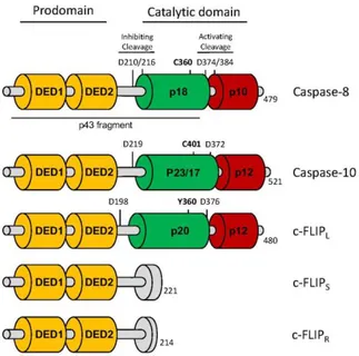
Caspase-8 activation occurs through a two-step model. Following procaspase-8 dimerization, the enzyme is first cleaved between the large and small catalytic subunits, at Asp374 and Asp387, to generate p41/43 and p10 fragments. Two isoforms of procaspase-8 are mainly expressed in mammalian cells explaining the p41 and p43 fragments (i.e., procaspase-8a and -8b). Second, p41/43 is cleaved at Asp210 and Asp216 to release the pro-domain and a p18 fragment. Mature caspase-8 is released from the DISC as a tetramer composed of two p18 fragments and two p10 fragments (p182
-p102) [104] (Figure 2). The first cleavage represents a sort of “substrate switch” because, while
procaspase-8 dimers possess a limited repertoire of substrates, including c-FLIP and procaspase-8 itself, the first cleavage step stabilizes the caspase-8 dimers and gives them access to different substrates involved in the apoptotic pathway, such as Bid and caspase-3. Of note, the cleavage at Asp210/216 attenuates the enzymatic activity of caspase-8, as a non-cleavable mutant of caspase-8 (D210A/D216A) shows a 4-fold increase in catalytic activity compared to wild-type caspase-8 [23]. Therefore, different caspase-8-containing structures can be distinguished in the cell with different localizations, signaling strengths, and substrate subsets.
As mentioned, the dependency of type II cells on the mitochondrial pathway to trigger death ligand-induced apoptosis was partly explained by the expression of anti-apoptotic factors. The mechanism for the failure of DISC formation in type II cells remains unclear, but it has been reported that these cells exhibit high PI3K activity [105, 106]. PI3K signaling has been shown to prevent the aggregation of CD95 by retaining the receptor outside of lipid rafts [105, 107, 108]. Aggregation of CD95 into these membrane subdomains seems to be required for caspase-8 recruitment and efficient induction of the apoptotic program [109], whereas the raft localization is not required for activation of the MAPK pathway [110]. To further limit caspase-8 recruitment to the DISC, activation of PI3K/Akt also leads
Accepted
Article
to phosphorylation of PEA-15 at serine 116 [44]. PEA-15 is composed of an N-terminal DED and a C-terminal tail containing two important serines: serine 104 and serine 116. Whereas the phosphorylation of serine 104 by PKC regulates the interaction of PEA-15 with the extracellular-related kinases (ERK1/2), the phosphorylation of serine 116 by Akt or calcium/calmodulin-dependent protein kinase (CAMK)II induces the binding of PEA-15 to FADD, thereby reducing the recruitment of procaspase-8 and thus inhibiting DISC formation [111].
In addition to this regulation of caspase-8 recruitment, caspase-8 activity can also be regulated by caspase-8 analogues such as procaspase-10 and cellular FLICE-like inhibitory protein (c-FLIP) (Figure 2). These proteins have been shown to interact with procaspase-8-FADD complexes and to modulate DISC activity [112-116]. Caspase-10 is a homologue of caspase-8 and when recruited into the DISC, it shares many substrates with caspase-8 [117]. Unexpectedly, a recent report demonstrated that knock-down of caspase-10 sensitizes cells to CD95-mediated apoptosis [22]. The authors observed that the presence of caspase-10 not only impairs DISC formation but also regulates the magnitude by which genes are modulated upon CD95L stimulation [22]. Nonetheless, because caspase-8 is only weakly inhibited by caspase-10, and because no caspase-10 orthologue exists in mice even though the apoptotic and non-apoptotic signaling pathways are preserved in this organism, caspase-10 seems to be a modulator of this signal rather than its toggle.
c-FLIP is also structurally related to caspase-8 but is devoid of enzymatic activity. Three main isoforms of c-FLIP can be found: two short isoforms (c-FLIPR/S),which only contain the pro-domain
encompassing the two DED domains, and a long isoform (c-FLIPL). c-FLIPL also contains a
C-terminal caspase domain that is inactive due to the replacement of a cysteine residue in the protease site with tyrosine [118]. Among the short isoforms, c-FLIPR (for Raji) is mainly detected in T and B
cells and exhibits similar functions as c-FLIPS (short), the expression of which is more ubiquitous
[119]. All c-FLIP isoforms can be recruited to the DISC by caspase-8, and whereas c-FLIPS
recruitment blocks caspase-8 activation [120], c-FLIPL canstimulate procaspase-8 proteolytic activity
[121-123] through the formation of c-FLIP/procaspase-8 heterodimers. C-FLIPL/procaspase-8
Accepted
Article
including RIP1 [124, 125]; cleavage of RIP1 leads to destabilization of the RIP1-RIP3 complex and inhibition of necroptosis. Accordingly, genetic experiments elegantly demonstrated that although deletion of casp8 is lethal at the embryonic stage in mice (ED 10.5), combined ablation of casp8 and
ripk3 results in viable mouse pups, confirming a close interplay between apoptosis and necroptosis
[64, 65]. In the heterodimers, c-FLIPL is processed into p43 and p12 fragments, which can interact
with Raf1, TNF receptor-associated factor (TRAF)2, and RIP1 to induce MAPK and NF-κB activation [126-128]. Of note, the mechanistic base for c-FLIPL and c-FLIPS modulation of the
CD95-mediated NF-κB activation remains a subject of debate and has to be clarified because several groups demonstrated that all c-FLIP isoforms efficiently abrogate the CD95L/TRAIL-mediated non-apoptotic signaling pathways (i.e., MAPK and NF-κB) [129, 130].
Alternatively, c-FLIP can be recruited directly by FADD to DISC, with the DEDs of c-FLIP and caspase-8 docking to different surfaces within the DED of FADD. While DED-1 of caspase-8 preferentially binds helices 2 and 5 of DED-FADD, DED-2 of c-FLIP (L or S) interacts with a surface covering helices 1 and 4 [131]. Inside the tripartite c-FLIP/FADD/Caspase-8 complex, the remaining caspase-8 DED can bind c-FLIPL and its caspase-like domain to generate an active but
membrane-restricted caspase-8 (c-FLIP p43/Caspase-8 p41/p43), or c-FLIPS blocking caspase-8 activation. When
the expression levels of c-FLIP drop, helices 1 and 4 of FADD are free to bind caspase-8 and this tripartite (FADD/Caspase-8/Caspase-8) containing a capase-8 homodimer releases a soluble and fully activated caspase-8 (p18/p10) triggering the apoptotic signaling pathway [131]. These tripartite complexes indicate that therapeutic approaches might exist to selectively inhibit the death receptor-mediated non-apoptotic signaling pathways by disturbing c-FLIP recruitment without affecting caspase-8/FADD interaction (apoptotic signal).
Caspase-8 maturation relies on the expression level of c-FLIPL. While c-FLIPL blocks
caspase-8-driven apoptotic signaling at high concentrations, low and intermediate concentrations of c-FLIP enhance caspase-8 recruitment and activation, thus facilitating the induction of apoptosis [123]. As an explanation for this observation, Hughes et al. proposed a hierarchical and co-operative model in which procaspase-8, docked to FADD, preferentially recruits c-FLIPLover a second procaspase-8to
Accepted
Article
form the first active protease at the DISC. When the cellular level of c-FLIPL declines, procaspase-8 is
recruited to the DISC through DED interactions to form procaspase-8 homodimers. Consequently, the protein level of c-FLIPL and, more importantly, the ratio of c-FLIPL to c-FLIPS are crucial
determinants of whether caspase-8 activation results in apoptotic or non-apoptotic signaling.
4.2 Dynamic CD95 signaling complexes
Unlike CD95 and TNF-related apoptosis-inducing ligand (TRAIL), TNFR1 preferentially induces the activation of pro-inflammatory responses over apoptotic signaling. Mechanistically, this preference is due to the sequential formation of two complexes: Complex I, which promotes NF-κB activation, and Complex II, which is responsible for the induction of apoptosis [132]. Upon TNF- binding, TNFR1-associated death domain protein (TRADD) is first recruited through its DD domain to TNFR1 at the plasma membrane. There, TRADD further associates with signaling proteins such as cellular inhibitor of apoptosis protein 1 and 2 (cIAP1/2), TNFR-associated factor 2 (TRAF2), and RIP1 to form Complex I. In this complex, the E3 ubiquitin ligases cIAP1/2 polyubiquitinate themselves and RIP1 through lysines K63, K48, and K11 [133-135]. The polyubiquitin chains serve as a docking platform for the recruitment of another set of proteins, including the linear ubiquitin assembly complex (LUBAC), which generates additional linear ubiquitin chains on Complex I [136]. The assembly of these polyubiquitin chains allows for the recruitment of the IκB kinase (IKK) complex and the transforming growth factor-β (TGF-β)-activated kinase (TAB-TAK1) complex, and the subsequent activation of the NF-κB and MAPK signaling pathways [137-140]. The activation of these signaling cascades will induce the expression of anti-apoptotic factors including the caspase-8 inhibitor c-FLIP. In conclusion, while ubiquitination is instrumental in the complex I formation, elimination of the ubiquitin moieties from the substrates by deubiquitinases including A20, Cezanne or Cylindromatosis (CYLD) promotes the switch to complex II (for review see [141]). Complex II formation requires the release of TNFR1, and the recruitment of FADD by TRADD through a DD/DD interaction serving as a platform to bind procaspase-8 [132]. Importantly, complex II assembly occurs through the
Accepted
Article
internalization of the different components in RIP1-dependent or -independent mechanism [141]. In this complex consisting of deubiquitinated RIP1, caspase-8 and adaptors TRADD and FADD, caspase-8 is activated, leading to apoptosis of the cell. CD95 stimulation seems also to form an internalized “complex II”, but the biological function of this complex remains to be addressed because both DISC (complex I) and complex II trigger a caspase-8-dependent apoptotic signaling pathway [142]. One hypothesis is that the cell localization of the two complexes might propose a different set of substrates to the activated caspase-8 leading to different signaling pathways.
Although TRAF2 can induce the activation of NFB, it plays a different role in the CD95 signaling pathway. Indeed, ubiquitination can control the CD95-mediated apoptotic and non-apoptotic signaling pathways through K48 and K63 ubiquitination of caspase-8 [143]. While cullin-3-driven K63 ubiquitination at lysine 461 of the small subunit enhances the apoptotic signaling pathway, TRAF2-dependent K48 ubiquitination at lysines 224, 229, and 231 of the large subunit inhibits apoptotic signaling by causing proteasome-dependent degradation of activated caspase-8 [143, 144]. In the molecular ordering of ubiquitination, TRAF2 is acting downstream of cullin3, suggesting that the latter activates and the former extinguishes caspase-8 activity, thereby tightly controlling this process [143]. Although TRAF2 docks onto caspase-8, it is not known whether this interaction is direct and/or whether the cullin3-dependent K63 ubiquitination of caspase-8 is a prerequisite for TRAF2 binding.
CD95 and TRAIL-R (DR4 and DR5) cannot bind TRADD. Nevertheless, the formation of dynamic complexes has also been suggested. Indeed, a recent study demonstrated that TRAIL stimulation can induce the formation of a cytosolic signaling complex that has been called the “FADDosome.” This complex, which is composed of FADD, FLIP, caspase-8, caspase-10, TAK1, A20, NF-B essential modulator (NEMO) and RIP1, is responsible for NF-κB activation and pro-inflammatory cytokine production [68]. Interestingly, the non-enzymatic activity of caspase-8 is essential for the formation of the complex because catalytically inactive caspase-8 was able to restore TRAIL-induced non-apoptotic signaling in caspase-8 knockout cells. This complex was further confirmed by work from the Walczak group showing that this complex also contained TRAF2, c-IAP1/2, and LUBAC [145]. Surprisingly, this study also demonstrated that this complex is formed at the plasma membrane and
Accepted
Article
that the LUBAC complex is a critical determinant of cell fate by restricting caspase-8 activation through ubiquitination while activating the NF-κB pathway.
After CD95 stimulation, a cytosolic complex containing FADD and caspase-8 was first observed in studies focusing on the apoptotic function of the receptor [142, 146]. Nonetheless, similarly to TRAIL, this soluble complex can also be involved in non-apoptotic functions. Indeed, a cytosolic FADD/caspase-8/RIP1/cIAP complex was shown to be essential in the activation of the NF-κB signaling pathway and the production of “find me” cytokines [147]. Caspase-8 functioned as a scaffold in this response as well because inhibition of its enzymatic activity did not alter this non-apoptotic signaling program. Regulation of caspase-8 activation through recruitment of DED-containing proteins such as c-FLIP/caspase-10 could therefore be central to the formation of these dynamic signaling complexes.
5.3 Soluble and transmembrane CD95L control the CD95 signaling pathway
Finally, the choice between the apoptotic and non-apoptotic response could be driven by CD95L itself. Unlike transmembrane CD95L (m-CD95L), which favors DISC formation and apoptosis, s-CD95L leads to the formation of a molecular complex devoid of FADD and caspase-8 that instead recruits and activates the src kinase c-yes via NADPH oxidase 3 (NOX3) and ROS production [59, 148]; this unconventional receptosome has been designated the “motility-inducing signaling complex” (MISC) (Figure 3) [59, 149].
M-CD95L can be cleaved by different metalloproteases generating s-CD95L, which fails to trigger cell death [150, 151] but instead, implements non-apoptotic signaling pathways [73, 152]. The differences in the kinetics of CD95 aggregation, or its internalization and/or membrane distribution [153], may account for the induction of one cue at the expense of another. As aforementioned, while s-CD95L exists as a homotrimer [150], its membrane-bound counterpart is a multi-aggregated homotrimers [154]. Therefore, the difference in their stoichiometry and/or their conformational effect
Accepted
Article
on the receptor (natural ligand versus agonistic antibodies) might explain the differences in the activated cell signaling pathways [27, 155]. Different forms of s-CD95L have been reported and although the s-CD95L generated by MMP7-driven cleavage of its 113ELR115 sequence induces apoptosis [156], its counterpart (cleaved between serine 126 and leucine 127) does not [78, 150, 152]. Moreover, s-CD95L in the bronchoalveolar lavage (BAL) fluid of patients suffering from acute respiratory distress syndrome (ARDS) can undergo oxidation at methionines 224 and 225, promoting its aggregation and thereby rendering it cytotoxic [157]. The stalk region of CD95L covers amino acid residues 103 to 136 and contains all metalloprotease cleavage sites described in the literature. Of note, in ARDS BAL fluid, CD95L also undergoes an oxidation at methionine 121, that prevents its cleavage by MMP7 and potentially explains why this cytotoxic ligand retains its stalk region and contributes to disease pathology [157]. Nonetheless, preservation of this region raises the question of whether an as-yet-unidentified MMP7-independent cleavage site exists in the membrane-proximal region of CD95L, or whether the ligand detected in ARDS patients corresponds in fact to the full-length form of CD95L embedded in exosomes [158, 159]. Indeed, exosome-bound CD95L can be produced by human prostate cancer cells (i.e., the LNCaP cell line), and triggers apoptosis [160]. In addition to MMPs, plasmin can also cleave the m-CD95L expressed by neo-vessels in cancers [161]. Urokinase-type plasminogen activator (uPA) converts plasminogen into its active form, plasmin, which in turn cleaves CD95L between the amino acid residues Arg144 and Lys145 within its trimerization domain, releasing a soluble ligand, which surprisingly triggers the death of endothelial cells [161]. Because no analysis of the s-CD95L stoichiometry has been realized in this study, it is difficult to determine whether the plasmin-driven s-CD95L corresponds to an aggregate of homotrimers or whether cleavage by plasmin releases a homotrimeric ligand associated with other components promoting its apoptotic effect.
Overall, these findings emphasize the importance of finely characterizing the stoichiometry of naturally processed CD95L in the serum of patients affected by cancers or chronic/acute inflammatory disorders to predict its biological role (i.e., apoptotic vs non-apoptotic).
Accepted
Article
Although caspase-8 is not detected in the MISC, it has been reported to participate in cell migration. Indeed, phosphorylation of caspase-8 at Tyr380 by src family kinase (SFK) inhibits its activity while promoting its scaffold function [106]. This phosphorylation notably allows for recruitment of the p85 alpha subunit of PI3K and the activation of the PI3K signaling pathway [99]. Nonetheless, CD95-induced migration and invasion can also occur through a DD-independent mechanism (reviewed in [162]), suggesting that a caspase-8-independent mode of cell migration exists in cells exposed to CD95L.
Exposure of T cells to s-CD95L triggers MISC formation and the recruitment of PLC1 to the calcium-inducing domain (CID) of CD95, leading to calcium release-activated calcium modulator (CRACM)1-driven Ca2+ entry and PI3K activation [61, 163]. Of note, while CD95-induced PLC1 activation and Ca2+ signaling stems from the CD95 CID [152], PI3K activation relies on the CD95 DD [152], suggesting that a complex interplay between the two domains regulates apoptotic versus non-apoptotic signaling. The identification of the distinct roles of these two domains encouraged us to design molecules selectively targeting the CID to inhibit the non-apoptotic signaling pathway without affecting the apoptotic response, which is essential for the elimination of viruses and tumors. Treatment of lupus-prone mice with the CID-targeting inhibitor reduced the severity of the pathology, validating the CD95 CID as a drug target for SLE patients. Of note, it has been reported that the CD95-mediated Ca2+ response might also affect the DD-dependent interactome. For instance, calmodulin docks onto the CD95-DD in a Ca2+-dependent manner and prevents FADD recruitment [164].
TRIP6 is another possible component of the MISC. Indeed, the CD95 CID can bind TRIP6 to induce migration of glioblastoma cells. When phosphorylated by src at Tyr55, TRIP6 interferes with the binding of FADD to CD95 and favors the activation of the NF-κB pathway [165].
In triple-negative breast cancer (TNBC), the recruitment of the tyrosine kinase receptor (RTK) epidermal growth factor receptor (EGFR) is required for activation of the PI3K pathway and cell migration [73]. Importantly, EGFR recruitment occurs independently of EGF and is mediated by the
Accepted
Article
c-yes kinase in response to s-CD95L [73]. Consequently, neutralizing anti-EGF/EGFR antibodies, including cetuximab, fail to prevent CD95-mediated cell migration, while erlotinib, a receptor tyrosine kinase inhibitor, inhibited this metastatic process [73].
Because EGFR can also induce tyrosine phosphorylation of caspase-8 at Tyr380, the CD95-EGFR interaction may also prime certain cancer cells to become unresponsive to the apoptotic signal triggered by cytotoxic CD95L while promoting cell migration, an essential step in cancer metastasis. Similarly, CD95 has been shown to trigger colon cancer cell metastasis by recruiting platelet-derived growth factor receptor (PDGFR), another RTK. In this context, the RTK activated PLC1 by phosphorylation on Tyr783, triggering a cofilin-mediated invasion process [166]. These findings suggest that RTK recruitment by CD95 inhibits the apoptotic signal by phosphorylating caspase-8 and simultaneously promotes cell motility.
Conclusions and perspectives
In summary, although the CD95/CD95L interaction can eliminate malignant cells by promoting formation of the DISC, it can also promote carcinogenesis by sustaining inflammation, regulating immune cells homeostasis, and/or inducing metastatic dissemination [59, 60, 72, 148, 167-169]. The molecular mechanisms underlying the switch between these different signaling pathways remain enigmatic. Important questions to be addressed are how the magnitude of CD95 aggregation regulates the formation of apoptotic versus motility signaling complexes and whether the different signaling responses originate from the same or distinct receptor domains. If this latter option proves to be the case, it may be possible to engineer inhibitors that are selective for the non-apoptotic signaling pathways, thereby preventing the induction of the pro-inflammatory/oncogenic signaling processes without apoptosis, which contributes to the anti-tumor and anti-infectious response. Answering these questions will lead to the development of new therapeutic agents with the ability to limit chronic inflammation and carcinogenesis, at least in pathologies associated with increased soluble CD95L,
Accepted
Article
such as certain cancers (e.g., pancreatic cancer [170], large granular lymphocytic leukemia, breast cancer [149], and NK cell lymphoma [171]) and autoimmune disorders (e.g., rheumatoid arthritis and osteoarthritis [172], graft-versus-host disease [173, 174], and SLE [59, 175]).
Acknowledgments
This work was supported by grants from INCa PLBIO, Fondation ARC, La Ligue contre le Cancer (Comités d’Ille-et-Vilaine/du Morbihan/des Côtes d’Armor/du Finistère/du Maine et Loire), Cancéropole Grand Ouest, Région Bretagne, and Rennes Métropole.
REFERENCES
1. Itoh, N., Yonehara, S., Ishii, A., Yonehara, M., Mizushima, S.-I., Sameshima, M., Hase, A., Seto, Y. & Nagata, S. (1991) The polypeptide encoded by the cDNA for human cell surface antigen Fas can mediate apoptosis, Cell. 66, 233-243.
2. Loetscher, H., Pan, Y.-C. E., Lahm, H.-W., Gentz, R., Brockhaus, M., Tabuchi, H. & Lesslauer, W. (1990) Molecular cloning and expression of the human 55 kd tumor necrosis factor receptor, Cell. 61, 351-359.
3. Pan, G., Bauer, J. H., Haridas, V., Wang, S., Liu, D., Yu, G., Vincenz, C., Aggarwal, B. B., Ni, J. & Dixit, V. M. (1998) Identification and functional characterization of DR6, a novel death domain-containing TNF receptor, FEBS Letters. 431, 351-356.
4. Trauth, B., Klas, C., Peters, A., Matzku, S., Moller, P., Falk, W., Debatin, K. & Krammer, P. (1989) Monoclonal antibody-mediated tumor regression by induction of apoptosis, Science. 245, 301-305.
5. Chtanova, T., Tangye, S. G., Newton, R., Frank, N., Hodge, M. R., Rolph, M. S. & Mackay, C. R. (2004) T Follicular Helper Cells Express a Distinctive Transcriptional Profile, Reflecting Their Role as Non-Th1/Th2 Effector Cells That Provide Help for B Cells, The Journal of Immunology. 173, 68-78.
6. Basu, R., O’Quinn, Darrell B., Silberger, Daniel J., Schoeb, Trenton R., Fouser, L., Ouyang, W., Hatton, Robin D. & Weaver, Casey T. (2012) Th22 Cells Are an Important Source of IL-22 for Host Protection against Enteropathogenic Bacteria, Immunity. 37, 1061-1075.
Accepted
Article
7. Gerlach, K., Hwang, Y., Nikolaev, A., Atreya, R., Dornhoff, H., Steiner, S., Lehr, H.-A., Wirtz, S., Vieth, M., Waisman, A., Rosenbauer, F., McKenzie, A. N. J., Weigmann, B. & Neurath, M. F. (2014) TH9 cells that express the transcription factor PU.1 drive T cell–mediated colitis via IL-9 receptor signaling in intestinal epithelial cells, Nature Immunology. 15, 676-686.
8. Strasser, A., Jost, P. J. & Nagata, S. (2009) The many roles of FAS receptor signaling in the immune system, Immunity. 30, 180-92.
9. Marsters, S. A., Pitti, R. M., Donahue, C. J., Ruppert, S., Bauer, K. D. & Ashkenazi, A. (1996) Activation of apoptosis by Apo-2 ligand is independent of FADD but blocked by CrmA, Curr Biol. 6, 750-2.
10. Ratner, A. & Clark, W. R. (1993) Role of TNF-alpha in CD8+ cytotoxic T lymphocyte-mediated lysis, J Immunol. 150, 4303-14.
11. Chu, J. L. (1995) Massive upregulation of the Fas ligand in lpr and gld mice: implications for Fas regulation and the graft-versus-host disease-like wasting syndrome, Journal of Experimental
Medicine. 181, 393-398.
12. Nagata, S. & Golstein, P. (1995) The Fas death factor, Science. 267, 1449-56.
13. Yang, Y. (1995) Fas and activation-induced Fas ligand mediate apoptosis of T cell hybridomas: inhibition of Fas ligand expression by retinoic acid and glucocorticoids, Journal of Experimental
Medicine. 181, 1673-1682.
14. Lu, M., Lawrence, D. A., Marsters, S., Acosta-Alvear, D., Kimmig, P., Mendez, A. S., Paton, A. W., Paton, J. C., Walter, P. & Ashkenazi, A. (2014) Opposing unfolded-protein-response signals converge on death receptor 5 to control apoptosis, Science. 345, 98-101.
15. Alnemri, E. S., Livingston, D. J., Nicholson, D. W., Salvesen, G., Thornberry, N. A., Wong, W. W. & Yuan, J. (1996) Human ICE/CED-3 Protease Nomenclature, Cell. 87, 171.
16. Papoff, G., Hausler, P., Eramo, A., Pagano, M. G., Di Leve, G., Signore, A. & Ruberti, G. (1999) Identification and Characterization of a Ligand-independent Oligomerization Domain in the
Extracellular Region of the CD95 Death Receptor, Journal of Biological Chemistry. 274, 38241-38250.
17. Siegel, R. M., Frederiksen, J. K., Zacharias, D. A., Chan, F. K., Johnson, M., Lynch, D., Tsien, R. Y. & Lenardo, M. J. (2000) Fas preassociation required for apoptosis signaling and dominant
inhibition by pathogenic mutations, Science. 288, 2354-7.
18. Edmond, V., Ghali, B., Penna, A., Taupin, J.-L., Daburon, S., Moreau, J.-F. & Legembre, P. (2012) Precise Mapping of the CD95 Pre-Ligand Assembly Domain, PLoS ONE. 7, e46236.
19. Boldin, M. P., Varfolomeev, E. E., Pancer, Z., Mett, I. L., Camonis, J. H. & Wallach, D. (1995) A Novel Protein That Interacts with the Death Domain of Fas/APO1 Contains a Sequence Motif Related to the Death Domain, Journal of Biological Chemistry. 270, 7795-7798.
20. Chinnaiyan, A. M., O'Rourke, K., Tewari, M. & Dixit, V. M. (1995) FADD, a novel death domain-containing protein, interacts with the death domain of fas and initiates apoptosis, Cell. 81, 505-512.
Accepted
Article
21. Kischkel, F. C., Hellbardt, S., Behrmann, I., Germer, M., Pawlita, M., Krammer, P. H. & Peter, M. E. (1995) Cytotoxicity-dependent APO-1 (Fas/CD95)-associated proteins form a death-inducing signaling complex (DISC) with the receptor, Embo J. 14, 5579-88.
22. Horn, S., Hughes, M. A., Schilling, R., Sticht, C., Tenev, T., Ploesser, M., Meier, P., Sprick, M. R., MacFarlane, M. & Leverkus, M. (2017) Caspase-10 Negatively Regulates Caspase-8-Mediated Cell Death, Switching the Response to CD95L in Favor of NF-κB Activation and Cell Survival, Cell
Reports. 19, 785-797.
23. Hughes, M. A., Harper, N., Butterworth, M., Cain, K., Cohen, G. M. & MacFarlane, M. (2009) Reconstitution of the death-inducing signaling complex reveals a substrate switch that determines CD95-mediated death or survival, Mol Cell. 35, 265-79.
24. Li, P., Nijhawan, D., Budihardjo, I., Srinivasula, S. M., Ahmad, M., Alnemri, E. S. & Wang, X. (1997) Cytochrome c and dATP-dependent formation of Apaf-1/caspase-9 complex initiates an apoptotic protease cascade, Cell. 91, 479-89.
25. Scaffidi, C. (1998) Two CD95 (APO-1/Fas) signaling pathways, The EMBO Journal. 17, 1675-1687.
26. Algeciras-Schimnich, A., Pietras, E. M., Barnhart, B. C., Legembre, P., Vijayan, S., Holbeck, S. L. & Peter, M. E. (2003) Two CD95 tumor classes with different sensitivities to antitumor drugs, Proc
Natl Acad Sci U S A. 100, 11445-50.
27. Chaigne-Delalande, B., Mahfouf, W., Daburon, S., Moreau, J. F. & Legembre, P. (2009) CD95 engagement mediates actin-independent and -dependent apoptotic signals, Cell Death Differ. 16, 1654-64.
28. Scaffidi, C., Fulda, S., Srinivasan, A., Friesen, C., Li, F., Tomaselli, K. J., Debatin, K. M., Krammer, P. H. & Peter, M. E. (1998) Two CD95 (APO-1/Fas) signaling pathways, Embo J. 17, 1675-87.
29. Yin, X. M. (2000) Signal transduction mediated by Bid, a pro-death Bcl-2 family proteins, connects the death receptor and mitochondria apoptosis pathways, Cell Res. 10, 161-7.
30. Yin, X. M., Wang, K., Gross, A., Zhao, Y., Zinkel, S., Klocke, B., Roth, K. A. & Korsmeyer, S. J. (1999) Bid-deficient mice are resistant to Fas-induced hepatocellular apoptosis, Nature. 400, 886-91.
31. Jost, P. J., Grabow, S., Gray, D., McKenzie, M. D., Nachbur, U., Huang, D. C., Bouillet, P., Thomas, H. E., Borner, C., Silke, J., Strasser, A. & Kaufmann, T. (2009) XIAP discriminates between type I and type II FAS-induced apoptosis, Nature. 460, 1035-9.
32. Roy, N., Deveraux, Q. L., Takahashi, R., Salvesen, G. S. & Reed, J. C. (1997) The IAP-1 and c-IAP-2 proteins are direct inhibitors of specific caspases, Embo J. 16, 6914-25.
33. Deveraux, Q. L., Takahashi, R., Salvesen, G. S. & Reed, J. C. (1997) X-linked IAP is a direct inhibitor of cell-death proteases, Nature. 388, 300-4.
34. Deveraux, Q. L., Roy, N., Stennicke, H. R., Van Arsdale, T., Zhou, Q., Srinivasula, S. M., Alnemri, E. S., Salvesen, G. S. & Reed, J. C. (1998) IAPs block apoptotic events induced by caspase-8 and cytochrome c by direct inhibition of distinct caspases, Embo J. 17, 2215-23.
Accepted
Article
35. Suzuki, Y., Nakabayashi, Y. & Takahashi, R. (2001) Ubiquitin-protein ligase activity of X-linked inhibitor of apoptosis protein promotes proteasomal degradation of caspase-3 and enhances its anti-apoptotic effect in Fas-induced cell death, Proc Natl Acad Sci U S A. 98, 8662-7.
36. Du, C., Fang, M., Li, Y., Li, L. & Wang, X. (2000) Smac, a mitochondrial protein that promotes cytochrome c-dependent caspase activation by eliminating IAP inhibition, Cell. 102, 33-42.
37. Sun, X. M., Bratton, S. B., Butterworth, M., MacFarlane, M. & Cohen, G. M. (2002) Bcl-2 and Bcl-xL inhibit CD95-mediated apoptosis by preventing mitochondrial release of Smac/DIABLO and subsequent inactivation of X-linked inhibitor-of-apoptosis protein, J Biol Chem. 277, 11345-51. 38. Beneteau, M., Pizon, M., Chaigne-Delalande, B., Daburon, S., Moreau, P., De Giorgi, F., Ichas, F., Rebillard, A., Dimanche-Boitrel, M. T., Taupin, J. L., Moreau, J. F. & Legembre, P. (2008)
Localization of Fas/CD95 into the lipid rafts on down-modulation of the phosphatidylinositol 3-kinase signaling pathway, Mol Cancer Res. 6, 604-13.
39. Peacock, J. W., Palmer, J., Fink, D., Ip, S., Pietras, E. M., Mui, A. L., Chung, S. W., Gleave, M. E., Cox, M. E., Parsons, R., Peter, M. E. & Ong, C. J. (2009) PTEN loss promotes mitochondrially dependent type II Fas-induced apoptosis via PEA-15, Mol Cell Biol. 29, 1222-34.
40. Varadhachary, A. S., Edidin, M., Hanlon, A. M., Peter, M. E., Krammer, P. H. & Salgame, P. (2001) Phosphatidylinositol 3'-kinase blocks CD95 aggregation and caspase-8 cleavage at the death-inducing signaling complex by modulating lateral diffusion of CD95, J Immunol. 166, 6564-9. 41. Pizon, M., Rampanarivo, H., Tauzin, S., Chaigne-Delalande, B., Daburon, S., Castroviejo, M., Moreau, P., Moreau, J. F. & Legembre, P. (2011) Actin-independent exclusion of CD95 by PI3K/AKT signalling: implications for apoptosis, Eur J Immunol. 41, 2368-78.
42. Condorelli, G., Vigliotta, G., Cafieri, A., Trencia, A., Andalo, P., Oriente, F., Miele, C., Caruso, M., Formisano, P. & Beguinot, F. (1999) PED/PEA-15: an anti-apoptotic molecule that regulates FAS/TNFR1-induced apoptosis, Oncogene. 18, 4409-15.
43. Renganathan, H., Vaidyanathan, H., Knapinska, A. & Ramos, J. W. (2005) Phosphorylation of PEA-15 switches its binding specificity from ERK/MAPK to FADD, Biochem J. 390, 729-35.
44. Trencia, A., Perfetti, A., Cassese, A., Vigliotta, G., Miele, C., Oriente, F., Santopietro, S., Giacco, F., Condorelli, G., Formisano, P. & Beguinot, F. (2003) Protein kinase B/Akt binds and
phosphorylates PED/PEA-15, stabilizing its antiapoptotic action, Mol Cell Biol. 23, 4511-21.
45. Strasser, A., Harris, A. W., Huang, D. C., Krammer, P. H. & Cory, S. (1995) Bcl-2 and Fas/APO-1 regulate distinct pathways to lymphocyte apoptosis, Embo J. Fas/APO-14, 6Fas/APO-136-47.
46. Lacronique, V., Mignon, A., Fabre, M., Viollet, B., Rouquet, N., Molina, T., Porteu, A., Henrion, A., Bouscary, D., Varlet, P., Joulin, V. & Kahn, A. (1996) Bcl-2 protects from lethal hepatic apoptosis induced by an anti-Fas antibody in mice, Nat Med. 2, 80-6.
47. Rodriguez, I., Matsuura, K., Khatib, K., Reed, J. C., Nagata, S. & Vassalli, P. (1996) A bcl-2 transgene expressed in hepatocytes protects mice from fulminant liver destruction but not from rapid death induced by anti-Fas antibody injection, J Exp Med. 183, 1031-6.
Accepted
Article
48. Kang, S. M., Schneider, D. B., Lin, Z., Hanahan, D., Dichek, D. A., Stock, P. G. & Baekkeskov, S. (1997) Fas ligand expression in islets of Langerhans does not confer immune privilege and instead targets them for rapid destruction, Nat Med. 3, 738-43.
49. Chen, J. J., Sun, Y. & Nabel, G. J. (1998) Regulation of the proinflammatory effects of Fas ligand (CD95L), Science. 282, 1714-7.
50. Buonocore, S., Flamand, V., Claessen, N., Heeringa, P., Goldman, M. & Florquin, S. (2004) Dendritic cells overexpressing Fas-ligand induce pulmonary vasculitis in mice, Clin Exp Immunol. 137, 74-80.
51. Matsuno, H., Yudoh, K., Watanabe, Y., Nakazawa, F., Aono, H. & Kimura, T. (2001)
Stromelysin-1 (MMP-3) in synovial fluid of patients with rheumatoid arthritis has potential to cleave membrane bound Fas ligand, J Rheumatol. 28, 22-8.
52. Vargo-Gogola, T., Crawford, H. C., Fingleton, B. & Matrisian, L. M. (2002) Identification of novel matrix metalloproteinase-7 (matrilysin) cleavage sites in murine and human Fas ligand,
Archives of Biochemistry and Biophysics. 408, 155-161.
53. Kiaei, M., Kipiani, K., Calingasan, N., Wille, E., Chen, J., Heissig, B., Rafii, S., Lorenzl, S. & Beal, M. (2007) Matrix metalloproteinase-9 regulates TNF-α and FasL expression in neuronal, glial cells and its absence extends life in a transgenic mouse model of amyotrophic lateral sclerosis,
Experimental Neurology. 205, 74-81.
54. Kirkin, V., Cahuzac, N., Guardiola-Serrano, F., Huault, S., Lückerath, K., Friedmann, E., Novac, N., Wels, W. S., Martoglio, B., Hueber, A. O. & Zörnig, M. (2007) The Fas ligand intracellular domain is released by ADAM10 and SPPL2a cleavage in T-cells, Cell Death and Differentiation. 14, 1678-1687.
55. Schulte, M., Reiss, K., Lettau, M., Maretzky, T., Ludwig, A., Hartmann, D., de Strooper, B., Janssen, O. & Saftig, P. (2007) ADAM10 regulates FasL cell surface expression and modulates FasL-induced cytotoxicity and activation-FasL-induced cell death, Cell Death and Differentiation.
56. Holler, N., Tardivel, A., Kovacsovics-Bankowski, M., Hertig, S., Gaide, O., Martinon, F., Tinel, A., Deperthes, D., Calderara, S., Schulthess, T., Engel, J., Schneider, P. & Tschopp, J. (2003) Two Adjacent Trimeric Fas Ligands Are Required for Fas Signaling and Formation of a Death-Inducing Signaling Complex, Molecular and Cellular Biology. 23, 1428-1440.
57. Schneider, P., Holler, N., Bodmer, J.-L., Hahne, M., Frei, K., Fontana, A. & Tschopp, J. (1998) Conversion of Membrane-bound Fas(CD95) Ligand to Its Soluble Form Is Associated with
Downregulation of Its Proapoptotic Activity and Loss of Liver Toxicity, The Journal of Experimental
Medicine. 187, 1205-1213.
58. Suda, T., Hashimoto, H., Tanaka, M., Ochi, T. & Nagata, S. (1997) Membrane Fas Ligand Kills Human Peripheral Blood T Lymphocytes, and Soluble Fas Ligand Blocks the Killing, The Journal of
Experimental Medicine. 186, 2045-2050.
59. Tauzin, S., Chaigne-Delalande, B., Selva, E., Khadra, N., Daburon, S., Contin-Bordes, C., Blanco, P., Le Seyec, J., Ducret, T., Counillon, L., Moreau, J.-F., Hofman, P., Vacher, P. & Legembre, P. (2011) The Naturally Processed CD95L Elicits a c-Yes/Calcium/PI3K-Driven Cell Migration Pathway, PLoS Biology. 9, e1001090.
Accepted
Article
60. LA, O. R., Tai, L., Lee, L., Kruse, E. A., Grabow, S., Fairlie, W. D., Haynes, N. M., Tarlinton, D. M., Zhang, J. G., Belz, G. T., Smyth, M. J., Bouillet, P., Robb, L. & Strasser, A. (2009) Membrane-bound Fas ligand only is essential for Fas-induced apoptosis, Nature. 461, 659-63.
61. Poissonnier, A., Sanseau, D., Le Gallo, M., Malleter, M., Levoin, N., Viel, R., Morere, L., Penna, A., Blanco, P., Dupuy, A., Poizeau, F., Fautrel, A., Seneschal, J., Jouan, F., Ritz, J., Forcade, E., Rioux, N., Contin-Bordes, C., Ducret, T., Vacher, A. M., Barrow, P. A., Flynn, R. J., Vacher, P. & Legembre, P. (2016) CD95-Mediated Calcium Signaling Promotes T Helper 17 Trafficking to Inflamed Organs in Lupus-Prone Mice, Immunity. 45, 209-23.
62. Cullen, S. P., Henry, C. M., Kearney, C. J., Logue, S. E., Feoktistova, M., Tynan, G. A., Lavelle, E. C., Leverkus, M. & Martin, S. J. (2013) Fas/CD95-induced chemokines can serve as "find-me" signals for apoptotic cells, Mol Cell. 49, 1034-48.
63. Green, D. R., Oberst, A., Dillon, C. P., Weinlich, R. & Salvesen, G. S. (2011) RIPK-dependent necrosis and its regulation by caspases: a mystery in five acts, Mol Cell. 44, 9-16.
64. Kaiser, W. J., Upton, J. W., Long, A. B., Livingston-Rosanoff, D., Daley-Bauer, L. P., Hakem, R., Caspary, T. & Mocarski, E. S. (2011) RIP3 mediates the embryonic lethality of caspase-8-deficient mice, Nature. 471, 368-72.
65. Oberst, A., Dillon, C. P., Weinlich, R., McCormick, L. L., Fitzgerald, P., Pop, C., Hakem, R., Salvesen, G. S. & Green, D. R. (2011) Catalytic activity of the caspase-8-FLIP(L) complex inhibits RIPK3-dependent necrosis, Nature. 471, 363-7.
66. Senft, J., Helfer, B. & Frisch, S. M. (2007) Caspase-8 interacts with the p85 subunit of phosphatidylinositol 3-kinase to regulate cell adhesion and motility, Cancer Res. 67, 11505-9. 67. Finlay, D., Howes, A. & Vuori, K. (2009) Critical role for caspase-8 in epidermal growth factor signaling, Cancer Res. 69, 5023-9.
68. Henry, C. M. & Martin, S. J. (2017) Caspase-8 Acts in a Non-enzymatic Role as a Scaffold for Assembly of a Pro-inflammatory "FADDosome" Complex upon TRAIL Stimulation, Mol Cell. 65, 715-729 e5.
69. Fernandes-Alnemri, T., Armstrong, R. C., Krebs, J., Srinivasula, S. M., Wang, L., Bullrich, F., Fritz, L. C., Trapani, J. A., Tomaselli, K. J., Litwack, G. & Alnemri, E. S. (1996) In vitro activation of CPP32 and Mch3 by Mch4, a novel human apoptotic cysteine protease containing two FADD-like domains, Proc Natl Acad Sci U S A. 93, 7464-9.
70. Muzio, M., Chinnaiyan, A. M., Kischkel, F. C., O'Rourke, K., Shevchenko, A., Ni, J., Scaffidi, C., Bretz, J. D., Zhang, M., Gentz, R., Mann, M., Krammer, P. H., Peter, M. E. & Dixit, V. M. (1996) FLICE, a novel FADD-homologous ICE/CED-3-like protease, is recruited to the CD95 (Fas/APO-1) death--inducing signaling complex, Cell. 85, 817-27.
71. Dupont, P. J. & Warrens, A. N. (2007) Fas ligand exerts its pro-inflammatory effects via neutrophil recruitment but not activation, Immunology. 120, 133-9.
72. Letellier, E., Kumar, S., Sancho-Martinez, I., Krauth, S., Funke-Kaiser, A., Laudenklos, S., Konecki, K., Klussmann, S., Corsini, N. S., Kleber, S., Drost, N., Neumann, A., Levi-Strauss, M., Brors, B., Gretz, N., Edler, L., Fischer, C., Hill, O., Thiemann, M., Biglari, B., Karray, S. &