RESEARCH OUTPUTS / RÉSULTATS DE RECHERCHE
Author(s) - Auteur(s) :
Publication date - Date de publication :
Permanent link - Permalien :
Rights / License - Licence de droit d’auteur :
Dépôt Institutionnel - Portail de la Recherche
researchportal.unamur.be
University of Namur
Glycan-foraging systems reveal the adaptation of Capnocytophaga canimorsus to the
dog mouth
Renzi, Francesco; Manfredi, Pablo; Dol, Mélanie; Fu, Jian; Vincent, Stéphane; Cornelis, Guy
Richard
Published in:
mBio
DOI:
10.1128/mBio.02507-14
Publication date:
2015
Document Version
Publisher's PDF, also known as Version of record
Link to publication
Citation for pulished version (HARVARD):
Renzi, F, Manfredi, P, Dol, M, Fu, J, Vincent, S & Cornelis, GR 2015, 'Glycan-foraging systems reveal the
adaptation of Capnocytophaga canimorsus to the dog mouth', mBio, vol. 6, no. 2, e02507-14.
https://doi.org/10.1128/mBio.02507-14
General rights
Copyright and moral rights for the publications made accessible in the public portal are retained by the authors and/or other copyright owners and it is a condition of accessing publications that users recognise and abide by the legal requirements associated with these rights. • Users may download and print one copy of any publication from the public portal for the purpose of private study or research. • You may not further distribute the material or use it for any profit-making activity or commercial gain
• You may freely distribute the URL identifying the publication in the public portal ? Take down policy
If you believe that this document breaches copyright please contact us providing details, and we will remove access to the work immediately and investigate your claim.
Glycan-Foraging Systems Reveal the Adaptation of Capnocytophaga
canimorsus to the Dog Mouth
Francesco Renzi,aPablo Manfredi,bMélanie Dol,aJian Fu,cStéphane Vincent,cGuy Richard Cornelisa
Département de Biologie, Université de Namur, Namur, Belgiuma; Biozentrum der Universität Basel, Basel, Switzerlandb; Département de Chimie, Université de Namur,
Namur, Belgiumc
ABSTRACT
Capnocytophaga canimorsus is known to form two kinds of cells on blood agar plates (coccoid and bacillary),
evok-ing phase variation. When grown in coculture with animal cells these bacteria appeared only as bacilli, but in the presence of
vancomycin they were round, indicating that coccoid shapes likely result from weakening of the peptidoglycan layer.
Polysac-charide utilization locus 5 (PUL5) and sialidase mutant bacteria, unable to retrieve glycans from glycoproteins, grew less than
wild-type bacteria and also appeared polymorphic unless GlcNAc was added, suggesting that C. canimorsus is unable to
synthe-size GlcNAc, an essential component of peptidoglycan. Accordingly, a genome analysis was conducted and revealed that C.
cani-morsus strain 5 lacks the GlmM and GlmU enzymes, which convert glucosamine into GlcNAc. Expression of the Escherichia coli
GlmM together with the acetyltransferase domain of GlmU allowed PUL5 mutant bacteria to grow normally, indicating that
C. canimorsus is a natural auxotroph that relies on GlcNAc harvested from the host N-glycoproteins for peptidoglycan synthesis.
Mucin, a heavily O-glycosylated protein abundant in saliva, also rescued growth and the shape of PUL5 mutant bacteria.
Utiliza-tion of mucin was found to depend on Muc, a Sus-like system encoded by PUL9. Contrary to all known PUL-encoded systems,
Muc cleaves peptide bonds of mucin rather than glycosidic linkages. Thus, C. canimorsus has adapted to build its peptidoglycan
from the glycan-rich dog’s mouth glycoproteins.
IMPORTANCE
Capnocytophaga canimorsus is a bacterium that lives as a commensal in the dog mouth and causes severe
infec-tions in humans. In vitro, it forms two kinds of cells (coccoid and bacillary), evoking phase variation. Here, we show that cell
rounding likely results from weakening of the peptidoglycan layer due to a shortage of N-acetylglucosamine (GlcNAc). C.
cani-morsus cannot synthesize GlcNAc because of the lack of key enzymes. In its niche, the dog mouth, C. canicani-morsus retrieves
Gl-cNAc by foraging glycans from salivary mucin and N-linked glycoproteins through two different apparatuses, Muc and Gpd,
both of which are related to the Bacteroides starch utilization system. The Muc system is peculiar in the sense that the enzyme of
the complex is a protease and not a glycosylhydrolase, as it cleaves peptide bonds in order to capture glycan chains. This study
provides a molecular genetic demonstration for the complex adaptation of C. canimorsus to its ecological niche, the oral cavity
of dogs.
Received 17 January 2015 Accepted 23 January 2015 Published 3 March 2015
Citation Renzi F, Manfredi P, Dol M, Fu J, Vincent S, Cornelis GR. 2015. Glycan-foraging systems reveal the adaptation of Capnocytophaga canimorsus to the dog mouth. mBio
6(2):e02507-14. doi:10.1128/mBio.02507-14.
Editor R. John Collier, Harvard Medical School
Copyright © 2015 Renzi et al. This is an open-access article distributed under the terms of theCreative Commons Attribution-Noncommercial-ShareAlike 3.0 Unported license, which permits unrestricted noncommercial use, distribution, and reproduction in any medium, provided the original author and source are credited.
Address correspondence to Guy Richard Cornelis, guy.cornelis@unamur.be.
This article is a direct contribution from a Fellow of the American Academy of Microbiology.
C
apnocytophaga canimorsus is a capnophylic gliding
Gram-negative bacterium from the Bacteroidetes phylum that is part
of the normal flora of the dog mouth (1–5). Although it has not
been reported to cause infections in dogs, it causes rare but severe
infections in humans who are in contact with dogs (6, 7). The
usual syndrome is septicemia, with mortality in the range of 50%.
Patients are generally older than 40 years old, and roughly half of
them have had a splenectomy or abused alcohol. However, the
other half have had no medical history (for review, see references
8 to 10). C. canimorsus 5, a strain isolated from a patient with
septic shock (11), has a lipopolysaccharide that triggers little
in-flammation (2, 11–13) and confers significant resistance to
phagocytosis (2), antimicrobial peptides (12), and complement
killing (2). Its genome, which consists of a single 2.5-Mb replicon
(14), contains 13 so-called polysaccharide utilization loci (PUL)
(15), a hallmark of Bacteroidetes bacteria (16). PUL typically
en-code polysaccharide-harvesting complexes made of several
lipo-proteins anchored in the outer membrane and that face the
out-side of the cell, as well as a TonB-dependent transporter. Some
lipoproteins are devoted to glycan binding, while others are
spe-cific hydrolases. The archetype of such complexes is the
Bacte-roides thetaiotaomicron starch utilization system (Sus), initially
described by Salyers and colleagues (17–20). C. canimorsus grows
readily on blood agar plates and in coculture with animal cells but,
surprisingly, it does not grow as well in the absence of animal cells
or when it is separated from animal cells by a membrane, indicating
that it relies on cells for nutrition (21). Unexpectedly, C. canimorsus
does not damage the cells but instead deglycosylates the cellular
mbio.asm.org
on September 2, 2015 - Published by
surface-exposed N-glycoproteins via its Gpd complex, encoded by
PUL5 (15, 21, 22). The Gpd complex consists of a SusC-like porin
(GpdC), a SusD-like binding protein (GpdD), two ancillary
lipopro-teins (GpdEF), and an endo--N-acetylglucosaminidase (GpdG)
which cleaves the N-linked oligosaccharide after the first
N-acetylglucosamine (GlcNAc) residue (22). The Gpd complex
deglycosylates not only surface glycoproteins but also soluble
glyco-proteins, like human IgG (22) and transferrin (23). Deglycosylation is
required for growth in coculture with animal cells but also for survival
in the mouse (15, 21). In the initial description of the species, Brenner
et al. (1) reported that C. canimorsus cultivated on blood agar plates
appeared as bacilli but also in different forms, including longer rods,
filaments that are often curved, spindle shaped, and coccoid (1),
which suggests that C. canimorsus undergoes some form of phase
variation.
In the present manuscript, we show that C. canimorsus 5 is a
natural auxotroph for GlcNAc that is missing the glmM and glmU
genes, which enable bacteria to convert glucosamine into GlcNAc.
The lack of GlcNAc leads to impaired peptidoglycan (PG)
assem-bly and bacterial death, explaining the polymorphism seen on
blood agar plates in the original description of the species (1).
C. canimorsus harvests GlcNAc not only from host N-linked
gly-coproteins through the PUL5-encoded Gpd complex but also
from mucin through the PUL9-encoded Muc complex. We
pro-pose that the GlcNAc auxotrophy, which contributes to virulence
(21), results from the adaptation of C. canimorsus as a commensal
of the glycan-rich dog mouth.
RESULTS
C. canimorsus polymorphism is due to N-acetylglucosamine
starvation. As reported early on (1), wild-type (wt) C. canimorsus
5 bacteria grown for 2 days on blood agar plates have different
morphologies, ranging from filaments to coccoid forms (Fig. 1A).
The coccoid forms strongly resemble the cells formed by
Esche-richia coli mutant strains that are unable to synthesize
aminosug-ars (24). When glucosamine or GlcNAc is not provided in the
medium, these mutants cannot synthesize glucosamine-6-P, and
they form spheroplasts due to a reduction in PG synthesis (24).
We thus speculated that the polymorphism observed with C.
cani-morsus 5 could be due to a similar reason. In order to see whether
this alteration of cell morphology was due to aminosugar
starva-tion and not some kind of phase variastarva-tion, we supplemented the
blood agar plates with 0.005% GlcNAc. As shown in Fig. 1A, the
addition of GlcNAc significantly reduced the formation of
abnor-mal forms, indicating that aminosugar starvation, rather than
phase variation, could be responsible for the observed
polymor-phism of wt C. canimorsus bacteria grown on blood agar plates.
PUL5 mutants grown on HEK293 cells are polymorphic due
to a defect in peptidoglycan synthesis. In coculture with HEK293
cells, C. canimorsus 5 bacteria deprived of the PUL5-encoded Gpd
complex reach a biomass that is 10-fold lower than that of wt
C. canimorsus 5 bacteria (15, 22) (Fig. 1B). Since this growth defect
can be rescued by supplementation of the medium with GlcNAc
(15) (Fig. 1B), we hypothesized it could mimic the growth defect
of wt bacteria on blood agar plates, and we examined the
mor-phology of wt and PUL5 mutant bacteria after 24 h of coculture
with HEK293 cells. Interestingly, while wt bacteria showed a
bac-illary form, PUL5 mutants appeared elongated, enlarged, or
com-pletely round (Fig. 1C). As shown in Fig. S1A and B in the
supple-mental material, PUL5 mutants progressively changed their
morphology while they stopped dividing, suggesting that growth
arrest and aberrant morphology are two linked phenomena. In
contrast, in the presence of GlcNAc, PUL5 mutant bacteria had
the same morphology as wt bacteria (Fig. 1C), suggesting that
GlcNAc starvation affects PG biosynthesis. In order to see whether
a defect in PG biosynthesis determines the same growth arrest and
cell rounding, we added vancomycin (25) in the coculture of
HEK293 cells and wt C. canimorsus 5. As shown in Fig. 1D (see also
Movie S1 in the supplemental material), the addition of
vancomy-cin transformed wt C. canimorsus 5 bacteria into round cells,
sim-ilar to those observed with PUL5 mutant bacteria (see Movie S2 in
the supplemental material). These results suggest that PUL5
mu-tant bacteria, which cannot harvest host N-linked glycans, stop
growing and change their cell shape because starvation of
amino-sugars leads to defects in peptidoglycan synthesis.
Sialidase mutants also have a peptidoglycan synthesis defect.
The PUL5-encoded Gpd complex works in concert with a
periplasmic sialidase (SiaC) whose activity is crucial for the
sub-sequent processing of the cleaved glycan chains (22). Since a siaC
deletion mutant also shows reduced growth on HEK293 cells
which can be rescued by addition of aminosugars (21), we asked
whether the bacterial cell shape of a siaC mutant would also be
affected. Indeed, a siaC mutant cocultivated with HEK293 cells for
24 h also showed aberrant morphologies: elongated, enlarged, and
completely round (Fig. 1E). Addition of GlcNAc restored the
nor-mal cell shape (Fig. 1E). We concluded from this result that the
inability to metabolize host N-linked glycans, due to either
ade of glycan chain cleavage and import (PUL5 deletion) or
block-ade of the first step of glycan processing in the periplasm (siaC
deletion), results in aminosugar starvation. This starvation is
likely to affect PG synthesis, leading to the appearance of aberrant
cell shapes and growth arrest.
C. canimorsus 5 is missing the genes glmM and glmU. Since
C. canimorsus 5 appears to be unable to synthesize GlcNAc, we
performed a Delta BLAST search of its genome (26) for homologs
of E. coli enzymes involved in PG biosynthetic pathways. As shown
in Fig. 2A, we could identify genes encoding all the PG
biosyn-thetic enzymes except for three: GlmS, GlmU, and GlmM, which
convert fructose-6-P into UDP-N-acetylglucosamine
(UDP-GlcNAc). GlmS is a glucosamine-6-P-synthase responsible for the
conversion of fructose-6-P into glucosamine-6-P; GlmM is a
phosphoglucosamine mutase that converts glucosamine-6-P into
glucosamine-1-P; GlmU is a bifunctional enzyme, with a
C-terminal glucosamine-1-P–N-acetyltransferase domain that
converts glucosamine-1-P into N-acetylglucosamine-1-P and an
N-terminal N-acetylglucosamine-1-P uridyltransferase domain
that converts GlcNAc-1-P into UDP-GlcNAc (27). Since the lack
of any of these enzymatic activities is lethal in E. coli (27), we
assumed that it is the lack of these enzymes that makes C.
cani-morsus dependent on exogenous GlcNAc for growth. If this
hy-pothesis were correct, expression of these three enzymes in PUL5
mutant bacteria would enable them to synthesize GlcNAc and
restore growth and bacterial shape in coculture with HEK293
cells. We cloned the three E. coli glm genes alone and in
combina-tion in a C. canimorsus expression vector (23) and monitored the
growth and morphology of recombinant PUL5 mutants in
cocul-ture with HEK293 cells. PUL5-deficient bacteria expressing both
GlmU and GlmM grew to the same extent as wt and showed a
normal shape, indicating that these enzymatic activities allow
C. canimorsus to synthesize GlcNAc (Fig. 2C and D).
mbio.asm.org
on September 2, 2015 - Published by
The fact that the expression of GlmM and GlmU alone was
sufficient to make PUL5 mutant bacteria independent of GlcNAc
suggested that the C. canimorsus 5 genome encodes a glucosamine
synthase (GlmS) that was not identified by our BLAST search.
Furthermore, since the addition of GlcNAc to the coculture of
HEK293 cells completely restored the growth of PUL5 mutant
bacteria, we inferred that C. canimorsus is able to synthesize
UDP-GlcNAc from UDP-GlcNAc, implying that a C. canimorsus 5 gene
en-codes a GlcNAc-1-P uridyltransferase. If this is correct, the
phospho-glucosamine mutase GlmM and only the phospho-
glucosamine-1-P⫺N-acetyltransferase domain of GlmU would be sufficient to compensate
for the PUL5 deletion. To test this hypothesis, we generated two
trun-cated GlmU protein variants: one with the first 77 residues deleted
(GlmU del78), consisting only of the
glucosamine-1-P⫺N-acetyltransferase activity, and one with the last 125 residues deleted
(GlmU Tr331), consisting only of the
N-acetylglucosamine-1-P-uridyltransferase activity (28) (Fig. 2B). We expressed in PUL5
mutants either GlmM and GlmU del78 or GlmM and GlmU
FIG 1 N-acetylglucosamine starvation affects growth and cell shape. (A) Bright-field microscopy pictures of wt (C. canimorsus 5) bacteria grown for 48 hourson blood agar plates in the absence or in the presence of GlcNAc (0.005%). (B) Counts of wt (C. canimorsus 5) and PUL5 (⌬PUL5) knockout bacteria after 23 hours growth in cDMEM with or without HEK293 cells (MOI, 0.05) and in the presence or in the absence of GlcNAc (0.005%). The averages from three independent experiments are shown. Error bars represent 1 standard deviation from the mean. See also Fig. S1A in the supplemental material. HEK293 cells provided GlcNAc captured by the PUL5-encoded Gpd complex. Note that HEK293 cells provided more than GlcNAc to wt C. canimorsus 5. (C) Bright-field microscopy pictures of wt (C. canimorsus 5) and PUL5 (⌬PUL5) bacteria after 23 hours growth on HEK293 cells. See also Fig. S1B. (D) Bright-field microscopy pictures of wt (C. canimorsus 5) bacteria grown on HEK293 cells for 12 hours and an additional 4 hours with or without vancomycin (5g/ml). See also Movies S1 and S2 in the supplemental material. (E) Bright-field microscopy pictures of sialidase mutant bacteria (⌬siaC) grown for 23 hours on HEK293 cells (MOI, 0.05) in the absence or in the presence of GlcNAc (0.005%).
mbio.asm.org
on September 2, 2015 - Published by
Tr331, and we monitored the growth and bacterial shapes in
co-culture with HEK293 cells. As shown in Fig. 2C and D, the
expression of GlmM and the GlmU
glucosamine-1-P–N-acetyltransferase domain (GlmU del78) was sufficient to allow
PUL5 mutant bacteria to behave similarly to wt. Expression of GlmM
and the uridyltransferase domain of GlmU (GlmU Tr331) restored
neither the growth nor the bacterial shape of PUL5 mutants (Fig. 2C
and D). Quite expectedly, the expression of GlmM and GmU del78
had the same rescuing effect on siaC mutants (Fig. 2E and F). The
slight but significant differences between the growth and the cell
shape observed in PUL5 and siaC mutants expressing GlmM and
GlmU del78 and the wt could be ascribed to the reduced enzymatic
activity of GlmU del78 compared to the full-length GlmU, as already
described by Pompeo and colleagues (28).
Taken together, these results indicate that the inability of
C. canimorsus 5 to synthesize GlcNAc is due to the lack of two
enzymes, a phosphoglucosamine mutase and a glucosamine-1-P–
N-acetyltransferase.
C. canimorsus 5 encodes a new
N-acetylglucosamine-1-P-uridyltransferase. In order to identify the unknown
N-acetyl-FIG 2 Heterologous expression of GlmU and GlmM in PUL5 and siaC mutant bacteria restores growth and normal cell shape. (A) Peptidoglycan biosynthetic
pathway showing C. canimorsus 5 orthologs of the E. coli enzymes. (B) Schematic representation of the E. coli GlmU bifunctional protein. The protein domains are boxed. The recombinant monofunctional proteins are indicated by the arrows. (C) Growth in coculture with HEK293 cells of PUL5 (⌬PUL5) mutant bacteria producing different combinations of GlmS, GlmM, GlmU, and monofunctional GlmU (MOI, 0.05; 23 h of growth). The averages from three independent experiments are shown. Error bars represent 1 standard deviation from the mean. *, P⬍ 0.05. (D) Bright-field microscopy pictures of bacteria grown for 23 h with HEK293 cells (MOI, 0.05). (E) Growth on HEK293 cells of of wt (C. canimorsus 5) and siaC (⌬siaC) bacteria expressing GlmM and monofunctional GlmU (MOI, 0.05; 23 h of growth). The averages from three independent experiments are shown. Error bars represent 1 standard deviation from the mean. **, P⬍ 0.01. (F) Bright-field microscopy pictures of bacteria grown for 23 h on HEK293 cells (MOI, 0.05).
mbio.asm.org
on September 2, 2015 - Published by
glucosamine-1-P-uridyltransferase of C. canimorsus 5, we searched
the genome for gene products with an enzymatic domain similar to
that of the N-terminal N-acetylglucosamine-1-P-uridyltransferase
domain of the E. coli GlmU. It turned out that Ccan_15070 encodes a
protein with a conserved glycosyltransferase domain (Fig. 3A).
At-tempts to knock out Ccan_15070 were unsuccessful unless C.
cani-morsus 5 could synthesize the E. coli GlmU Tr331 protein, suggesting
that Ccan_15070 is an essential gene and indeed encodes an
N-acetylglucosamine-1-P-uridyltransferase.
We then generated a PUL5 Ccan_15070 double-knockout
strain complemented with the E. coli glmM and glmU genes and
monitored growth and cell morphology. As shown in Fig. 3B and
C, the strain grew to the same extent as wt and had a normal cell
shape, indicating that the deletion of Ccan_15070 could be fully
complemented by the N-acetylglucosamine-1-P-uridyltransferase
activity of the E. coli GlmU.
Finally, to test the Ccan_15070 enzymatic activity, we
ex-pressed the protein with a C-terminal Strep tag in E. coli and we
added purified Ccan_15070 to a solution of GlcNAc-1-P and
UTP. The analysis of the reaction products via
high-performance liquid chromatography (HPLC) confirmed the
production of UDP-GlcNAc (Fig. 3D). Thus, Ccan_15070
en-codes an N-acetylglucosamine-1-P-uridyltransferase, which
we named GlmU
Cc.
GlcNAc is also harvested through a PUL-encoded system
tar-geting mucin. The loss of the capacity to synthesize aminosugars
during evolution implies that C. canimorsus relies on aminosugars
from the dog mouth. In the oral cavity, one of the most abundant
glycoproteins is mucin, which is heavily glycosylated and harbors
mainly O-linked glycans. The PUL5-encoded Gpd system can
thus not harvest the mucin sugars. In order to determine if the
aminosugars from mucin O-glycans can be used to synthesize PG,
we took advantage of the fact that PUL5 mutant bacteria grow less
well than wt in coculture with HEK293 cells. Hence, we tested
whether the addition of purified bovine submaxillary mucin
re-stored the growth of PUL5 bacteria. As shown in Fig. 4G and H,
FIG 3 Ccan_15070 encodes an N-acetylglucosamine-1-P uridyltransferase. (A) Schematic representation of the C. canimorsus 5 Ccan_15070 gene-encoded N-acetylglucosamine-1-P-uridyltransferase (Uniprot ID F9Yr13). (B) Growth on HEK293 cells of wt (C. canimorsus 5), PUL5 (⌬PUL5), and PUL5/Ccan_15070(⌬PUL5/⌬Ccan_15070) double-mutant bacteria expressing GlmM and GlmU (MOI, 0.05; 23 h of growth). The averages from three independent experiments are shown. Error bars represent 1 standard deviation from the mean. (C) Bright-field microscopy picture of PUL5 Ccan_15070 double mutant bacteria expressing GlmM and GlmU grown for 23 h on HEK293 cells (MOI, 0.05). (D) Detection of UDP-GlcNAc production by HPLC. Incubation of recombinant Ccan_15070 with UTP and GlcNAc-1-P resulted in the production of UDP-GlcNAc. (E) No UDP-GlcNAc formation could be detected in the absence of recombinant Ccan_15070.
mbio.asm.org
on September 2, 2015 - Published by
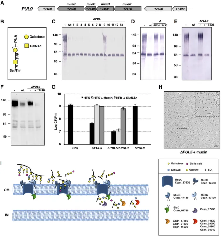
FIG 4 PUL9 gene products allow mucin degradation and deglycosylation. (A) Schematic representation of the PUL9 locus. (B) Schematic drawing of the PNA
lectin target oligosaccharides. (C) PNA lectin staining of human saliva after incubation in the presence of wt C. canimorsus 5 and the 13 PUL knockout strains. The upper band represents mucin, while the lower band represents sIgA. (D) Saliva PNA staining after incubation with wt bacteria, PUL9, and Ccan_17430 knockout bacteria. (E) Saliva PNA staining after incubation with wt bacteria, PUL9, or PUL9 bacteria expressing the only Ccan_17430 protein. (F) Western blot analysis of human mucin 7 in saliva after incubation with wt bacteria, PUL9, or PUL9 bacteria expressing the only Ccan_17430 protein. See also Fig. S3 in the supplemental material. (G) Growth after 23 h of wt (C. canimorsus 5), PUL5 (⌬PUL5), PUL9 (⌬PUL9), and PUL5/PUL9 (⌬PUL5 ⌬PUL9) mutant bacteria in the presence of HEK293 cells with or without bovine submaxillary gland mucin (1 mg/ml) or GlcNAc (0.005%) (MOI, 0.005). The averages from three independent experiments are shown. Error bars represent 1 standard deviation from the mean. (H) Bright-field microscopy picture of PUL5 (⌬PUL5) mutant bacteria grown for 23 h on HEK293 cells in the presence of bovine mucin (1 mg/ml) (MOI, 0.05). (I) Functional model of mucin degradation and deglycosylation. Mucin is bound at the bacterial surface by the MucCDEG surface protein complex. The MucG protease cleaves mucin into glycosylated peptides that are imported into the periplasm through the MucC pore. Terminal sialic acid is cleaved by sialidase (SiaC), and terminal sulfate groups are removed by the sulfatase. The glycan is further processed by the sequential activity of several periplasmic exoglycosidases that allow the liberation of monosaccharides.
mbio.asm.org
on September 2, 2015 - Published by
addition of bovine mucin fully restored the growth and the
mor-phology of PUL5 mutant bacteria.
To ascertain that salivary mucins are deglycosylated by C.
cani-morsus 5, we incubated bacteria with filtered fresh human saliva
and we analyzed the salivary glycoproteins before and after
incu-bation, by using sodium dodecyl sulfate-polyacrylamide gel
elec-trophoresis followed by staining with peanut agglutinin (PNA).
PNA recognizes terminal galactose linked to GalNAc in O-linked
oligosaccharides, a glycan structure named T-antigen (Fig. 4B)
that is abundant in mucins (29). A saliva glycoprotein with a
mo-lecular mass around 100 to 140 kDa turned out to be
deglycosy-lated after incubation with wt C. canimorsus 5 (Fig. 4C). In
con-trast, the glycosylation state of a protein with a molecular mass of
around 60 kDa was unaffected (Fig. 4C). LC-mass spectrometry
(LC/MS) analysis of the two proteins identified the
higher-molecular-mass protein as salivary mucin (MUC7 or MG2;
Uni-prot accession number Q8tax7) and the lower-molecular-mass
one as secreted IgA (sIgA, or Ig
␣-1 chain region; Uniprot
acces-sion number P01876) (data not shown). This indicated that
C. canimorsus 5 specifically targeted MG2 glycans rather than the
sIgA ones. We reasoned that one of the 13 PUL mutants could be
responsible for the T-antigen cleavage from MG2, and we tested
the 13 C. canimorsus 5 PUL knockout mutants (15). Of all the PUL
mutants, only PUL9 knockout bacteria were impaired in MG2
T-antigen deglycosylation (Fig. 4C). To ensure that mucin
utili-zation was PUL9 dependent, we generated a PUL5 PUL9
double-knockout strain and tested whether addition of mucin restored
the growth of this mutant. As expected, addition of mucin did not
restore the growth defect of the double mutant, indicating that
mucin degradation and utilization are PUL9 dependent (Fig. 4G).
PUL9 encodes a bona fide Sus-like complex. PUL9 encodes
seven proteins (Ccan_17420 to Ccan_17490) (Fig. 4A). The
neighboring Ccan_17470 and Ccan_17450 genes encode typical
SusC-like (TonB-dependent receptor) and SusD-like proteins.
Ccan_17480 is annotated as a
-galactosidase, while Ccan_17490
is a putative sulfatase. Both carry a signal peptide I and hence are
predicted to be periplasmic. Ccan_17420 is a predicted
lipopro-tein annotated as a carboxypeptidase. Ccan_17430 is predicted to
be a mucinase (30) with two BACON (Bacteroidetes-associated
carbohydrate often N-terminal domain) modules (31) and one
F5/8 type C (32) carbohydrate binding module. Ccan_17440 is the
most abundant PUL-encoded protein of the C. canimorsus 5
sur-fome (15) and also harbors an F5/8 type C carbohydrate binding
module. Ccan_17430 (mucinase) and Ccan_17450 (SusD-like)
were also found in the surfome of C. canimorsus 5 (15). In order to
determine whether all these proteins form with Ccan_17470
(SusC-like), a surface complex similar to the one formed by the
PUL5 products, we tagged the C terminus of the porin
Ccan_17470 and carried out a His-Strep tandem affinity
purifica-tion (see Fig. S2 in the supplemental material). The LC/MS
analysis of the eluate identified 35 proteins (see Table S1 in the
supplemental material), among which were Ccan_17450,
Ccan_17440, and Ccan_17430. Thirteen proteins were identified
in the mock eluate, of which 11 were also present in the
Ccan_17470 –Strep-His eluate. No PUL9-encoded product was
identified in the mock eluate (see Table S1). These data suggest
that Ccan_17470, Ccan_17450, Ccan_17440, and Ccan_17430
form a complex at the bacterial surface. Hence, PUL9 encodes a
bona fide Sus-like complex, and we propose to call MucC the
SusC-like protein, MucD the SusD-like protein, MucG the
puta-tive mucinase, and MucE the four surface proteins (Fig. 4A).
In-terestingly, among the proteins that eluted with the Muc complex,
we also identified a sialidase. This periplasmic enzyme was
previ-ously found to be associated with the Gpd complex and involved
in N-glycan processing (22). Thus, we could also envision
involve-ment of this protein in mucin glycan processing.
The hydrolytic enzyme from the complex is not a glycan
hydrolase but a mucin peptidase. As already mentioned,
Ccan_17430 is predicted to be a mucinase (30) with two BACON
(31) and one F5/8 type C (32) carbohydrate binding modules.
Since PUL-encoded complexes are usually devoted to the
hy-drolysis of complex carbohydrates (33), the presence of a protein
hydrolase was unexpected. In order to verify the function of
Ccan_17430, we incubated human saliva with wt and Ccan_17430
mutant bacteria and monitored mucin glycosylation via PNA
staining. Deletion of Ccan_17430 abolished mucin
deglycosyla-tion (Fig. 4D). Furthermore, expression of Ccan_17430 in a PUL9
deletion background led to a complete loss of mucin staining by
PNA, suggesting that Ccan_17430 alone is responsible for the
ob-served phenotype (Fig. 4E). We then monitored the presence of
mucin MG2 by Western blotting and found that it was
dramati-cally decreased, indicating that Ccan_17430 indeed encodes a
pro-tease that cleaves mucin (Fig. 4F). In agreement with this, purified
recombinant Ccan_17430 fully degraded MG2 from human saliva
(see Fig. S3 in the supplemental material). Addition of EDTA
completely blocked the mucinase activity, indicating that
Ccan_17430 is a metallopeptidase. Removal of the N-terminal
conserved BACON and F5_F8 type C glycan binding domains led
to reduced mucin degradation activity, suggesting that these
do-mains could play a role in mucin binding, even though the
trun-cated protein was still able to bind and degrade MG2.
Thus, the PUL9-encoded system appears to be quite different
from the canonical ones, such as the Sus (33) and Gpd (22)
sys-tems, where a glycosylhydrolase at the bacterial surface is
respon-sible for the cleavage of complex sugars like starch or N-linked
glycans. In the case of PUL9, it is a protease that attacks mucin at
the bacterial surface, generating glycosylated peptides.
DISCUSSION
Here, we addressed the question of the phenotypic polymorphism
of C. canimorsus (1), and we found that it is linked to the
adapta-tion of C. canimorsus to its host. We showed that C. canimorsus
does not undergo a kind of phase variation but, instead, endures
aminosugar starvation, which leads to bacterial growth arrest and
cell shape change. This phenotype is a consequence of the
block-ade of peptidoglycan synthesis. These findings are explained by
the fact that C. canimorsus relies on exogenous aminosugars
be-cause it is unable to synthesize its own GlcNAc. Bacteria generally
synthesize GlcNAc starting from fructose-6-P through a series of
metabolic steps (Fig. 2A). In silico analysis of the genome of
C. canimorsus 5 did not identify orthologs of all the necessary
genes. Furthermore, coexpression of the E. coli GlmM and the
GlmU glucosamine-1-P–N-acetyltransferase domain completely
restored the growth and cell morphology of PUL5 mutant
bacte-ria. We conclude from these results that C. canimorsus is unable
to synthesize GlcNAc because of the lack of these two
enzy-matic activities. Furthermore, we identified and characterized a
novel N-acetylglucosamine-1-P-uridyltransferase that is able
to synthesize UDP-GlcNAc from GlcNAc-1-P. Differently from
E. coli and other well-studied bacteria, where the
glucosamine-mbio.asm.org
on September 2, 2015 - Published by
1-P
⫺N-acetyltransferase and the
N-acetylglucosamine-1-P-uridyltransferase activities are found in the same protein (GlmU)
(27), the C. canimorsus enzyme (GlmU
Cc) is monofunctional,
har-boring only the N-acetylglucosamine-1-P-uridyltransferase
activ-ity and missing the glucosamine-1-P
⫺N-acetyltransferase one.
We reasoned that the loss of capacity to synthesize GlcNAc
could be a common feature in the phylum Bacteroidetes. We
per-formed an in silico search for orthologs of GlmU
Ccin bacteria and
found that, indeed, in Bacteroidetes, 30 out of 45 genera have a
monofunctional ortholog of GlmU
Cc(see Fig. S4 in the
supple-mental material). This suggests that, like C. canimorsus 5, most
Bacteroidetes are unable to synthesize GlcNAc and so rely on
ex-ogenous aminosugars.
The loss of the capacity to synthesize N-acetylated aminosugars
during evolution implies that C. canimorsus relies on aminosugars
from its ecological niche, the dog mouth. In agreement with this
hypothesis, we showed that C. canimorsus 5 degrades salivary
mu-cin, a protein that is heavily O-glycosylated and very abundant in
saliva. Mucin degradation was found to depend on a mucin
pro-tease (MucG), which is, unexpectedly, part of a Sus-like surface
complex encoded by the PUL9 locus. Nevertheless, mucin
degra-dation rescued the growth of a PUL5 mutant, indicating that
mu-cin aminosugars can be taken up and metabolized. Given the
structure of mucin, a protease is probably as efficient as a glycosyl
hydrolase in liberating carbohydrates. This is nevertheless the first
example of a PUL-encoded complex endowed with a protease
ac-tivity. It is also the first example of an oral commensal that utilizes
mucin via a PUL-encoded system. The C. canimorsus 5 PUL9
shows high similarity in gene composition and organization to the
Bacteroides thetaiotaomicron BT4240-50 PUL, which is induced by
the presence of mucin O-glycans (16). Like PUL9, besides the
SusC and SusD-like proteins, the BT4240-50 PUL encodes a
mu-cinase (BT4244) that has been shown to target bovine mucin in
vitro (30). However, the C. canimorsus 5 and BT mucinases share
only 27% identity. Both loci encode periplasmic
-galactosidases
(Ccan_17480 and BT4241) that are 34% identical and likely to be
involved in mucin glycan processing. In B. thetaiotaomicron, the
-galactosidase has been found to be constitutively expressed
(16). PUL9 also encodes a putative periplasmic sulfatase
(Ccan_17490), which is similar (47% identity) to the
mucin-desulfating sulfatase BT3177 (34). We reason that these two
en-zymes could act on mucin glycans once the glycosylated peptides
are transferred into the periplasm through the Ccan_17470 MucC
pore. Mucin O-glycans are mainly composed of core 1- and core
2-type O-glycans that can harbor terminal sulfate groups (SO
3)
and sialic acid residues (29). One can thus envision a mucin
deg-radation and deglycosylation model where mucin is first bound at
the bacterial surface by the MucCDGE surface complex, then
cleaved by the activity of the MucG mucinase, which liberates
glycosylated peptides that are translocated into the periplasm
through the MucC pore. Once in the periplasm, the glycopeptides
are subsequently processed by periplasmic enzymes; the removal
of terminal sialic acid residues by sialidase (Ccan_04780) and of
terminal sulfate groups by the Ccan_17490 sulfatase make
galac-tose accessible to
-galactosidases (Ccan_17480, Ccan_01530,
and Ccan_15520) whose activities allow the
-N-acetyl-glucosaminidases (Ccan_16820, Ccan_20090, Ccan_03860, and
Ccan_04040) to liberate the GlcNAc residues present in core
2-type O-glycans (Fig. 4I). The PUL9 locus also encodes,
down-stream from MucG, a putative lipoprotein with a
carboxypepti-dase domain (Ccan_17420) (Fig. 4A). The presence of this enzyme
in the locus suggests an involvement in the processing of mucin
glycopeptides once in the periplasm, but this was not investigated.
Our data thus characterize a novel PUL-encoded system which
allows harvesting of mucin glycan by the activity of a mucinase at
the bacterial surface.
Overall, this work illustrates how a commensal that has evolved
to adapt to a specific niche becomes dependent on that niche.
C. canimorsus adapted to the dog mouth by acquiring or evolving
two Sus-like machines that are tuned to harvest sugars from saliva
and possibly from the oral epithelium. Since these machines
pro-vide abundant aminosugars, C. canimorsus could lose the capacity
to synthesize GlcNAc. Interestingly, C. canimorsus has two
Sus-like machines, allowing it to retrieve aminosugars. One of these,
the Muc machine, can only be of use in the mouth, but the other
one, the Gpd machine, is able to sustain growth in other
compart-ments of a host and probably represents a virulence factor (15, 21).
Recently, it was shown that several members of the genus
Bac-teroides, which are able to utilize different polysaccharides,
liber-ate polysaccharide breakdown products that can be consumed by
bacteria from other species unable to grow on the polysaccharide
alone (35). These findings indicate that, in the human gut
micro-bial ecosystem, there are syntrophic interactions between different
bacteria. Similar syntrophic interactions might well occur in the
dog mouth, since the biology of C. canimorsus resembles that of
B. thetaiotaomicron, but the dog oral ecosystem is quite different
from the intestine in the sense that digestion does not start in the
dog mouth and hence the microbial competition may be more
fierce. In addition, PUL5-deficient C. canimorsus cannot be
cross-fed by wt C. canimorsus (21), which can be explained by the
mech-anisms of uptake and processing of host glycans by the Gpd
appa-ratus where N-glycans are cleaved and likely immediately
imported into the periplasm (22). It is likely that the same applies
to the Muc apparatus, but this was not investigated. For C.
cani-morsus, we can thus envision a scenario that is different from the
one of the human gut. Capnocytophaga could have a more
“self-ish” behavior, as it harvests, takes up, and utilizes its own glycans,
avoiding release of polysaccharide breakdown products, which
could be utilized by competitor bacteria.
MATERIALS AND METHODS
Bacterial strains and growth conditions. (i) Conventional bacterial growth conditions and selective agents. The strains used in this study are
listed in Table S2 in the supplemental material. Escherichia coli strains were routinely grown in LB broth at 37°C. C. canimorsus bacteria were routinely grown on heart infusion agar (HIA; Difco) supplemented with 5% sheep blood (Oxoid) for 2 days at 37°C in the presence of 5% CO2. To
select for plasmids, antibiotics were added at the following concentra-tions: 10g/ml erythromycin (Em), 10 g/ml cefoxitin (Cf), 20 g/ml gentamicin (Gm), 100g/ml ampicillin (Ap), 50 g/ml kanamycin (Km), and 10g/ml tetracycline (Tc). GlcNAc (catalog number A8625; Sigma) was added at a final concentration of 0.005% when indicated.
(ii) Growth of C. canimorsus 5 bacteria with HEK293 cultured cells.
HEK293 cells were cultured in Dulbecco’s modified Eagle’s medium (DMEM; Invitrogen) with 10% (vol/vol) fetal calf serum (Invitrogen) and 1 mM sodium pyruvate (cDMEM). Cells were grown in medium without antibiotics in a humidified atmosphere enriched with 5% CO2at 37°C.
Bacteria were harvested by gently scraping colonies off the agar surface and resuspended in phosphate-buffered saline (PBS). A total of 1⫻ 104
bacteria were incubated with 2⫻ 105HEK293 cells (multiplicity of
infec-tion [MOI], 0.05) in a final volume of 1 ml medium devoid of antibiotics for 23 h.
mbio.asm.org
on September 2, 2015 - Published by
(iii) Monitoring bacterial growth in HEK293 cell cultures. A total of
1⫻ 104bacteria were added to 2⫻ 105HEK293 cells (MOI, 0.05) in a final
volume of 1 ml medium devoid of antibiotics. At different times, the medium containing bacteria and cells was collected and serially diluted. Dilutions were plated on HIA 5% sheep blood plates. After 48 h of incu-bation at 37°C with 5% CO2, bacterial colonies were counted.
(iv) Growth in HEK293 cell cultures in the presence of bovine mu-cin. A total of 1⫻ 104bacteria were incubated with 2⫻ 105HEK293 cells
(MOI, 0.05) in a final volume of 1 ml medium devoid of antibiotics and with 1 mg of mucin from bovine submaxillary glands (catalog number M3895; Sigma-Aldrich).
Mutagenesis and allelic exchange. Mutagenesis of C. canimorsus 5 wt
was performed as described in reference 36 with slight modifications. The oligonucleotides used in this study are listed in Table S4 in the supplemen-tal material. Briefly, replacement cassettes with flanking regions spanning approximately 500 bp homologous to the direct target gene-framing re-gion were constructed with a three-fragment overlapping-PCR strategy. First, two PCRs were performed on 100 ng of C. canimorsus 5 genomic DNA with primers A and B for the upstream flanking regions and with primers C and D for the downstream regions. Primers B and C contained an additional 5= 20-nucleotide extension homologous to the resistance
erm(F) insertion cassette or to the tet(Q) insertion cassette. The erm(F)
resistance cassette was amplified from plasmid pmm106 DNA (36) with primers 5502 and 5503. The tet(Q) resistance cassette was amplified from plasmid pfl38 DNA (see Table S3 in the supplemental material) with primers 7180 and 7181. All three PCR products were cleaned and then mixed in equal amounts for PCR using Phusion polymerase (Finnzymes). The initial denaturation was at 98°C for 2 min, followed by 12 cycles without primers to allow annealing and then elongation of the overlap-ping fragments (98°C for 30 s, 50°C for 40 s, and 72°C for 2 min). After the addition of external primers (A and D), the program was continued with 20 cycles (98°C for 30 s, 50°C for 40 s, and 72°C for 2 min 30 s) and finally 10 min at 72°C. Final PCR products consisted of the target gene with
erm(F) or tet(Q) insertion cassettes and were then digested with PstI and
SpeI for cloning into the appropriate sites of the C. canimorsus suicide vector pMM25 (36). Resulting plasmids were transferred by RP4-mediated conjugative DNA transfer from E. coli S17-1 to C. canimorsus 5 to allow integration of the insertion cassette. Transconjugants were then selected for the presence of the erm(F) or tet(Q) cassettes on erythromycin or tetracycline plates, respectively, and then checked for sensitivity to cefoxitin. Correct cassette insertions were confirmed by sequencing.
Construction of complementation and expression plasmids.
Plas-mids and oligonucleotides used in this study are listed in Tables S3 and S4 in the supplemental material.
(i) Construction of GlmM-, GlmU-, and GlmS-expressing vectors.
Full-length glmS and glmU were amplified from E. coli MG1655 DNA with primers 7412/7413 and 7406/7407, respectively, and cloned into plasmid pPM5 (23) into NcoI and XhoI restriction sites, leading to plasmids pFR11 and pFR12. Full-length glmU and glmS were coamplified from E. coli MG1655 DNA by using primers 7406 and 7413 and cloned into pPM5 by using NcoI and XhoI restriction sites, leading to plasmid pFR13. Full-length E. coli glmM was amplified from E. coli MG1655 DNA with primers 7415 and 7416 and cloned downstream of the glmS gene into pFR11 by using XhoI and SpeI restriction sites, leading to plasmid pFR14. Full-length E. coli glmM was amplified from E. coli MG1655 DNA with primers 7415 and 7416 and cloned downstream of the glmS gene into pFR13 by using XhoI and SpeI restriction sites, leading to plasmid pFR15. Full-length E. coli glmM was amplified from E. coli MG1655 DNA with primers 7415 and 7416 and cloned downstream of the glmU gene into pFR12 by using XhoI and SpeI restriction sites, leading to plasmid pFR16. E. coli glmU with the first 77 codons deleted (GlmU del78) was amplified with primers 7422 and 7407 and cloned into pFR16 by using NcoI and XhoI restriction sites, leading to plasmid pFR17. E. coli glmU with codons 332 to 456 deleted (GlmU Tr331) was amplified from E. coli MG1655 DNA with
primers 7406 and 7421 and cloned into pFR16 by using NcoI and XhoI restriction sites, leading to plasmid pFR18.
(ii) Construction of GlmUCc- and MucG-overexpressing vectors. To generate GlmUCc with a C-terminal Strep tag, the glmUCc gene
(Ccan_15070) was amplified with primers 7442 and 7450 and cloned into pET22B(⫹) by using NcoI and XhoI restriction sites, leading to plasmid pFR19.
mucG (Ccan_17430), devoid of the first 21 codons and encoding a
C-terminal Strep tag, was amplified with primers 7020 and 7021 and cloned into pET22b(⫹) by using NcoI and XhoI restriction sites, leading to plasmid pFR22. mucG (Ccan_17430), devoid of the first 355 codons and encoding a C-terminal Strep tag, was amplified with primers 7019 and 7021 and cloned into pET22b(⫹) by using NcoI and XhoI restriction sites, leading to plasmid pFR23.
(iii) Construction of mucG complementation vector. The 340-bp
re-gion upstream of the mucC (Ccan_17470) starting codon sequence, con-taining the putative mucC promoter, was cloned into pMM47A.1 vector (36) by using SalI and NcoI restriction sites, leading to plasmid pFR20. Full-length mucG (Ccan_17430) was amplified with primers 6923 and 6925 and cloned into pFR20 by using NcoI and XbaI restriction sites, leading to plasmid pFR21.
(iv) Engineering MucC-Strep-His-D, -E, and -G and MucC, -D, -E, and -G expression vectors. To engineer MucC (Ccan_17470) with a
C-terminal Strep tag, the gene mucC (Ccan_17470) was amplified with primers 6842 and 6786 and cloned into pFR20 with NcoI and XhoI restric-tion sites in frame with the 6⫻ His in the vector, leading to plasmid pFR24.
Full-length mucD, mucE, and mucG (Ccan_17450-30) genes were am-plified with primers 6846 and 6845 and cloned into pFR24 downstream from mucC-Strep-His by using SpeI restriction sites, leading to plasmid pFR26. Full-length mucC (Ccan_17470) was amplified with primers 6842 and 6805 and cloned into pFR20 with NcoI and XhoI restriction sites, leading to plasmid pFR25. Full-length mucD, mucE, and mucG (Ccan_17450-30) were amplified with primers 6846 and 6845 and cloned into pFR25 downstream from mucC with SpeI restriction sites, leading to plasmid pFR27.
(v) Construction of a TetQ expression vector. The tet(Q) gene was
amplified from pMM104 (36) DNA with primers 7156 and 7157 and cloned into pPM5 by using NcoI and XbaI restriction sites, leading to plasmid pFL38.
Microscopy pictures and movies. For each experiment, bacterial
sus-pensions were deposited onto 1% agarose pads. All microscopy images were captured with an Axioscop (Zeiss) microscope with an Orca-Flash 4.0 camera (Hamamatsu) and Zen 2012 software (Zeiss). Images were processed using ImageJ software.
(i) Growth curves of bacteria in cocultures with HEK293 cells. For
each time point, 10-l aliquots of bacterium-cell suspensions were trans-ferred onto a 1% agarose pad made with PBS, and images were captured on the microscope.
(ii) Vancomycin treatment of wt bacteria in cocultures with HEK293 cells. wt bacteria were grown for 12 h in HEK293 cell cultures.
Vancomycin hydrochloride was then added at a final concentration of 5g/ml. Bacteria were collected after 4 h, 10 l of the bacterium-cell suspension was transferred onto a 1% agarose pad made with PBS, and images were collected on the microscope.
(iii) Microscopy movies of wt bacteria treated with vancomycin.
Bacteria were grown to exponential phase on HEK293 cell cultures for 12 h in 1-ml final volumes and then resuspended. Ten microliters of the bacterium-cell suspension was transferred onto a 1% agarose pad made with DMEM with 10% (vol/vol) heat-inactivated human serum (S1-Liter; EMD Millipore) and vancomycin hydrochloride (Sigma-Aldrich) at a fi-nal concentration of 5g/ml. Time-lapse movies were generated by im-aging the cells every 2 min for 12 h. During all microscopy experiments, the stage was kept at 37°C to allow bacterial growth. Movies were gener-ated from time-lapse images using Zen 2012 software (Zeiss).
mbio.asm.org
on September 2, 2015 - Published by
(iv) Microscopy movies of PUL5 mutant bacteria. PUL5 bacteria
were grown to exponential phase in HEK293 cell cultures for 12 h in 1-ml final volumes and then resuspended. Ten microliters of the bacterium-cell suspension was transferred onto a 1% agarose pad made with DMEM with 0.5% (vol/vol) heat-inactivated human serum (S1-Liter; EMD Mil-lipore), a condition we found allowed a few bacterial generations before leading to growth arrest and shape change. Time-lapse movies were gen-erated by imaging the cells every 2 min for 12 h. During all microscopy experiments, the stage was kept at 37°C to allow bacterial growth. Movies were generated from time-lapse images by using Zen 2012 software (Zeiss).
Recombinant Ccan_15070 (GlmUCc) and Ccan_17430 (MucG)
overexpression and purification. E. coli BL21 bacteria harboring plasmid
pFR19, expressing a C-terminal Strep-tagged Ccan_15070, or pFR22, ex-pressing a C-terminal Strep-tagged Ccan_17430 or harboring plasmid pFR23, expressing C-terminal Strep-tagged Ccan_17430 devoid of the BACON and F5/8 type C domains, were inoculated at an optical density at 600 nm (OD600) of 0.05 in 300 ml LB medium with ampicillin and grown
for 2 h at 37°C with agitation. To induce protein expression, isopropyl-
-D-thiogalactopyranoside was added to a final concentration of 1 mM, and
cells were grown for 2 more hours. Bacteria were collected by centrifuga-tion at 5,000⫻ g, resuspended in PBS, and lysed in a French press. Lysates were centrifuged at 15,000⫻ g for 15 min, and supernatants were col-lected.
Each supernatant was loaded into a column containing 1 ml of a 50% slurry (0.5 ml CV) Strep-Tactin Superflow resin (catalog number 2-1206-002; IBA). The flowthrough was reloaded into the resin 2 more times. The resin was then washed 4 times with 10 CV of buffer W (100 mM Tris, 150 mM NaCl, 1 mM EDTA; pH 8), and proteins were eluted in 3 steps with 0.5 ml elution buffer (100 mM Tris, 150 mM NaCl, 1 mM EDTA, 2.5 mM desthiobiotin; pH 8). The proteins present in the elution fractions were visualized by Coomassie staining and immunoblotting with mouse anti-Strep tag antibodies (MCA2489P; AbD Serotec).
UDP-GlcNAc synthesis by recombinant Ccan_15070 (GlmUCc). The activity and substrate specificity of the enzyme Ccan_15070 was assessed by incubating 30g of purified recombinant Ccan_15070 protein with 1,245g of UTP (catalog number U6625; Sigma) and 500 g of GlcNAc-1-P (A2142; Sigma) in 1 ml 100 mM Tris-HCl (pH 7.5) and 10 mM MgCl2
solution for 16 h at 37°C. As a control, the same amount of substrate was incubated in the absence of recombinant purified Ccan_15070. UDP-GlcNAc production was detected by HPLC using a Waters 600 E device with a C18Atlantis T3, 5m, 4.60- by 250-mm column. Elution was done
with 50 mM triethylimine acetic acid, pH 6.8, 0.5% CH3CN; detection was
at 262 nm and at a flow rate of 1 ml/min.
Human salivary mucin deglycosylation and degradation analyses.
Fresh human saliva was collected from healthy volunteers and sterilized by filtration using 0.22-m filters (Millipore). Bacteria were collected from blood agar plates and resuspended in PBS at an optical density at OD600of 1. Aliquots of 100l of bacterial suspensions (corresponding to
5⫻ 107bacteria) were incubated with 100l of human saliva for 240 min
at 37°C. As a negative control, 200l of 1:2-diluted human saliva solution alone was incubated for 240 min at 37°C. Samples were then centrifuged for 5 min at 13,000⫻ g, and supernatant collected and loaded in a 10% SDS gel. Samples were analyzed by lectin staining with PNA according to the manufacturer’s recommendations (DIG glycan differentiation kit, catalog number 11210238001; Roche). MG2 mucin degradation was monitored by Western blotting with mouse anti human MG2 antibodies (ab55542; Abcam).
Mucinase assays with purified recombinant MucG. One hundred
microliters of human saliva was incubated with 1g of purified MucG protein for 1.5 or 3.5 h at 37°C in the presence or absence of 50 mM EDTA. Samples were loaded onto 10% SDS-PAGE gels and analyzed by lectin staining with PNA according to the manufacturer’s recommendations (DIG glycan differentiation kit; Roche).
Muc proteins copurification. C. canimorsus 5⌬mucC bacteria
har-boring plasmid pFR26 expressing a C-terminal Strep-His double-tagged MucC, or harboring plasmid pFR27, expressing MucC without any tag (mock), were grown on sheep blood plates for 2 days at 37°C in the pres-ence of 5% CO2. Bacteria from 6 plates were scraped and lysed in 35 ml of 25 mM Tris-HCl, 150 mM NaCl, 0.2% Triton, 1% NP-40, 1% sodium deoxycholate, pH 7.6.
For His affinity purification, the lysates were clarified by centrifuga-tion (10 min at 18,500⫻ g at room temperature), and the supernatant was diluted 1:2 in PBS, 10 mM imidazole in the presence of proteinase inhib-itor (cOmplete Mini, EDTA-free protease Inhibinhib-itor cocktail tablets; Roche). Aliquots of 3.5 ml of a 50% slurry of chelating Sepharose fast flow beads (GE Healthcare) were first coupled to Ni2⫹according to the
man-ufacturer’s instructions, and then 1.75 ml of resin was added to the solu-tion and the mixture was incubated overnight at 4°C on a rotating wheel. The solution was then loaded into a column, and the resin was washed first with 25 column volumes (CV) of high-salt buffer (50 mM Tris, 500 mM NaCl; pH 8) and then with 5 CV of low-salt buffer (50 mM Tris, 100 mM NaCl; pH 8). Proteins were then eluted from the resin with 2 CV of elution buffer (50 mM Tris, 100 mM NaCl, 350 mM imidazole; pH 8). The ma-terial eluted from the Ni2⫹column was then diluted 1:2 in PBS, and 1 ml
of a 50% slurry (0.5 ml CV) of Strep-Tactin superflow resin (catalog number 2-1206-002; IBA) was added. The solution was then incubated overnight at 4°C on a rotating wheel. Next, the solution was loaded into a column, and the flowthrough was reloaded into the resin 2 more times. The resin was then washed 4 times with 10 CV of buffer W (100 mM Tris, 150 mM NaCl, 1 mM EDTA; pH 8), and proteins were eluted in 3 steps with 0.5 ml of elution buffer (100 mM Tris, 150 mM NaCl, 1 mM EDTA, 2.5 mM desthiobiotin; pH 8). Tagged MucC was detected by immuno-blotting using mouse anti-His antibody (catalog number 27-4710-01; Amersham Bioscience), and the proteins present in the elution fractions were identified by LC/MS.
LC /MS identification of proteins in the elution fractions. Proteins
eluted from the MucC-Strep-His and MucC (mock) pulldowns were sep-arated on a 12% SDS gel. The proteins were silver stained and excised with a razor blade. Proteins were trypsin digested and analyzed by LC/MS as described in reference 15.
Phylogenetic analyses of GlmUCc. The orthologous cluster of KEGG:
ml:Ccan_15070 (GlmUCc) was initially searched in the SSDB (KEGG) database within the bacterial kingdom with a best-best rule. Only hits above a threshold of 150 were further considered (137 hits). Because of low similarity levels between GlmUCcand most of its bifunctional ho-mologs, the cluster of orthologs was extended by searching the SSDB with the bifunctional GlmU ortholog, with the highest similarity score (i.e., the closest bifunctional relative of GlmUCc:dte:Dester_1461, 27% similarity). The second search clustered 2,403 hits (including ml:Ccan_15070). The two merged sets resulted in a cluster of 2,532 protein sequences that were submitted for Pfam domain identification (http://pfam.xfam.org/ search#tabview⫽tab1). A total of 554 consensual 16S rRNA sequences (derived from 2 to 20,843 [Bacillus] individual sequences from the Ribo-somal Database Project website [37]) from the genera identified here were used to determine a 16S rRNA-based phylogeny. The evolutionary history was inferred by using the neighbor-joining method (38). The bootstrap consensus tree inferred from 1,000 replicates (39) was taken to represent the evolutionary history of the taxa analyzed (40). Branches correspond-ing to partitions reproduced in less than 70% bootstrap replicates were collapsed. The analysis involved 554 nucleotide sequences. All positions with less than 50% site coverage were eliminated. That is, fewer than 50% alignment gaps, missing data, and ambiguous bases were allowed at any position. There were a total of 1,453 positions in the final data set. Evolu-tionary analyses were conducted with MEGA6 (41).
SUPPLEMENTAL MATERIAL
Supplemental material for this article may be found athttp://mbio.asm.org/ lookup/suppl/doi:10.1128/mBio.02507-14/-/DCSupplemental.
Figure S1, TIF file, 3 MB.
mbio.asm.org
on September 2, 2015 - Published by
Figure S2, TIF file, 1.3 MB. Figure S3, TIF file, 1.1 MB. Figure S4, TIF file, 1 MB. Movie S1, MOV file, 8.3 MB. Movie S2, MOV file, 8.1 MB. Table S1, DOCX file, 0.1 MB. Table S2, DOCX file, 0.1 MB. Table S3, DOCX file, 0.1 MB. Table S4, DOCX file, 0.1 MB.
ACKNOWLEDGMENTS
We thank Frédéric Lauber for stimulating discussions.
We are grateful to Damien Hermand and Regis Hallez for assistance with the microscopy and to Paul Jenoe and Suzette Moes for LC/MS.
F.R. is a postdoctoral research fellow (Chargé de Recherche) of the Belgian Fonds National de la Recherche Scientifique. This work was fi-nanced by advanced grant 293605-CAPCAN from the European Research Council.
ADDENDUM IN PROOFS
During the reviewing process of this manuscript a paper was published in Nature (doi:10.1038/nature13995) showing that human gut Bacteroidetes can also utilize glycans via a selfish mechanism avoiding releasing polysaccharide breakdown products which could be utilized by competitor bacteria.
REFERENCES
1. Brenner DJ, Hollis DG, Fanning GR, Weaver RE. 1989. Capnocytophaga
canimorsus sp. nov. (formerly CDC group DF-2), a cause of septicemia
following dog bite, and C. cynodegmi sp. nov., a cause of localized wound infection following dog bite. J Clin Microbiol 27:231–235.
2. Shin H, Mally M, Meyer S, Fiechter C, Paroz C, Zaehringer U, Cornelis
GR. 2009. Resistance of Capnocytophaga canimorsus to killing by human
complement and polymorphonuclear leukocytes. Infect Immun 77: 2262–2271.http://dx.doi.org/10.1128/IAI.01324-08.
3. Blanche P, Bloch E, Sicard D. 1998. Capnocytophaga canimorsus in the oral flora of dogs and cats. J Infect 36:134.http://dx.doi.org/10.1016/ S0163-4453(98)93918-4.
4. Dilegge SK, Edgcomb VP, Leadbetter ER. 2011. Presence of the oral bacterium Capnocytophaga canimorsus in the tooth plaque of canines. Vet Microbiol 149:437– 445.http://dx.doi.org/10.1016/j.vetmic.2010.12.010. 5. Suzuki M, Kimura M, Imaoka K, Yamada A. 2010. Prevalence of
Cap-nocytophaga canimorsus and CapCap-nocytophaga cynodegmi in dogs and cats
determined by using a newly established species-specific PCR. Vet Micro-biol 144:172–176.http://dx.doi.org/10.1016/j.vetmic.2010.01.001. 6. Bobo RA, Newton EJ. 1976. A previously undescribed gram-negative
bacillus causing septicemia and meningitis. Am J Clin Pathol 65:564 –569. 7. Butler T, Weaver RE, Ramani TK, Uyeda CT, Bobo RA, Ryu JS, Kohler
RB. 1977. Unidentified gram-negative rod infection. A new disease of man.
Ann Intern Med 86:1–5.http://dx.doi.org/10.7326/0003-4819-86-1-1. 8. Gaastra W, Lipman LJ. 2010. Capnocytophaga canimorsus. Vet Microbiol
140:339 –346.http://dx.doi.org/10.1016/j.vetmic.2009.01.040.
9. Janda JM, Graves MH, Lindquist D, Probert WS. 2006. Diagnosing
Capnocytophaga canimorsus infections. Emerg Infect Dis 12:340 –342. http://dx.doi.org/10.3201/eid1202.050783.
10. Tierney DM, Strauss LP, Sanchez JL. 2006. Capnocytophaga canimorsus mycotic abdominal aortic aneurysm: why the mailman is afraid of dogs. J Clin Microbiol 44:649 – 651.http://dx.doi.org/10.1128/JCM.44.2.649-651.2006. 11. Shin H, Mally M, Kuhn M, Paroz C, Cornelis GR. 2007. Escape from
immune surveillance by Capnocytophaga canimorsus. J Infect Dis 195: 375–386.http://dx.doi.org/10.1086/510243.
12. Ittig S, Lindner B, Stenta M, Manfredi P, Zdorovenko E, Knirel YA, dal
Peraro M, Cornelis GR, Zähringer U. 2012. The lipopolysaccharide from Capnocytophaga canimorsus reveals an unexpected role of the
core-oligosaccharide in MD-2 binding. PLoS Pathog 8:e1002667.http:// dx.doi.org/10.1371/journal.ppat.1002667.
13. Zähringer U, Ittig S, Lindner B, Moll H, Schombel U, Gisch N, Cornelis
GR. 2014. NMR-based structural analysis of the complete rough-type
li-popolysaccharide isolated from Capnocytophaga canimorsus. J Biol Chem
289:23963–23976.http://dx.doi.org/10.1074/jbc.M114.571489. 14. Manfredi P, Pagni M, Cornelis GR. 2011. Complete genome sequence of the
dog commensal and human pathogen Capnocytophaga canimorsus strain 5. J Bacteriol 193:5558 –5559.http://dx.doi.org/10.1128/JB.05853-11.
15. Manfredi P, Renzi F, Mally M, Sauteur L, Schmaler M, Moes S, Jenö P,
Cornelis GR. 2011. The genome and surface proteome of Capnocytophaga canimorsus reveal a key role of glycan foraging systems in host
glycopro-teins deglycosylation. Mol Microbiol 81:1050 –1060.http://dx.doi.org/ 10.1111/j.1365-2958.2011.07750.x.
16. Martens EC, Chiang HC, Gordon JI. 2008. Mucosal glycan foraging enhances fitness and transmission of a saccharolytic human gut bacterial symbiont. Cell Host Microbe 4:447– 457.http://dx.doi.org/10.1016/ j.chom.2008.09.007.
17. Shipman JA, Berleman JE, Salyers AA. 2000. Characterization of four outer membrane proteins involved in binding starch to the cell surface of
Bacteroides thetaiotaomicron. J Bacteriol 182:5365–5372.http:// dx.doi.org/10.1128/JB.182.19.5365-5372.2000.
18. Shipman JA, Cho KH, Siegel HA, Salyers AA. 1999. Physiological char-acterization of SusG, an outer membrane protein essential for starch uti-lization by Bacteroides thetaiotaomicron. J Bacteriol 181:7206 –7211. 19. Reeves AR, D’Elia JN, Frias J, Salyers AA. 1996. A Bacteroides
thetaiotao-micron outer membrane protein that is essential for utilization of
maltoo-ligosaccharides and starch. J Bacteriol 178:823– 830.
20. Reeves AR, Wang GR, Salyers AA. 1997. Characterization of four outer membrane proteins that play a role in utilization of starch by Bacteroides
thetaiotaomicron. J Bacteriol 179:643– 649.
21. Mally M, Shin H, Paroz C, Landmann R, Cornelis GR. 2008.
Capnocy-tophaga canimorsus: a human pathogen feeding at the surface of epithelial
cells and phagocytes. PLoS Pathog 4:e1000164.http://dx.doi.org/10.1371/ journal.ppat.1000164.
22. Renzi F, Manfredi P, Mally M, Moes S, Jenö P, Cornelis GR. 2011. The N-glycan glycoprotein deglycosylation complex (Gpd) from
Capnocy-tophaga canimorsus deglycosylates human IgG. PLoS Pathog 7:e1002118. http://dx.doi.org/10.1371/journal.ppat.1002118.
23. Manfredi P, Lauber F, Renzi F, Hack K, Hess E, Cornelis GR. 2015. A new iron acquisition system in Bacteroidetes. Infect Immun 83:300 –310.
http://dx.doi.org/10.1128/IAI.02042-14.
24. Sarvas M. 1971. Mutant of Escherichia coli K-12 defective in
D-glucosamine biosynthesis. J Bacteriol 105:467– 471.
25. Huang KC, Mukhopadhyay R, Wen B, Gitai Z, Wingreen NS. 2008. Cell shape and cell-wall organization in gram-negative bacteria. Proc Natl Acad Sci U S A 105:19282–19287. http://dx.doi.org/10.1073/ pnas.0805309105.
26. Boratyn GM, Schäffer AA, Agarwala R, Altschul SF, Lipman DJ,
Mad-den TL. 2012. Domain enhanced lookup time accelerated BLAST. Biol
Direct 7:12.http://dx.doi.org/10.1186/1745-6150-7-12.
27. Barreteau H, Kovac A, Boniface A, Sova M, Gobec S, Blanot D. 2008. Cytoplasmic steps of peptidoglycan biosynthesis. FEMS Microbiol Rev
32:168 –207.http://dx.doi.org/10.1111/j.1574-6976.2008.00104.x. 28. Pompeo F, Bourne Y, van Heijenoort J, Fassy F, Mengin-Lecreulx D.
2001. Dissection of the bifunctional Escherichia coli N-acetylglucosamine-1-phosphate uridyltransferase enzyme into autonomously functional do-mains and evidence that trimerization is absolutely required for glucosamine-1-phosphate acetyltransferase activity and cell growth. J Biol Chem 276:3833–3839.http://dx.doi.org/10.1074/jbc.M004788200. 29. Prakobphol A, Thomsson KA, Hansson GC, Rosen SD, Singer MS,
Phillips NJ, Medzihradszky KF, Burlingame AL, Leffler H, Fisher SJ.
1998. Human low-molecular-weight salivary mucin expresses the sialyl Lewis X determinant and hasL-selectin ligand activity. Biochemistry 37: 4916 – 4927.http://dx.doi.org/10.1021/bi972612a.
30. Nakjang S, Ndeh DA, Wipat A, Bolam DN, Hirt RP. 2012. A novel extracellular metallopeptidase domain shared by animal host-associated mutualistic and pathogenic microbes. PLoS One 7:e30287. http:// dx.doi.org/10.1371/journal.pone.0030287.
31. Mello LV, Chen X, Rigden DJ. 2010. Mining metagenomic data for novel domains: BACON, a new carbohydrate-binding module. FEBS Lett 584: 2421–2426.http://dx.doi.org/10.1016/j.febslet.2010.04.045.
32. Baumgartner S, Hofmann K, Chiquet-Ehrismann R, Bucher P. 1998. The discoidin domain family revisited: new members from prokaryotes and a homology-based fold prediction. Protein Sci 7:1626 –1631.http:// dx.doi.org/10.1002/pro.5560070717.
33. Martens EC, Koropatkin NM, Smith TJ, Gordon JI. 2009. Complex glycan catabolism by the human gut microbiota: the Bacteroidetes Sus-like paradigm. J Biol Chem 284:24673–24677.http://dx.doi.org/10.1074/ jbc.R109.022848.
mbio.asm.org
on September 2, 2015 - Published by
34. Xu J, Bjursell MK, Himrod J, Deng S, Carmichael LK, Chiang HC,
Hooper LV, Gordon JI. 2003. A genomic view of the human-Bacteroides thetaiotaomicron symbiosis. Science 299:2074 –2076.http://dx.doi.org/ 10.1126/science.1080029.
35. Rakoff-Nahoum S, Coyne MJ, Comstock LE. 2014. An ecological net-work of polysaccharide utilization among human intestinal symbionts. Curr Biol 24:40 – 49.http://dx.doi.org/10.1016/j.cub.2013.10.077. 36. Mally M, Cornelis GR. 2008. Genetic tools for studying Capnocytophaga
canimorsus. Appl Environ Microbiol 74:6369 – 6377.http://dx.doi.org/ 10.1128/AEM.01218-08.
37. Cole JR, Wang Q, Cardenas E, Fish J, Chai B, Farris RJ,
Kulam-Syed-Mohideen AS, McGarrell DM, Marsh T, Garrity GM, Tiedje JM, Ribo-somal Database Project. 2009. Improved alignments and new tools for
rRNA analysis. Nucleic Acids Res 37:D141–D145.http://dx.doi.org/ 10.1093/nar/gkn879.
38. Saitou N, Nei M. 1987. The neighbor-joining method: a new method for reconstructing phylogenetic trees. Mol Biol Evol 4:406 – 425.
39. Felsenstein J. 1985. Confidence limits on phylogenies: an approach using the bootstrap. Evolution 39:783.http://dx.doi.org/10.2307/ 2408678.
40. Tamura K, Nei M, Kumar S. 2004. Prospects for inferring very large phylogenies by using the neighbor-joining method. Proc Natl Acad Sci U S A 101:11030 –11035.http://dx.doi.org/10.1073/pnas.0404206101. 41. Tamura K, Stecher G, Peterson D, Filipski A, Kumar S. 2013. MEGA6:
molecular evolutionary genetics analysis version 6.0. Mol Biol Evol 30: 2725–2729.http://dx.doi.org/10.1093/molbev/mst197.
mbio.asm.org
on September 2, 2015 - Published by