The use of mesenchymal stromal cells in solid
organ transplantation
Céline Gregoirea, Alexandra Briquetb, François Jouretc, Chantal Lechanteurb, Etienne Baudouxb, Olivier Gietb, Olivier Delloyeb, Frédéric Barona, Olivier Detryd, and Yves Beguina,b,∗
a
Department of Hematology, Le Centre Hospitalier Universitaire of Liège and University of Liège, Liège, Belgium
b
Laboratory of Cell and Gene Therapy, Le Centre Hospitalier Universitaire of Liège and University of Liège, Liège, Belgium
cDepartment of Nephrology and Transplantation, Le Centre Hospitalier Universitaire of Liège and University of Liège, Liège, Belgium dDepartment of Abdominal Surgery and Transplantation, Le Centre Hospitalier Universitaire of Liège and University of Liège, Liège, Belgium
Chapter menu
56.1 Introduction, 825
56.2 Potential effects of mesenchymal stromal cells in solid organ transplantation, 825
56.3 Immunomodulation, 826 56.4 Tissue and organ regeneration, 826 56.5 Prevention of ischemia–reperfusion injury, 826 56.6 Mesenchymal stromal cell administration in solid organ
transplantation, 826
56.6.1 Kidney transplantation, 826
56.1 Introduction
Organ transplantation is the only definitive treatment for many critical diseases of the liver, kidney, heart, pancreas, and lungs. Although it is the primary therapeutic option at present, trans planted patients have to deal with the numerous side effects of life-long dependence on immunosuppressive drugs, and these drugs still fail to prevent chronic rejection of the transplanted organ in many cases. The risk of developing cancer and oppor tunistic infections is also markedly increased in solid organ transplant (SOT) recipients receiving long-term immuno suppressive therapy. Cancer and opportunistic infections cannot be completely avoided since they result from the immuno suppressive drugs used posttransplant that affect not only the anti-graft response but also the entire immune response. Finding a way to establish donor-specific immunological tolerance with out the need for nonspecific immunosuppression remains one of the major goals in transplantation medicine [1,2]. Another
∗Corresponding author: University of Liège, Department of Hematology, CHU
Sart Tilman, 4000 Liège, Belgium. Email:[email protected].
56.6.2 Liver transplantation, 829 56.6.3 Heart transplantation, 831 56.6.4 Lung transplantation, 831
56.6.5 Pancreas and islet transplantation, 831 56.6.6 Bowel transplantation, 832
56.7 Conclusions, 832 References, 832
important aim is the improvement of graft survival and function. Overall, graft survival is about 15 years, but the increasing shortage of organs has led to the use of expanded criteria for donor organs often donated by older individuals, which are less robust organs than those donated by younger donors.
Mesenchymal stromal cells (MSCs) are currently being eval uated in SOT with the hope of achieving more selective immu nosuppression, better graft function, and longer graft survival.
56.2 Potential effects of mesenchymal stromal cells in solid organ
transplantation
Potential MSC benefits to SOT could be mediated through three main mechanisms: immunomodulation, tissue repair, and anti-oxidative effects. Moreover, MSCs are able to selectively home to the sites of tissue injury and inflammation, including the intes tine, kidney, lung, liver, thymus, and skin [3]. Mechanisms underlying this homing include interactions between soluble mediators such as stromal cell-derived factor-1, hyaluronic acid, macrophage inflammatory protein-1(α), hepatocyte growth The Biology and Therapeutic Application of Mesenchymal Cells, First Edition. Edited by Kerry Atkinson.
© 2017 John Wiley & Sons, Inc. Published 2017 by John Wiley & Sons, Inc.
factor (HGF) and their respective MSC receptors, C–X–C che mokine receptor type 4 (CXCR4), CD44, C–C chemokine recep tor 1, and c-Met [4] (and see Chapters 22 and 23).
56.3 Immunomodulation
The immunosuppressive properties of MSCs have been widely studied and include inhibition of T cell proliferation and func tion, inhibition of dendritic cell (DC) maturation, and induction of regulatory T cells (Tregs), DCs, and macrophages. For exam ple, Bartholomew et al. demonstrated the ability of MSCs to inhibit lymphocyte proliferation in vitro and to prolong skin graft survival in vivo [5]. The importance of immunomodulation by MSCs in SOT is illustrated by the fact that donor-derived, autologous, and unrelated third-party MSCs seem to have dif ferent effects in transplantation models [6]. Transplantation of human leukocyte antigen (HLA)-nonidentical MSCs is enabled by their low immunogenicity. However, while some authors performed mismatched allogeneic MSC transplantation with no signs of alloreactivity against donor MSCs [7,8], others observed induction of memory T cell responses and immune rejection of MSCs in major histocompatibility complex mis matched hosts [9–11]. Pretransplant infusion of donor-derived MSCs may lead to sensitization to donor antigens and hence to accelerated graft rejection, or, on the contrary, may desensitize the host to donor antigens. Dosing and timing of MSC infusion may play a role in this immune balancing act [12]. So far, some data suggest that MSCs sharing major histocompatibility com plex antigens with the donor graft may be superior for inducing long-term tolerance [6]. The effects of MSCs on the innate immune system and the adaptive immune system are described in detail in Chapters 33 and 34 respectively.
56.4 Tissue and organ regeneration
The regenerative effects of MSCs were initially attributed to their multilineage differentiation capacity, such as in liver disease in which MSC administration enabled liver regeneration in animal models of terminal liver failure or metabolic diseases [13,14]. This could be useful in partial liver transplantation (LT) (split graft or living donor LT), a technique that is being increasingly used because of the shortage of donor organs. MSCs also demonstrated multiple beneficial effects in repairing acute kid ney injury in animal models of acute renal failure through differentiation toward endothelial or smooth muscle cell line ages [15] or through a reduction of apoptosis and an increased proliferation of renal cells [16].
More recently it has been demonstrated that the beneficial effects of MSCs in these settings are mediated by their ability to secrete trophic factors that induce proliferation and differentia tion of progenitor cells at the site of injury, rather than from transdifferentiation [6]. For example, allogeneic human MSCs
are able to restore alveolar fluid clearance in human lungs through the secretion of keratinocyte growth factor [17]. The importance of these soluble factors secreted by MSCs is also illustrated by the fact that MSC-conditioned medium (MSC CM) has been found effective in reducing inflammation and improving survival in an animal model of LT [18]. Further information on the MSC secretome is given in Chapters 18 and 19.
56.5 Prevention of ischemia–reperfusion injury
The term ischemia–reperfusion injury (IRI) defines inflamma tory lesions observed when tissues are reperfused after an ischemic period, as happens after declamping in transplant surgery. Mechanisms involve endothelium dysfunction with increased permeability, production of oxygen radicals and inflammatory mediators, and expression of adhesion molecules. These phenomena lead to a stimulation of both innate and adaptive immune responses that are responsible for organ damage [19].
Because of their ability to migrate into hypoxic or inflamed tissues and to downregulate excessive immune responses, the potential beneficial effects of MSCs on IRI have been investigated in animal models of kidney [20–23], liver [24,25], and lung [26] IRI with promising results. For example, it has been demon strated that hyaluronic acid accumulating in the kidney following ischemic injury stimulates the migration of MSCs through interaction with the surface receptor CD44 [27]. Complex paracrine interactions seem involved in this protective effect, with downregulation of proinflammatory signals such as tumor necrosis factor-α, transforming growth factor-β, interleukin (IL) 1β, and IL-6 and upregulation of anti-inflammatory cytokines such as IL-10 [24], resulting in MSC-mediated suppression of oxidative stress and inhibition of apoptosis [25].
56.6 Mesenchymal stromal cell administration in solid organ transplantation
56.6.1 Kidney transplantation
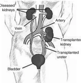
Kidney transplantation is the best option for patients suffering from end-stage kidney diseases, improving survival and quality of life compared with maintenance dialysis [28,29]. The site of placement of a transplanted kidney is shown in Figure 56.1.
However, long-term immunosuppressive treatments are nec essary to prevent graft rejection. Induction therapy generally includes an IL-2 receptor antagonist such as basiliximab or a lymphocyte-depleting agent such as rabbit antithymocyte glob ulin (ATG) or muromonab-CD3 (OKT-3). A combination of immunosuppressive medications is used as maintenance ther apy, including calcineurin inhibitors (CNIs) such as tacrolimus
Figure 56.1Kidney location after transplantation. Source:nih.gov.
or cyclosporine (CSP), antiproliferative agents such as myco phenolate or azathioprine, corticosteroids (prednisolone) and/or mammalian target of rapamycin inhibitors such as sirolimus or everolimus. The main causes of posttransplant morbidity and mortality are cardiovascular diseases, infections, malignancies, and graft dysfunction.
56.6.1.1 Animal studies
MSCs have demonstrated their ability to downregulate the immune response after kidney transplantation in several animal studies. Zhang et al transplanted Lewis rats with Wistar rat kidney and bone marrow (BM)-MSCs. Intravenous 1× 10⁷ BM-MSCs were given 1 week before transplantation, immediately after reperfusion, and 1 and 2 weeks posttransplant and com pared with the effects of MSCs alone, CSP alone, and a combi nation of MSCs and CSP. They found that MSCs were able to preserve graft function, as assessed by serum creatinine level, and to prolong animal survival, but were not as effective as CSP [30]. Monotherapy with third-party BM-MSCs infused directly into the renal artery of the transplanted kidney (3× 10⁶cells injected soon after reperfusion) was studied in a syngeneic and allogeneic model of rat kidney transplantation. Compared with placebo, MSC-treated rats had better kidney function as assessed by serum creatinine level, creatinine clearance rate, proteinuria and diuresis, reduced histological damage as assessed by the degree of tubulitis and vasculitis, and a reduced inflammatory infiltrate with fewer ED1+
and CD8+ cells [31]. ED1 is a rat marker of activated monocytes, macrophages, DCs, and micro glial cells and is also known as CD68. Tubulitis is a defining feature for the diagnosis and management of acute renal allograft rejection.
The efficacy of syngeneic MSCs to prevent IRI was also demonstrated in a rat kidney transplantation model with pro longed cold ischemia. Rats received CSP 1.5 mg/kg/per day for 2 days and three intravenous injections of 2.5× 10⁶or 5.0× 10⁶ MSCs 7 days before, immediately after, and 1 day after
transplantation. MSC administration resulted in reduced intra-graft gene expression of proinflammatory cytokines, chemo kines, and intercellular adhesion molecule-1, and in a reduced infiltration of antigen-presenting cells in the renal allograft [32]. The mechanisms of MSC-induced allograft tolerance are not yet fully elucidated, but generation of Tregs and tolerogenic DCs and production of indoleamine 2,3-dioxygenase appear to be involved in mice [33]. The importance of the route of adminis tration has been pointed out in rats, as intra-arterial injection was shown to be more efficient than intravenous injection, with a more rapid functional recovery as assessed by serum creatinine levels and reduced inflammatory infiltrate in the graft [34]. These findings suggested that a high concentration of MSCs at the site of immunological injury may be crucial for the efficacy of MSCs in this setting.
With regard to the timing of MSC administration, experi ments in mice have shown that syngeneic MSCs injected intra venously after transplantation 2 days posttransplant localized mainly in the graft, where they were responsible for local inflammatory reaction and failed to enhance graft survival, while MSCs injected 7 days and 1 day before transplantation localized in lymphoid organs, promoting Treg expansion and significantly prolonging allograft survival [35]. However, it has been shown in rats that a delayed third-party MSC injection was also beneficial, protecting the kidney allograft from injury development as shown by reduced interstitialfibrosis and tubular atrophy, lower inflammatory cell infiltration, and improved balance between proinflammatory and anti-inflammatory cytokines [36].
The long-term safety of MSC administration has been ques tioned by one group, who observed an unexpected increase in MSC-related life-threatening events in rats, including throm botic microangiopathy, infarctions, and infections [37]. So far there has been no other report of such MSC-related adverse events in transplant recipients.
56.6.1.2 Human studies
The first studies on human cells showed that in vitro donor-derived adipose MSCs were able to significantly inhibit recipient antidonor reactivity and proliferation of CD4+ and CD8+ T lymphocyte subsets in mixed lymphocyte reactions [38]. Com bined MSC and hematopoietic stem cell co-transplantation compared with hematopoietic stem cell transplantation alone produced significantly higher peripheral blood chimerism, better kidney transplant function as assessed by serum creatinine levels, and improved survival [39]. Intra-osseous injection of donor MSCs into the BM at a dose of 1× 10⁶cells/kg at the time of kidney transplantation was evaluated in a pilot study of seven patients undergoing living donor kidney transplantation. The procedure was safe, but three patients had biopsy-proven acute rejection that was controlled with corticosteroid pulse therapy. Donor-specific T cell proliferation was observed in two patients, while Treg priming responses were observed in two others associated with increased IL-10 levels in the peripheral blood [40].
56.6.1.3 Mesenchymal stromal cells versus conventional induction therapy
Perico et al. evaluated the pretransplant infusion of autologous BM-MSCs in two living-related kidney transplant recipients. One patient had corticosteroid-sensitive acute cellular rejection of the kidney 2 weeks posttransplant, but both patients had excellent renal function at 1 year follow-up. Circulating memory CD8+T cell numbers and donor-specific CD8+T cell cytolytic responses were reduced. Compared with patients receiving basiliximab induction therapy, CD4+FoxP3+ Treg expansion was not increased [41].
Tan et al. reported a randomized controlled trial of living donor kidney transplantation in 156 patients who were divided into three groups: a control group receiving antibody induction therapy versus two groups receiving autologous BM-MSCs at a dose of 1–2 × 10⁶/kg administered at the time of kidney graft
reperfusion and 2 weeks later plus either low-dose or standard-dose CNI treatment. Graft survival was similar, but MSC infusion resulted in fewer acute rejections at 6 months, no corticosteroid-resistant rejection, accelerated renal function recovery, and a reduced rate of opportunistic infections. How ever, acute rejection rates and glomerularfiltration rates were not different at 1 year postransplant [42].
56.6.1.4 Mesenchymal stromal cells versus conventional maintenance therapy
Perico et al. also investigated the effect of autologous BM-MSCs in two living-related kidney transplant recipients 7 days after transplantation. Increasing levels of CD4+ CD25high FoxP3+ CD127+Tregs and inhibition of memory/effector CD8+T cell expansion were observed, resulting in a long-term tolerogenic environment compared with the control group. However, patients suffered from engraftment syndrome with transient renal dysfunction a few days after MSC administration, and biopsies showed a focal inflammatory infiltrate with neutrophils and MSCs and complement C3 deposition [43]. (Engraftment syndrome characterized by fever, rash, pulmonary edema, weight gain, liver and renal dysfunction, and/or encephalopathy usually occurs at the time of neutrophil recovery after hematopoietic cell transplantation).
Another recent pilot study with 12 patients receiving living-donor related kidneys compared living-donor-derived BM-MSC infu sions with afirst dose of 5 × 10⁶cells into the renal allograft artery at the time of kidney transplantation and a second intravenous dose of 2× 10⁶cells/kg 1 month later plus low-dose (50% of usual) tacrolimus versus a control group who received no MSCs and standard doses of tacrolimus. Patients also received myco phenolate mofetil (MMF) and methylprednisolone. All patients had stable renal function at 1 year posttransplant despite reduced tacrolimus doses in the MSC group, and only one episode of acute rejection occurred (in the control group). Significantly increased B cell levels were observed in the MSC group 3 months after transplantation. There were no MSC-related adverse events [44].
56.6.1.5 Mesenchymal stromal cells as treatment for kidney rejection
In a phase I trial, Reinders et al. studied two intravenous infusions of 106autologous BM-MSCs per kilogram as treatment for kidney rejection in six patients. MSC infusions were well tolerated, and no treatment-related serious adverse events were reported. Biopsies were performed after MSC administration in two patients and showed resolution of tubulitis without intersti tialfibrosis/tubular atrophy. Five patients had downregulation of peripheral blood mononuclear cell proliferation and three patients developed an opportunistic viral infection [45].
56.6.1.6 Mesenchymal stromal cells as a desensitizing treatment
In a report of three cases, infusion of 50× 106donor-derived MSCs led to successful desensitization of patients with a positive lymphocyte cross-match before kidney transplantation [46].
56.6.1.7 Ongoing trials
56.6.1.7.1 Mesenchymal stromal cells in addition to conven tional therapy
The Liège trial (NCT01429038) is a single-center, open-label prospective phase I–II study that includes 10 kidney transplant recipients receiving a kidney from a deceased donor (and 10 liver transplant recipients who will be described later). In addition to induction therapy of standard immunosuppression with anti-IL 2 antibodies and maintenance therapy of tacrolimus, MMF, and corticosteroids, patients receive an intravenous infusion of third-party MSCs at a dose of 1.5–3.0 × 10⁶
/kg on postoperative day 3 (±2 days). The 10 study patients will be compared at 1 year posttransplant with a control group of 10 similar kidney trans plant recipients. The primary endpoint is safety of the MSC infusion, including infusional toxicity and incidence of infections and cancers. Secondary endpoints are patient and graft survival; graft function assessed by the number of posttransplant hemo dialysis episodes required and serum creatinine levels; biopsy-proven rejection rates using the Banff classification; feasibility and safety of weaning or decreasing immunosuppression, recip ient immune function as assessed by T cell blood populations, including Tregs, T-cell receptor excision circle quantification, Vβ repertoire diversity, pathogen-specific T cells, anti-organ donor HLA antibodies; and potential development of anti-MSC donor HLA antibodies.
Thefirst Bergamo trial (NCT00752479) is studying the effects of syngeneic ex vivo expanded MSCs in HLA-mismatched (one or two haplotype mismatches) living donor kidney transplanta tion. Patients are randomized into two groups. One group receives an intravenous injection of 2× 10⁶ MSCs/kg at the time of kidney transplantation. All patients receive conventional induction treatment with basiliximab, rabbit ATG and methyl prednisolone and maintenance treatment with CSP and MMF. Patients will be followed for 12 months. Primary outcomes include percentage inhibition of memory and/or naive T cell responses, induction of donor-reactive T cell anergy, and levels of
circulating Tregs. Secondary outcomes are safety of the MSC infusion, graft function, and graft rejection.
The second Bergamo trial (NCT02012153) is investigating the effect of autologous ex vivo expanded BM-MSCs in HLA-mis matched (one or two haplotype mismatches) living donor kidney transplantation. MSCs are administered intravenously on the day before kidney transplantation at a dose of 2× 10⁶MSCs/kg. Patients are followed for trial parameters for 1 year. Outcomes evaluated at 6 and 12 months include levels of circulating naive cells, memory cells, and Tregs, T cell function in mixed lympho cyte reaction, urinary forkhead box P3 (FOXP3) mRNA expres sion, and adverse events.
56.6.1.7.2 Mesenchymal stromal cells versus conventional induction therapy
The Fuzhou study (NCT00658073) is evaluating autologous MSCs as an alternative to antibody induction therapy in renal transplantation. Patients are randomized into three groups, two of them receiving two intravenous injections of MSCs immedi ately after releasing the renal artery clamp and 2 weeks after transplantation, while the third group receives standard induc tion therapy with anti-IL-2 receptor antibody. All patients will receive maintenance therapy with CNIs, MMF, and cortico steroids, but one of the MSC groups receives a reduced dose (80% less) of CNIs. Patients will be followed for trial parameters for 1 year. Primary outcomes are incidence of acute rejection and early renal function recovery. Secondary outcomes are patient and graft survival, and prevalence of adverse events.
56.6.1.7.3 Mesenchymal stromal cells versus conventional maintenance therapy
In a Leiden trial (NCT02057965), intravenous infusions of autologous BM-MSCs are being evaluated as maintenance ther apy in renal transplantation with a kidney from a deceased, a living unrelated, or a HLA-nonidentical living-related donor older than 50 years of age. Patients are randomized into two groups. One group will receive MSCs and everolimus versus tacrolimus and everolimus. Patients in the MSC group will receive two injections of 1–2 × 10⁶
MSCs/kilogram at 6 and 7 weeks after transplantation. After the second MSC infusion, tacrolimus is tapered off and withdrawn over a 2-week period. The primary outcome is kidneyfibrosis at 6 months compared with 4 weeks posttransplant. Secondary outcomes evaluated at 6 months posttransplant are renal function and proteinuria, complications (including opportunistic infections and cardiovas cular disease), adverse effects, a composite endpoint of efficacy failure (biopsy-proven acute rejection, graft loss, or death, and immunological parameters such as donor-specific antibodies.
56.6.1.7.4 Mesenchymal stromal cells as treatment for kidney rejection
Another Leiden trial (NCT00734396) is studying the safety and efficacy of autologous BM-MSCs to treat renal transplant rejec tion in HLA-DR mismatched kidney graft recipients. Patients
with subclinical rejection and/or an increase in interstitialfibro sis/tubular atrophy in the renal biopsy at 4 weeks or 6 months receive two intravenous injections of 1–2 × 10⁶
MSCs/kg 7 days apart. Primary outcomes are safety and feasibility of MSC administration at 2 years, while secondary outcomes are efficacy parameters, including the presence of late acute rejection in the 6-month biopsy compared with the 4-week biopsy, Sirius red staining for renal cortical matrix accumulation, and immuno logic response before and after MSC infusion.
56.6.2 Liver transplantation
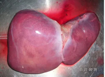
Since the pioneering experience in the 1960s LT has become established as the standard treatment of many end-stage liver diseases, including cirrhosis, primary liver cancer, fulminant hepatic failure, and a number of other metabolic or congenital hepatic diseases. Expected patient survival after LT is now more than 80% at 1 year and 60–70% at 5 years, dependent on the different indications for transplantation [47]. The excellent results of LT have led to a shortage of liver grafts that has been partially fulfilled by the development of living-related LT, and the procurement of liver grafts from brain-dead donors, aged cadaveric donors, or donors procured after cardiac death [48]. A human liver photographed at autopsy is shown in Figure 56.2.
Immunosuppressive regimens include CNIs, antimetabolites, mammalian target of rapamycin inhibitors, corticosteroids, and antibody-based therapies. A combination of CNIs and antime tabolites with tapering doses of corticosteroids during thefirst month is generally used to prevent graft rejection, while treat ment of rejection usually comprises boluses of intravenous corticosteroids or ATG for corticosteroid-resistant rejection.
56.6.2.1 Animal studies
Syngeneic BM-MSCs have shown their ability to home to the liver and to alleviate acute immunologic rejection in an orthotopic (normal location) rat LT model [49]. Similarly,
Figure 56.2 Human liver. Source:http://commons.wikimedia.org/wiki/File: Human_Hepar.jpg#/media/File:Human_Hepar.jpg.
adipose-tissue-derived MSCs infused intravenously at day 7 and day+3 as well as during the operation via the portal vein were able to significantly decrease acute rejection as assessed by reduced serum IL-2 levels and increased serum IL-10 levels as well as reduced hepatocyte apoptosis [50]. MSCs also demon strated their efficacy in a liver xenotransplantation model (ortho topic guinea pig to rat LT), enabling increased survival and alleviating acute rejection compared with placebo or with the powerful corticosteroid dexamethasone [51].
MSCs from different donors have shown equivalent effects in a rat LT model. Recipient-, donor-, or third-party-derived MSCs were infused intravenously at the time of surgery and once daily for 3 days thereafter. MSC administration resulted in signifi cantly improved recipient survival, inhibition of allograft rejec tion based on histological analysis and induction of CD4+CD25+ Foxp3+Tregs, with no significant difference between the three MSC-treated groups [52].
Du et al. studied the efficacy of the intravenous injection of MSC-CM in a 50% reduced-size rat LT model. In the MSC-CM group the authors observed a significantly lower release of liver injury biomarkers, a reduction in hepatocyte apoptosis, an increased proliferation of hepatocytes and sinusoidal endothelial cells, and reduced signs of inflammation in the graft as assessed by lower expression of several proinflammatory cytokines, reduced neutrophil infiltration and Kupffer cell activation and increased expression of vascular endothelial growth factor and matrix metallopeptidase (or metalloproteinase) 9. These obser vations were associated with increased survival in the MSC-CM group compared with the control group [18]. (Kuppfer cells were first observed by Karl Wilhelm von Kupffer in 1876 and are hepatic macrophages.)
In an attempt to improve MSC homing, the same group used MSCs overexpressing CXCR4 in a 50% reduced size rat LT model. MSCs reduced the release of liver injury biomarkers and apoptosis of hepatocytes. Although unable to further reduce liver injury, CXCR4 overexpression successfully enhanced MSC homing and hepatocyte proliferation, resulting in a significant survival benefit [53].
MSCs were also efficient in preventing, but not treating, acute graft-versus-host disease (GVHD) following LT. Rats that received donor- or recipient-derived BM-MSCs from day 0 to day 6 after LT (5× 10⁶ MSCs/day) had a significantly longer survival and no typical LT-associated GVHD symptoms, while administration of MSCs after the onset of symptoms was ineffective for treating GVHD. The efficacy of MSC therapy was associated with higher Treg ratios in the peripheral blood [54].
Another use of MSCs in LT has been the in vivo delivery of engineered growth factors or cytokines. Intraportal infusion of 5× 10⁶syngeneic HGF-expressing MSCs immediately after liver reperfusion was performed in a 30% small-for-size rat LT model. HGF-expressing MSCs prevented liver failure and improved liver function and liver weight recovery. Moreover, HGF enhanced the incorporation of MSCs into injured livers, and
the engrafted cells and their progeny produced albumin [55]. In another study the same group showed that HGF-expressing MSCs significantly inhibited the formation of liver fibrosis in rats undergoing small-for-size rat LT with synergistic effects of the MSCs and HGF [56].
Similarly, genetic delivery of IL-10 by IL-10-engineered-MSCs has been tested. Compared with placebo, intravenous injection of syngeneic IL-10-transduced MSCs 30 min after transplantation resulted in longer survival, lower Banff scores, increased expres sion of the Treg-associated cytokines IL-10 and TGF-β1, and the transcription factor FOXP3, as well as decreased expression of T helper (Th)17 T-cell-related cytokines IL-17, IL-6, interferon-γ, tumor necrosis factor-α, and IL-23 and the retinoic-acid related orphan receptor gamma (RORγ) transcription factor (RORγt) [57]. RORγ is a protein that in humans is encoded by the RORC (RAR-related orphan receptor C) gene. RORγ is member of the nuclear receptor family of transcription factors. It is essential for lymphoid organogenesis, in particular lymph nodes and Peyer’s patches, but not the spleen. RORγ also plays an important regulatory role in thymopoiesis, by reducing apoptosis of thymocytes and promoting thymocyte differentia tion into proinflammatory Th17 cells. It also plays a role in inhibiting apoptosis of undifferentiated T cells and promoting their differentiation into Th17 cells, possibly by downregulating the expression of Fas ligand and IL2 respectively. RORγt drives production of the cytokine granulocyte-macrophage colony-stimulating factor in Th cells, which is essential for the effector phase of autoimmune neuroinflammation.
56.6.2.2 Ongoing trials in humans
The Liège trial (NCT01429038), mentioned earlier, is a single-center, open-label prospective phase I–II study that includes 10 liver transplant recipients (as well as the 10 kidney transplant recipients described earlier). In addition to standard immuno suppression with tacrolimus, MMF, and corticosteroids, patients receive an intravenous infusion of third-party MSCs at a dose of 1.5–3.0 × 10⁶MSCs/kg on the third postoperative day (±2 days).
The 10 study patients will be followed for trial parameters for 1 year and compared with a control group of 10 similar liver recipients. The primary endpoints are safety of the MSC infusion and the incidence of infections and cancers. The secondary endpoints are patient and graft survival; graft function as assessed by serum bilirubin levels, prothrombin time, hepatic transaminase, and GGT levels; biopsy-proven (Banff classifica tion) rejection rates; feasibility and safety of weaning or decreas ing immunosuppression; recipient immune function as assessed by T cell blood populations, including Tregs, T-cell receptor excision circle quantification, Vβ repertoire diversity, pathogen-specific T cells, and anti-organ donor HLA antibodies; and potential development of anti-MSC donor HLA antibodies.
The Beijing trial (NCT01690247) and the Guangdong trial (NCT02223897) are studying the addition of intravenous injec tions of 1× 10⁶umbilical cord MSCs/kg in addition to conven tional therapy. MSCs are administered once every fourth week
for 12 weeks in thefirst trial and once per week for the first month followed by once per month for 6 months in the second trial. In the Beijing trial, patients are followed for trial parameters for 48 weeks. Outcomes are rates of acute rejection and early liver function recovery, patient and graft survival, and incidence of adverse events. The Guangdong trial is focused on ischemic-type biliary lesions, and outcomes after 18 months include the inci dence of ischemic-type biliary lesions, and changes in hepatic enzymes and biliary blood supply as assessed by contrast-enhanced ultrasound examination.
56.6.3 Heart transplantation
Since the first human heart transplantation in 1967 this treatment has been the best option for selected patients with end-stage heart failure not remediable by more conserv ative measures. Despite significant improvements in survival since the introduction of current immunosuppression and better prevention and treatment of infections, there remain major complications, including allograft vasculopathy and other classical complications of solid organ transplantation, including graft failure, graft rejection, opportunistic infections, and malignancies.
56.6.3.1 Animal studies
In rats, mesenchymal progenitor cells have demonstrated their ability to migrate into heart allografts after systemic administra tion [58] and to participate in tissue repair during chronic rejection [59]. The effects of MSC administration on heart allograft survival are still unknown. Some authors have shown that in rats MSCs did not prolong allograft survival [60] and even accelerated allograft rejection when used alone [61] or when added to low-dose CSP [62], while other studies demonstrated increased heart allograft survival using MSCs alone [63–66] or when MSCs were added to MMF [60,67]. A combination of MSCs and low-dose sirolimus has enabled long-term heart graft survival of more than 100 days with normal histology [65]. The mechanisms of MSC effects on heart allografts are not yet elucidated, but seem to include CD4+ CD25+ Foxp3+ Treg and tolerogenic DC expansion [64,65], interferon-γ-induced suppression of T cell proliferation [60,61], and inhibition of endothelial activation with intercellular adhesion molecule 1 and E-selectin downregulation [61].
No trials in humans have been conducted yet.
56.6.4 Lung transplantation
Single or bilateral lung transplantation is a viable treatment option for patients with various end-stage lung diseases, such as chronic obstructive pulmonary disease, idiopathic pulmonary fibrosis, cystic fibrosis, emphysema due to alpha-1 antitrypsin deficiency, and idiopathic pulmonary arterial hypertension. Lung transplant recipients are exposed to complications that include primary graft dysfunction, infections, malignancies, and both acute and chronic rejection. Chronic lung allograft manifests as a bronchiolitis obliterans syndrome (BOS).
Endothelin-1-stimulated MSCs may play a pathologic role in this phenomenon by differentiating locally into myofibroblastic cells [68]. However, another group demonstrated that resident lung MSCs isolated from human lung allografts inhibited T cell proliferation in vitro [69].
There is only one preclinical study of the administration of MSCs in lung transplantation, and this demonstrated the ability of IL-10-engineered MSCs to prevent lung IRI in mice [26].
The immunomodulatory properties of MSCs are currently being assessed in patients with BOS after lung transplantation. In the Prince Charles Hospital trial in Brisbane, Australia (NCT01175655), patients with BOS will receive MSCs from a third-party donor at a dose of 2× 10⁶/kg twice weekly for 2 weeks. Safety, pulmonary function, and survival will be eval uated at 12 months. In a Mayo Clinic trial (NCT02181712), patients with BOS will receive an intravenous injection of 2–4 × 10⁶
allogeneic MSCs/kg. Adverse events and pulmonary function will be assessed after 2 weeks.
56.6.5 Pancreas and islet transplantation
Pancreas or islet transplantation is performed in order to restore glucose-regulated endogenous insulin secretion, which allows insulin independence and improves glycemic control in patients with diabetes mellitus. However, pancreas or islets available for transplantation are scarce, and long-term pancreatic transplant survival rates are unsatisfactory, partly due to the lack of markers for pancreas graft rejection. As in other organ transplantations, long-term immunosuppression with several drugs is often required posttransplant.
MSCs have the ability to differentiate into insulin-producing cells both in vitro [70] and in vivo in animals [71,72] and humans [73,74]. Moreover, autologous [75], syngeneic [76–78] or allogeneic [78–80] MSCs have demonstrated immunomodu latory properties in animal models of islet transplantation. Complex mechanisms are involved including secretion of soluble factors (such as matrix metalloproteinase-2 and -9 [76] and IL 10 [81]), improvement of graft revascularization [77], and effects on immune cells (including the inhibition of maturation and function of DCs [82], downregulation of memory T cells [80], and induction of regulatory T cells [83]). Moreover, in non human primates, co-transplantation of allogeneic donor MSCs and islet cells led to significantly enhanced islet engraftment and function at 1 month posttransplant, and additional infusions of donor or third-party MSCs successfully treated rejection [84]. In addition to these effects, MSCs provide a supportive micro environmental niche through the secretion of paracrine factors and the deposition of extracellular matrix in vitro [85,86], in pre-transplantation co-cultures [87], and in vivo [88]. Finally, as in LT, MSCs are able to prevent IRI in animal models of islet transplantation [89,90].
There is only one ongoing clinical trial on co-transplantation of allogeneic islets and autologous MSCs. Primary outcomes are exogenous insulin requirement, glycated hemoglobin, and glu cose and C-peptide levels. Secondary outcomes are liver and
kidney function, portal vein ultrasound appearance, auto antibodies, and complete blood count (NCT00646724).
56.6.6 Bowel transplantation
There is one case report of successful administration of third-party-derived BM-MSCs to treat severe refractory bowel dys function secondary to infection after bowel transplantation [91]. More recently, Doğan et al. gave an intra-arterial infusion of autologous BM-MSCs (three doses of 1× 10⁶MSCs/kg) at the time of reperfusion and on days 15 and 30 posttransplant in six patients undergoing small intestine transplantation. All patients received anti-thymocyte immunoglobulin and corticosteroids as induction therapy and a CNI using low-dose tacrolimus as maintenance therapy. Four patients developed acute rejection. Three did not survive, of whom one did not respond to bolus corticosteroid therapy and two developed severe fungal and bacterial infections after corticosteroids. Two patients did not experience rejection and had no complications with good quality of life during the 30 months of trial follow-up [92].
56.7 Conclusions
Studies are providing increasing evidence of MSC benefits in solid organ transplantation. These benefits appear to be obtained by a combination of three major properties of MSCs: immuno modulation, tissue/organ repair, and prevention of IRI. Complex mechanisms are involved, and further research is required to elucidate them. Convincing animal studies in kidney transplan tation and LT, and to a lesser extent in lung and islet transplan tation, have led to the initiation of human trials. Initial results are promising in kidney transplantation. In heart transplantation, conflicting data have been obtained and more research is needed in animal models before human clinical trials can be initiated. Major goals in thisfield are reduction of graft rejection rates, improvement of organ and patient survival, and avoidance of treatment-related adverse effects through dose reduction or withdrawal of immunosuppressive drugs.
References
1 N. Pilat, C. Klaus, E. Schwaiger, T. Wekerle. Hurdles to the induction of tolerogenic mixed chimerism. Transplantation 2009;87:S79–S84. 2 F. C. Popp, E. Eggenhofer, P. Renner, et al. Mesenchymal stem cells can affect solid organ allograft survival. Transplantation 2009;87:S57–S62. 3 S. M. Devine, C. Cobbs, M. Jennings, et al. Mesenchymal stem cells distribute to a wide range of tissues following systemic infusion into nonhuman primates. Blood 2003;101:2999–3001.
4 L. A. Marquez-Curtis, A. Janowska-Wieczorek. Enhancing the migration ability of mesenchymal stromal cells by targeting the SDF-1/CXCR4 axis. Biomed Res Int 2013;2013:561098.
5 A. Bartholomew, C. Sturgeon, M. Siatskas, et al. Mesenchymal stem cells suppress lymphocyte proliferation in vitro and prolong skin graft survival in vivo. Exp Hematol 2002;30:42–48.
6 M. J. Hoogduijn, F. C. Popp, A. Grohnert, et al. Advancement of mesenchymal stem cell therapy in solid organ transplantation (MISOT). Transplantation 2010;90:124–126.
7 K. Le Blanc, C. Götherström, O. Ringdén, et al. Fetal mesenchymal stem-cell engraftment in bone after in utero transplantation in a patient with severe osteogenesis imperfecta. Transplantation 2005;79:1607–1614.
8 M. Sundin, O. Ringdén, B. Sundberg, et al. No alloantibodies against mesenchymal stromal cells, but presence of anti-fetal calf serum antibodies, after transplantation in allogeneic hematopoietic stem cell recipients. Haematologica 2007;92:1208–1215.
9 A. J. Nauta, G. Westerhuis, A. B. Kruisselbrink, et al. Donor-derived mesenchymal stem cells are immunogenic in an allogeneic host and stimulate donor graft rejection in a non-myeloablative setting. Blood 2006;108:2114–2120.
10 K. H. Grinnemo, A. Månsson, G. Dellgren, et al. Xenoreactivity and engraftment of human mesenchymal stem cells transplanted into infarcted rat myocardium. J Thorac Cardiovasc Surg 2004;127: 1293–1300.
11 N. Eliopoulos, J. Stagg, L. Lejeune, et al. Allogeneic marrow stromal cells are immune rejected by MHC class I- and class II-mismatched recipient mice. Blood 2005;106:4057–4065.
12 M. Crop, C. Baan, W. Weimar, M. Hoogduijn. Potential of mesen chymal stem cells as immune therapy in solid-organ transplantation. Transpl Int 2009;22:365–376.
13 F. C. Popp, P. Piso, H. J. Schlitt, M. H. Dahlke. Therapeutic potential of bone marrow stem cells for liver diseases. Curr Stem Cell Res Ther 2006;1:411–418.
14 T. K. Kuo, S. P. Hung, C. H. Chuang, et al. Stem cell therapy for liver disease: parameters governing the success of using bone marrow mesenchymal stem cells. Gastroenterology 2008;134:2111–2121. 15 J. Chen, H. C. Park, F. Addabbo, et al. Kidney-derived mesenchymal
stem cells contribute to vasculogenesis, angiogenesis and endothelial repair. Kidney Int 2008;74:879–889.
16 M. Morigi, M. Introna, B. Imberti, et al. Human bone marrow mesenchymal stem cells accelerate recovery of acute renal injury and prolong survival in mice. Stem Cells 2008;26:2075–2082.
17 D. F. McAuley, G. F. Curley, U. I. Hamid, et al. Clinical grade allogeneic human mesenchymal stem cells restore alveolar fluid clearance in human lungs rejected for transplantation. Am J Physiol Lung Cell Mol Physiol 2014;306:L809–L815.
18 Z. Du, C. Wei, K. Cheng, et al. Mesenchymal stem cell-conditioned medium reduces liver injury and enhances regeneration in reduced-size rat liver transplantation. J Surg Res 2013;183:907–915 19 P. Erpicum, J. M. Krzesinski, F. Jouret. Place de l’AMP-activated
protein kinase dans le preconditionnement ischemique renal. Neph rol Ther 2013;10:17–24.
20 P. Semedo, P. M. Wang, T. H. Andreucci, et al. Mesenchymal stem cells ameliorate tissue damages triggered by renal ischemia and reperfusion injury. Transplant Proc 2007;39:421–423.
21 F. Tögel, Z. Hu, K. Weiss, et al. Administered mesenchymal stem cells protect against ischemic acute renal failure through differenti ation-independent mechanisms. Am J Physiol Renal Physiol 2005;289:F31–F42.
22 F. Tögel, K. Weiss, Y. Yang, et al. Vasculotropic, paracrine actions of infused mesenchymal stem cells are important to the recovery from acute kidney injury. Am J Physiol Renal Physiol 2007;292: F1626–F1635.
23 P. Erpicum, O. Detry, L. Weekers, et al. Mesenchymal stromal cell therapy in conditions of renal ischaemia/reperfusion. Nephrol Dial Transplant 2014;29:1487–1493.
24 C. K. Sun, C. L. Chang, Y. C. Lin, et al. Systemic administration of autologous adipose-derived mesenchymal stem cells alleviates hepatic ischemia–reperfusion injury in rats. Crit Care Med 2012;40:1279–1290.
25 G. Jin, G. Qiu, D. Wu, et al. Allogeneic bone marrow-derived mesenchymal stem cells attenuate hepatic ischemia–reperfusion injury by suppressing oxidative stress and inhibiting apoptosis in rats. Int J Mol Med 2013;31:1395–1401.
26 E. Manning, S. Pham, S. Li, et al. Interleukin-10 delivery via mesenchymal stem cells: a novel gene therapy approach to prevent lung ischemia-reperfusion injury. Hum Gene Ther 2010;21:713–727. 27 M. B. Herrera, B. Bussolati, S. Bruno, et al. Exogenous mesenchymal stem cells localize to the kidney by means of CD44 following acute tubular injury. Kidney Int 2007;72:430–441.
28 P. Schnuelle, D. Lorenz, M. Trede, F. J. Van Der Woude. Impact of renal cadaveric transplantation on survival in end-stage renal failure: evidence for reduced mortality risk compared with hemodialysis during long-term follow-up. J Am Soc Nephrol 1998;9:2135–2141. 29 F. K. Port, R. A. Wolfe, E. A. Mauger, et al. Comparison of survival
probabilities for dialysis patients vs cadaveric renal transplant recipients. JAMA 1993;270:1339–1343.
30 W. Zhang, C. Qin, Z. M. Zhou. Mesenchymal stem cells modulate immune responses combined with cyclosporine in a rat renal transplantation model. Transplant Proc 2007;39:3404–3408. 31 M. De Martino, S. Zonta, T. Rampino, et al. Mesenchymal stem cells
infusion prevents acute cellular rejection in rat kidney transplanta tion. Transplant Proc 2010;42:1331–1335.
32 Y. Hara, M. Stolk, J. Ringe, et al. In vivo effect of bone marrow-derived mesenchymal stem cells in a rat kidney transplantation model with prolonged cold ischemia. Transpl Int 2011;24:1112–1123.
33 W. Ge, J. Jiang, J. Arp, et al. Regulatory T-cell generation and kidney allograft tolerance induced by mesenchymal stem cells associated with indoleamine 2,3-dioxygenase expression. Transplantation 2010;90:1312–1320.
34 S. Zonta, M. De Martino, G. Bedino, et al. Which is the most suitable and effective route of administration for mesenchymal stem cell-based immunomodulation therapy in experimental kidney transplan tation: endovenous or arterial? Transplant Proc 2010;42:1336–1340. 35 F. Casiraghi, N. Azzollini, M. Todeschini, et al. Localization of mesenchymal stromal cells dictates their immune or proinflamma tory effects in kidney transplantation. Am J Transplant 2012;12: 2373–2383.
36 M. Franquesa, E. Herrero, J. Torras, et al. Mesenchymal stem cell therapy prevents interstitialfibrosis and tubular atrophy in a rat kidney allograft model. Stem Cells Dev 2012;21:3125–3135. 37 M. Koch, A. Lehnhardt, X. Hu, et al. Isogeneic MSC application in a
rat model of acute renal allograft rejection modulates immune response but does not prolong allograft survival. Transpl Immunol 2013;29:43–50.
38 M. J. Crop, C. C. Baan, S. S. Korevaar, et al. Donor-derived mesenchymal stem cells suppress alloreactivity of kidney transplant patients. Transplantation 2009;87:896–906.
39 A. V. Vanikar, H. L. Trivedi, A. Feroze, et al. Effect of co-transplan tation of mesenchymal stem cells and hematopoietic stem cells as
compared to hematopoietic stem cell transplantation alone in renal transplantation to achieve donor hypo-responsiveness. Int Urol Nephrol 2011;43:225–232.
40 H. Lee, J. B. Park, S. Lee, et al. Intra-osseous injection of donor mesenchymal stem cell (MSC) into the bone marrow in living donor kidney transplantation; a pilot study. J Transl Med 2013; 11:96.
41 N. Perico, F. Casiraghi, E. Gotti, et al. Mesenchymal stromal cells and kidney transplantation: pretransplant infusion protects from graft dysfunction while fostering immunoregulation. Transpl Int 2013;26:867–878.
42 J. Tan, W. Wu, X. Xu, et al. Induction therapy with autologous mesenchymal stem cells in living-related kidney transplants: a randomized controlled trial. JAMA 2012;307:1169–1177. 43 N. Perico, F. Casiraghi, M. Introna, et al. Autologous mesenchymal
stromal cells and kidney transplantation: a pilot study of safety and clinical feasibility. Clin J Am Soc Nephrol 2011;6:412–422. 44 Y. Peng, M. Ke, L. Xu, et al. Donor-derived mesenchymal stem cells
combined with low-dose tacrolimus prevent acute rejection after renal transplantation: a clinical pilot study. Transplantation 2013;95:161–168.
45 M. E. Reinders, J. W. de Fijter, H. Roelofs, et al. Autologous bone marrow-derived mesenchymal stromal cells for the treatment of allograft rejection after renal transplantation: results of a phase I study. Stem Cells Transl Med 2013;2:107–111.
46 G. Saadi, F. Fadel, M. El Ansary, S. A. El-Hamid. Mesenchymal stem cell transfusion for desensitization of positive lymphocyte cross-match before kidney transplantation: outcome of 3 cases. Cell Prolif 2013;46:121–126.
47 R. Adam, E. Hoti. Liver transplantation: the current situation. Semin Liver Dis 2009;29:3–18.
48 O. Detry, V. Donckier, V. Lucidi, et al. Liver transplantation from donation after cardiac death donors: initial Belgian experience 2003–2007. Transpl Int 2010;24:611–618.
49 Z. F. Hong, X. J. Huang, Z. Y. Yin, et al. Immunosuppressive function of bone marrow mesenchymal stem cells on acute rejection of liver allografts in rats. Transplant Proc 2009;41:403–409.
50 C. D. Wan, R. Cheng, H. B. Wang, T. Liu. Immunomodulatory effects of mesenchymal stem cells derived from adipose tissues in a rat orthotopic liver transplantation model. Hepatobiliary Pancreat Dis Int 2008;7:29–33.
51 J. W. Wang, Y. B. Liu, B. Xu, et al. The study on immunomodulation of donor mesenchymal stem cells on discordant liver xeno transplantation. Zhonghua Wai Ke Za Zhi 2005;43:1254–1258 (in Chinese).
52 Y. Wang, A. Zhang, Z. Ye, et al. Bone marrow-derived mesenchymal stem cells inhibit acute rejection of rat liver allografts in association with regulatory T-cell expansion. Transplant Proc 2009;41: 4352–4356.
53 Z. Du, C. Wei, J. Yan, et al. Mesenchymal stem cells overexpressing C–X–C chemokine receptor type 4 improve early liver regeneration of small-for-size liver grafts. Liver Transpl 2013;19:215–225. 54 X. Xia, W. Chen, T. Ma, et al. Mesenchymal stem cells administered
after liver transplantation prevent acute graft-versus-host disease in rats. Liver Transpl 2012;18:696–706.
55 Y. Yu, A. H. Yao, N. Chen, et al. Mesenchymal stem cells over-expressing hepatocyte growth factor improve small-for-size liver grafts regeneration. Mol Ther 2007;15:1382–1389.
56 Y. Yu, L. Lu, X. Qian, et al. Antifibrotic effect of hepatocyte growth factor-expressing mesenchymal stem cells in small-for-size liver transplant rats. Stem Cells Dev 2010;19:903–914.
57 J. Niu, W. Yue, Y. Song, et al. Prevention of acute liver allograft rejection by IL-10-engineered mesenchymal stem cells. Clin Exp Immunol 2014;176:473–484.
58 G. D. Wu, J. A. Nolta, Y. S. Jin, et al. Migration of mesenchymal stem cells to heart allografts during chronic rejection. Transplantation 2003;75:679–685.
59 G. D. Wu, M. E. Bowdish, Y. S. Jin, et al. Contribution of mesenchy mal progenitor cells to tissue repair in rat cardiac allografts under going chronic rejection. J Heart Lung Transplant 2005;24: 2160–2169.
60 E. Eggenhofer, P. Renner, Y. Soeder, et al. Features of synergism between mesenchymal stem cells and immunosuppressive drugs in a murine heart transplantation model. Transpl Immunol 2011;25: 141–147.
61 E. Eggenhofer, J. F. Steinmann, P. Renner, et al. Mesenchymal stem cells together with mycophenolate mofetil inhibit antigen presenting cell and T cell infiltration into allogeneic heart grafts. Transpl Immunol 2011;24:157–163.
62 S. Inoue, F. C. Popp, G. E. Koehl, et al. Immunomodulatory effects of mesenchymal stem cells in a rat organ transplant model. Transplan tation 2006;81:1589–1595.
63 H. P. Zhou, D. H. Yi, S. Q. Yu, et al. Administration of donor-derived mesenchymal stem cells can prolong the survival of rat cardiac allograft. Transplant Proc 2006;38:3046–3051.
64 F. Casiraghi, N. Azzollini, P. Cassis, et al. Pretransplant infusion of mesenchymal stem cells prolongs the survival of a semiallogeneic heart transplant through the generation of regulatory T cells. J Immunol 2008;181:3933–3946.
65 W. Ge, J. Jiang, M. L. Baroja, et al. Infusion of mesenchymal stem cells and rapamycin synergize to attenuate alloimmune responses and promote cardiac allograft tolerance. Am J Transplant 2009;9:1760–1772.
66 S. M. Wu, W. X. Zhang, M. H. Wang, et al. Proteomic analysis of the immunosuppressive effects of mesenchymal stem cells in a rat heart transplantation model. Adv Clin Exp Med 2013;22:785–794. 67 F. C. Popp, E. Eggenhofer, P. Renner, et al. Mesenchymal stem cells
can induce long-term acceptance of solid organ allografts in synergy with low-dose mycophenolate. Transpl Immunol 2008;20:55–60. 68 M. Salama, P. Jaksch, O. Andrukhova, et al. Endothelin-1 is a useful
biomarker for early detection of bronchiolitis obliterans in lung transplant recipients. J Thorac Cardiovasc Surg 2010;140:1422–1427. 69 L. Jarvinen, L. Badri, S. Wettlaufer, et al. Lung resident mesenchymal stem cells isolated from human lung allografts inhibit T cell prolif eration via a soluble mediator. J Immunol 2008;181:4389–4396. 70 Y. Sun, L. Chen, X. G. Hou, et al. Differentiation of bone
marrow-derived mesenchymal stem cells from diabetic patients into insulin-producing cells in vitro. Chin Med J (Engl) 2007;120:771–776. 71 X. H. Wu, C. P. Liu, K. F. Xu, et al. Reversal of hyperglycemia in
diabetic rats by portal vein transplantation of islet-like cells gener ated from bone marrow mesenchymal stem cells. World J Gastro enterol 2007;13:3342–3349.
72 Y. Zhang, W. Shen, J. Hua, et al. Pancreatic islet-like clusters from bone marrow mesenchymal stem cells of human first-trimester abortus can cure streptozocin-induced mouse diabetes. Rejuvenation Res 2010;13:695–706.
73 S. M. Phadnis, M. V. Joglekar, M. P. Dalvi, et al. Human bone marrow-derived mesenchymal cells differentiate and mature into endocrine pancreatic lineage in vivo. Cytotherapy 2011;13:279–293. 74 E. Karaoz, A. Okcu, Z. S. Ünal, et al. Adipose tissue-derived mesenchymal stromal cells efficiently differentiate into insulin-producing cells in pancreatic islet microenvironment both in vitro and in vivo. Cytotherapy 2013;15:557–570.
75 M. G. Solari, S. Srinivasan, I. Boumaza, et al. Marginal mass islet transplantation with autologous mesenchymal stem cells promotes long-term islet allograft survival and sustained normoglycemia. J Autoimmun 2009;32:116–124.
76 M. Figliuzzi, R. Cornolti, N. Perico, et al. Bone marrow-derived mesenchymal stem cells improve islet graft function in diabetic rats. Transplant Proc 2009;41:1797–1800.
77 T. Ito, S. Itakura, I. Todorov, et al. Mesenchymal stem cell and islet co-transplantation promotes graft revascularization and function. Transplantation 2010;89:1438–1445.
78 B. Longoni, E. Szilagyi, P. Quaranta, et al. Mesenchymal stem cells prevent acute rejection and prolong graft function in pancreatic islet transplantation. Diabetes Technol Ther 2010;12:435–446. 79 H. Wu, D. Wen, R. I. Mahato. Third-party mesenchymal stem cells
improved human islet transplantation in a humanized diabetic mouse model. Mol Ther 2013;21:1778–1786.
80 F. R. Li, X. G. Wang, C. Y. Deng, et al. Immune modulation of co transplantation mesenchymal stem cells with islet on T and dendritic cells. Clin Exp Immunol 2010;161:357–363.
81 Y. H. Kim, Y. M. Wee, M. Y. Choi, et al. Interleukin (IL)-10 induced by CD11b+cells and IL-10-activated regulatory T cells play a role in immune modulation of mesenchymal stem cells in rat islet allografts. Mol Med 2011;17:697–708.
82 F. R. Li, C. Y. Deng, X. G. Wang, et al. The effect of co-transplantation of bone marrow-derived mesenchymal stem cells and islet on maturation and function of bone marrow-derived dendritic cells in recipient mice. Xi Bao Yu Fen Zi Mian Yi Xue Za Zhi 2010;26:646–649 (in Chinese).
83 D. M. Xu, X. F. Yu, D. Zhang, et al. Mesenchymal stem cells differentially mediate regulatory T cells and conventional effector T cells to protect fully allogeneic islet grafts in mice. Diabetologia 2012;55:1091–1102.
84 D. M. Berman, M. A. Willman, D. Han, et al. Mesenchymal stem cells enhance allogeneic islet engraftment in nonhuman primates. Dia betes 2010;59:2558–2568.
85 T. Bhaiji, Z. L. Zhi, J. C. Pickup. Improving cellular function and immune protection via layer-by-layer nanocoating of pancreatic islet beta-cell spheroids cocultured with mesenchymal stem cells. J Biomed Mater Res A 2012;100:1628–1636.
86 N. E. Davis, L. N. Beenken-Rothkopf, A. Mirsoian, et al. Enhanced function of pancreatic islets co-encapsulated with ECM proteins and mesenchymal stromal cells in a silk hydrogel. Biomaterials 2012; 33:6691–6697.
87 C. L. Rackham, P. K. Dhadda, P. C. Chagastelles, et al. Pre-culturing islets with mesenchymal stromal cells using a direct contact config uration is beneficial for transplantation outcome in diabetic mice. Cytotherapy 2013;15:449–459.
88 K. S. Park, Y. S. Kim, J. H. Kim, et al. Trophic molecules derived from human mesenchymal stem cells enhance survival, function, and angiogenesis of isolated islets after transplantation. Transplant 2010;89:509–517.
89 Y. Lu, X. Jin, Y. Chen, et al. Mesenchymal stem cells protect islets 91 C. D. Ceresa, R. N. Ramcharan, P. J. Friend, A. Vaidya. from hypoxia/reoxygenation-induced injury. Cell Biochem Funct Mesenchymal stromal cells promote bowel regeneration after
2010;28:637–643. intestinal transplantation: myth to mucosa. Transpl Int 2013;
90 J. Zhao, S. He, C. Wang, et al. Protective effect of bone marrow 26:e91–e93.
mesenchymal stem cells on islets from hypoxia/reoxygenation- 92 S. M. Doğan, S. Kılınç, E. Kebapçı, et al. Mesenchymal stem cell induced injury. Zhongguo Xiu Fu Chong Jian Wai Ke Za Zhi therapy in patients with small bowel transplantation: single center 2013;27:910–915. (in Chinese). experience. World J Gastroenterol 2014;20:8215–8220.