1
T
T
H
H
È
È
S
S
E
E
En vue de l'obtention duD
D
O
O
C
C
T
T
O
O
R
R
A
A
T
T
D
D
E
E
L
L
’
’
U
U
N
N
I
I
V
V
E
E
R
R
S
S
I
I
T
T
É
É
D
D
E
E
T
T
O
O
U
U
L
L
O
O
U
U
S
S
E
E
Délivré par l’Université Toulouse III – Paul Sabatier Discipline ou spécialité : Cancérologie
JURY
Pr Thierry LEVADE, Professeur des Universités Président Dr Fabienne FOUFELLE, Directeur de Recherche, INSERM Rapporteur Dr Frederic BOST, Directeur de Recherche, CNRS Rapporteur Pr Charles DUMONTET, Professeur des Universités Rapporteur Pr Guo-sheng Ren, Professeur de l`Universités Medicale de Chongqing Invités
Pr Catherine Muller, Professeur des Universités Directeur de thèse Pr Philippe Valet, Professeur des Universités Directeur de thèse
Ecole doctorale : Ecole doctorale Biologie Santé Biotechnologies Unité de recherche : IPBS CNRS UMR5089
Directeurs de Thèse : Pr Catherine Muller et Pr Philippe Valet
Rapporteurs: Fabienne FOUFELLE, Frederic BOST, Charles DUMONTET
Présentée et soutenue par Yuan Yuan WANG Le 25 September 2013
Titre : Deciphering the crosstalk between breast cancer cells and tumour-surrounding
2
Acknowledgment
First and foremost, I would like to express the deepest appreciation to my “science parents”: Prof. Catherine Muller and Prof. Philippe Valet for inspiring me and for providing such an interesting project for me to work during these four years. Thank you for your direction and your help in the development of my scientific mind: innovative concept, insistence, enthusiasm, strict for the science, and how to give a speech in front of people. I am honored to have worked with you and I have learned more from you than you will know.
Thanks to the past members in MIKA team: thanks to Bea not only for teaching me many experiment skills from my very beginning and start this interesting project, but also teaching me how to make crêpe and Moelleux a l`ananas et noix de coco, and also all the “useful French words” . I am really happy to have you such a kind and straight “little boss” and French friend. Thanks to Lud for picking me at the train station when I first arrived in Toulouse and helping me a lot from the life to the work at the beginning. Thanks to Cathy Botanch for your welcome cake and your kindness.
Thanks to the present members in our team, you are really helpful and friendly: Steph for teaching me how to use confocal microscopy and all the IF analysis; Laurence, for reviewing my thesis manuscript in spite of your very busy schedule and I am enjoying discussing with you; The “Prostate boys”: Victor and Adrien, the rest “Breast girls” Camille Lehood, hey chouette! and Delphine, “Melanoma princess” Ikrame and “Hypoxia queen” Fred, Your lively discussion at group meeting have made science come to life, I have truly valued our scientific interaction. I treasure all the fun we have made during these years and each time we got together, running, eating and drinking… and you are also very good French teacher “Ca roule ma poule” “c’est parti mon kiki” I will frequently repeat them in order to keep link with you
Thanks to “Adipolab” members: especially, “Mice queen” Sophie for helping and teaching me the in vivo experiment, Camille for the β-oxidation detection and Cedric for providing me several antibodies.
Thanks to Madama Escourrou Ghislaine for all Immunohistochemistry analysis and I learnt a lot about “the microcosmic world” of breast cancer.
Thanks to other person in IPBS: Leyre, Guillaume, Piere, Emma, Ying, Jin, Zhong, Jiahui for you kind help.
Thanks to my thesis committee members: for your encouragement, insightful comments and hard questions.
I would like to acknowledge the financial support from CSC for 4 years that made my Ph.D work possible.
I have been fortunate to come across many funny and good Chinese friends: SuSu, Liang, Tian, Xiaoqian, Jian, Hang, Han, Lijun,Mingchun et al. without you, life would be bleak. Thanks for the movies, dinners, sports, concerts and plays we enjoy together.
My sincere thanks also go to Prof. Ren GuoSheng, for offering me this opportunity to study abroad. Lastly, I am profoundly thankful for the love, support and understanding from my parents, my grandma!
3 Abbreviation ACC ACL ACS ADF ADRP ADSCs ANLS AMPK ANP APC AOX AT ATGL ARF BMP7 BMAT BNP CAA CAC CAF CACT C/EBP CNP CK1 CLS COLVI CPT CSF-1 CGI-58 DFAT DC DGAT DG DLBCL EGF ER ECM EMT FABP FAP FAS FFA FGF FN FSP-1
Acetyl CoA carboxylase ATP citrate lyase
Acyl-CoA synthase
Adipocyte-Derived Fibroblast
Adipocyte differentiation-related protein Adipocyte-Derived Stem cells
Astrocyte-neuron shuttle AMP Activated Protein Kinase Atrial natriuretic peptide Adenomatous Polyposis Coli Acyl-CoA oxidase
Adipose tissue
Adipocyte Triglyceride Lipase ADP-ribosylation factor Bone morphogenetic protein 7 Bone marrow adipose tissue Brain natriuretic peptide Cancer-Associated Adipocyte Cancer associated cachexia Cancer-Associated Fibroblast Carnitine acylcarnitine translocase CAAT/Enhancer Binding Protein C-type natriuretic peptide
Casein Kinase 1 Crown like structure Collagen VI
Carnitine palmitoyltransferase
Macrophage Colony-Stimulating Factor-1 Comparative gene identification-58 Dedifferentiated Fat Cell
Dendritic Cell
Diacylglycerol acyltransferase Diacylglycerol
Diffuse large B cell lymphoma Epidermal Growth Factor Estrogen receptor
Extracellular matrix
epithelial-to-mesenchymal transition Fatty acid binding protein
Fibroblast Activated Protein Fatty Acid Synthase
Free fatty acid
Fibroblast Growth Factor Fibronectine
4 GSK-3β HDAC-1 HER2/neu HFD HGF HIF HSL IFN IGF-1 iNOS IL IMC KLF KG LD LDH-A LEF/TCF LCAD LHYD LKAT LRP MAGL MAPK MAT MCP MEC MMP MIC-1 MUFA mTOR NG2 NK NPR OB-R PAI1 PDGF PI3K PPAR PGC1α PLD PKA PDH PDK
Glycogene Synthase Kinase-3β Histone deacetylase-1
Human epidermal growth factor receptor 2 High fat diet
Hepatocyte Growth Factor Hypoxia-inducible factor Hormone sensitive lipase Interferon
Insuline-like Growth Factor-1 Inducible nitric oxide synthase Interleukine
Indice de masse corporelle
Kruppel-Like zinc finger transcription Factor Ketoglutarate
Lipid droplet
Lactate dehydrogenase A
Lymphoid-Enhancer-binding Factor/T-Cell-specific transcription Factor Long chain acyl-CoA dehydrogenase
Long chain enoyl-CoA hydratase Long chain 3-ketoacyl-CoA thiolase
Low-density lipoprotein Receptor-related protein MonoAcylGlycerol Lipase
Mitogen Activated Protein Kinase Mammary adipose tissue
Monocyte Chemoattractant Protein Matrice Extra-Cellulaire
Matrix MetalloProtease
Macrophage inhibitory cytokine 1 Monounsaturated fatty acid Mammalian target of rapamycin Neuron-Glial Antigen-2
Natural Killer
Natriuretic peptide receptor Leptin receptor
Plasminogen activator inhibitor 1 Platelet-Derived Growth Factor PhosphoInositol-3-kinase
Peroxisome Proliferator Activated Receptor PPARγ co-activator 1α
Phospholipase D Protein kinase A
Pyruvate dehydrogenase
5 PML ROS SAT SCO2 SCD S1P SHBG αSMA SREBP STAT SETDB1 SVF TADC TAM TCF TEC TAG TGF TNC TNF TFP TIP47 TIGAR TLR4 UCP1 USP2a VAT VEGF Wnt Promyelocytic leukaemia Reactive oxygen species Subcutaneous adipose tissue
Synthesis of cytochrome C oxidase protein Stearoyl-CoA desaturase
Sphingosine 1-Phosphate Sex Hormone Binding Globulin α-Smooth Muscle Actin
Sterol Regulatory Element Binding Protein Signal Transducer and activators of transcription SET domain bifurcated 1
Stroma vascular fraction
Tumor Associated Dendritic Cell Tumor Associated Macrophage T cell-specific transcription factor Tumor Associated endothelial cell Triacylglycerol
Transforming Growth Factor Tenascine-c
Tumor Necrosis Factor Trifunctional protein
Tail-interacting protein of 47kDa
TP53-Induced Glycolysis and Apoptosis Regulator Toll like receptor
Uncoupling protein 1
Ubiquitin-specific protease 2a Visceral adipose tissue
Vascular Endothelium Growth Factor Wingless-type MMTV integration site
6
Summary
INTRODUCTION ... 8
GENERAL INTRODUCTION ... 9
INTRODUCTION GENERALE ... 11
PART I: THE TUMOR MICROENVIRONMENT. ... 13
1. Abnormalities in the microenvironment during tumor progression ...13
2. Complex cellular components in solid tumors ...15
2.1 Vascular cells ... 15
2.2 Inflammatory/Immune cells ... 16
2.3 Fibroblasts ... 17
2.4 Adipocytes ... 22
PART II: ADIPOSE TISSUE AND CANCER INTERACTION... 23
1. The adipose tissue (AT) overview ...23
2. The adipocytes ...25
2.1 The cellular origins of adipocytes and their differentiation ... 27
2.2 Model systems used to study adipocyte in vitro ... 31
3. Physiological functions of adipose tissue ...32
3.1 Lipid droplet biogenesis ... 33
3.2 Fat mobilization from lipid droplets ... 35
3.3 Paracrine and endocrine role of adipose tissue ... 39
4. Different depots of adipose tissue ...42
4.1 Subcutaneous/Visceral Adipose Tissue (VAT/SAT) ... 42
4.2 Mammary adipose tissue (MAT) ... 42
4.3 Bone marrow adipose tissue (BMAT) ... 43
5. Adipose tissue in the obese individual ...44
6. Relationship between obesity and breast cancer ...47
6.1 Breast cancer at a glance ... 48
6.2 How do adipocytes influence breast cancer biology? ... 49
7. Paracrine role of adipocytes in tumor progression ...55
7.1 Evidence of adipocytes participate in breast cancer progression ... 55
7.2 Paracrine role of adipocytes ... 56
8. Contribution of ADSCs to breast cancer progression ...59
9. Oncological risk after autologous lipofilling grafting in breast cancer patients ...60
PART III: CANCER METABOLISM... 61
7
2. Diversity of energy production pathways of cancer cells ...64
2.1 Glutamine metabolism ... 64
2.2 Mitochondrial oxidative phosphorylation (OXPHOS) ... 67
3. Fatty acid metabolism in the limelight ...69
3.1 De novo fatty acid synthesis and the relationship with cancer ... 69
3.2 Cellular uptake and transport of long-chain fatty acids ... 72
3.3 Fatty acid β-oxidation ... 75
3.4 Mitochondrial fatty acid β-oxidation in cancer ... 81
RESULTATS ... 88 REVIEW I: ... 89 REVIEW II: ... 90 ARTICLE I: ... 91 ARTICLE II: ... 92 ARTICLE III: ... 93
CONCLUSIONS AND PERSPECTIVES ... 94
BIBLIOGRAPHIE... 104
8
9
GENERAL INTRODUCTION
It is increasingly being recognized that human cancers in vivo do not behave as homogeneous collections of neoplastic cells but as complex multicellular systems in which malignant cells establish bidirectional interactions with neighbouring non-malignant cells. This interplay between tumor cells and the stroma—which comprises non-malignant cells and extracellular components— has been recognized as constituting a ‘tumor microenvironment’, and is now considered to be a hallmark of cancer biology. Among the many different cell types in stroma, mature adipocyte received relatively little attention according to the view that it is merely an energy-storing cell, relatively inert to its surrounding. However, adipocyte has been identified as a highly endocrine cell that can secrete various adipokines which are potentially able to modulate tumor behavior through heterotypic signaling process (Review 1). Since current epidemiological evidence convincingly shows that obesity is associated with greater cancer specific mortality and poor prognosis outcome, the involvement of fat cells in the progression of cancer appears of major interest.
The purpose of my PhD study is to shed in light the crosstalk between adipocytes and breast cancer cells and underscore the need to fully understand the role of this heterotypic signaling in modulating tumor behavior.
First of all, I participated to the efforts of my team to demonstrate that invasive cancer cells have a dramatic impact on surrounding adipocytes. Both in vitro and in vivo, these adipocytes exhibit a decrease of lipid content, a decreased expression of adipocyte markers and an activated state by the overexpression of proinflammatory cytokines. We named them “Cancer-Associated Adipocytes” (CAAs) (Experimental article 1).
Then, using combined in vitro and in vivo approaches, we identified for the first time that part of the cancer-associated fibroblasts (CAFs) which actively participate in tumor progression originate from the “dedifferentiation” of mature adipocytes. We named this new stromal cell population “Adipose-Derived Fibroblasts” (ADFs). They could participate in the desmoplastic reaction composed of several fibroblasts embedded in a rich extracellular matrix, commonly found in breast cancer and promote tumor dissemination (Experimental article 2).
Finally, we noticed that one of the key features of CAAs is the loss of lipid content observed both in
vitro and in human breast tumors. Given that tumor cells possess variety of metabolic
transformation according to their energy demand and the tumor microenvironment, CAAs may play a role in determining the metabolic phenotype of tumor cells. My work is to characterize a new metabolic crosstalk between breast cancer cells and tumor-surrounding adipocytes. During our investigation, we observed that part of this delipidation is due to the ability of tumor, but not normal
10 breast epithelial cells to induce lipolysis in adipocytes. We showed that the FFAs released during the lipolysis process by adipocytes are transferred, processed and stored in tumor cells as triglycerides (TG), contained in lipid droplets. When needed, these TG can be released as FFAs by an ATGL-dependent lipolytic pathway specifically present in tumor cells as shown both in vitro and in human tumors. These released FFAs are used for fatty acid β-oxidation (FAO), an activity present in cancer, but not normal, breast epithelial cells and regulated by coculture with adipocytes. Inhibition of these lipolytic/FAO coupled pathways either by pharmacological or gene repression strategies strongly decreases the invasive abilities of tumor cells that have been cocultivated with adipocytes (Experimental article 3).
Therefore, our results strongly reinforced the concept of a metabolic crosstalk is established between adipocyte and tumor cells. For the first time, we showed ATGL, an enzyme that plays a key role in regulating lipid network and invasiveness of breast cancer cells in an adipocyte-rich microenvironment.
In conclusion, such results provide an important concept in the understanding of the poor prognosis of breast cancer in the increasing obese population and warning about the oncological risk after autologous lipoaspirate grafting in breast cancer patients (Review 2) as well. Understanding the mechanisms of symbiosis between cancer cells and adipocytes should reveal new therapeutic possibilities.
11
INTRODUCTION GENERALE
Il est maintenant établi que les tumeurs humaines ne se comportent pas comme un ensemble homogène de cellules néoplasiques, mais comme des systèmes complexes pluricellulaires où les cellules malignes établissent des interactions bidirectionnelles avec les cellules qui les entourent. Ces interactions entre les cellules tumorales et les cellules du stroma (microenvironnement) sont désormais considérées comme des facteurs majeurs impliqués dans la biologie du cancer. Parmi les nombreux types cellulaires du stroma, les adipocytes matures ont à ce jour reçu relativement peu d'attention, en effet ces derniers ont longtemps étaient considérés comme de simples cellules de stockage d'énergie, relativement inertes à leur environnement. Cependant, l’adipocyte a été identifié comme une cellule à fortes capacités endocrines sécrétant diverses adipokines pouvant potentiellement moduler le comportement de la tumeur via un processus de signalisation hétérotopique (voir la revue Wang et al, 2012). Les données épidémiologiques actuelles montrent de façon convaincante que l'obésité est associée à une plus grande mortalité par cancer ainsi qu’à une faible survie sans progression. L'implication des cellules graisseuses dans la progression du cancer apparaît donc d'un intérêt majeur.
Le but de mon travail de thèse a été de mettre en lumière le dialogue bidirectionnel entre les adipocytes et les cellules cancéreuses mammaires et de souligner la nécessité de bien comprendre le rôle de cette signalisation hétérotopique dans la modulation du comportement de la tumeur. Tout d'abord, nous avons démontré que les cellules cancéreuses invasives ont un impact important sur les adipocytes environnants la tumeur. Aussi bien in vitro qu’in vivo, ces adipocytes présentent une diminution de leur contenu lipidique, une diminution de l'expression des marqueurs adipocytaires et un état activé par la surexpression de cytokines pro-inflammatoires. Nous les avons appelé les "adipocytes associés au cancer» (CAA) (voir le document Dirat et al, 2011). Par la suite, en combinant des approches in vitro et in vivo, nous avons identifié pour la première fois qu'une partie des fibroblastes associés au cancer (CAF) qui participent activement à la progression tumorale proviennent de la «dédifférenciation» d’adipocytes matures. Nous avons nommé cette nouvelle population de cellules stromales "fibroblastes du tissu adipeux» (ADF). Ces derniers pourraient participer à la réaction desmoplastique composée de fibroblastes intégrés dans une matrice extracellulaire riche, communément trouvées dans le cancer du sein et pourraient ainsi promouvoir la dissémination tumorale (voir le document Bochet et al, 2013). Enfin, nous avons remarqué que l'une des caractéristiques clés des CAA est la perte de leur contenu en lipides observée à la fois in vitro et dans des tumeurs mammaires humaines. Étant donné que les cellules tumorales possèdent une grande plasticité métabolique en fonction de leur besoin énergétique et du microenvironnement tumoral, les CAA pourraient jouer un rôle dans la détermination du phénotype
12 métabolique des cellules tumorales. Mon travail se porte sur la caractérisation d’un nouveau dialogue métabolique entre les cellules du cancer du sein et les adipocytes environnants. Au cours de nos recherches, nous avons constaté qu’une partie de cette délipidation est due à la capacité de la tumeur (mais pas des cellules épithéliales mammaires normales) à induire la lipolyse dans les adipocytes. Les acides gras libres (FFA) libérés au cours du processus de lipolyse sont ensuite transférés, modifiés et stockées dans les cellules tumorales sous la forme de triglycérides (TG), contenu dans les gouttelettes lipidiques. En cas de besoin, ces TG peuvent être libérés sous forme de FFA par une voie lipolytique spécifiquement présente dans les cellules tumorales. Ces FFA libérés peuvent être utilisés pour la β-oxydation mitochondriale, une activité présente dans les cancers, mais pas dans les cellules épithéliales mammaires saines. De manière intéressante, cette β-oxydation des lipides est positivement régulée dans les cellules tumorales co-cultivées avec des adipocytes et son inhibition, par des approches pharmacologiques ou de répression d’expression, diminue fortement, à la fois in vitro et in vivo, les capacités invasives des cellules tumorales qui ont été co-cultivées avec des adipocytes. Pour la première fois, dans des tumeurs primaires du sein, nos résultats ont identifié l'une des principales lipases, ATGL, comme étant fortement exprimée dans les cellules tumorales en contact des adipocytes comparé aux cellules situées à distance de la tumeur. Cette surexpression est intimement corrélée à un mauvais pronostic pour les patientes atteintes d’un cancer du sein. Ainsi, cette forte expression d’ATGL dans les cellules cancéreuses agressives leur confère une activité lipolytique élevée leur permettant d’utiliser les FFA dérivés des adipocytes pour augmenter leurs propriétés malignes via la β-oxydation des lipides (voir manuscrit Wang et al, 2013).
En conclusion, notre travail a permis de fournir un concept important dans la compréhension du mauvais pronostic du cancer du sein dans la population grandissante de sujets obèses, mais également d'alerter sur le risque oncologique lié à une autogreffe de tissu adipeux (lipoaspiration) (voir la revue Wang, 2013). La compréhension des mécanismes symbiotiques entre les cellules cancéreuses et les adipocytes devrait révéler de nouvelles possibilités thérapeutiques.
13
PART
I:
THE TUMOR MICROENVIRONMENT
.
1. Abnormalities in the microenvironment during tumor progression
In the past four decades, many researchers have focused their work primarily on tumor cells. Mutations either on oncogenes or tumor suppressor genes are important for tumor initiation. However, the concept of “seed and soil” that an appropriate host microenvironment (the soil) is needed for the optimal growth of tumor cells (the seed) has been also proposed long time ago [1]. Evidences now indicate that solid tumors comprise not only neoplastic cells but also normal stromal cells that have been recognized as constituting a ‘tumor microenvironment’. The crosstalk between cancer cells and stromal cells plays an important role in tumor survival, growth, metastasis, and response to antitumor therapy by secreting growth factors, chemokines, extracellular matrix (ECM) components [2];[3];[4]. The surrounding and interwoven stroma provides a connective-tissue framework to the tumor tissue. This framework includes a specific type of Extracellular matrix (ECM)—the tumor matrix—as well as cellular components such as fibroblasts, immune and inflammatory cells, and blood-vessel cells. As its constitution resembles that of the granulation tissue formed during wound healing, Hal Dvorak even defined a tumor as “a wound that never heals” [5].
Cancer cells themselves can alter their adjacent stroma to form a permissive and supportive environment for tumor progression by secreting growth factors and proteases, which can act in autocrine and paracrine manners. Concomitantly, cancer cells initiate the secretion of specific pro-migratory and –invasive ECM components, along with their respective receptors, and reduce the expression of protease inhibitors. The imbalance between proteases and their inhibitors then lead to the degradation of ECM components, resulting in the mobilization of matrix-bound growth factors and the generation of reactive ECM fragments. Together with the tumor-derived growth factors, these factors then induce angiogenesis as well as recruit and activate stromal inflammatory cells, fibroblasts and adipocytes, leading to the secretion of additional growth factors and proteases to amplify these signals. This cascade results in the establishment of an activated stroma that promotes malignant tumor progression (Figure 1).
The relative amount of stroma and its composition vary considerably from tumor to tumor and do not correlate with the degree of tumor malignancy [6]. But the interactive signaling between tumor and stroma contributes to the formation of a complex multicellular organ. In a manner similar to the development and function of normal organs, which occurs through reciprocal communication between different cell types, the interaction between cancer cells and their microenvironment can largely determine the phenotype of the tumor. For example, recent studies have shown that the establishment of human breast tumor xenografts in mice depends on the presence of human
tumor-14 derived stromal fibroblasts [7]. We will then first describe the main cellular component of the cancer stromal (vascular cells, inflammatory cells and fibroblasts) and their relative contribution in tumor progression.
Figure 1. Crosstalk between tumor cells and their activated stromal surroundings. Tumor cells initiate
the secretion of growth factors, ECM components, along with the imbalance between proteases and their inhibitors induce angiogenesis as well as recruit and activate stromal inflammatory cells, fibroblasts and adipocytes, leading to the secretion of additional growth factors and proteases to amplify these signals. (Mueller MM and Fusenig NE, 2004, Nat Rev Cancer).
15
2. Complex cellular components in solid tumors
2.1 Vascular cells
Like normal vessels, tumor blood vessels are composed of endothelial cells, mural cells (pericytes or smooth muscle cells) and basement membrane. However, all of these components are morphologically and/ or functional different from their normal counterparts [8].
Tumor-associated endothelial cells (TECs) are the major player in the formation of tumor vasculature through sprouting from existing blood vessels (a process called ‘angiogenesis’). During blood vessel formation, endothelial cells proliferate, migrate and form the inner layer of a lumen, followed by basement membrane formation and pericyte attachment. Angiogenesis is stimulated by excessive pro-angiogenic factors secreted by tumor or stromal cells in an oxygen-depleted microenvironment. Hypoxia stabilizes the hypoxia-inducible transcription factors (HIF), one of the key roles of HIF in hypoxia is the induction of pro-angiogenic factors, including VEGF, angiopoietin-2, PDGF and FGF, and downregulation of anti-angiogenic factors, such as thrombospondin [9];[10];[11]. New blood vessel formation is critical for tumor development and progression, as it delivers nutrients and oxygen to growing tumor and removes metabolic wastes. In addition, vascular endothelial cells form a barrier between circulating blood cells, tumor cells and the extracellular matrix (ECM), thus playing a central role in regulating the trafficking of leukocytes and tumor cells [12]. In this regard, endothelial cells are critical for boosting a host immune defense against cancer cells and for controlling tumor metastasis. However, the ‘gate-keeping’ function of endothelial cells in tumors is heavily compromised. TECs are not tightly associated with each other, resulting in wider interendothelial junctions that cause plasma leakage and hemorrhage [13]. Consequently, tumor vasculature is often leaky and less efficient in blood perfusion, leading to increase interstitial fluid pressure, hypoxia and acidic extracellular pH that may significantly affect the delivery and efficiency of chemotherapeutic drugs. The leaky blood vessels also facilitate the intravasation of tumor cells and promote tumor metastasis.
Several experiments have shown the difference between “normal” endothelial cells and endothelial cells within a tumor-context. For example, it has been reported that human hepatocellular carcinoma-derived endothelial cells, when compared to the ones from adjacent normal liver tissue, show increased apoptosis resistance, enhanced angiogenic activity and acquire more resistance to the combination of angiogenesis inhibitor with chemotherapeutic drugs [14]. Studies have also revealed distinct gene expression profiles of TECs and identified cell-surface markers distinguishing tumor versus normal endothelial cells [15].
In blood vessels, pericytes are smooth muscle cell-like cells that cover the vascular tube. They are intimately associated with endothelial cells and embedded within the vascular basement membrane, and play an important role in the maintenance of blood vessel integrity. Pericytes in tumors are
16 different from normal ones: in tumors, pericytes are often less abundant and more loosely attached to the endothelial layer [16];[17]. The abnormality in pericytes weakens the vessel wall and increases vessel leakiness. Thus, angiogenesis is the first compartment that as a good target for cancer therapies [18].
2.2 Inflammatory/Immune cells
Tumors are often infiltrated by inflammatory cells, such as macrophages, neutrophils, lymphocytes, mast cells, and myeloid progenitors. This phenomenon was initially observed by Rudolf Virchow more than a century ago and thought to be an immunological response attempting to eliminate cancer cells. Whereas immune cells play a role in recognizing and eradicating early cancer cells [19], mounting evidence has also shown that inflammatory cells within tumors can enhance tumor initiation and progression by helping cancer cells to acquire hallmark capabilities [4].
Epidemiologic and clinical studies show that approximately 25% of all human cancers in adults result from chronic inflammation [20]. For example, chronic viral or bacterial infection in stomach, liver and cervical, repeated solar irradiation of the skin, and prolonged exposure to tobacco smoke or other environmental chemicals in lung, contribute significantly to the chronic tissue injury and subsequently oncogenesis of these organs [21]. During this oncogenic event, persistent tissue injury result in changes in the cellular architecture of the epithelium and the surrounding stromal elements and promote genetic mutations and epigenetic alterations of epithelia cells. This change in tissue homeostasis can in turn lead to a chronic inflammatory response that never fades away, which then further promotes tumor growth, angiogenesis, invasion and metastasis through the activation of surrounding stromal cells and recruitment of different inflammatory cells.
Among the abundant inflammatory cells, macrophages form a major inflammatory population in most tumors and are important determinants of the inflammatory milieu. Broadly speaking, macrophages can be defined as classically (M1) or alternatively (M2) activated. M1 macrophages are involved in Type 1 reactions and are classically activated by microbial products, killing microorganisms and producing reactive oxygen and nitrogen intermediates. In contrast, M2 cells (Tumor associated macrophages; TAMs) are important component of infiltrated leukocytes in most malignant tumors, M2 macrophages can be identified using a number of markers, since they express arginase, mannose receptor, high-level IL-10, low level MHC II and IL-12. They produce many tumor-promoting factors, like EGF and VEGF, release cytokines and enzymes that promote angiogenesis and local and distant invasion, like VEGF and MMP9 and down-regulate expression of anti-angiogenic factors, like IL-12 to facilitate tumor progression [22];[23];[24]. Macrophages are derived from CD34+ bone marrow progenitors that continually proliferate and shed for their progeny into the bloodstream as pro-monocytes. In breast cancer, infiltrating TAMs correlate with
17 poor prognostic features, higher tumor grade, high vascular grade, increased necrosis and decreased disease-free survival [25]. Recently, one study found that CCL18 was highly expressed in TAMs and promoted the invasion and metastasis of cancer cells by triggering integrin clustering and enhancing their adherence to the ECM [26].
Other components of the inflammatory infiltrate also modulate tumor behavior. T-regulatory (T-reg) cells were first identified in 1971 by Gershon and Kondo [27], followed by reports of T cells suppressing the anti-tumor immune response, and then by identification of CD4+ T cells that suppressed autologous cytotoxic anti-tumor immune response [28]. These T cells are now classified as CD4+, CD25+, FoxP3+ and are thought to protect the host from autoimmune disease by suppressing self-reactive cells and therefore also blocking anti-tumor responses. Neutrophils also generate ECM degrading enzymes, reactive oxygen and nitrogen species. These bioactive factors promote cancer cell proliferation, invasion and resistance to apoptosis through, for instance, the interleukin-JAK/STAT pathway [29], and induce new blood vessel formation in the tumor. Extracellular matrix-degrading enzymes promote cancer cell invasion and metastasis, whereas accumulation of reactive oxygen and nitrogen species can cause DNA mutations, suppress DNA repair enzymes, increase genomic instability, and aggravate cancer progression.
While the tumor-promoting effects of infiltrating inflammatory cells have been well documented, certain types of immune cells, particularly cytotoxic T cells and natural killer cells, exhibit anti-tumor activities. The high numbers of these cells within a anti-tumor predict a favorable prognosis [30];[31]. Immune surveillance is considered as an important mechanism to inhibit carcinogenesis and maintain tumor dormancy [19]. Evading immune destruction by downregulating tumor antigens, suppressing immune cell function and other means is an emerging hallmark of cancer cells and plays important roles in cancer progression and metastasis [4].
2.3 Fibroblasts
2.3.1 Origin and markers of cancer-associated fibroblasts (CAFs)
Fibroblasts are elongated cells that show a fusiform or spindle-like shape in profile. Fibroblasts were first described in the late 19th century. They are the vascular, epithelial and non-inflammatory cells of the connective tissue and are its principal cellular component [32]. They are embedded within the fibrillar matrix of the connective tissue and are, to a large extent, responsible for its synthesis. Fibroblasts in mammals are highly heterogeneous, a recent study comparing the genome-wide expression patterns of 50 human fibroblast cultures isolated from 16 different sites showed that gene-expression patterns among fibroblast populations from distinct anatomical sites are as divergent as the gene-expression patterns observed among distinct lineages of white blood cells [33]. The major functions of fibroblasts include the deposition of ECM, the regulation of
18 epithelial differentiation, and the inflammatory responses [34]. Such increased activity is referred to as ‘activation’ (figure 2). Once the wound is repaired, the number of activated fibroblasts decreases significantly and the resting phenotype is thought to be restored. Unlike wound healing, but similar to organ fibrosis, the fibroblasts at the site of a tumor remain perpetually activated. This leads to tumor desmoplasia through excessive ECM deposition.
It has been demonstrated that cancer-associated fibroblasts (CAFs) are extremely abundant in the stroma of many tumors, including breast, prostate and pancreatic carcinomas [35];[36]. Of note is that CAFs are heterogeneous populations and their relative composition is greatly different among different tumors. The main activation markers are α-Smooth Muscle Actin (α-SMA), Fibroblast-Specific Protein (FSP), Platelet-Derived Growth Factor (PDGF) receptor-β and Fibroblast Activation Protein (FAP) [37];[35]. This large heterogeneity in marker expression for CAF subpopulations or in CAFs originating from different tumors may be explained by their possible miscellaneous origin. Indeed, CAFs are variously reported to stem from resident fibroblasts, bone marrow-derived progenitor cells, or from endothelial or cancer cells through endothelial-mesenchymal transition or epithelial-mensenchymal transition (EMT) [38] or a brand new origination from adipocytes (see article 2). At present, neither the origin of these cells, nor the extent to which their origin determines their contribution to tumor progression can be unanimously agreed upon.
The normal stroma in most organs contains a minimal number of fibroblasts in association with a physiological ECM whereas the reactive stroma is associated with an increased number of fibroblasts, enhanced capillary density and type-I-collagen and fibrin deposition. To date, transforming growth factor-β (TGFβ) and CXCL12/SDF-1 are regarded as the major tumor cell-derived factors affecting CAF activation through a TGFβ and CXCL12/SDF-1 autocrine signaling loop. Other profibrotic factors releases by cancer cells like epidermal growth factor (EGF), platelet-derived growth factor (PDGF) and fibroblast growth factor 2 (FGF2) can also act on resident fibroblasts and induce their activation [39]. Another important mechanism in the activation of CAFs is the downregulation of tumor suppressor genes such as p53, p21, PTEN, and caveolin-1 (CAV-1) [25].
2.3.2 The role of CAFs in cancer progression: Secretion of soluble factors
CAFs produce growth factors, cytokines, chemokines and ECM proteases to stimulate angiogenesis and cancer cell proliferation and invasion (figure 3) [40]. The existence of a large number of CAFs in tumors is often associated with poor prognosis [41]. Hepatocyte growth factor (HGF), EGF, bFGF, as well as cytokines such as SDF-1 and IL-6, are all substantially expressed by CAFs upon
19 contact with different tumor histotypes. For example, CAFs secret elevated levels of stromal cell-derived factor 1 (SDF-1; also called CXCL12) that facilitates angiogenesis by recruiting endothelial progenitor cells into the tumor [42]. SDF-1 can also interact with the CXCR4 receptor expressed on the surface of cancer cells, thus stimulating tumor cell growth and promoting tumor progression in vivo [43]. Furthermore, in breast adenocarcinoma, SDF-1 secreted by CAFs has been found driven by hypoxia-inducible factor-1 (HIF-1), which is in turn activated by hypoxia-mediated oxidative stress [44]. CAFs might contribute to epithelial-to-mesenchymal transition (EMT) in nearby cancer cells and promote their invasiveness [40]. TGFβ, another factor produced by CAFs, is a critical mediator of the EMT; IL-6 has been found to be 100-fold increased expression in CAFs compared with non-cancer-associated fibroblasts and also induces EMT in ER-positive breast cancer cell lines (MCF7, T47D) [45].
CAFs are also able to secrete several members of the MMP family as well as plasminogen activators. These enzymes may act mainly through 3 pathways below: 1) direct degradation of ECM; 2) cleavage of membrane-bound growth factors or cytokines as well as their receptors; 3) cleavage of cell adhesion molecules like cadherins, leading to an increased motility and EMT [46]. The expression of tumor (MMP1, MMP2 and MMP14) and stromal (MMP9, MMP13 and MMP14) matrix metalloproteinases is mandatory for squamous cell carcinoma progression [47]. MMP9 directly cleaves the extra-cellular domain of E-cadherin, promoting normal mammary epithelial cells to disaggregate and undergo EMT, promoting cancer-cell invasiveness [48]. MMP13 secreted by CAFs promotes tumor angiogenesis by releasing VEGF from the ECM, thereby leading to the increased invasion of squamous cell carcinoma or melanoma [49]. MMP1 also shows such a tumor-promoting effect. The protease-activated receptor PAR1, which is a tethered-ligand receptor, is activated by proteolytic cleavage of the extracellular domain by MMP1, promoting cancer cell migration and invasion through PAR1-dependent Ca2+ signals [50].
Alongside MMPs, CAFs from colon and breast carcinoma also express uPA and its receptor uPAR [51]. Cancer cells engage with their stromal fibroblasts a specific feed-forward loop mediated by paracrine bFGF and EGF, which induce uPA transcription in the fibroblasts. In turn, the serine protease system helps in activating these paracrine factors from the tumor cell surface/ECM [52].
20
Figure 2. Activated fibroblasts. a. Normal fibroblasts are embedded within the fibrillar ECM of connective
tissue, which consists largely of type I collagen and fibronectin. b. Fibroblasts can acquire an activated phenotype, which is associated with an increased proliferatvie activity and enhanced secretion of ECM protein such as type I collagen and tenascin C, and also fibronectin that contains the extra domain a (EDA-fibronectin) and SPARC (secreted protein acidic and rich in cysteine) (Kalluri and Zeisberg, 2006, Nat Rev Cancer).
Figure 3. Origins and functions of CAFs. A number of origins have been proposed for CAFs, including
bone marrow-derived cells (BDMC), response of normal fibroblasts to tumor-derived signals, genetic and epigenetic alterations in normal fibroblasts, or epithelial-mesenchymal transition (EMT) of normal or tumor epithelial cells. They have many effects on tumor cells (Allen M and Louise Jones J, 2011, J Pathol).
21 ECM remodelling
CAFs also generate an altered ECM environment. Most solid tumors exhibit a very different profile of ECM proteins in the stroma compared to their normal counterparts, and many of these proteins interact directly with tumor cells, via integrins and other cell surface receptors, to influence functions such as proliferation, apoptosis, migration and differentiation [53]. CAFs retain a major role in ECM remodeling since they are mainly responsible for the production of ECM proteins (i.e. collagens, fibronectin) as well as proteases and other enzymes involved in the posttranslational modification of ECM proteins themselves. Invasive carcinoma is often associated with expansion of the tumor stroma and increased deposition of ECM. Such increased deposition of ECM in tumors is known as desmoplasia. Indeed, many cancers are associated with desmoplasia, a fibrotic state characterized by an accumulation of type I and III collagens and by a degradation of type IV collagen [54];[55]. Tumor desmoplasia has been associated with the poor prognosis of cancers and it has also been observed in metastatic sites [56].
One way in which tensile strength can be modulated is via lysyl oxidase (LOX), an enzyme secreted primarily by fibroblasts that serves to cross-link collagens and elastin, increasing the insoluble matrix and contributing to tensile strength [57]. Recently, in a mouse model of breast carcinoma, treatment with LOX inhibitors led to a decrease of ECM cross links, preventing ECM stiffening and delaying tumor progression [58]. LOX pre-conditioning and stiffening of the mouse mammary fat pad also resulted in growth and invasion of premalignant mammary cells, suggesting that increased ECM stiffness can promote tumorigenesis as well as alter established tumor behavior [58]. In addition, ECM rigidity in tumor contributes together with impaired vascular and lymphatic mesh, to increase interstitial fluid pressure, leading to a reduced chemotherapeutics delivery inside tumors [59]. Together with ECM stiffness, interstitial flow is an important mechanical stress in the tumor stroma. It drives TGF-β1 and MMP-dependent fibroblast tissue invasion which, in turn, enhances tumor cell invasion by ECM remodeling [60]. These findings confirm that CAFs may serve as guidance structures that direct the migration of epithelial cancer cells by inducing protease-mediated ECM remodeling.
Fibronectin (FN) and hyaluronan—two other ECM components, mostly produced by CAFs, have been shown to play a role in tumor microenvironment. In tumors, there is frequent up-regulation of the ‘oncofetal’ forms of FN that contain extra-domain (ED)A, (ED)B, and IIICS sequences [61]. FN-EDA is required for the transduction of TGFβ signals, and the conversion of fibroblasts to myofibroblasts [62], whereas FN-EDB is particularly associated with neovascular structures in many different tumor types [63]. Stromal FN is positively associated with human tumor metastatic potential and MMP secretion [56], in addition, FN regulates ovarian cancer metastatic potential by promoting a ligand-independent activation of the c-Met proto-oncogene through binding to α5β1
22 receptor [64]. Vascular endothelial growth factor (VEGF) can be released by cancer cells themselves, but fibroblasts and inflammatory cells are also the principal source of host-derived VEGF. It induces microvascular permeability, which leads to the extravasation of plasma proteins such as fibrin, which in turn attract an influx of fibroblasts, inflammatory cells and endothelial cells. These cells produce ECM that is rich in fibronectin and type I collagen, both of which are conducive to initiating tumor angiogenesis [65]. CAFs mediated the overexpression of hyaluronan within the tumor microenvironment, and it has been shown to have a role in the recruitment of tumor-associated macrophages, which are key regulatory cells involved in tumor neovascularization through endothelial cell recruitment [66].
Tumor metabolism remodeling
Cancer cells mainly use glucose by glycolysis, producing lactate even in the presence of oxygen, a metabolic phenomenon called: Warburg Effect or Aerobic Glycolysis, whilst normal cells fully exploit glucose by oxidative phosphorylation. Recently, accumulating evidence showed that CAFs participate in the complex metabolism of tumors, engaging a bidirectional liaison with tumor cells, prompting them to overcome energy depletion due to the Warburg effect (This specific role of CAFs will be described in the third part of my thesis).
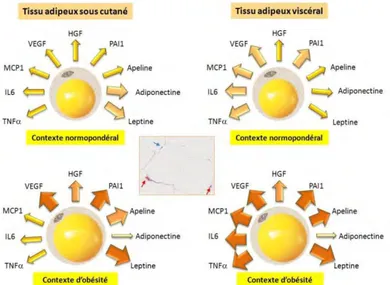
2.4 Adipocytes
Among the different cell types frequently found at close proximity of evolving tumors, little attention has been given to cells that compose the adipose tissue although a growing interest can be noted in recent years. Adipose tissue (AT) is consisting of mainly mature adipocytes and progenitors (preadipocytes and adipose-derived stem cells, ADSCs) that are contained in stroma vascular fraction (SVF). The role of adipose tissue, and more specifically adipocytes, in tumor initiation, growth, and metastasis, is a relatively new area of investigation. Emerging studies including our results clearly indicate that a bidirectional crosstalk is established between cellular components of AT and cancer cells and that the tumor-surrounding AT contributes to inflammation [67];[68], ECM remodeling [69];[70] as well as energy supply within the tumors [71]. Moreover, obesity is an epidemic problem nowadays in the world and is associated with several healthy problems including cancer. In next part, we will make an overall description of AT composition and function with special emphasis on the specificity of adipose depots, key aspects that need to be taken in account when paracrine effects of AT on tumor progression is considered. Try to deciphering the cellular and molecular mechanisms involved in it and to better understand the link between obesity and the poor prognosis of some cancers.
23
PART
II:
ADIPOSE TISSUE AND CANCER INTERACTION
1. The adipose tissue (AT) overview
For decades, adipose tissue was considered an inert mass of stored energy with some advantageous properties, such as its function as an insulating substance and as a mechanical support for more important structures. However, over the last two decades has seen a surge of interest in the study of adipose tissue, from its developmental biology to its physiology. What accounts for this new found respect for adipocytes? The discovery of leptin in 1994 presaged a growing awareness that adipocytes are essential regulators of whole-body energy homeostasis. These cells have been established as a dynamic organ that carries out several important physiological processes as diverse as haemostasis, blood pressure, immune function, angiogenesis and energy balance [72]. Another reason for the surge in interest in adipocytes is the awareness of abnormal fat accumulation is associated with healthy problems. For example, it is well known that overweight and obese individuals have a substantially greater risk of developing chronic diseases, such as cardiovascular disease (mainly ischemic heart disease and stroke), diabetes, respiratory failure, musculoskeletal disorders (especially osteoarthritis), and also cancer (endometrial, breast, kidney and colon) [73]. Hence, it is clear that a better understanding of the mechanisms linking adipose tissue development, function and expansion is required to improve our chances of identifying the most successful therapeutic approaches.
Adipose tissues are organized to form a large organ with discrete anatomy, specific vascular and nerve supplies, complex cytology, and high physiological plasticity [74];[75]. There are two main types of adipocyte, brown and white, which differ in several important properties (as discuss below). Even among white adipocytes, cells from different locations can have distinct molecular and physiological properties. The various adipose depots are the subcutaneous depots (inguinal, femoral, abdominal) which present in the deep layer of the skin, the visceral depots (mesenteric, omental, retroperitoneal, epididymal, gonadal, perivascular, and pericardial depots) [76], mammary depots and the bone marrow depots (Figure 4). All these specific regional depots exhibit differences in structure, function, composition, and secretion profiles. This is more evidently demonstrated by the links revealed between increased deep abdominal or visceral, but not peripheral, fat extent, and the metabolic disturbances associated with obesity [77].
24
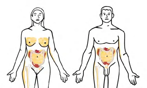
Figure 4. Distribution of adipose tissue in human body. White adipose tissue, mainly composed of
adipocytes, is located in subcutaneous (inguinal femoral in women) and in abdomen where it surrounds the internal organs (visceral adipose tissue). The adipose tissue is also found in bone, women-breast and around the men`s prostate (Laurent V et al, 2013, Médecine/sciences)
Figure 5. Components of adipose tissue. Adipocytes are the main cellular component of adipose tissue, the
other cell types that are present are precursor cells (including pre-adipocytes), fibroblasts, vascular cells and immune cells and these cells constitute the stromal vascular fraction of adipose tissue (SVF). (Ouchi N et al., 2011,Nat Rev Immunol)
25
2. The adipocytes
Adipose tissue is largely composed of adipocytes, but also contains a stromal vascular fraction made up of pericytes, endothelial cells, monocytes, macrophages, and pluripotent stem cells (Figure 5) [78];[79]. Two main types of adipocytes are easy to distinguish by morphology: white adipocytes are spherical cells with ~90% of their volume comprising a single cytoplasmic lipid droplet and a ‘squeezed’ nucleus, whereas brown adipocytes are polygonal cells with a roundish nucleus and several cytoplasmic lipid droplets (Figure 6). Brown adipocytes are also characterized by numerous large mitochondria packed with cristae. Mitochondria in brown adipocytes are marked by the expression of uncoupling protein 1 (UCP1), a unique protein that uncouples oxidative phosphorylation from ATP synthesis and thereby results in the production of heat (thermogenesis) [80];[81];[82]. Thus, white and brown adipocytes are quite different in their morphology and physiology: white adipocytes store energy for the metabolic needs of the organism, whereas brown adipocytes burn energy for thermogenesis. Both cell types are contained in the multiple depots of the adipose organ [83];[84]. White adipocytes are considered the dominant adipocyte subtype in adult humans with different sizes present in subcutaneous depots (mainly large adipocytes) and visceral depots (mainly small adipocytes) [85];[86]. Most brown adipose tissue in rodents is localized to the interscapular region. In human fetuses and newborns, BAT is found in axillary, cervical, perirenal, and periadrenal regions [80] but decreases shortly after birth and has traditionally been considered insignificant in adults, except perhaps in patients with pheochromocytoma, where adrenergic activity is extremely high [87], or in outdoor workers in northern climes subject to prolonged cold exposure [88]. A third type of adipocyte, the ‘beige’ or ‘brite’ (brown in white) adipocyte, has recently been identified which are the cells with intermediate morphology between that of white and brown adipocytes and has characteristics of both white and brown adipocytes. Like white adipocytes, basal UCP1 expression is low in beige adipocytes. However, like brown adipocytes, beige adipocytes respond to cyclic AMP (cAMP) stimulation by an increase of UCP1 expression [89];[90];[91].
26
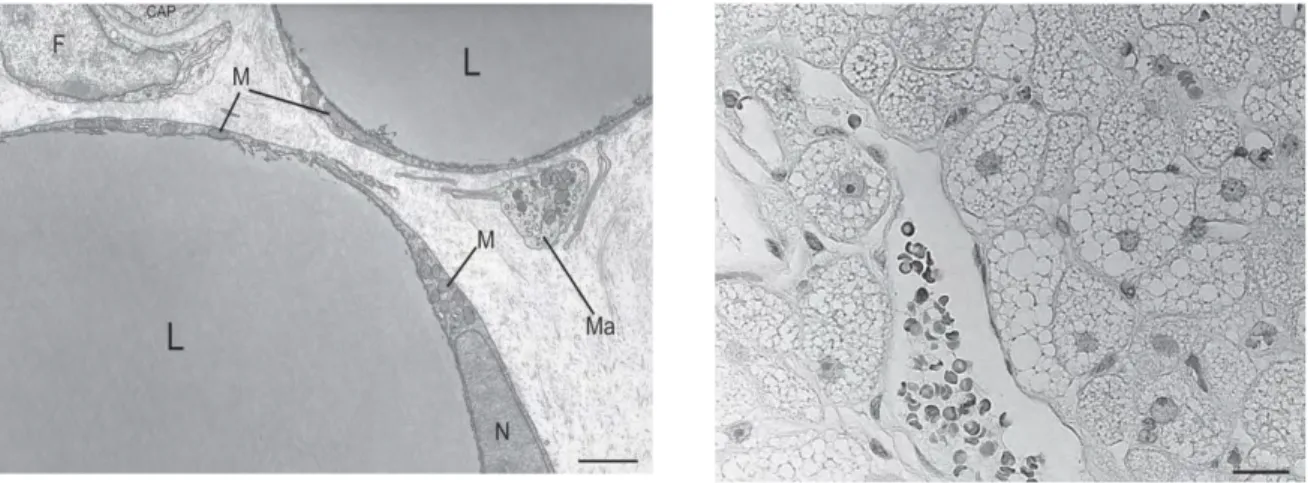
Figure 6. Left, Electron microscopy of murine white adipose tissue. The small and elongated
mitochondria in the perinuclear area and in the thin rim of cytoplasm surrounding the large unilocular lipid droplet. F, fibroblast; CAP, capillary lumen; Ma, macrophage; M, mitochondria; N, nucleus; L, liquid droplet. Bar = 2 μm
Right, Light microscopy of human brown adipose tissue. The characteristic multilocular lipid
27
2.1 The cellular origins of adipocytes and their differentiation
In the recent past, it was largely accepted that both brown and white adipocytes arose from resident mesenchymal progenitor cells that were present in adipose tissue. This idea was supported by evidence indicating that both brown and white adipocytes require PPARγ for development. However, the notion that brown and white cells arise from a similar origin is outdated. Brown adipocytes instead share a common MYF5+PAX7+ precursor with muscle cells [92]. The MYF5+PAX7+ precursor is driven to brown adipocyte terminal differentiation by bone morphogenetic protein 7 (BMP7), peroxisome proliferator activated receptor-γ (PPARγ) and CCAAT/enhancer-binding proteins (C/EBPs) cooperating with the transcriptional co-regulator PR domain-containing 16 (PRDM16). By contrast, PRDM16 does not affect white adipogenesis. White adipocytes can also be stimulated to display characteristics of brown adipocytes by β-adrenergic signaling, cold exposure and thiazolidinediones, which seem to function through the indicated factors (Figure 7). These results suggest that brown cells in BAT and brown fat-like cells in WAT have different cellular origins, and the transcriptional modulator PRDM16 plays an important role in both of these cell types to regulate genes required for thermogenesis [93]. There is also evidence to indicate that bone marrow progenitor-derived adipocytes and adipocyte progenitors can arise from hematopoietic cells via the myeloid lineage [94]. Collectively, these recent studies suggest that our understanding of the origin of adipocytes is rapidly changing, and additional studies will be necessary to clarify our understanding of this emerging area of biology.
Despite the functional, developmental and locational differences between white and brown adipocytes, these two cell types share many common differentiation features. All adipocytes, along with osteoblasts, myocytes and chondrocytes, differentiate from mesenchymal stem cells (MSCs) [95] in a process known as adipogenesis. Adipogenesis can be divided into two main phases: determination and terminal differentiation. The first phase involves the commitment of a pluripotent stem cell to the adipocyte lineage. Determination results in the conversion of the stem cell to a pre-adipoctye, which cannot be distinguished morphologically from its precursor cell but has lost the potential to differentiate into other cell types. In the second phase, the pre-adipocyte takes on the characteristics of the mature adipocyte—it acquires the machinery that is necessary for lipid transport and synthesis, insulin sensitivity and the secretion of adipocyte-specific proteins. Over the past two decades, attention has centred on the role of the nuclear receptor PPARγ and members of the C/EBP family in adipogenesis. We briefly discuss the important functions of these factors in adipogenesis.
28
Figure 7. Relationship between white and brown adipogenesis. Historically, white and brown adipocytes
were thought to derive from the same precursor cell. However, brown adipocytes instead share a common MYF+PAX7+ precursor with muscle cells. MYF+PAXA7+ precursor is driven to brown adipocyte terminal differentiation by BMP7, PPARγ and C/EBPs with PRDM16. White adipocytes can also be stimulated to display characteristics of brown adipocytes by β-adrenergic signaling, cold exposure and thiazolidinediones. MSC, mesenchymal stem cell; MYF5, myogenic factor 5; PAX7, paired-box 7; PGC1α, PPARγ co-activator 1α (Ana G. Cristancho and Mitchell A. Lazar, 2011, Nat Rev Mol Cell Biol).
29 PPARγ: the master regulator of adipogenesis.
PPARγ is a member of the nuclear-receptor superfamily, and is both necessary and sufficient for adipogenesis. Forced expression of PPARγ is sufficient to induce adipocyte differentiation in fibroblasts [96], and no factor has been discovered that promotes adipogenesis in the absence of PPARγ. These findings are consistent with the observation that most pro-adipogenic factors seem to function at least in part by activating PPARγ expression or activity. Studies using in vitro models of adipogenesis have consistently shown that PPARγ mRNA is induced by several transcription factors, including C/EBPβ, C/EBPδ, EBF1, and KLF5. Repressors of adipogenesis such as GATA2, KLF2, and CHOP have been shown to attenuate PPARγ expression [97]. Moreover, crucial signaling pathways in adipogenesis converge on the regulation of PPARγ expression or activity. PPARγ proteins are expressed in two forms, PPARγ1 and PPARγ2, which are produced by a combination of differential promoter usage and alternative splicing [98]. PPARγ1 is expressed at low levels in multiple tissues, whereas PPARγ2 is highly expressed in fat cells and differs from PPARγ1 by an amino-terminal extension of 30 amino acids [99]. Two studies have addressed the relative adipogenic activity of the PPARγ isoforms without coming to a consensus. Ren et al have shown that ectopic expression of PPARγ2 rescues adipogenesis, whereas expression of PPARγ1 does not [100]. By contrast, Mueller et al showed that both PPARγ1 and PPARγ2 can promote differentiation in Pparγ-/-fibroblasts; however, PPARγ2 is slightly more efficient at promoting
adipogenesis [101].
In vivo studies have shown that mice with adipose tissue-specific loss of PPARγ display decreased
fat pad size and insulin resistance in adipose tissue and liver [102]. Although PPARγ is required for adipogenesis, there is evidence to suggest that this nuclear receptor may not be needed to maintain the differentiated state of the cell after adipogenesis [103]. The importance of PPARγ in human adipose tissue has also been established, and subjects with mutations in the PPARγ gene can develop severe insulin resistance and lipodystrophy [104];[105]. Taken together, these studies show the requirement for PPARγ in adipocyte differentiation and whole-body insulin sensitivity.
C/EBPs
C/EBPs comprise another family of basic leucine zipper transcription factors. There are six C/EBP isoforms, all of which possess a highly conserved bZIP domain that serves as a point of interaction for homodimerization or heterodimerization with other family members [106]. The most common nomenclature for the members is C/EBPs α, β, δ, γ, ε and ζ. The temporal expression of these factors during adipocyte differentiation indicates a cascade whereby early induction of C/EBPβ and C/EBPδ leads to induction of C/EBPα. The notion is further supported by sequential binding of these transcription factors to several adipocyte promoters during differentiation. Loss of function
30 experiments in 3T3-L1 and NIH-3T3 cell lines confirmed the importance of C/EBPβ and –δ, as inhibition of either of these C/EBPs resulted in the attenuation of adipogenesis [107]. C/EBPβ and C/EBPδ promote adipogenesis at least in part by inducing C/EBPα and PPARγ. The amounts of C/ebpa and Pparg mRNA are normal in the remaining adipocytes of these double-knockout mice in contrast to C/EBPβ- and C/EBPδ- deficient MEFs, which do not express C/EBPα and PPARγ [108]. Despite the importance of C/EBPs in adipogenesis, these transcription factors can`t function efficiently in the absence of PPARγ. For example, C/EBPβ cannot induce expression of C/EBPα in the absence of PPARγ, which is required to release histone deacetylase-1 (HDAC1) form the C/ebpa promoter [109]. Furthermore, the ectopic expression of C/EBPα cannot rescue adipogenesis in Pparg-/- fibroblasts [110]. However, C/EBPα has an important role in differentiated adipocytes. Expression of exogenous PPARγ in C/EBPα-deficient cells showed that, although C/EBPα is not required for accumulation of lipid and the expression of many adipocyte genes, it is necessary for the acquisition of insulin sensitivity [111].
WNT signaling
WNT family members are secreted glycoproteins that have key roles during development. Canonical WNT signaling is activated following the binding of WNT ligands to the heterodimeric cell surface receptors low-density lipoprotein receptor-related 5 (LRP5), LRP6 and Frizzled. This induces the family of T cell-specific transcription factors (TCFs) to recruit a β-catenin-dependent co-activator complex to activate target gene transcription. Canonical WNT signaling has been shown to inhibit adipogenesis [112]. Mice expressing WNT10B, the main WNT ligand expressed by preadipocytes, in adipocytes show a decrease in WAT and BAT mass [113]. WNT10B promotes osteogenesis in MSCs, indicating that canonical WNT signaling also regulates brown adipogenesis and MSC cell fate [114]. WNT signaling maintains preadipocytes in an undifferentiated state through inhibition of the adipogenic transcription factors C/EBPα and PPARγ [115]. Repression of WNT10B in white primary preadipocytes is required for adipocyte differentiation [116].However, there is evidence that the canonical WNT pathway is essential for the survival of pre-adipocyte. WNT10B levels increase in confluent cultures of 3T3-L1 cells [115], and WNT1 can protect preadipocytes from apoptosis [117].
WNT ligands can also signal through β-catenin-independent pathways, a process known as non-canonical signaling, by signaling through alternative cell surface receptors and activating different intracellular pathways. The non-canonical WNT ligand WNT5A activates the histone methyltransferase SET domain bifurcated 1 (SETDB1) which involve in inhibiting the ability of PPARγ. Thus, WNT5A inhibit the adipogenesis and it can promote osteogenesis in MSCs [118]. By
31 contrast, the non-canonical WNT ligand WNT5B through preventing nuclear translocation of β-catenin to inhibit the canonical WNT pathway, thereby promoting adipocyte differentiation [119]. Despite these main factors gave the field a starting point, other new factors like signal transducers and activators of transcription (STATs), Kruppel-like factor (KLF) proteins, TGFβ superfamily signaling, the composition and stiffness of the ECM and cell-cell contact and cell shape also play a key role to promote preadipocyte differentiation into mature adipocytes [120]; [92].
2.2 Model systems used to study adipocyte in vitro
A variety of cellular model systems are used to study the molecular pathways of adipogenesis and adipocyte function in vitro. These models can mainly be separated in two categories. The first group includes pluripotent fibroblasts that have the ability to differentiate into several cell types, including myocytes, chondrocytes, and adipocytes. This group includes the 10T1/2, BALB/c-3T3, RCJ3.1, and CHEF/18 fibroblast cell lines. The second group of model systems comprises fibroblast like preadipocytes that are committed to differentiating into adipocytes and includes L1, 3T3-F442A, 1246, Ob1771, TA1, and 30A5 preadipocytes. The latter group represents the cells used most frequently to study adipogenesis [120].
3T3-L1 and 3T3-F442A are the two most extensively characterized and used preadipocyte cell lines. Both of these lines were derived from disaggregated 17- to 19-day-old Swiss 3T3 mouse embryos [121];[122]. However, unlike 3T3-L1 cells, 3T3-F442A preadipocytes do not require glucocorticoid-supplemented differentiation cocktail to induce adipogenesis [123];[124];[125]. It is commonly believed that 3T3-F442A precursors are at a more advanced stage of commitment than 3T3-L1`s. Implantation of 3T3-F442A, but not 3T3-L1, preadipocytes into athymic mice results in the generation of ectopic fat that is histologically [126] and biochemically [127] indistinguishable from adipose tissue. An original 2D coculture system setup in our laboratory using human breast cancer cell lines and in vitro differentiated 3T3-F442A adipocytes mimics the adipocytes changes observed at the invasive front of breast tumors [68], underscoring the validity of these mouse models in cancer studies. Mature adipocytes can also be obtained from the in vitro differentiation of the SVF of adipose tissue (that contains ADSCs) from human and rodents in defined culture conditions either in 2D or 3D culture systems [128];[129].
A clear limitation of the use of these human and murine pre-adipocyte cell lines and primary precursors present in the SVF fraction is that they are not suitable models to study obesity-related condition due to their inability to undergo hypertrophy in vitro. Therefore, the use of mature adipocytes isolated from AT of lean and obese subjects should be considered. Indeed, the isolation of adipocytes from the extracellular matrix and the SVF fraction is a technique available in many laboratories whatever the AT considered including MAT [68].Briefly, collagenase treatment of AT