HAL Id: inserm-01845482
https://www.hal.inserm.fr/inserm-01845482
Submitted on 20 Jul 2018
HAL is a multi-disciplinary open access
archive for the deposit and dissemination of sci-entific research documents, whether they are pub-lished or not. The documents may come from teaching and research institutions in France or abroad, or from public or private research centers.
L’archive ouverte pluridisciplinaire HAL, est destinée au dépôt et à la diffusion de documents scientifiques de niveau recherche, publiés ou non, émanant des établissements d’enseignement et de recherche français ou étrangers, des laboratoires publics ou privés.
Biological challenges for regeneration of the degenerated
disc using cellular therapies
Michael Bendtsen, Cody Bunger, Pauline Colombier, Catherine Le Visage,
Sally Roberts, Daisuke Sakai, Jill Urban
To cite this version:
Michael Bendtsen, Cody Bunger, Pauline Colombier, Catherine Le Visage, Sally Roberts, et al.. Bi-ological challenges for regeneration of the degenerated disc using cellular therapies. Acta Orthopaed-ica, Informa Healthcare, 2017, 87 (sup363), pp.39 - 46. �10.1080/17453674.2017.1297916�. �inserm-01845482�
Full Terms & Conditions of access and use can be found at
http://www.tandfonline.com/action/journalInformation?journalCode=iort20
ISSN: 1745-3674 (Print) 1745-3682 (Online) Journal homepage: http://www.tandfonline.com/loi/iort20
Biological challenges for regeneration of the
degenerated disc using cellular therapies
Michael Bendtsen, Cody Bunger, Pauline Colombier, Catherine Le Visage,
Sally Roberts, Daisuke Sakai & Jill P G Urban
To cite this article: Michael Bendtsen, Cody Bunger, Pauline Colombier, Catherine Le Visage, Sally Roberts, Daisuke Sakai & Jill P G Urban (2016) Biological challenges for regeneration of the degenerated disc using cellular therapies, Acta Orthopaedica, 87:sup363, 39-46, DOI: 10.1080/17453674.2017.1297916
To link to this article: https://doi.org/10.1080/17453674.2017.1297916
© 2017 The Author(s). Published by Taylor & Francis on behalf of the Nordic Orthopedic Federation.
Published online: 13 Mar 2017.
Submit your article to this journal
Article views: 478
Acta Orthopaedica 2016; 87 (eSuppl 363): 39–46 39
Biological challenges for regeneration of the degenerated
disc using cellular therapies
Michael BENDTSEN 1, Cody BUNGER 1, Pauline COLOMBIER 2*, Catherine LE VISAGE 2, Sally ROBERTS 3, Daisuke SAKAI 4, and Jill P G URBAN 5
1 Department of Orthopaedics, Aarhus University Hospital, Denmark; 2 INSERM UMR 1229, Regenerative Medecine and Skeleton, University of Nantes, France; 3 Spinal Studies and ISTM (Keele University), Robert Jones and Agnes Hunt Orthopaedic Hospital, Oswestry, UK; 4 Department of Orthopaedics, Tokai University Hospital, Japan; 5 Department of Physiology, Anatomy and Genetics, Oxford University, Oxford, UK. *current address: Cardiovascular Research Institute, University of California, San Francisco, CA, USA.
Correspondence: [email protected] Submitted 2016-03-15. Accepted 2017-01-07.
© 2017 The Author(s). Published by Taylor & Francis on behalf of the Nordic Orthopedic Federation. This is an Open Access article distributed under the terms of the Creative Commons Attribution-Non-Commercial License (https://creativecommons.org/licenses/by-nc/3.0)
DOI 10.1080/17453674.2017.1297916
Interest in the biology of the intervertebral disc has grown signifi cantly over the past 2 decades, driven mainly by stud-ies aimed at developing biological therapstud-ies for repairing degenerate discs (Alini et al. 2002, Sakai and Grad 2015). Most interest has focused on cellular therapies, where cells, capable of synthesizing appropriate disc tissue, are implanted into the damaged tissue to replace resident cells that have died or have acquired a degenerative phenotype. This appears to be an attractive strategy, and has led to a signifi cant increase in information about disc cellular biology. It follows the approach used clinically for repairing damaged cartilage (Hunziker et al. 2015); however, cell therapy for the disc faces more obstacles than that for cartilage repair and has not yet entered routine clinical practice.
In this review, we discuss some of the challenges in suc-cessful cellular repair of the disc. We fi rst review the function, organization, and composition of a normal disc, outline the changes that occur in degeneration, and consider how these might infl uence function. We then summarize cell therapy approaches to repairing the disc in relation to the choice of cells and cell support. We outline the challenges facing the implanted cells in the degenerate disc, and ask whether these therapies can be evaluated in animal models. Finally, we out-line the important, but often neglected, problem of patient selection.
The disc is complex in structure, composition, and function. What are we aiming to repair/ regenerate?
The normal disc
Morphology and composition
The intervertebral discs are large load-bearing cartilaginous tissues that lie interspersed between the bony vertebral bodies.
Morphologically, the disc appears to consist of 2 main regions (Figure 1), with an inner, more gelatinous region, the nucleus pulposus (or nucleus), encircled by a stiffer, collagenous annu-lus fi brosus (or annuannu-lus), consisting of concentric lamellae. The nucleus and annulus are separated from the bone by a thin (approx. 1-mm) layer of hyaline cartilage, the cartilage end-plate; annulus insertions anchor the disc to the bone (Nosikova et al. 2012). The normal disc is virtually avascular, with blood vessels and nerves being found only in the periphery of the annulus.
The composition and organization of the macromolecules that make up its extracellular matrix enable the disc to fulfi ll its mechanical role. Fibrillar collagens provide the structural framework of the disc (Eyre et al. 1991). The collagen network
Figure 1. A schematic view of the vertebral joint. Here it is partly cut away to show the annulus fi brosus (AF) surrounding the nucleus pulp-osus (NP) of the intervertebral disc, the cartilaginous endplate (CEP) and bony endplate (BEP) interspersed between the disc and vertebral body (VB), and the spinal canal (SC) lying behind the vertebral bodies and the disc. The spinal canal—surrounded by the discs, the spinal processes (SP), and apophyseal joints (AJ)—encloses the spinal cord which gives rise to the nerve roots (NR) running adjacent to the poste-rior portion of the disc. (Adapted from Urban and Roberts 1986).
SP CEP NP BEP AJ NR VB SC 10191 Bendtsen D.indd 39 10191 Bendtsen D.indd 39 3/9/2017 3:07:19 PM3/9/2017 3:07:19 PM
of the nucleus is formed from fi ne fi brils of (mainly type-II) collagen. Parallel bundles of fi brils (mainly type-I), running obliquely between the adjacent vertebral bodies, form the concentric lamellae of the annulus (Takeda 1975, Pezowicz et al. 2006). The lamellae are held together by elastic proteins (Yu et al. 2015), which help to give the disc its fl exibility. Aggrecan, the other major macromolecular component, is a large polyanionic proteoglycan that imparts a high osmotic pressure to the disc matrix (Sivan et al. 2006); the matrix thus tends to imbibe water, infl ating the collagen network until the osmotic swelling pressure balances the applied load. Apart from aggrecan and collagens, the disc matrix also contains a large number of other proteins (Figure 2A), which, although present in low concentrations, are also important in regulat-ing the stability and function of the disc matrix (Feng et al. 2006).
Disc cells
The human disc contains a small population of resident cells (Pattappa et al. 2012) that make and maintain the disc’s mac-romolecules. The cells also produce proteases that are capable of degrading all matrix components. In a healthy disc, the rates at which the macromolecules are made and broken down are in balance (Figure 2B), but because of the low cell density, the turnover in human discs is very slow (Sivan et al. 2014a).
The cell type—and hence the composition—of the matrix synthesized varies across the disc and changes with age. The
nucleus pulposus of all mammals is initially populated by clus-ters of large notochordal cells that produce a highly hydrated, aggrecan-rich, collagen-poor matrix. In humans and in some other species, the cell phenotype changes during growth, with the notochord cells being replaced by several phenotypically distinct but poorly characterized subpopulations of chondro-cyte-like cells (Molinos et al. 2015). These chondrochondro-cyte-like cells produce matrix that becomes more collagenous and less hydrated during development in humans. In the outer annu-lus, fi broblast-like cells synthesize the highly organized col-lagen-rich lamellae. The disc also contains a small number of progenitor cells that are potentially able to differentiate into the appropriate disc cell phenotypes (Henriksson et al. 2009, Sakai et al. 2012, Gruber et al. 2016). Little is known about the cells of the cartilage endplate.
The degenerate disc
Disc degeneration is a loose term that encompasses pro-gressive biochemical, cellular, and structural changes to the disc—with consequent changes in its load-bearing properties. Although little is understood about the factors that initiate disc degeneration, the process appears to be driven by changes in the behavior of its resident cells, which begin to increase the production of proteases and reduce production of the matrix macromolecules. Hence, macromolecules are degraded and lost from the disc at a faster rate than they can be replaced.
Information on the changes in disc composition and organi-Figure 2. A. Schematic illustration of assemblies of matrix proteins in the intervertebral disc. Aggrecan monomer is synthesized intracellularly and secreted into the ECM where it forms supramolecular aggregates with HA that are stabilized by link proteins. Collagen synthesis involves removal of the N- and C-terminal propeptides from procollagen to generate tropocollagen which self-assembles into polymeric collagen fi brils. Cartilage oligomeric matrix protein (COMP) acts as a catalyst in collagen fi brillogenesis, and small leucine-rich proteoglycans (SLRPs; e.g. decorin, big-lycan, fi bromodulin, lumican) and collagen IX regulate fi bril thickness and interfi brillar spacing. CS: chondroitin sulfate; KS: keratan sulfate; HA: hyaluronan; HS-PG, heparan sulfate proteoglycan; MAT: matrilin; PRELP: proline arginine-rich end leucine-rich repeat protein. (Reproduced from Feng et al. (2006) with permission). B. Schematic illustration depicting the synthesis and degradation of the disc extracellular matrix. In normal, healthy discs, there is a fi ne balance between matrix synthesis, assembly, and turnover, which becomes perturbed during disc degeneration. Aggrecanases (ADAMTS-4 and -5) within the ECM cause cleavage and fragmentation of the aggrecan core protein. Degradation of collagen fi brils occurs through the activity of collagenases (MMP-1 and -13) and gelatinases (MMP-2 and -9). α5β1: α5β1 integrin (fi bronectin receptor); CS: chondroitin sulfate; CD44: hyaluronic acid receptor; G1, G2, and G3: globular domains of aggrecan; GF growth factors: cytokines and other bioactive signaling molecules; HA: hyaluronic acid; KS, keratan sulfate. (Reproduced from Sivan et al. (2014a) with permission).
A B
10191 Bendtsen D.indd 40
41
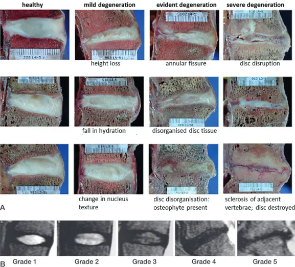
zation with degeneration has been obtained from examination of discs taken at autopsy or removed at surgery (Lyons et al. 1981, Boos et al. 2002, Roberts et al. 2006). Degenerate discs have high concentrations of proteases that tend to degrade the macromolecules of the disc, particularly aggrecan—the con-centration of which falls on disc degeneration (Sivan et al. 2014b) (Figure 2B); degenerate discs thus retain less water and lose it faster under load. As the disc degrades and becomes more dehydrated, the lamellae become disorganized and the disc loses structural integrity, with formation of fi ssures and defects at the bone-disc interface (Figure 3A). The cartilagi-nous endplate tends to calcify, decreasing nutrient transport to the cells; many of them become senescent and die (Kletsas 2009). Blood vessels and nerves invade the previously avascu-lar, aneural disc along with infl ammatory cells such as mac-rophages. The changes seen in disc degeneration vary from individual to individual, may start early in life, appear to be strongly genetic (Boos et al 2002, Battié et al. 2009), and are an ongoing process with the severity and number of
degenera-nance imaging (MRI). It is often classifi ed using MRI scores (Pfi rrmann et al. 2001) (Figure 3B), based on changes in disc height and signal intensity without considering other degra-dative features. MRI grade-3 discs, for instance, may include discs with very different degrees of endplate irregularity, disc bulge, or radial or circumferential tears (Figure 3A). Currently, degenerative changes at the tissue and cellular level cannot be detected non-invasively.
What degenerative changes are the biological thera-pies aimed at repairing?
Currently, disc cell therapies are mostly aimed at restoring macromolecular components, with aggrecan in the nucleus being the major focus, as the mechanical consequences of its loss are very apparent. However, while desirable mechanical properties for repair have been defi ned (Cortes et al. 2014), little is known about what other components of the complex matrix—apart from collagens—are necessary for functional repair. Moreover, while restoration of nucleus hydration is
Grade 1 Grade 2 Grade 3 Grade 4 Grade 5
A
B
Figure 3. A. Sagittal sections of human lumbar intervertebral discs at various stages of degeneration. Features such as height loss, fall in water content, annular tears, osteophytes, and endplate sclerosis observed at different stages of degeneration are indicated (Adapted from Galbursera et al. 2014). B. MRIs showing discs at different stage of Pfi rrmann degeneration grade. Grading is based on signal intensity, distinction between nucleus and annulus, degree of homogeneity of disc structure, and loss of disc height. Features which are apparent morphologically (Figure 3a), such as fi ssures, changes in the endplate and even herniations are not taken into account in this grading scheme (adapted from Pfi rrmann et al. 2001)
tive changes increasing with age.
Functional changes in disc degen-eration
The morphological and bio-chemical changes resulting from disc degeneration infl uence the mechanical behavior of the disc, and therefore of the whole spinal column (Adams 2004, Galbusera et al. 2014, Von Forell et al. 2015, Muriuki et al. 2016). Degeneration, with its loss of aggrecan, results in a fall in hydration and a reduction in disc height, an increase in disc bulge, and a change in stiffness. Loss of the integrity of the disc results in instability of the spinal motion segment, possibly leading to spondylolisthesis. Inappropriate loads are thus transmitted to other spinal structures such as the facet joints, which may become osteo-arthritic—and also to the posterior ligaments, which may thicken, leading to spinal stenosis. Pro-found degenerative changes in the spinal column triggered by a series of these degenerative events may end in onset of complex spinal deformities.
Diagnosis of disc degeneration in vivo
In vivo, disc degeneration is detected using magnetic
reso-10191 Bendtsen D.indd 41
10191 Bendtsen D.indd 41 3/9/2017 3:07:21 PM3/9/2017 3:07:21 PM
the aim of many studies, fewer studies have examined repair of the annulus (Sakai and Grad 2015) or cartilage endplate (Bendtsen et al. 2011, Nosikova et al. 2012), yet the integrity of these structures is also essential for disc health. Thus, would functional and stable cellular disc repair require an approach that integrates all disc regions (Nosikova et al. 2012)?
Cellular repair
Which cells are appropriate for cellular repair of the disc?
It is a challenge to fi nd an appropriate source of cells for disc repair (Kregar-Velikonja et al. 2014, Sakai and Andersson 2015). Human disc cells can only be harvested during sur-gical procedures. As no autologous cells from healthy discs are available, cells from other cartilages have been used for animal studies, while the use of notochord cells to stimulate resident cells is under investigation (Arkesteijn et al. 2015). Most researchers have, however, concentrated on differentiat-ing stem cells or progenitor cells towards a nucleus pulpo-sus-like cell type. Many studies have investigated the use of autologous mesenchymal stem cells (MSCs); allogenic MSCs are being tested in clinical trials (Table). A few studies have investigated differentiation of progenitor cells, or embryonic or induced pluripotent stem cells, towards the notochord- or adult nucleus pulposus cell phenotype. Success in differen-tiation is judged by expression of phenotypic nucleus pulpo-sus markers (Risbud et al. 2015), which may not be specifi c
(Thorpe et al. 2016), and through expression of matrix mac-romolecules such as collagen II and aggrecan, which are also expressed by other cartilages.
Currently, strategies tend to implant only 1 cell type into the disc—albeit that there are different cellular subpopulations even in the nucleus—and disc degeneration almost invariably involves more than 1 disc region (Figure 3A). Will stem cells implanted directly into the disc differentiate into the popula-tions required to regenerate a stable nucleus, and repair the annulus and endplate? Strategies such as the use of notochord cells and chondrocyte-like cells generated from human stem cells may restore the dialogue between both cell types, based on the secretion of growth factors including TGF-β, CTGF, and SHH, and lead to the survival of nucleus cells and an increase in proteoglycan synthesis (Dahia et al. 2012). Would such differentiation strategies be suffi cient, or would each region have to be directly targeted with appropriate cells?
Can implanted cells survive and function in the chal-lenging environment found in degenerate discs?
As the dense matrix of the cartilaginous endplate and matrix of the normal disc acts as a permeability barrier between the disc cells and circulating macromolecules, the activity of the disc cells is governed to a large extent by their extracellular physical environment, and by signals from contacts with the extracellular matrix.
Nutrient levels limit the number of viable cells that can be implanted into the disc
Clinical trials of cellular therapies for intervertebral disc repair
ClinicalTrials.gov
Title Place identifi er Status
Autologous adipose derived stem cell therapy for Bundang CHA Hospital, NCT02338271 Recruiting intervertebral disc degeneration Korea
Treatment of degenerative disc disease with allogeneic Hospital Clinico Universitario, NCT01860417 Ongoing, not recruiting mesenchymal stem cells Valladolid, Spain
Autologous adipose tissue derived mesenchymal stem Biostar, Korea University NCT01643681 Unknown cell transplantation in patient with lumbar intervertebral Anam Hospital
disc degeneration
Safety and preliminary effi cacy study of mesenchymal Mesoblast Ltd. NCT01290367 Completed but no precursor cells (MPCs) in subjects with lumbar back pain results posted Safety and effi cacy study of rexlemestrocel-L (viz. allogenic Mesoblast Ltd. NCT02412735 Recruiting MSCs) in subjects with chronic discogenic lumbar back pain
Lumbar degenerative disc disease treatment with bone Red de Terapia Celular, NCT02440074 Withdrawn marrow autologous mesenchymal stem cells (MSV) Spain
Human autograft mesenchymal stem cell mediated Trinity Stem Cell Institution, NCT02529566 Enrolling by invitation stabilization of the degnerative lumbar spine Odessa, Florida, USA
Adipose cells for degenerative disc disease Bioheart Inc. NCT02097862 Recruiting
Safety and effi cacy with NOVOCART disc plus (ADCT) for Tetec AG NCT01640457 Ongoing, not recruiting the treatment of degenerative disc disease in lumbar
spine (NDisc)
A study comparing the safety and effectiveness of cartilage ISTO Technologies Inc., NCT01771471 Ongoing, not recruiting cells injected into the lumbar disc as compared to a placebo USA
10191 Bendtsen D.indd 42
43
Extracellular nutrient concentrations are of particular impor-tance in the avascular disc (Figure 4A) (Grunhagen et al. 2011), which obtains its energy by aerobic glycolysis. Nutri-ent levels fall with distance from the blood supply and must remain above critical levels (0.2 mM glucose, pH 6.7) for cells to remain viable. Although much interest has been expressed in the hypoxic environment of the disc and the role of HIF-1 and HIF-2 (Risbud et al. 2010), nucleus cells can survive with-out oxygen; even so, they consume it, and matrix synthesis is affected by oxygen concentrations. As in other avascular car-tilages (Stockwell 1971), viable cell density varies inversely with disc height, being only 1–5 million cells/mL in healthy human lumbar discs but over 50 million cells/mL in mouse discs (Figure 4B).
The supply of nutrients thus limits the number of viable cells that can be implanted into even a healthy disc. In degen-erate discs, calcifi cation of the endplate further restricts nutri-ent supply and the number of viable cells (Figure 4A). Cells implanted into a degenerate disc may therefore have limited access to nutrients, compromising their activity and survival.
Signals from the matrix are disturbed in degenerate discs
Disc cells are sensitive to the level of extracellular osmolarity, which is regulated by aggrecan concentrations. Loss of aggre-can and hence osmolarity in degenerate discs both reduces rates of matrix production (Takeno et al. 2007) and initiates infl ammatory changes (van Dijk et al. 2015). In addition, cells in degenerate discs produce more active proteases (Roberts et al. 2000, Pockert et al. 2009), which will tend to work against the ability of implanted cells to produce new matrix.
The infl ammatory environment of degenerate discs can have an adverse effect on implanted cells
Infl ammation is almost invariably encountered in degener-ate discs (Risbud and Shapiro 2014). Infl ammatory cytokines upregulate matrix degradation, thus slowing the rates of matrix accumulation and hindering attempts at repair; they can also induce pain. Moreover, these cytokines lead to further nutri-tional stresses, increasing rates of glycolysis, and thus further reducing glucose levels and pH levels—thereby compromis-ing the activity and viability of implanted cells (Wuertz et al. 2009). Infl ammation therefore appears to provide an unfavor-able environment for implanted cells.
Can scaffolds drive cells towards repair?
The highly hydrated networks of hydrogels make them par-ticularly suitable as a cell support for nucleus regeneration. While synthetic scaffolds with mechanical properties match-ing those of the nucleus are of interest, natural biopolymers have advantages in mimicking the native extracellular envi-ronment regarding mechanical, permeability, and biochemi-cal properties—and in providing a bioresorbable temporary 3-dimensional microenvironment. Some, such as injectable alginate (Zeng et al. 2015) and hyaluronan hydrogels (Pero-glio et al. 2013), may optimize stem cell differentiation and synthesis of an appropriate extracellular matrix. However, there are still no hydrogels that are able to fulfi ll needs regard-ing both cell biocompatibility and load-bearregard-ing capacity, and yet can also act as a reservoir of bioactive molecules.
0 10 20 30 40 50 60 70 80 0 1 2 3 4 5 6 7
Cell density (cells/mm3)
Disc half-height (mm)
Rat Rabbit Dog Cat Human Mouse Pig
Figure 4. A. Schematic illustration showing nutrient pathways in a normal disc (a) and changes seen in disc degeneration (b). Most of the disc is supplied with nutrients by diffusion from capillaries arising in the vertebral body, which penetrate the subchondral plate and terminate at the junc-tion with the cartilage endplate. Nutrients diffuse from these capillaries, through the cartilage endplate and disc matrix to the cells, which, in the center of a human disc, may be up to 8 mm from the nearest capillary. Nutrient supply is adversely affected in disc degeneration; disc degeneration is associated with atherosclerosis of the lumbar arteries and calcifi cation of the cartilaginous endplate. Loss of nutrient supply leads to a fall in the number of active and viable cells that can be supported in the disc. (Reproduced from Huang et al. (2014) with permission). B. The inverse relation-ship between disc cell density across the nucleus pulposus and disc height. Cell density was measured in histological sections of discs taken from mice, rats, rabbits, cats, dogs, pigs, and humans. Here it has been plotted against disc half-height (adapted from Holm and Nachemson 1983).
A B
10191 Bendtsen D.indd 43
10191 Bendtsen D.indd 43 3/9/2017 3:07:23 PM3/9/2017 3:07:23 PM
Results from animal models may be misleading
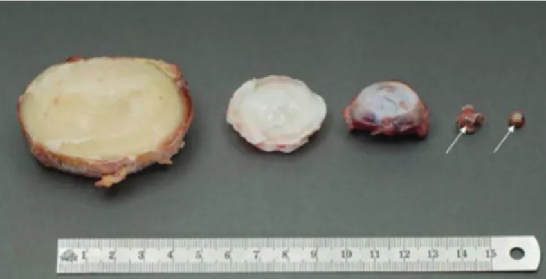
Numerous in vivo studies have examined the process of cellu-lar repair in animals ranging from mice to cellu-larger animals such as pigs and goats (Sakai and Andersson 2015), with apparently favorable outcomes. However, can such promising results be expected in humans? The discs of these animals, even those of cattle, are considerably smaller than human lumbar discs (Figure 5). The animal discs can consequently support a much greater cell density than human discs (Figure 4B). Moreover, the animals used are generally young or even immature, with degeneration produced by an acute intervention that may not produce infl ammatory changes similar to those seen in humans, and may leave the nutrient supply unimpaired. Here, implanted cells appear to be able to survive and produce repair tissue relatively rapidly (in weeks or months). By contrast, the half-life of aggrecan in a degenerate human disc is around 4 years, and that of collagen and elastin is more than 50 years (Sivan et al. 2014b). Hence, results from animal models must be viewed with caution (Alini et al. 2008).
Which patients would benefi t from disc repair?
The important question of which patients would be suitable for cellular therapies has seldom been addressed (Kandel et al. 2008, Tibiletti et al. 2014, Benneker et al. 2014, Sakai and Andersson 2015). Patients come to see a clinician because they have back pain, not because they are worried about disc degeneration. Indeed, many people with even severe disc degeneration are asymptomatic and are unaware of having any spinal problems (Brinjikji et al. 2015). Thus, should pain rather than disc degeneration be the clinical target?
Currently, there is no reliable means of diagnosing whether a disc is the source of pain or not; discography has been discred-ited and may indeed cause harm (Carragee et al. 2009), and
there are no validated MRI indications. In most cases, it is not known whether low back pain even arises from the disc; other structures such as the facets may also be involved, so regenerat-ing the disc alone may not be effective. Moreover neuropathic pain, central nervous system changes, and disorders of muscu-lar control are evident in many back pain patients (Freynhagen and Baron 2009, Yu et al. 2014, Schabrun et al. 2015), so even complete regeneration of the disc may not cure the pain.
Summary
Because of the complex nature of degenerative changes, bio-logical repair of the disc invokes challenges in many areas. An integrated approach that involves not only the choice of appro-priate cells and scaffolds for the different regions of the disc (including the endplate), but also targets infl ammation and nutrient supply, might be necessary for successful and stable repair—and restoration of function. Although small clinical studies using single cell populations have been published showing apparent success (Meisel et al. 2006, Yoshikawa et al. 2010, Orozco et al. 2011, Mochida et al. 2015), information on outcomes is still awaited from randomized clinical trials (Table), which are currently in progress.
Conclusions
Over the past decade, the growing interest in the development of cell therapies has led to real progress with not only some promising results in this fi eld in animal studies, but also in fur-thering our understanding of the biology of the intervertebral disc in general. However, a number of biological challenges must be overcome before these cellular therapies can be put into routine clinical use in humans.
One challenge is to improve characterization of the pheno-type of the various disc cell populations, and then to deter-mine how they interact under normal conditions and also in the nutrient-poor and infl ammatory environment of degener-ate discs—and importantly, to characterize the matrix macro-molecules that they produce at the protein level. Without this information, it would be diffi cult to develop rational strategies for differentiation of stem or progenitor cells into cell pheno-types that can survive implantation and produce a stable and functional matrix.
Another challenge is to develop strategies for coping with the long repair process (years) in large human discs. This might necessitate designing scaffolds that, as well as sup-porting cells, would be able to restore load-bearing function to the degenerate disc and that can be maintained safely in the tissue until an appropriate matrix is synthesized by the low number of viable cells that are able to survive in human lumbar discs.
Yet another challenge, as in other regenerative cell-based Figure 5. Relative sizes of intervertebral discs from different species.
From left to right: human lumbar L4–L5 disc; bovine tail C1–C2 disc; sheep thoracic T11–T12 disc; rat lumbar and tail discs (with arrows showing the intervertebral disc location). (Reproduced from Alini et al. 2008 with permission).
10191 Bendtsen D.indd 44
45
therapies, is to reduce costs. Currently, the high cost of autolo-gous donor cell preparations, and regulatory barriers, prevent routine clinical application for disorders such as disc degen-eration.
Probably the most diffi cult challenge is to improve diagno-sis in order to determine which patients would benefi t most from disc regeneration, remembering that patients seek medi-cal help for pain, not for disc degeneration. Even though cur-rent strategies using anti-TNF antibodies to treat pain have not always met with success (Cohen et al. 2009, Freeman et al. 2013), patients might still be better served by developing cel-lular therapies that are aimed at damping down infl ammation and pain (Pettine et al. 2015,Willems et al. 2015), rather than through therapies aimed at biological regeneration of the disc.
Adams M A. Biomechanics of back pain. Acupunct Med 2004; 22(4): 178-88. Alini M, Roughley P J, Antoniou J, Stoll T, Aebi M. A biological approach
to treating disc degeneration: not for today, but maybe for tomorrow. Eur Spine J 2002; 11 Suppl 2: S215-S220.
Alini M, Eisenstein S M, Ito K, Little C, Kettler A A, Masuda K, Melrose J, Ralphs J, Stokes I, Wilke H J. Are animal models useful for studying human disc disorders/degeneration? Eur Spine J 2008; 17(1): 2-19. Arkesteijn I T, Smolders L A, Spillekom S, Riemers F M, Potier E, Meij B P,
Ito K, Tryfonidou M A. Effect of coculturing canine notochordal, nucleus pulposus and mesenchymal stromal cells for intervertebral disc regenera-tion. Arthritis Res Ther 2015; 17: 60.
Battié M C, Videman T, Kaprio J, Gibbons L E, Gill K, Manninen H, Saarela J, Peltonen L. The Twin Spine Study: Contributions to a changing view of disc degeneration. Spine J2009; 9(1): 47-59.
Bendtsen M, Bunger C E, Zou X, Foldager C, Jorgensen H S. Autologous stem cell therapy maintains vertebral blood fl ow and contrast diffusion through the endplate in experimental intervertebral disc degeneration. Spine (Phila Pa 1976) 2011; 36(6): E373-E379.
Benneker L M, Andersson G, Iatridis J C, Sakai D, Hartl R, Ito K, Grad S. Cell therapy for intervertebral disc repair: advancing cell therapy from bench to clinics. Eur Cell Mater 2014; 27: 5-11.
Boos N, Weissbach S, Rohrbach H, Weiler C, Spratt K F, Nerlich A G. Clas-sifi cation of age-related changes in lumbar intervertebral discs: 2002 Volvo Award in basic science. Spine 2002; 27(23): 2631-44.
Brinjikji W, Luetmer P H, Comstock B, Bresnahan B W, Chen L E, Deyo R A, Halabi S, Turner J A, Avins A L, James K, Wald J T, Kallmes D F, Jarvik J G. Systematic literature review of imaging features of spinal degeneration in asymptomatic populations. AJNR Am J Neuroradiol 2015; 36(4): 811-6. Carragee E J, Don A S, Hurwitz E L, Cuellar J M, Carrino J A, Herzog R.
2009 ISSLS Prize Winner: Does discography cause accelerated progres-sion of degeneration changes in the lumbar disc: a ten-year matched cohort study. Spine (Phila Pa 1976 ) 2009; 34(21): 2338-45.
Cohen S P, Bogduk N, Dragovich A, Buckenmaier C C, III, Griffi th S, Kuri-hara C, Raymond J, Richter P J, Williams N, Yaksh T L. Randomized, double-blind, placebo-controlled, dose-response, and preclinical safety study of transforaminal epidural etanercept for the treatment of sciatica. Anesthesiology 2009; 110(5): 1116-26.
Cortes D H, Jacobs N T, DeLucca J F, Elliott D M. Elastic, permeability and swelling properties of human intervertebral disc tissues: A benchmark for tissue engineering. J Biomech 2014; 47(9): 2088-94.
Dahia C L, Mahoney E, Wylie C. Shh signaling from the nucleus pulposus is required for the postnatal growth and differentiation of the mouse interver-tebral disc. PLoS One 2012; 7(4): e35944.
Eyre D R, Caterson B, Benya P, Heinegard D, Oegema T R, Pearce R H, Pope M H, Urban J P G. The intervertebral disc. In: New Perspectives on Low Back Pain. (Eds. Gordon S, Frymoyer J). Am Inst Orthop Surg., Philadel-phia, 1991; 147-209.
Feng H, Danfelter M, Stromqvist B, Heinegard D. Extracellular matrix in disc degeneration. J Bone Joint Surg Am 2006; 88 Suppl 2: 25-9.
Freeman B J, Ludbrook G L, Hall S, Cousins M, Mitchell B, Jaros M, Wyand M, Gorman J R. Randomized, double-blind, placebo-controlled, trial of transforaminal epidural etanercept for the treatment of symptomatic lumbar disc herniation. Spine (Phila Pa 1976 ) 2013; 38(23): 1986-94.
Freynhagen R, Baron R. The evaluation of neuropathic components in low back pain. Curr Pain Headache Rep 2009; 13(3): 185-190.
Galbusera F, van R M, Ito K, Huyghe J M, Brayda-Bruno M, Wilke H J. Ageing and degenerative changes of the intervertebral disc and their impact on spinal fl exibility. Eur Spine J 2014; 23 Suppl 3: S324-S332.
Gruber H E, Riley F E, Hoelscher G L, Ingram J A, Bullock L, Hanley E N, Jr. Human annulus progenitor cells: Analyses of this viable endogenous cell population. J Orthop Res 2016; 34(8): 1351-60.
Grunhagen T, Shirazi-adl A, Fairbank J C, Urban J P. Intervertebral disk nutrition: a review of factors infl uencing concentrations of nutrients and metabolites. Orthop Clin North Am 2011; 42(4): 465-77, vii.
Henriksson H, Thornemo M, Karlsson C, Hagg O, Junevik K, Lindahl A, Brisby H. Identifi cation of cell proliferation zones, progenitor cells and a potential stem cell niche in the intervertebral disc region: a study in four species. Spine (Phila Pa 1976 ) 2009; 34(21): 2278-87.
Hunziker E B, Lippuner K, Keel M J, Shintani N. An educational review of cartilage repair: precepts & practice--myths & misconceptions--progress & prospects. Osteoarthritis Cartilage 2015; 23(3): 334-50.
Kandel R, Roberts S, Urban J P G. Tissue engineering and the intervertebral disc: the challenges. Eur Spine J 2008; 17 Suppl 4: S480–S491.
Kletsas D. Senescent cells in the intervertebral disc: numbers and mecha-nisms. Spine J 2009; 9(8): 677-8.
Kregar-Velikonja N, Urban J, Frohlich M, Neidlinger-Wilke C, Kletsas D, Potocar U, Turner S, Roberts S. Cell sources for nucleus pulposus regen-eration. Eur Spine J 2014; 23 Suppl 3: S364-S374.
Lyons G, Eisenstein S M, Sweet M B. Biochemical changes in intervertebral disc degeneration. Biochim Biophys Acta 1981; 673(4): 443-53.
Meisel H J, Ganey T, Hutton W C, Libera J, Minkus Y, Alasevic O. Clinical experience in cell-based therapeutics: intervention and outcome. Eur Spine J 2006; 15 Suppl 3: S397-S405.
Mochida J, Sakai D, Nakamura Y, Watanabe T, Yamamoto Y, Kato S. Inter-vertebral disc repair with activated nucleus pulposus cell transplantation: a three-year, prospective clinical study of its safety. Eur Cell Mater 2015; 29: 202-12.
Molinos M, Almeida C R, Goncalves R M, Barbosa M A. Improvement of bovine nucleus pulposus cells Isolation leads to identifi cation of three phe-notypically distinct cell subpopulations. Tissue Eng Part A 2015; 21(15-16): 2216-27.
Muriuki M G, Havey R M, Voronov L I, Carandang G, Zindrick M R, Lorenz M A, Lomasney L, Patwardhan A G. Effects of motion segment level, Pfi r-rmann intervertebral disc degeneration grade and gender on lumbar spine kinematics. J Orthop Res 2016: 34(8): 1389-98.
Nosikova Y S, Santerre J P, Grynpas M, Gibson G, Kandel R A. Characteriza-tion of the annulus fi brosus-vertebral body interface: identifi caCharacteriza-tion of new structural features. J Anat 2012; 221(6): 577-89.
Orozco L, Soler R, Morera C, Alberca M, Sanchez A, Garcia-Sancho J. Inter-vertebral disc repair by autologous mesenchymal bone marrow cells: a pilot study. Transplantation 2011; 92(7): 822-828.
Pattappa G, Li Z, Peroglio M, Wismer N, Alini M, Grad S. Diversity of inter-vertebral disc cells: phenotype and function. J Anat 2012; 221(6): 480-96. Peroglio M, Eglin D, Benneker L M, Alini M, Grad S. Thermoreversible
hyaluronan-based hydrogel supports in vitro and ex vivo disc-like differen-tiation of human mesenchymal stem cells. Spine J 2013; 13(11): 1627-39.
10191 Bendtsen D.indd 45
10191 Bendtsen D.indd 45 3/9/2017 3:07:25 PM3/9/2017 3:07:25 PM
Pettine K A, Murphy M B, Suzuki R K, Sand T T. Percutaneous injection of autologous bone marrow concentrate cells signifi cantly reduces lumbar discogenic pain through 12 months. Stem Cells 2015; 33(1): 146-56. Pezowicz C A, Robertson P A, Broom N D. The structural basis of
interlamel-lar cohesion in the intervertebral disc wall. J Anat 2006; 208(3): 317-30. Pfi rrmann C W, Metzdorf A, Zanetti M, Hodler J, Boos N. Magnetic
reso-nance classifi cation of lumbar intervertebral disc degeneration. Spine 2001; 26(17): 1873-8.
Pockert A J, Richardson S M, Le Maitre C L, Lyon M, Deakin J A, Buttle D J, Freemont A J, Hoyland J A. Modifi ed expression of the ADAMTS enzymes and tissue inhibitor of metalloproteinases 3 during human intervertebral disc degeneration. Arthritis Rheum 2009; 60(2): 482-91.
Risbud M V, Shapiro I M. Role of cytokines in intervertebral disc degenera-tion: pain and disc content. Nat Rev Rheumatol 2014; 10(1): 44-56. Risbud M V, Schipani E, Shapiro I M. Hypoxic regulation of nucleus pulposus
cell survival: from niche to notch. Am J Pathol 2010; 176(4): 1577-1583. Risbud M V, Schoepfl in Z R, Mwale F, Kandel R A, Grad S, Iatridis J C, Sakai
D, Hoyland J A. Defi ning the phenotype of young healthy nucleus pulposus cells: Recommendations of the Spine Research Interest Group at the 2014 Annual ORS Meeting. J Orthop Res 2015; 33(3): 283-93.
Roberts S, Caterson B, Menage J, Evans E H, Jaffray D C, Eisenstein S M. Matrix metalloproteinases and aggrecanase: their role in disorders of the human intervertebral disc. Spine 2000; 25(23): 3005-13.
Roberts S, Evans H, Trivedi J, Menage J. Histology and pathology of the human intervertebral disc. J Bone Joint Surg Am 2006; 88 Suppl 2: 10-4. Sakai D, Andersson G B. Stem cell therapy for intervertebral disc
regenera-tion: obstacles and solutions. Nat Rev Rheumatol 2015; 11(4): 243-256. Sakai D, Grad S. Advancing the cellular and molecular therapy for
interverte-bral disc disease. Adv Drug Deliv Rev 2015; 84: 159-171.
Sakai D, Nakamura Y, Nakai T, Mishima T, Kato S, Grad S, Alini M, Risbud M V, Chan D, Cheah K S, Yamamura K, Masuda K, Okano H, Ando K, Mochida J. Exhaustion of nucleus pulposus progenitor cells with ageing and degeneration of the intervertebral disc. Nat Commun 2012; 3: 1264. Schabrun S M, Elgueta-Cancino E L, Hodges P W. Smudging of the motor
cortex is related to the severity of low back pain. Spine (Phila Pa 1976 ) 2015. [Epub ahead of print].
Sivan S, Merkher Y, Wachtel E, Ehrlich S, Maroudas A. Correlation of swell-ing pressure and intrafi brillar water in young and aged human interverte-bral discs. J Orthop Res 2006; 24(6): 1292-8.
Sivan S S, Hayes A J, Wachtel E, Caterson B, Merkher Y, Maroudas A, Brown S, Roberts S. Biochemical composition and turnover of the extracellular matrix of the normal and degenerate intervertebral disc. Eur Spine J 2014a; 23 Suppl 3: S344-S353.
Sivan S S, Wachtel E, Roughley P. Structure, function, aging and turnover of aggrecan in the intervertebral disc. Biochim Biophys Acta 2014b; 1840(10): 3181-9.
Stockwell R. The inter-relationship of cell density and cartilage thickness in mammalian articular cartilage. J Anat 1971; 109: 411-22.
Takeda T. Three-dimensional observations of collagen framework of human lumbar discs. J Japan Orthop Assoc 1975; 49: 45-57.
Takeno K, Kobayashi S, Negoro K, Uchida K, Miyazaki T, Yayama T, Shi-mada S, Baba H. Physical limitations to tissue engineering of intervertebral disc cells: effect of extracellular osmotic change on glycosaminoglycan production and cell metabolism. Laboratory investigation. J Neurosurg Spine 2007; 7(6): 637-44.
Thorpe A A, Binch A L, Creemers L B, Sammon C, Le Maitre C L. Nucleus pulposus phenotypic markers to determine stem cell differentiation: fact or fi ction? Oncotarget 2016; 7(3): 2189-200.
Tibiletti M, Kregar V N, Urban J P, Fairbank J C. Disc cell therapies: critical issues. Eur Spine J 2014; 23 Suppl 3: S375-S384.
Urban J P, Roberts S. Development and degeneration of the intervertebral discs. Mol Med Today 1995; 1(7): 329-35.
van Dijk B, Potier E, van D M, Langelaan M, Papen-Botterhuis N, Ito K. Reduced tonicity stimulates an infl ammatory response in nucleus pulposus tissue that can be limited by a COX-2-specifi c inhibitor. J Orthop Res 2015; 33(11): 1724-31.
Von Forell G A, Stephens T K, Samartzis D, Bowden A E. Low Back Pain: A Biomechanical Rationale Based on “Patterns” of Disc Degeneration. Spine (Phila Pa 1976 ) 2015; 40(15): 1165-1172.
Willems N, Yang H Y, Langelaan M L, Tellegen A R, Grinwis G C, Kranen-burg H J, Riemers F M, Plomp S G, Craenmehr E G, Dhert W J, Papen-Botterhuis N E, Meij B P, Creemers L B, Tryfonidou M A. Biocompat-ibility and intradiscal application of a thermoreversible celecoxib-loaded poly-N-isopropylacrylamide MgFe-layered double hydroxide hydrogel in a canine model. Arthritis Res Ther 2015; 17: 214.
Yoshikawa T, Ueda Y, Miyazaki K, Koizumi M, Takakura Y. Disc regenera-tion therapy using marrow mesenchymal cell transplantaregenera-tion: a report of two case studies. Spine (Phila Pa 1976 ) 2010; 35(11): E475-E480. Yu R, Gollub R L, Spaeth R, Napadow V, Wasan A, Kong J. Disrupted
func-tional connectivity of the periaqueductal gray in chronic low back pain. Neuroimage Clin 2014; 6: 100-8.
Yu J, Schollum M L, Wade K R, Broom N D, Urban J P. ISSLS Prize Winner: A Detailed examination of the elastic network leads to a new understand-ing of annulus fi brosus organization. Spine (Phila Pa 1976 ) 2015; 40(15): 1149-57.
Zeng Y, Chen C, Liu W, Fu Q, Han Z, Li Y, Feng S, Li X, Qi C, Wu J, Wang D, Corbett C, Chan B P, Ruan D, Du Y. Injectable microcryogels reinforced alginate encapsulation of mesenchymal stromal cells for leak-proof deliv-ery and alleviation of canine disc degeneration. Biomaterials 2015; 59: 53-65.
10191 Bendtsen D.indd 46