Molecular Meclianism of insulin-enliancing and
—mimetic action of Vanadium Compounds
par
Mohamad Z. Mehdi
Département de Sciences Biomédicales
Faculté de Médecine
Thèse présentée à la Faculté des études supérieures en vue de l’obtention du grade de Philosophi Doctor (Ph.D.)
en Sciences Biomédicales
Décembre 2005
-r’
Université
ll
de Montré al
Direction des bîblïothèques
AVIS
L’auteur a autorisé l’Université de Montréal à reproduite et diffuser, en totalité ou en partie, par quelque moyen que ce soit et sur quelque support que ce soit, et exclusivement à des fins non lucratives d’enseignement et de
recherche, des copies de ce mémoire ou de cette thèse.
L’auteur et les coauteurs le cas échéant conservent la propriété du droit
d’auteur et des droits moraux qui protègent ce document. Ni la thèse ou le mémoire, ni des extraits substantiels de ce document, ne doivent être
imprimés ou autrement reproduits sans l’autorisation de l’auteur.
Afin de se conformer à la Loi canadienne sur la protection des renseignements personnels, quelques formulaires secondaires, coordonnées
ou signatures intégrées au texte ont pu être enlevés de ce document. Bien
que cela ait pu affecter la pagination, il n’y a aucun contenu manquant.
NOTICE
The author of this thesis or dissertation has granted a nonexciusive license
allowing Université de Montréal to reproduce and publish the document, in
part or in whole, and in any format, soleïy for noncommercial educational and research purposes.
The author and co-authors if applicable retain copyright ownership and moral
rights in this document. Neither the whole thesis or dissertation, nor
substantiaf extracts from it, may be printed or otherwise reproduced without the author’s permission.
In compliance with the Canadian Privacy Act some supporting forms, contact information or signatures may have been removed from the document. While this may affect the document page count, it does flot represent any loss of
Université de Montréal Faculté des études supérieures
Cette thèse intitulée:
Molecular Mechanism of insulin-enhancing and —mimetic action of Vanadium Compounds
Présentée par: Moharnad Z. Mehdi
a été évaluée par un jury composé des personnes suivantes
Dr. John S.D. Chan. président-rapporteur Dr. Ashok K. Srivastava, directeur de recherche
Dr. Sergei Orlov, membre du jury Dr. Ghassan Bkaily, examinateur externe Dre Céline Huot, représentante du doyen de la FES
Abstract
Vanadium lias emerged as an extremely potent agent witli insulin-like properties. These insulin-like properties have been demonstrated in isolated celis, tissues, different animal models of type I and type 2 diabetes as well as in a limited number of human subjects. Vanadium treatment lias been found to improve abnormalities of carbohydrate and lipid metabolism and of gene expressionin rodent moUds ofdiabetes. In isolated ceils, it enhances glucose transport, glycogen and lipid synthesis. and inhibits gluconeogenesis and lipolysis. The molecular mechanisms responsible for the insulin-like effects of vanadium compounds have been shown to involve the activation ofseveral key components of insulin signaling pathways, which include the rnitogen-activated protein kinases (MAPKs). extracellular signal-regulated kinasel/2 (ERKT/2) and phosphatidylinositol 3-kinase (P13-K) / protein 3-kinase B (PKB). It is interesting that the vanadium effect is associated with enhanced tyrosine phosphorylation of insulin receptor substrate-1 (IRS-1) whule the requirernent for insulin receptor (IR) protein tyrosine kinase (PTK) activity in vanadium-rnediated effects is stili controversial. Since the MAPK and P13-K’PKB pathways are implicated in mediating the mitogenic and metabolic effects of insulin respectively, it is plausible that vanadium-induced activation of these pathways serves as a mechanism for its insulin-like responses. The work presented in this thesis has been designed to explore the cellular mechanisms of action of vanadium compounds as insulin enhancing and -mimicking agents.
In the first pari of our studies. we have investigated the molecular mechanism by which vanadyl sulfate (VS) prolongs and enhances insulin action. We demonstrated that VS enhances the magnitude and duration of insulin-induced ERKI/2 and P13-K activities, which is associated with prolonged interaction between IRS-1 and the p8 subunit of P13-K. Since ERKI/2 and P13-K are key components of the insulin signaling patliway. these
studies have suggested that prolonged activation of these two kinases contribute to the molecular mechanisrns ofthe insulin-enhancing effect of VS.
In the second part, we have investigated the mechanism responsible for a stronger insulinomimetic effect of organo-vanadium compounds (OVC) over inorganic vanadium saits. Our studies have demonstrated that OVC are stronger inhibitors of protein tyrosine phosphatases (PiPases) and this effect is associated with robust tyrosine phophorylation of several cellular proteins, including 1R13-subunit and IRS-l. In addition, the OVC are superior to VS in augmenting the association between IRS-1 and p. leading to a potent activation of PKB/glycogen synthase kinase-
313
(GSK-313) phosphorylation. Taken together, these studies indicate that the high PTPase inhibitory potential of OVC translates into greater phosphorylation of PKB and GSK-313, which may in turn contribute to greater effectiveness of OVC in ameliorating glucose horneostasis and insulin sensitivity in rodent models of diabetes mellitus.In the third part of our studies, we have attempted to identify the putative protein tyrosine kinase (PTK) as an upstream modulator of vanadium (IV) oxo bis (maltolato) (BMOV) (an OVC which has been shown to be a more potent insulino mimetic/antidiabetic agent than inorgani e vanadium salts)-induced PKB phosphorylation. These studies have revealed that among several receptor PTKs, the activation of insulin like growth factor receptor type 1 (IGF-1R)-PTK plays an important role in provoking BMOV-induced PKB phosphorylation. We have also dernonstrated an involvement of PKC6 as a mediator in BMOV-induced PKB phosphorylation by using pharmacological isozyme-specific inhibitors ofPKC.
Hydrogen peroxide (H202), a reactive oxygen species (ROS). has been shown to mimic insulin action, and many sttidies have indicated that vanadium treatment of celis resuits in the generation of ROS such as H202. Vanadium-induced production of ROS has been rcported to mediate its cellular effects in many cells. Therefore, in the las, part of these studies, we have examined if the phosphorylation of PKB and ERK1/2 induced by H202 shares some common features with vanadium with regard to a role for IR-PTK as an
upstream modulator of this effect. These studies have demonstrated that in contrast to vanadium compounds, H202-induced activation of ERK1/2 and PKB requires both tyrosine kinase activity of IR as well as c-Src. but the role of c-Src-PTK is more dominant in this process.
In conclusion. the studies presented in this thesis have identified that vanadium induced prolonged interaction of IRS-Y/p85, resulting in sustained activation of ERK1/2/P13-K is important in enhancing insulin action. We have also demonstrated that, compared to inorganic vanadium saits the OVC are more potent inhibitors of PTPases and stronger activators of P13-KJPKB. This observation may form the basis for the high efficacy of OVC in regulating glucose homeostasis in diabetic rodents. An important role for IGF-1 R activation as an upstream PTK to evoke 3MO V-induced PKB phosphorylation was also identifled in these studies. Finally, we have established that H202, another insulin-mimetic agent, utilizes the IR and c-Src-PTK to initiate P13-KJPKB and ERK1/2 activation.
Keywords: Diabetes; Vanadium; Insulin Signaling; Insulin Receptor; Protein Tyrosine Kinase; Protein Kinase B; Extracellular Signal-Regulated Kinasel/2; Protein Tyrosine Phosphatase
Résumé
Le vanadium se comporte comme un agent extrêmement efficace ayant des propriétés comparables à celles de 1insuline. Ces propriétés comparables à celles de l’insuline ont été démontrées dans des cellules isolées, des tissus, chez différents types d’animaux ayant un diabète de type I et de type 2. aussi bien que chezun nombre limité de personnes humaines. Le traitement au vanadium améliore les anomalies du métabolisme des glucides et des Lipides et de l’expression des gènes chez les modèles animaux diabétiques. Dans les cellules isolées, il rehausse le transport du glucose, la synthèse du glycogène et des lipides, et empêche la gluconéogenèse et la lipolyse. Il a été démontré que les mécanismes moléculaires responsables des effets des composés de vanadium comparables à ceux induits par l’insuline impliquent l’activation de plusieurs composantes clés de la voie de signalisation de l’insuline. Celles-ci incluent les protéines mitogen activated-protein kinases (MAPKs). les extracellular signal-regulated kinasel/2 (ERK1/2) et le phosphatidylinositol 3-kinase (P13-K) / la protéine kinase B (PKB). Il est intéressant de constater que l’effet du vanadium est associé à la phosphorylation de la tyrosine du substrat du récepteur d’insuline de type Ï (IRS-l) tandis que le recours à l’activité de la protéine kinase de la tyrosine (PTK) du récepteur de l’insuline (IR) pour les effets du vanadium est sujet de controverse. Puisque les voies de la MAPK et de la P13-K / PKB sont impliquées dans la médiation des effets mitogènes et métaboliques de l’insuline respectivement, il est plausible que l’activation de ces voies par le vanadium serve de mécanisme pour ces réponses. Le travail présenté dans cette thèse a été conçu pour explorer les mécanisme cellulaires de l’action des composés du vanadium comme agents de rehaussement et d’imitation de l’insuline.
Dans la première partie de notre étude, nous avons étudié le mécanisme moléculaire par lequel le sulfate de vanadyl (VS) prolonge et rehausse l’action de l’insuline. Nous avons démontré que le VS rehausse l’ampleur et la durée des ERKI/2 et l’activité de
la P13-K induites par l’insuline, associées à l’interaction prolongée entre l’IRS-l et la sous-unité p8 de la P13-K. Puisque les ERK1I2 et la P13-K sont des composantes clés de la voie de signalisation de l’insuline, ces études ont suggéré qu’une activation prolongée de ces deux kinases contribue au mécanismes moléculaires de l’effet de rehaussement de
I’ insuline par le VS.
Dans la deuxième partie, nous avons étudié le mécanisme responsable d’un effet
insulino-mimétique plus fort des composés organiques de vanadium (COV) versus les sels inorganiques de vanadium. Nos études ont démontré que les COV sont des inhibiteurs plus puissants des protéines phosphatases de la tyrosine (PiPase) et que cet effet est associé à une phosphorylation plus robuste de la tyrosine de plusieurs protéines cellulaires, y compris la sous-unité 1R13 et l’IRS-l. En outre, les COV sont supérieurs au VS pour augmenter l’association entre l’IRS-l et la p85 menant à une activation plus efficace de la phosphorylation de la PKB et de la glycogen synthase kinase 33 (GSK-3j3). Dans son ensemble, cette étude a indiqué que l’inhibition plus marquée de la PTPase par les CVO se traduit par une plus grande phosphorylation de la PK3 et de la GSK-3t3. qui. à tour de rôle. peuvent contribuer à une plus grande efficacité des COV en améliorant l’homéostasie du glucose et la sensibilité à l’insuline chez les animaux diabétiques.
Dans la troisième partie de notre étude, nous avons essayé d’identifier la protéine kinase de la tyrosine (PTK) putative comme modulateur de la phosphorylation de la PKB induite par le vanadium (IV) oxo bis maïtolato (BMOV) (un COV qui s’est avéré un agent insulino-mimétique / antidiabétique plus efficace en comparaison avec les sels inorganiques de vanadium) en induisant la. Ces études ont indiqué que. parmi plusieurs récepteurs PTKs. l’activation du récepteur de Ï’insuÏin-like growlh factor type I (IGf- Ï R) joue un rôle important en provoquant la phosphorylation de la PKB induite par le BMOV. Nous avons également observé une participation de la PKC comme médiateur dans la phosphorylation de la PKB induite par le BMOV en utilisant des inhibiteurs pharmacologiques spécifiques d’isozymes de la PKC.
Le peroxyde d’hydogène (F1202), une espèce réactive oxygénée (ROS). a démontré une
au vanadium entraîne la génération de ROS . telle que le H202. Il a été démontré que la
production de ROS par le vanadium est impliquée dans la médiation de ses effets cellulaires. Par conséquent, dans la dernière partie de cette étude, nous avons examiné si la phosphorylation de la PKB et des ERK1/2 induites par le 11202 partage quelques caractéristiques communes avec le vanadium quant au rôle du récepteur de l’insuline (IR) PTK comme modulateur de cet effet. Ces études ont démontré que. contrairement au vanadium, l’activation des ERK1/2 et de la PKB par le 11202 requiert l’activité de la kinase de la tyrosine du IR. et celle du c-Src, mais la PTK du c-Src joue un rôle plus dominant dans ce processus.
En conclusion, les études présentées dans cette thèse ont identifié que le vanadium potentialise l’action de l’insuline via une interaction prolongée entre l’IRS-l et le p85 ayant pour résultat une activation soutenue des ERK1/2/ P13-K. Nous avons également démontré que, par rapport aux sels inorganiques de vanadium, les COV sont des inhibiteurs plus efficaces des PTPases et des activateurs forts de la P13-K / PKB. Cette observation peut servir de base pour interpréter l’efficacité plus marquée des COV pour maintenir l’homéostasie du glucose chez les animaux diabétiques. Un rôle important de l’activation de l’IGF-lR agissant comme PTK pour évoquer la phosphorylation de la PKB induite par le BMOV a également été identifié dans ces études. Nous avons également établi que le 11202, un autre agent insulino-mimétique, utilise la PTK du récepteur de l’insuline et du c-Src pour induire l’activation de la P13-K’ PKB et des ERK1/2.
Mots-clés : diabète, vanadium, signalisation de l’insuline, récepteur de l’insuline, protéine kinase de la tyrosine. protéine kinase B. extracellular signal-regulated kinasel/2, protéine phosphatase de la tyrosine
Table of contents
Abstract iii
Résumé vi
List of figures xiv
List ofabbreviations xv
Dedication xviii
Acknowledgements xix
CHAPTER 1 1
Introduction 1
I .1-Historical aspect of vanadium 1
I .2-Chemistry of vanadium 2
1.2.1 -Vanadium saits: Inorganic compounds 2
1.2.2-Vanadium complexes: Pervanadate and organovanadium compounds 2 1.3-Diabetes and Insulin-mimetic effects of vanadium compounds 6 1.3.1-Effect on animal models of type I diabetes mellitus $
1.3.1.1-Effect of inorganic vanadium saits $
1.3.1 .2-Effect of organic vanadium compounds 9
I.3.2-Effect on animal models of type II diabetes mellitus 10 I .3.3-Clinical studies in human diabetes mellitus 11 1.4-Mechanisrn ofthe hypoglycaernic effect of vanadium 12
I .4.1-Effect on glucose transport 13
1.4.2-Effect on glycogen metabolism 14
I .4.4-Effect on lipogenesis and lipolysis . 16
1.5-Vanadium toxicity 16
1 .6-Molecular mechanism of vanadium action 17
1 .6.1- The insulin signaling cascade 1 8
1.6.1.1- The insulin receptor 20
1 .6.1 .2-The insulin receptor substrates 24
1.6.1.3 -Phosphatidylinositol 3-Kinase (P13-K) 27
1.6.1.4-Protein kinase B (PKB) 31
1.6.1.4.1 -Role of PKB in glucose transport 33
I .6.1.4.2-Role ofPKB in glycogen synthesis 34
1.6.1 .4.3-Role of PKB in gluconeogenesis 34
1.6.l.4.4-Role ofPKB in protein synthesis 37
1.6.1.4.5-Role ofPKB in antilipolysis 37
1.6.1.4.6-Role ofPKB in lipogenesis 38
1.6.1.5- The Mitogen Activated Protein kinase (MAPK) pathway 3$ 1 .6.1 .6-Potential protein tyrosine phosphatases (PlPases) implicated in the insulin
signaling cascade 42
1.6.1.6.1-PTPIB 43
1.6.1.6.2-LAR 44
1.6.1 .6.3-RPTPa (LRP) and RPTPE 44
1.6.1.6.4 SHP2 44
1.6.1 .6.5-Mitogen-activated protein kinase phosphatases (MKPs) 46
1.6.1.6.6-PTEN 46
I .6.2-Effect of vanadium on IR and IRS-1 phosphorylation 47
I .6.3-Effect of vanadium on MAPK pathway 48
1.6.4-Effect of vanadium on the P13-K pathway 49
I .6.5-PTPases as targets of vanadium action 50
1.6.6-Reactive oxygen species (ROS) as a potential mediator of vanadium action .... 53
1.6.7.1 -Epidermal growth factor receptor (EGFR). 54
1 .6.7.2-Platelet growth factor receptor (PDGFR) 55
1 .6.7.3-Insulin-like growth factor I receptor 55
1.6.7.4-c-Src 55
1.7-Objectives ofthe present study 57
CHAPTER 2 59
Prolongation of insulin-induced activation of mitogen-activated proteïn kinases ERK 1/2 and phosphatidylinositol 3-kinase by vanadyl sulfate, a protein tyrosine
phosphatase inhibitor 59
2.1 Abstract 60
2.2-Introduction 61
2.3-Materials and Methods 64
2.4-Resuits and Discussion 67
2.5-References 75
2.6-figure legends 79
CHAPTER 3 89
Organo-vanadium compounds are potent activators of the protein kinase B signaling pathway and protein tyrosine phosphorylation: Mechanism of
insulinomimesis 89
3.1-Abstract 90
3.2-Introduction 92
3.3-Materials and Methods 94
3.4-Resuits 97
3.5-Discussion 100
3.7-References. 105
CHAPTER4 114
Invotvement of Insutin-Like Growth Factor type 1 Receptor and Protein Kinase C in Bis(maltolato)-oxovanadium (IV)-induced Phosphorylation of Protein kinase B in
HepG2 ceils 114
4.1-Abstract 116
4.2-Introduction 11 7
4.3-Materiats and Methods 119
4.4-Resuits 122
4.5-Discussions 127
4.6-figure legends 131
CHAPTER 5 150
H202-induced phosphorylation of ERKI/2 and PKB requires tyrosine kinase
activity of insulin receptor and c-Src 150
5.1-Abstract 151
5.2- INTRODUCTION 152
5.3-Materials and methods 154
5.4-Resuits 156 5.5-Discussions 159 5.6-f igure legends 163 5.7-References 166 CHAPTER 6 176 GENERAL DISCUSSION 176
6.1 Potential mechanism of VS as insulin-enhancer agent. 176
6.2 Mechanism of action of organo-vanadium cornpounds 178
6.3 Mechanism of BMOV-induced PKB phosphorylation 180
6.4 Involvement of Insulin Receptor (IR)- and C-SRC-PTK(s) in the mechanisrn of
action ofT-1202 I $2
Conclusion ami Perspectïves 184
References 186
List of figures
figure 1: Chemical structure of inorganic vanadium cornpounds . 3
Figure 2: Chemical structure of sorne organovanadium compounds 5 Figure 3: Schernatic model showing key elernents ofthe insulin-signaling cascade 19
Figure 4: Structure of the insulin receptor 22
Figure 5: Structure of the Insulin Receptor Substrates- 1 tIRS- 1) 25 Figure 6: Schernatic representation ofPKB activation and its physiological role 30 Figure 7: Recapitulative schema showing a roÏe of PKB in the regulation of carbohydrate
rnetabolism 35
Figure 8: Schematic diagram showing key steps invo!ved in insu!in-induced activation of
ERK1/2 41
Figure 9: Schematic mode! showing potential targets of vanadium (V) action in relation to
the insulin-signaling cascade 52
Figure 10: A model summarizing mechanism of insulin-enhancing and rnimetic of
List of abbreviations
AMP adenosine monophosphate
Ang II Angiotensin II
ASO antisense oligonucleotide
ATP adenosine triphosphate
BMOV vanadium (IV) oxo bis(maltolato) cAMP cyclic adenosine monophosphate
CHO-HIR chinese hamster ovary ceils overexpressing human insulin receptor EGFR epiderma growth factor receptor
ERK extraceÏlular signal-regulated kinase fKHR forkhead transcription factor
FA fatty acid
FAS FA synthase
G6Pase glucose-6-phosphatase
GDP guanosine diphosphate
GLUT-4 glucose transporter protein type 4 GPCR G-protein-coupled receptor
GTP guanosine triphosphate
Grb-2 growth factor receptor binder-2
GS glycogen synthase
GSK-3 glycogen synthase kinase- 3
H202 hydrogen peroxide
HepG2 human hepatorna celi HSL hormone sensitive lipase
10f-1 R insulin-like growth factor type I receptor
IR insulin receptor
Jak Janus tyrosine kinase
JNK Jun N-terminal kinase
kDa kiloDalton
LAR Leucocyte Antigen Receptor
MAPK mitogen activated protein kinase MEK mitogen extracellular regulated kinase
MKP mitogen activated protein kinase phosphatases rnTOR mammalian target ofraparnycin
NaVO3 NaMV sodium metavanadate Na3 VO4, NaOV sodium orthovanadate
OVC organo-vanadium compounds
70s6k
p70 ribosomal S6 kinase
90rsk
p90 ribosomal kinase
PDGfR Platelet derived growth factor receptor PDK phosphoinositide-dependent kinase PEPCK phosphoenolpyruvate carboxykinase
PH pleckstrin homology P13 -K phosphatidylinositol 3-kinase PI phosphatidylinositol P14.5 P2 phosphatidylinositol 4. 5 triphosphate P13, 4.5P3 phosphatidylinositol 3. 4. 5 triphosphate PKB protein kinase B PKC protein kinase C P13 phosphotyrosine binding
PTEN phosphatase and tension homologue deleted on chromosome 10 PTK protein tyrosine kinase
PIPi B protein tyrosine phosphatase- 13 PTPase protein tyrosine phosphatase
SAPK stress-activated protein kinase
SH2 src homology 2
SHC src hornology collagen
SHP2 SH2 domain-containing tyrosine phosphatase-2
SOS son of seven less
STZ streptozotocin
TNF-u turnor necrosis factor-Œ
VAC vanadium (IV) oxo bis (acetylacetone)
VET vanadium (IV) oxo bis (3-ethylacetylacetone)
Dedication
To the exceptional writer and the great philosopher!
To ihe One who ilium inatedour 1)7aterialiStic world
Wirh a ray oflight from the spheres oftÏie fternaÏ Spin!!
To tÏ?e Lord ofpurity, innnacuÏateness and love!
b ihe spirit ofmyTeacher, my Gtdde and mv Beloved Frophel! To Dr. Dahesh
I dedicate this thesis!
“In thisday, I ra,, ttp to the&eative force,
Askbigfor true strength of wiIl andfirm resolve.
In this day, Iput ail niy trustin God,
Laying to rest the past, together with its good and evil!
Inthis day! I turned my eyes toward the (High Ideals,):
Virttie, tr,tth, love, eternal life, justice and beautf
In this day, rnj’ heart turned toward heavenand to God!”
Dr Dahesh
Jerusalem, December $. 1935
A selection from his book The Broken Heart.
Dr. Dahesh (1909-1984) is a Lebanese author and philosopher. His writings consist of 150 works ranging over different literary genres. He proclaimed his doctrine, known as Daheshism, in Beirut on March 23, 1942. Daheshism expresses a belief in the essential Unity of religions and human brotherhood and in the necessity ofrenouncing violence and detestable sectarian and religious fanaticism.
Acknowledgements
First and foremost, I would like to express my gratitude to my supervisor Dr Ashok Srivastava for being an excellent mentor. for bis timely advice, for respecting rny work and ideas, for bis friendship and for providing me witb a great environment for learning and independence. With bis heip. this work was really a pleasure to complete.
My sincere thanks go to Dr. Lise Coderre for her encouragement and heip that she provided me witb at ail levels of the researcb project, certainly during the process of my direct passage to Ph.D. program. She also provided me with ber valuable insights during my scholarship applications.
I must atso thank Dr Suhayla Mukaddam-Daher who contributed with her valuable suggestions in most ofmy academic activities and thank Dr Sanjay Pandey for providing me with valuable experimental assistance at limes ofcritical need. I would also to thank Dr Jean Louis Chiasson for his scientific suggestions and for his interest in my research during the weekiy lab. meeting.
I am very grateful to ail the lab members for their friendship and help: Dr Nihar Pandey, Grace Bou Daou, George Vardatsikos. Ah Bouallegue and Zeina Azar. Speciai mention must also be given to Alexandre and Demiana who encouraged me in so many different ways. I would also hike to thank Ovid Da sua for bis editorial assistance.
I would like to thank ail the jury members for having provided their lime and effort in evaluating this thesis. Traineeships from the FRSQ, from Association Diabète Québec and from faculté des études supérieures of University of Montreal are greatly appreciated. I am deeply and forever indebted to my parents for their love, support and encouragement throughout my entire life. Many thanks also to rny parents-in law for their tremendous faith and constant support. I am gratefui to my dear uncle Majed and bis wife Taj for their generous aid. The encouragement and the help of my two spiritual brothers Dr Fawzi Burgass and Guy Naccache were appreciated.
Lastly, and most importantly, I would like to thank my wife Lina, who lias provided me with unconditional love, never-ending support, and tireless encouragement. Words cannot express my gratitude and appreciation to ber.
CHAPTER 1
Introduction
1.1-HISTORICAL ASPECT 0F VANADIUM
Vanadium. a group V trace elernent that belongs to the first transition series of elements. is ubiquitously distributed in the biosphere as well as in mammals and represents the 21 st most abundant element (about 0.02%) in the earths crust (1). Pure vanadium is a bright silver-white, soft, ductile metal. Andreas Manuel Del Rio was the flrst chemist to postulate the existence of this new element in 1801, but Nus Gabriel Sefstrom, a Swedish chernist, lias been credited for its discovery in 183 1 (2). It was named after Vanadis, the Norse goddess of beauty. youth and lustre, because its saits possess beautiful colours (2). Vanadium lias become the subject of interest arnong nutritionists since the discovery of this metal as an essentiaÏ eÏement in various species (3;4). WhiÏe the vanadium requirement in lower organisms has been established, its essential value in humans remains to be proven (5;6). Although most foods contain low arnounts of vanadium (<1 ng/g). they are a major source of exposure to vanadium for the general population (7). Many cereals, fisli, fresli fruits and vegetables contain the element: more than 40 mg per g. Daily vanadium intake has been estimated to be 10-160 tg. and its main food sources are black pepper. dill seeds.
mushrooms, parsley, shellfish and spinaci, which contain between 0.05 to 1.8 tgvanadium per g (7;8). Analysis of body fluids, organs and tissues has estimated that the total body pool of vanadium in liumans is between 100 and 200 tg (6;7), and it ranges from 0.014 to 7.2 jiM in mammalian cells (1 ;8).
J.2-CHEMISTRY 0F VANADIUM
1.2.1-Vanadium saits: Inorganic compounds
ihe vanadium element can exist in four valency states, 2, 3, 4 and 5. and, thus, its chemistry is complex (2;9). Vanadium occurs as vanadyl (VO2) below pH 3.5 andin basic solutions, its predominant forrn is orthovanadate (V043), which is chemically sirnilar to the phosphates (P043) (2;9). Vanadium presents as H2V04 in neutral solutions (7). Metavanadate (V03). the predominant species in body fluids (e.g. plasma). enters cells by an anion transport system and is reduced by glutathione (GSH) to the vanadyl state (V02+ forrn. Exogenously-adrninistered vanadyl suiphate (VS) and ammonium vanadate have been found to tightly bind serum transferrin, indicating that this protein may serve as a vanadium transporter (10). As vanadyl. it can interact potently with the phosphatases, and inhibition is attributed to a five-coordinate vanadate complex which mirnics the transition state of the phosphate ester hydrolysis reaction. Chemical structures of these vanadium saits are shown in Fig. 1.
1.2.2-Vanadium complexes: Pervanadate and organovanadium compounds
In an atternpt to improve potential therapeutic efflcacy, several vanadium complexes were synthesized by many laboratories. Such complexes include peroxovanadate generated upon mixing of vanadate with hydrogen peroxide (H202) (Il). These compounds. similar to vanadate, also have structural resemblance to the phosphate. Jnterestingly, adding peroxo group(s) sequentially causes an increase in their potency to inhibit protein tyrosine phosphatases (PTPases). ihis may be due to their enhanced ability to irreversibly oxidize the bound thiol groups on PiPase.
o
O
liv
liv
VN
HOY\
Q
-HO
Metavanadate OrthovanadateH2O%O H2
v.vlH2O
I
NDH2
o
s03-Vanadyt SulfatePeroxovanadate exerts redox activities (oxidation-reduction) which arise due to the formation of a complex upon reaction with II2O2. It has been reported that in the acidic,
basic and neutral pH ranges, vanadate forms mono-, tri- and diperoxo-vanadium complexes, respectively. In this regard, vanadate can act as an antioxidant because it rernoves F1202 from the surrounding upon chemical reaction. Diperoxovanadate. at physiological pH, is stable for several hours and degraded at a siower rate by catalase compared to free H202.
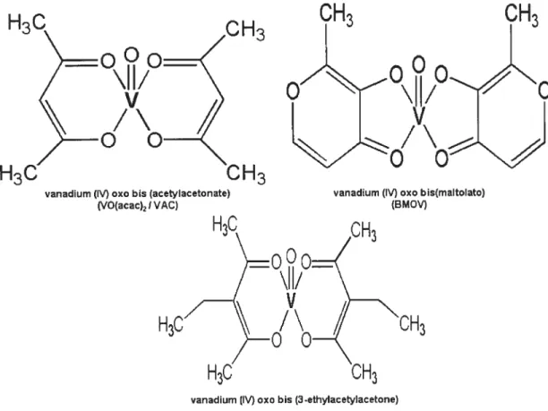
Other types of vanadium complexes. named organovanadium compounds (OVC), are synthesized by complexing vanadyl to organic ligands. These include vanadium (1V) oxo bi s(maltolato) (BMOV), vanadium (IV) oxo bis (acetylacetonate) (VO(acac)2/VAC), vanadium (IV) oxo bis (3-ethylacetylacetone) (VET). vanadium (IV) oxo bis (ethylmaltolato) (BEOV). vanadium (IV) oxo bis (6-methylpicolinato). and L-glutarnic acid -monohydroxarnate-NaOV complex (12-1$). The chernical structures of some of these
OVC are shown in Fig. 2. These OVC have been shown to be more absorbed and Iess toxic than inorganic salts (12:19-21). Subsequently. they were found to exhibit significantly enhanced insulin-mimetic activity in diabetic laboratory animais compared to inorganic
H3C
OH3
Eooo
\lI/
V
/\
H30
OH3
vanadium (IV) oxo bis (acetylacetonate) (VO(acac)2 $ VAC)
CH3
OH3
•0
vanadium (IV) oxo bis(maltolato) (BMOV)
H30
MJ
OH3
vanadium (IV) cxc bis (3-ethylacetylacetone) Wet)
7.3-DIABETES AND INSULIN-MIMETIC EFFECTS 0F VANADIUM COMPOUNDS
Diabetes has been known to afflict human populations since prehistoric times. Susruta, an Indian physician, described the diabetic syndrome in 400 B.C., calling it Madhumeh” or “honeyed urine” (22:23). Our modem era lias witnessed a surge in the incidence of diabetes. and it is estimated that at present approximately 150 million people world wide have the disease (24:25). According to the World Health Organisation (WHO). this number might double by 2025 (25).
Diabetes is caused by an absolute or relative lack of insulin secretion or action, and lias been classified into two major forms: type I. forrnerly known as insulin-dependent diabetes mellitus. and type II, that is. non-insulin-dependent diabetes mellitus. In type I diabetes. the absolute lack of insulin secretion is due primarily to the destruction of 13-cells by autoimmune mechanisms. On the other hand, in type 2 diabetes, f3-cells are able to produce insulin, but their insulin secretory response and insulin action on target tissues are defective, which results in hyperinsulinemia and insulin resistance (26). About 90% of the total diabetic population fails into the type II category. whule the remaining 10% are type I. Type II diabetes may be treated by diet control or by oral hypoglycernic agents. such as sulfonylurea, biguanides and thiazolidinediones (27-30). However, type I diabetics require regular daily injections of insulin to treat their diabetes, and some type II diabetics in the advanced stage of the disease also need insulin. Although the availability of highly purified insulin and the use of oral hypoglycaemic drugs as monotherapy or in combination with other agents (27:28) have greatly improved the management of diabetes. it still rernains a major health concem for humans with its prevalence increasing out of control (31). Thus. new therapeutic approaches are needed to more efficiently treat and hopefully cure diabetes.
In this regard, several studies performed in the Iast 2 decade have suggested that compounds of the trace element vanadium exert various insulin-mimetic and anti-diabetic effects in vitro and in vivo (32). The eariiest documented evidence of the insulin-like effects of the inorganic vanadium sait, sodium orthovanadate (Na3VO4), was published by Lyonnet et al. (33) in 1899, 22 years before the discovery of insulin. This group observed that oral Na3VO4 administration decreased glucosuria in 2 out of 3 diabetic patients (33). Their smdy went uimoticed for a long time. but the demonstration of an in vitro insuiin mimetic effect of vanadium salis by Tolman et al. (34) in 1979 sparked further interest. The latter group (34) showed that several inorganic vanadium compounds. simiiarly to insulin, stimulated glucose transport and oxidation in adipocytes, increased glycogen synthesis in the rat diaphragrn and hepatocytes. and inhibited gluconeogenesis in liver ceils. Since then. nurnerous studies have revealed various insulin-mimetic effects of vanadium compounds in vitro and in vivo. including the stimulation of glucose transport and glucose oxidation (34-3$), glycogen synthesis (3439-41), and lipogenesis (42;43) as well as the inhibition of lipolysis (37;43) and gluconeogenesis (34).
Among the in vivo actions of vanadium, the discovery that attracted the attention of diabetologists and endocrinologists was the serninal work of Heyliger et al. (19) which showed that Na3VO4 normalized hyperglycemia in an animaL model of diabetes mellitus. Since then, these findings were followed by those of several groups who confirmed and extended them to animal models of type 1 and type 2 diabetes mellitus as well as humans. Both inorganic salts. such as sodium orthovanadate (Na3VO4, NaOV). Vanadyl sulfate (VOSO4 3H20 / 4W0. VS). sodium metavanadate (NaVO3. NaMV) as weil as OVC such as BMOV, VAC, VET, or bis (6-methylpicoiinato) oxo vanadium were tested in most of these studies. Following section will summarizes some ofthese studies.
1.3.1-Effect on animal models oftypeI diabetes mellitus
1.3.1. 1-Effect of inorganic va,tadittn, saits
The studies of Heyliger et al. (19) were performed in female rats rendered diabetic by injection of streptozotocin (STZ), a compound that specifically destroys insulin-producing
13
ceils of the pancreas (44). STZ-diabetic rats are generally considered a mode! of type 1 diabetes mellitus (45). Orally-administered NaOV in drinking water (0.8 mg/ml). over a period of 42 days, was shown to decrease plasma glucose from 20 mM to 8.7 mM (19). Interestingly, in these studies, vanadium was not associated with an increase in plasma insulin. In fact, NaOV treatrnent lowered insulin levels even in normal rats without significantly altering plasma glucose (19). It was, therefore. suggested that the vanadium induced reduction of glycemia was independent of its effect on insulin secretion. These investigators also noted severe diarrhoea in some STZ-diahetic rats. This condition. however, was corrected by administering NaOV in 0.5% NaC1 solution (19).The studies of Heyliger et al. (19) were confirrned and expanded by Meyerovitch et al. (20) who demonstrated that NaMV (0.8 rng/ml in drinking water) decreased plasma glucose in STZ-diabetic male Wistar rats to almost normoglycemic (in some cases. hypoglycaemic) levels within 2 to 4 days of treatment (20). Subsequently, the giucose-lowering effects of vanadium compounds were validated by several other investigators. In ail these studies. hyperglycaemia was significantly reduced and. in many cases, virtually normalised by oral administration of vanadium compounds to diabetic rodents. The dose of vanadium salts required for exerting a maximum glucose-lowering effect varied between studies, but a median concentration of 0.5 mg/ml in drinking water appeared to be sufficient. The glucose-lowering action was rnaintained during the course of therapy, but the animals became hyperglycaemic within 2-4 days afler its cessation (20). In 1 experiment. however, where STZ-diabetic rats were given VS for 3 weeks, normoglycaemia was sustained for 13 weeks afier withdrawal oftreatment (46).
A comparative study of NaOV, VS and NaMV showed that regardless of the type of vanadium saits used, the decrease in plasma glucose levels, glucosuria and urinary volume and the improvement in oral glucose tolerance were similar in ail cases (47). These investigators also observed that although plasma glucose levels increased rapidly after treatment withdrawal, they remained significantly lower than those found in non-treated STZ-diabetics for at least an additional 4 weeks (47).
1.3. 1.2-Effect oforgaiiic vanadium coinpottnds
To overcome the gastrointestinal side-effects of vanadium and to enhance its absorption through the gut, McNeill et al. (12) pioneered the use of OVC in rat models of diabetes. Administration of a VS-maltol complex (BMOV, 1.5$ mM) to STZ-diabetic rats in drinking water for 4 weeks reduced plasma glucose from 13.9 to 7.0 mM and corrected the polydypsia associated with diabetes (12). BMOV elicited the glucose-lowering response within 1 day oftreatment. decreasing plasma glucose from 20 to 8 mM (21). in contrast to the use of inorganic vanadium salts, which required about 4-7 days for a similar glucose Iowering outcorne (19;20). Intraperitoneal injection of BMOV was even more effective than BMOV administration in drinking water because it decreased blood glucose to tess than 9 mM within 8 hours (21). The ED50 of BMOV was found to be 3 times lower than that of VS (0.08 versus 0.22 mmol/kg) and thus, it was 3 times more potent (21). In addition. BMOV was beffer tolerated. as evidenced by a lack of gastrointestinal toxicity (diarrhoea). and no mortality (48).
Subsequently, several new OVC were synthesised to test their antidiabetic potential (14-17). Among these complexes, the anti-diabetic potential of BMOV has been investigated most extensively (12;19-21).
Reul et al. (13) recently compared the effects of 3 different OVC, BMOV, VAC, and VET to inorganic VS on glucose metabolism in STZ-diabetic rats. Their studies showed that ah 3 OVC were more potent than VS in eliciting the hypoglycaemic response, and VAC appeared to be superior to VET and BMOV (13). The superior effect of VAC may be due
to its better intestinal absorption since a higher plasma vanadium level was achieved at 1 and 3 weeks in comparison to either BMOV or VET (13). Moreover, vanadium therapy of diabetic rats with OVC did flot evoke any marked toxicity on hepatic and renal functions (13). Thus. it appears that these complexes have an advantage over the inorganic saits as potential anti-diabetic agents.
In addition to inorganic and organic vanadium compounds. peroxovanadium compounds have been tested as insulin-mimetic agents (11 ;49). Intraperitoneal or intrajugular injection of bis-peroxovanadium into STZ-diabetic and biobreeder rats markedly reduces hyperglycaemia within 30 min (49;50). However, unlike other vanadium compounds, flot many studies have been performed with peroxovanadium compounds.
1.3.2-Effect on animal models oftype II diabetes mellitus
The anti-diabetic potential of various vanadium compounds bas also been examined in animal models of type 2 diabetes mellitus. Three well-characterised models, genetically obese, fatty (falfa) Zucker rats, genetically-diabetic C57 BL/KsJ-db/db (db/db) mice, and genetically-diabetic ob/ob mice (45), have been investigated. Administration to falfa rats of NaOV 0.5 mg/mI in drinking water and up to 0.25 rng/g in food for 3 rnonths considerably irnproved their glucose homeostasis as well as oral glucose tolerance (51). In addition. during an intravenous glucose tolerance test, the glucose disappearance rate, an index of glucose utilisation. was 50% higher in treated rats cornpared to the controls. whereas plasma insulin levels were reduced by 50% (51). In db/db mice, 12-week therapy with NaOV 0.6 mg/ml decreased plasma glucose from 24 to 7 mM (52), and only 0.25 mg/ml was sufficient to lower blood glucose from 15 to 8 mM (52). A similar response was exhibited in ob/ob mice where the glucose-lowering effect was evident within a week, reaching a maximum after 16 days oftreatment (53).
The impact of a diet mixed with NaOV (1 .5 rng/g) (rather than in drinking water) was also investigated, in a high-sucrose diet-induced hyperinsulinemic insulin-resistant rat mode!. and it was found that hyperglycaemia as well as hyperinsulinernia (54) were signiflcantly irnproved. In addition to NaOV. the organo-vanadium compound BMOV was found to reduce hyperinsulinemia. insulin resistance and glucose intolerance in falfa Zucker rats (55-57). On the other hand. the effect of peroxovanadium cornpounds on the above rodent modeis of type 2 diabetes mellitus bas flot yet been examined.
VS bas been investigated in Psammomys obesus. a gerbi! (nicknamed the ‘sand rat”) that represents a nutritiona!!y-induced mode! of diabetes and insulin resistance (58). Psammomys obesus receiving a high-energy diet become hyperglycaernic and hyperinsulinemic (59). VS at a dose of 5 rng/kg body weigbt for 5 days resulted in proionged restoration of normogiycaemia and normoinsulinemia in this model (5$). In addition. these changes were associated with a normal glucose toierance test and a decreased level of the hepatic gluconeogenic enzyme phosphoenol pyruvate carboxykinase (PEPCK) (5$). Interesting!y,
vs
was ineffective when administered to sand rats that had complete!y iost their insulin secretory capacity. indicating a requirement of !ow-!evel insulin for vanadium to work in these animais. Thus. vanadium appeared to be an insulin potentiator/enhancer rather than mirnicker in improving insulin resistance in sand rats (5$).1.3.3-Clinical studies in human diabetes mellitus
The demonstration of a beneficiai action of vanadium compounds in both type I and type 2 anima! modeis of diabetes mellitus encouraged severa! investigators to embark on research in hurnan diabetics. In earlier studies, srnall doses (50-125 mg/day) ofNaMV or VS were administered orally to a iimited number of type 1 or type 2 diabetic subjects for periods ranging from 2 to 4 weeks (60-62). In type I diabetics. NaMV (125 mg/day) for 2 weeks had no effect on fasting plasma glucose ievels. but caused a small yet significant deciine in
daily insulin requirements and improved glucose utilisation in 2 out of 5 patients (60). In type 2 diabetics, NaMv resu!ted in increased insuiin sensitivity due to enhanced
non-oxidative glucose disposai (60). Similarly. VS improved insulin resistance in type 2 diabetics accompanied by a slight decrease in fasting plasma glucose and glycosylated haemoglobin (HbA1), enhanced insulin-mediated glucose uptake and suppressed hepatic glucose production (HGP) (61;62).
Since only a limited number of subjects were treated with low doses of vanadium for a short duration in these eariy studies. 2 independent groups recently investigated the effect of long-term treatment with higher VS doses in a larger type 2 diabetic population (63:64). Goldfine et al. (63) treated 16 patients with VS doses varying from 75 to 300 rng/day for 6 weeks and observed that fasting glucose declined significantly only in the 300-mg VS group whereas HbA1 decreased in both the 150- and 300-mg groups. Interestingly, these treatments had no effect on either basal HGP or on the suppression of HGP in response to insulin. A sirnilar study conducted by Cusi et aI. (64). who treated 11 type 2 diabetics with VS at a dose level of 150 mg/day for 6 weeks, found that both HbA1 and fasting plasma glucose were significantly decreased. In these investigations, VS reduced endogenous glucose production and increased insulin-mediated glucose disposal. However, none of the clinical research undertaken so far demonstrated complete normalisation of hyperglycaemia, as has been the case in animal models of diabetes mellitus. The difference may be attributed to a much lower blood vanadium level reached in patients (1-10 iIM) than in animais as well as the duration oftherapy (63).
1.4-MECHANISM 0F THE HYPOGLYCAEMIC EFFECT 0F VANADIUM
The precise mechanism by which vanadium compounds improve hyperglycaemia and glucose homeostasis in diabetes remains unclear. Vanadium therapy in a type 1 model of diabetes mellitus slightiy but insigniflcantly increased plasma insulin (I 9;20). whereas a significant, up to 50% decrease in plasma insulin was observed in type 2 models (51-53;65;66). Clearly, this alteration in insulin levels can not be attributed to the anti-diabetic effects of vanadium compounds in type 1 diabetics, but may be beneficial for type 2
diabetics. The hypophagic influence of vanadium compounds miglit contribute to their hypoglycaemic outcome (67). but studies showing that organic or peroxovanadium compounds exert acute glucose-Iowering actions within a few minutes to hours after their administration (not sufficient to suppress feeding) (11;12;21;4$-50) do not support this hypothesis. Furthermore, both in vitro and in vivo, vanadium compounds modify glucose and Hpid metabolism in adipose tissue, muscle. liver and several cultured ceil unes (32:68), which may serve as a more plausible mechanism for the glucose regulatory effects of vanadium.
1.4.1-Effect on glucose transport
The stimulatory effect of NaOV on glucose transport lias been observed in rat adipocytes (36:3$;69), rat skeletal muscle (40:70). human skeletal muscle (71:72), 313-Li adipocytes (73), and L-6 myotubes (74:75). In most of these studies. a high (mM) vanadium concentration was required to enhance glucose transport. However, Dubyak and Kleinzeller (36) reported that long-terrn (3-hour) treatrnent with 300 1.iM NaOV stimulated glucose transport. which was 80% the value achieved with 17 nM insulin. Thus. longer incubation with low vanadium concentrations may be sufficient to evoke a physiologically relevant increase in glucose transport.
Since insulin-stimulated glucose transport is mediated by an insulin-sensitive glucose transporter protein type 4, GLUT-4 (76;77), the effect ofNaOV, VS or BMOV on GLUT-4 was investigated in diabetic rodent models (5$;7$-$2). In the basal state, GLUT-4 is localised in intracellular sites, and upon insulin stimulation. it is translocated to the celi surface to facilitate glucose uptake by target cells (76). Vanadium exposure of STZ diabetic rats lias been shown to restore the expression and/or cdl surface translocation of GLUT-4 in skeletal muscle (7$;79). and heart (80;81). In addition. VS treatment of a nutritionally-induced, insulin-resistant model of Psammomys obesus. lias been found to restore membrane-associated as well as total protein and mRNA content of GLUT-4 in the gastrocnemius muscle (52). On the other hand, in contrast to STZ-diabetic or sand rats,
NaOV in insulin-resistant falfa rats increased insulin-mediated glucose utilisation in muscle without any effect on either GLUT-4 protein or rnRNA (22). The discrepancy between the vanadium effect in STZ-diabetic or sand rat and insulin-resistant fa1fa rodents may be attributed to the fact that GLUI-4 content remains unaltered in insulin-resistant models (83;84) whereas SIZ-diabetic rats exhibit decreased GLUT-4 expression (78;$5;$6). Thus. it is difficult to establish a simple correlation between GLUT-4 content and glucose uptake and utilisation in vivo ($2). However, an increase in the intrinsic activity or enhanced targeting of GLUT-4 to the ccli surface might contribute to improved glucose uptake by vanadium (22). In isolated rat adipocytes ($7) and in cultured L-6 myotubes (75), NaOV bas been shown to stirnulate GLUT-4 transiocation to the cell surface. Ibus, vanadium compounds are capable of stirnulating glucose transport through an effect on GLUI-4.
1.4.2-Effect on glycogen metabolism
Another physiological response modulated by vanadium is mediated by its action on glycogen synthesis. NaOV and VS have been found to stimulate glycogen synthesis in in vitro systems. including the mouse diaphragm (41). rat hepatocytes and diaphragm (34;88), rat adipocytes (39;89). Chinese hamster ovary ceils over-expressing insulin receptor (CR0-HIR) (90) and 313-LI adipocytes (73). These cornpounds also improve depressed glycogen levels in the liver of SIZ-diabetic (91 ;92) as well as in other insulin-resistant rodent models (52;93). In addition, NaOV bas been demonstrated to restore the glycogen synthetic rate in 90% pancreatectomized, diabetic rats (94). On the other hand, treatment of 7-week STZ-diabetic rats to BMOV failed to improve insulin-stirnulated glycogen synthase activation in skeletal muscle (95) wbereas similar treatment enhanced it in fa!fa Zucker rats (96). Ihe reason for these discrepancies is not clear, but it is possible that the basal level of glycogen synthase activity is moduÏated differentiaÏÏy in a tissue-specific manner in various models ofdiabetes (93).
Since vanadate treatment normalises the decreased activity of the active glycogen synthase form in the liver of STZ-diabetic rats (91), and activates glycogen synthase in rat adipocytes (89), it has been suggested that this may be one of the mechanisms by which vanadium compounds enhance glycogen synthesis ($9;91). Vanadate has also been found to increase glucose-6-phosphate (G6P) by inhibiting glucose-6-phosphatase (G6Pase) activity in rat adipocytes (97;9$). Moreover, vanadate restores the depressed levels of G6P in the adipose tissue, liver and muscle of STZ-diabetic rats (97). Since G6P is an allosteric modulator of glycogen synthase activity (99), alteration of its levels may rnodify the activation status of glycogen synthase and, eventually, glycogen synthesis. Similarly to the effects in animal models of diabetes, 3-week VS therapy (100 mg/day) in type 2 diabetic patients has been associated with an increase in insulin-stimulated glycogen synthesis (61). However, in a more recent study, VS treatment (150 mg/day) for 6 weeks in humans, although it caused a 1 .5-fold enhancement of glycogen synthase fractional velocity (an indicator ofheightened glycogen synthase activation), failed to elicit any significant change in either basal or insulin-stimulated glycogen synthase activity (63). Thus, the impact of VS therapy on glycogen synthase activity in hurnans rernains controversial, and needs to be examined in detail.
1.4.3-Effect on gluconeogenesis
In addition to the stimulatory action on glucose uptake and utilisation, vanadium-induced suppression of hepatic glucose output can also contribute to improved glucose homeostasis. In fact, decreased hepatic glucose production has been noted in diabetic animals (94;100;101) and humans afier vanadium therapy (61;62;64;101), but some studies have failed to obtain this result (60;63). The reason for these discrepant findings is not clear at present, but the study model, duration of therapy, and vanadium dose rnight have been determining factors. An inhibitory action ofNaMV on glucose output bas been documented in the isolated, perfused rat liver (102), and VO(acac)2 bas been shown to suppress gluconeogenesis in isolated hepatocytes and kidney cortex tubules from control and diabetic rabbits (103). Consistent with these data on gluconeogenesis, vanadium treatment
decreased the heightened expression of the gluconeogenic enzymes PEPCK and G6Pase (13:98:104-109).
1.4.4-Effect on lipogenesis and lipolysis
In addition to their action on glucose metabolism. vanadium compounds have been reported to modulate Iipid metabolisrn both in vivo and in vitro. NaOV treatrnent of insulin resistant, sucrose-fed diabetic rats and fa!fa Zucker rats significantly lowered plasma triglycerols (93). Furthermore, NaMV and VS decreased plasma cholesterol levels in
hurnans (63:64:110), and this effect was associated with a decline of low- as well as high density lipoproteins (64); however, no change in either plasma free fatty acid or triglyceride fractions was noted (62;63). Vanadate has also been shown to reduce total and free cholesterol levels in normal subjects (111), which may be due to inhibition of the steps involved in cholesterol biosynthesis (1 12:113). In isolated hepatocytes (114) and adipocytes (37). NaOV modulated lipid metabolism by’ stimulating lipogenesis and suppressing lipolytic activity.
1.5-VANADIUM TOXICITY
Despite their impressive anti-diabetic properties, vanadium compounds have been associated with several toxic effects (115:116). The most common toxic effects are diarrhoea, decreased fluid and food uptake, dehydration and reduced body weight gain (20:67:78:91:94:106:117) which can, however, be corrected by adding NaC1 to the drinking water, adjusting the pH of the solution to neutrality, and by gradually increasing the dose of vanadium (20:41). Organic vanadium compounds are much safer as anti-diabetic agents than inorganic vanadium salts. and diabetic rats receiving organic compounds did not show any gastrointestinal side-effects and did flot develop diarrhoea (13). Administration of the chelater tiron was also found to minimise vanadate toxicity without altering its anti-diabetic actions (115).
In addition to gastrointestinal discomfort, other toxic effects of vanadium saits have been documented in the literature, whicli include hepatotoxicity, nephrotoxicity and teratogenicity as we!! as deve!opmenta!/reproductive toxicity (115; 116). Tliey stimulate mitogenesis and ce!! proliferation in cu!tured ce!!s (112) and, thus, have tlie potentia! to exert tumorigenic/carcinogenic activity. In contrast, recent work lias demonstrated that vanadium compounds inhibited serum- and growth factor-stimulated mitogenesis (119;120) and possess anti-tumor activity (121;122). Many other studies have, however, fai!ed to detect any change in the levels of urea, creatinine, g!utamic oxaloacetic transaminase, and g!utamic pyruvic transarninase (123), indices of kidney and !iver functions. Moreover, no significant changes in the histopatho!ogy of severa! tissues, inc!uding the liver, sp!een, stomach, heart and lung, have been observed among contro! and VS-treated anima!s (124). E!ectron microscopic examination of ob/ob mice treated with NaOV for 47 days did not reveal any sign of hepatotoxicity (20). Some reports have indicated that vanadium compounds cause behavioural changes in rats exposed to NaMV (125). In patients treated with vanadium saits, gastrointestinal discomfort was the most common toxic effect, which could be corrected by decreasing the dose !evel (60;61). Moreover, c!inica! studies have been of short duration (up to 6 weeks) and have uti!ised muci lower doses than those administered in animal experiments; thus, the !ong-term toxicity of vanadium in humans rernains to be explored. Clear!y, at present, there is no consensus on the toxic effects of vanadium compounds, and detailed and systernatic investigations are needed to evaluate the toxicity of various vanadium cornpounds before undertaking long-tenu c!inica! tria!s in humans.
1.6-MOLECULAR MECHANISM 0F VANADIUM ACTION
At the mo!ecu!ar !eve!, the precise mechanism by which vanadium e!icits insu!in-!ike and anti-diabetic effects remains poor!y characterised. Since vanadate is a potent PTPases inhibitor, and insu!in receptor (IR) activation requires increased tyrosine phosphorylation of its
f3
subunit (126; 127), it was be!ieved that vanadium caused an activation of IR bypreventing dephosphorylation of the IR-t3-subunit. The insulin like effects of vanadium salts were therefore attributed to be secondary to PTPase inhibition. In this manner one or more tyrosine kinases that participate in the insulin signaling cascade may be indirectly inftuenced. Thus, it is primordial to dissect the signal transduction pathways stimulated by insulin for better understanding of the mechanism of vanadium action at the cellular level.
1.6.1- The insulin signaling cascade
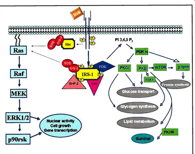
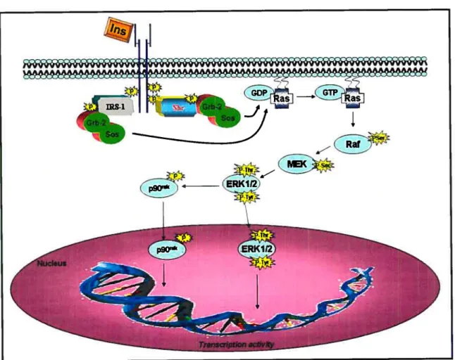
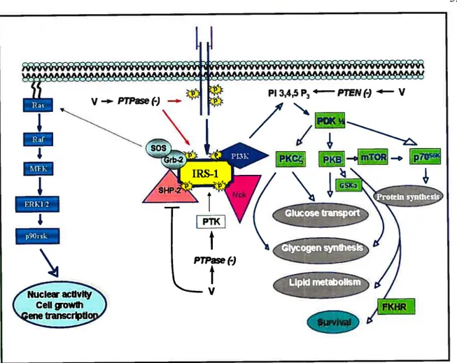
Insulin, the primary hormone implicated in blood glucose control, acts by stimulating glucose influx and metabolism in adipocytes and muscle and by inhibiting gluconeogenesis by the liver. The insulin action in target cells is propagated by binding to its receptor on celi membranes (Fig. 3). The IR consists of a heterodirneric Œ2, 132 structure. Insulin binding to the IR-Œ-subunit results in conformational changes, leading to enhanced intrinsic protein tyrosine kinase (PTK) activities of the 13-subunit by multi-site tyrosine phosphorylation. Once activated, IR-PTK can phosphorylate several cytosolic IR substrates, including insulin receptor subtrates (IRSs), Src hornology collagen (Shc) and adaptor protein with pleckstrin homology (PH) and Src homology 2 (SH2) domains (APS). The phosphorylated proteins dock downstream effector molecules that contain the SH2 domain (12$), which are then able to activate two key signaling pathways. In one pathway, the association of IRS type 1 (IRS- 1) with growth factor receptor binder-2 (Grb-2) complexed with mammalian son of seven less (mSOS) results in subsequent activation of the Ras, Raf, MEK and extracellular signal-regulated kinase (ERK) pathway. Activated ERK1/2 phosphorylates and activates a downstream ribosomal protein kinase, p9O. Both ERKI/2 and 90rsk can be translocated to the nucleus where they phosphorylate transcription factors, and contribute to the mitogenic and growth-promoting effects of insulin (12$;129).
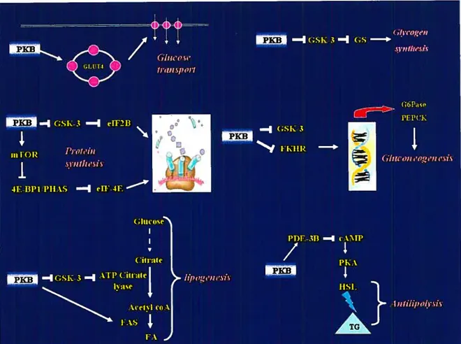
Figure 3: Schematic model showing key elements of the insulin-signaling cascade Insulin (Ins) -induced tyrosine pliosphorylation and activation of insulin receptor substrates (IRSs) by the protein tyrosine kinase (PTK) of -subunits lead to recruitment of Src homology (SH-2) dornain-containing signaling proteins, such as Grb-2-SOS, the p85 regulatory subunit of P13-K, a lipid and protein kinase. The IRS-associated complex initiates 2 signaling pathways. One pathway. known as the MAPK pathway, consists of Ras/Raf/MEK!ERKI/2 and p9Orsk. Both ERKI/2 and p9Orsk can be translocated to the nucleus where they can activate transcription subsequent to the phosphorylation of several transcription factors. Another pathway involves P13-K activation, which resuits in the generation of PIP3. PIP3 activates a variety of downstrearn signaling components, including PDKs, PK3, mTOR, p7Os6k, PKC, GSK-3 and fKHR. PKB and PKC are involved in a glucose transport system. mTOR regulates protein synthesis via p7Os6k. Glycogen synthesis is regulated by PKC directly and by PKB via GSK-3. PKB-rnediated phosphorylation of FKHR inhibits its effect on transcriptional activity. (Abbreviations are defined in the list of abbreviations).
* Adapted/Modffied from (12$;129;301).
1F
IRafi
IMEI
1F
I
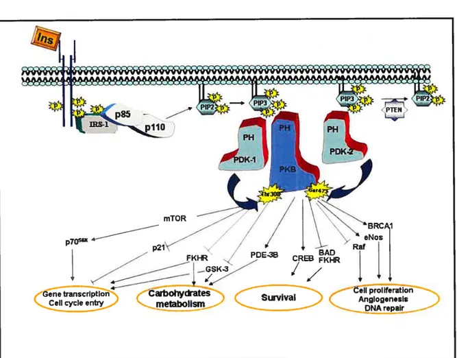
Ccli growth Gene transcriptionThe second pathway that radiates from the IRS complex upon insulin stimulation involves phosphatidylinositol 3-kinase (P13-K) activation (127;129) (fig. 3). P13-K phosphorylates phosphatidylinositol (PI) lipids at position 3 of the inositol ring, and generates 3-phosphorylated forms of PI, such as phosphatidylinositol 3, 4, 5 triphosphate (PIP3) (130). which is implicated in the activation of phosphoinositide-dependent kinase (PDK) and related serine/threonine protein kinases responsible for the phosphorylation and stimulation of several downstrearn signaling protein kinases. such as protein kinase B (PKB) (also known as Akt), p70 ribosomal S6 kinase (p70’6”)(l3l) and protein kinase C (PKC)-zeta (132). Activation of these protein serine/threonine kinases bas been hypothesized to mediate the metabolic effects of insulin at the level of glucose transport, GLUT-4 translocation, glycogen and protein synthesis (129). In addition to the P13-K-dependent pathway of insulin-induced glucose transport. recent studies have suggested a potential role of an alternate P13-K-independent mechanisrn of glucose transport (133). Ibis mechanism involves insulin-induced tyrosine phosphorylation of the protooncogene Cbl (133). Cbl associated protein (CAP) and APS are important components of this mechanisrn and contribute in the membrane targeting of tyrosine phosphorylated Cbl to lipid rafts which eventually activates a small 9 guanosine triphosphate (GIP) binding protein, TC 10 (134). Ihe activation of IC 10 has been suggested to be a crucial step in mediating insulin-induced GLUT-4 translocation (134:135).
The following sections provide a full description of sorne of the key components of the insulin signaling cascade.
1.6.1.1- The insiilln receptor
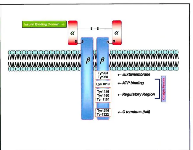
Ihe IR is present in all vertebrate tissues with varying degrees of expression. Its concentration ranges from 40 receptors on circulating erythrocytes to more than 200,000 receptors on myocytes. adipocytes and hepatocytes (128). The IR gene is present on the short ami of hurnan chromosome 19, is more than 150 kilobases, and contains 22 exons, which encode 4.2-kb cDNA (4). The IR is composed of two extracellular Œ-subunits and two transmembrane 3-subunits linked to each other by disulfide bonds (128) (fig. 4). Both
the c- and
f3-
subunits are derived from a single proreceptor by proteolytic processing at a cleavage site consisting of four basic amino acids. Alternative splicing sunounding exon11 resuits in two receptor isoforms differing by 12 arnino acids close to the COOl-1 terminus of the a-subunits. The a-subunit and
f3
subunits weigh 135 and 95 kilodalton (kDa) respectively. The Œ-subunit is located entirely outside of the ce!! and contains the insu!in-binding site with stoichiometry between J and 2 insulin molecules/receptor, whereas the intracellular section of the f3-subunit possesses intrinsic insulin-regulated tyrosine kinase activity. Different biochernical approaches have found that the distinct regions of the first 500 amino acids of the Œ-subunits contain ligand-binding determinants (136). Therefore, insulin-binding affinity is increased by replacing amino acids 64-137 of the insulin-like growth factor type 1 receptor (IGf-IR) Œ-subunits by corresponding residues from the IR. In addition. replacing amino acids ofresidues 325-524 from the IR in this chimera enhances insulin-binding (136137). The a-subunit of IR can also act as a regu!atory subunit of the tetrameric receptor because the unoccupied IR c&-subunits inhibit the tyrosine kinase activity of the f3-subunits. Depletion of the u-subunits by deletion mutagenesis or proteolytic cleavage or certain point mutations in the Œ-subunits (Arg$6 —Pro) abrogate this inhibition (138). The carboxy-terrninal of the Œ-subunits has four Cys residues, 647, 682, 683 and 684, which are involved in subunit disulfide linkage. Cys 647 to Ser 647 mutation did not cause any change in insulin binding, but these mutants were defective in insulin-stimulated kinase activity. Therefore. insulin-stimulated signa! transmission from ligand binding to kinase activation requires proper covalent interaction between the Œ- and
f3-
subunits for normal subunit communication (139; 140).Figure 4: Structure of the insulin receptor
The insulin receptor is cornposed of two o-subunits and two J3-subunits linked by disulfide bonds. The Œ-subunits are entirely extracellular and contain the insulin binding domains. while the 13-subunits penetrate through the plasma membrane into the cytoplasm. The
13-subunit has several tyrosine autophosphorylation sites. ATP binding dornain and regulatory region necessary for the activation of its intrinsic protein tyrosine kinase activity.* AdaptedlModified from (127).
a
s—sa
-Juxtamembrane %‘—ATPbinding—I
• lTyrll46I
Tyrll5D ‘—ReguIatoryRegionI
151 -Ctem7lnus (Ïa(OThe IR-f3 subunits consist of short extraceilular sites for glycosylation, a transmembrane dornain of 23 amino acids and an intracellular region which bas protein tyrosine kinase activity required for insulin action (Fig. 4). The IR-f3-subunits possesses different functional regions. inc luding the adenosine triphosphate (ATP)-binding domain and autophosphorylation sites in the intraceilular juxtamembrane region (Tyr 960, and possibÏy Tyr 953 and Tyr 972), a regulatory region (Tyr 1146. Tyr 1150 and Tyr 1151). and the C terminus (Tyr 13 16 and Tyr 1322). In vitro mutagenesis experiments have provided evidence that a key lysine residue at position 1030 (LyslO3O), which is an ATP-binding site, when replaced by other amino acids, causes complete loss of kinase activity of the
f3
subunit aÏthough insulin-binding is unaffected (141;142). A similar function for a lysine residue at position 1012 bas been found (143;144). The discrepancy in the position of lysine between these two studies is due to differences in the approacb used in numbering these residues.Tyrosine phosphorylation occurs through a transmechansim in which insulin binds to the x subunit of one Œf3-dimer and thus stimulates phosphorylation of the adjacent covalently linked f3-subunit (145). Autophosphorylation of ail tbree tyrosine residues ofthe regulatory region increases tyrosine kinase activity of the IR. Deletion of 1 or ail tyrosine residues in this region by mutation reduces insuiin-stimuiated kinase activity (146;147).
Thejuxtarnembrane region oftbe IR-f3-subunit containing Tyr 960 within NPXY (Asn-Pro any amino acid-Tyr) motifs plays a role in signal propagation. Repiacement of Tyr 960 by phenylalanine or alanine inhibits signai transmission without affecting autophosphorylation in the other regions and kinase activity in vitro (142:149). Anaiysis of Tyr960 point mutation shows a type of receptor that is impaired in internaiization in response to insulin (150). In addition, Tyr960 to Phe point mutation is unable to phosphoryiate IRS-1, a crucial IR substrate, which resuits in decreased P13-K activation and blocks glycogen and DNA synthesis (151). Thus, the juxtamembrane region is a criticai dornain required for
both insulin-stimulated signaling via IRS-1 phosphorylation as weil as receptor internalization.
The C terminus of the IR contains two autophosphorylation sites. Tyr 1316 and Tyr 1322, which may not be essentiai for kinase activation (152). In contrast, the C terminus lias serine and threonine residues phosphorylated in response to phorbol esters, cyclic adenosine monophosphate (cAMP) analogues. and insulin itself. thus Ïeading to decreased insulin-stimulated tyrosine kinase activity (153-155). Thus. the C terminus plays a reguÏatory role that is essential for signaling.
1.6.1.2- The insulln receptor siibstrates
As stated earÏier. activated IR-PTK mediates the insulin response through tyrosine phosphorylation of IRS proteins which serve as docking sites for effector molecuies responsibie for propagating the insuiin signal (156). At least four IRS proteins have been identified in mammais. Whiie IRS-1 and IRS-2 are wideiy expressedin ail tissues, IRS-4 is only expressed in the brain, thymus, kidney and f3-cells and IRS-3 is present in rodents and highly restricted to adipose tissue (157). Iwo other srnali proteins caiied. Gab and p62d have been shown to be substrates ofthe IR and are considered to be IRS proteins (156). IRS proteins have severai common features and are composed of a NI-12-terminal PH and/or phosphotyrosine binding (PTB) domain foliowed by a COOH-terminal tail comprised of multiple tyrosine and serine/threonine phosphorylation site residues. The generai structure ofIRS-l is presented in Fig. 5.
The roie of the PH dornain in IRS is flot known because physiologicaliy-relevant binding partners are undefined; however, phospholipids, acidic peptides, or specific proteins such as PH domain interacting protein (PHIP) are defined as PH domain-binding partners (15$), but the mechanism of this coupiing is unknown.
IRS-1 serves as a type of docking protein for recruitrnent and activation of other enzymes that ultirnately mediate insulin effects. The relative positions of the pleckstrin homology (PH) and phosphotyrosine binding (P13) domains are indicated. Potential tyrosine phosphorylation sites are indicated by Y while known phosphorylation motifs are enclosed in boxes below potential binding partners, including P13-K, Grb-2 and SHP-2. (Abbreviations are deflned in the list of abbreviations)
* Adapted/Modifled from (156).
SHP-2
ÉWHIE
Grb-2
cl243
The PTB domain was found to mediate interactions of IRSs with the IR, insulin-like growth factor type 1 receptor (IGF-1R) and interleukin-4 (IL-4) receptors through phosphorylated NPXY motifs located in these receptors.
The tyrosine phosphorylated residues in the COOH-terrninal serve as docking sites for SH2 domain-containing signal transducers. including p8, a regulatory subunit of P13-K. SH2 domain-containing tyrosine phosphatase (SHP2). Src-like kinase fyn or adapter rnolecules. such as Grb-2, nck, Crk, SHB and others that mediate downstream signais (129). Moreover, other partners including SV4O large T antigen. Œvt33 and 1 4-3-3 bind to IRS proteins through an unknown tryosine phosphorylation-independent mechanism (129). The role of serine/threonine phosphorylation sites bas not been fully unraveÏled. but severai studies have shown that factors. known to induce insulin resistance. such as free fatty acids (159), turnor necrosis factor-a (TNF-a) (160), angiotensin II (Ang TI) (161), endothelin-l (162) and chronic insulin treatrnent (163), lead to increased serine/threonine phosphorylation of IRS-1 and consequently to impainuent of insulin signai transduction. Many Ser/Thr-kinases which phosphorylate IRS-1 are identifled including Raf. MEK. mitogen-activated protein kinase (MAPK). p90ts, Rho kinase-Œ (R0K-), and PKC
isoforms and kinases downstream ofthe P13-K cascade such as PDK-1, PKB. mammalian target of raparnycin (mTOR), 7056K and glycogen synthase kinase-313 (GSK-3f3) (164). Specific Ser/Thr phosphorylation sites in IRS-1 identified in vitro include Ser-302, Ser-307, $er-612, Ser-636, Ser-639, Ser-731 and Ser-789. 0f these, Ser-307 phosphorylation has
been studied most intensiveÏy as a rnechanism for disrupting IR/ÏRS- I interactions (164). Importantly, a high level of phosphoryiated Ser-307 has been detected in vivo in human skeletal muscles (165) and in insulin-resistant rodent models (166). Serine phosphorylation of IRS-1 by some of these kinases has been shown to impair the ability of insulin to enhance IRS- 1 tyrosine phosphorylation (167) and lead to proteasome-mediated degradation (168). Taken together, Ser/Thr phosphorylation ofIRS-1 could represent one