425
DOI 10.1095/biolreprod.103.026724
Poly(A) RNA Is Reduced by Half During Bovine Oocyte Maturation but Increases
when Meiotic Arrest Is Maintained with CDK Inhibitors
1Anne Sophie Lequarre,
2Juan M Traverso, Joelle Marchandise, and Isabelle Donnay
Unite´ des Sciences Ve´te´rinaires, Institut des Sciences de la Vie, Universite´ Catholique de Louvain,
Louvain-la-Neuve B-1348, Belgium
ABSTRACT
Variations in the amount of different RNA species were in-vestigated during in vitro maturation of bovine oocytes. Total RNA content was estimated to be 2 ng before meiosis, and after meiosis resumption, no decrease was observed. Ribosomal RNA did not appear to be degraded either, whereas poly(A) RNA was reduced by half after meiosis resumption, from 53 pg to 25 pg per oocyte. Real-time polymerase chain reaction was performed on growth and differentiation factor-9 (GDF-9), on cyclin B1, and on two genes implicated in the resistance to oxidative stress, glucose-6-phosphate-dehydrogenase (G6PD) and peroxiredoxin-6 (PRDXperoxiredoxin-6). When these transcripts were reverse-transcribed with hexamers, the amplification results were not different be-fore or after in vitro maturation. But when reverse transcription was performed with oligo(dT), amplification was dramatically reduced after maturation, except for cyclin B1 mRNA, implying deadenylation without degradation of three transcripts. Al-though calf oocytes have a lower developmental competence, their poly(A) RNA contents were not different from that of cow oocytes, nor were they differently affected during maturation. When bovine oocytes were maintained in vitro under meiotic arrest with CDK inhibitors, their poly(A) RNA amount increased, but this rise did not change the poly(A) RNA level once matu-ration was achieved. The increase could not be observed under transcription inhibition and, when impeding transcription and adenylation, the poly(A) RNA decreased to a level normally ob-served after maturation, in spite of the maintenance of meiotic arrest. These results demonstrate the importance of adenylation and deadenylation processes during in vitro maturation of bo-vine oocytes.
gamete biology, gene regulation, kinases, meiosis, oocyte devel-opment
INTRODUCTION
During oocyte growth, when the follicle develops from
the primordial stage to the antral stage, transcription and
translation are intense and result in the production of RNAs
and protein, both for immediate use and for storage [1].
Messenger RNAs can be stored for long periods, with
half-lives of up to 28 days in mouse oocytes [2]. Stable storage
1This research was funded by the European Commission, Grant of the Vth Framework: QLK3-CT1999-00104 (Ex Ovo Omnia) and by ‘‘Actions de Recherche Concerte´es’’ de la Direction ge´ne´rale de la Recherche Scien-tifique, Communaute´ franc¸aise de Belgique2Correspondence: A.S. Lequarre, Unite´ Ve´te´rinaire, Batiment Carnoy, 5 place Croix du Sud, Louvain-la-Neuve, B-1348 Belgium.
FAX: 32 10 47 37 17; e-mail: Lequarre@vete.ucl.ac.be Received: 4 January 2004.
First decision: 30 January 2004. Accepted: 16 March 2004.
Q 2004 by the Society for the Study of Reproduction, Inc. ISSN: 0006-3363. http://www.biolreprod.org
of mRNAs implies variable length of their 3
9 poly(A) tail,
but also different association with specific RNA binding
proteins, especially the masking proteins [3, 4]. Once a
mouse oocyte has reached its full size (diameter 75–80
mm), it contains about 0.6 ng of total RNA [5], with 8%
being poly(A) RNA [6], whereas the poly(A) RNA
repre-sents only 1% of the total RNA in somatic cells. During
meiotic resumption of a mouse oocyte, the amount of
poly-adenylated RNA is reduced by more than half [7], and
con-comitantly, important changes occur in protein synthesis,
probably due to differential recruitment of stored mRNAs
[8]. Such a poly(A) RNA decrease is also observed during
maturation of Xenopus oocytes [9], but studies in other
spe-cies are scarce.
In cattle, once a follicle reaches 3 mm in diameter, the
oocyte has achieved maximum size, and the transcription
essentially ceases [10, 11]. The competence for completing
meiosis up to metaphase II is already acquired [12], but the
developmental competence will be further enhanced
through the subsequent steps of folliculogenesis [13]. The
molecular events occurring during that period are mostly
unknown. When a bovine oocyte is aspirated from an antral
follicle with a diameter
.3 mm, spontaneous meiotic
re-sumption occurs, but it requires a short burst of
transcrip-tion in the cumulus-oocyte-complex during the initial hours
of maturation [14]. Before chromatin condensation, some
transcription can be detected in the germinal vesicle (GV)
of the oocyte [15, 16], but it is no longer detectable after
GV breakdown (GVBD), whereas polyadenylation appears
intense at least up to metaphase I [16].
The goal of this study was to analyze variations in the
amount of different RNA species during in vitro maturation
of the bovine oocyte, because other studies had focused on
the growth period of the oocyte [17] or during
preimplan-tation embryonic development [18]. Therefore, we
mea-sured the total RNA content and estimated the proportion
of ribosomal RNA before and after in vitro maturation.
Pre-viously, poly(A) RNA quantification required large
num-bers of oocytes [19], but a more sensitive assay was
de-veloped (i.e., the poly(A) RNA detection system, by
Pro-mega, Madison, WI), which allows quantification with
few-er oocytes. Poly(A) RNA was quantified before and aftfew-er
in vitro maturation not only in cow oocytes, but also in calf
oocytes. The lower developmental potential of calf oocytes
has been largely documented [20–22]. Calf oocytes show
a reduced relative protein expression [23, 24] and delayed
ooplasmic maturation [21, 25] with biochemical failures
[26]. These cytoplasmic aspects could be related to a lower
amount or to a different processing of poly(A) RNA during
meiosis resumption.
To confirm the modifications of poly(A) tails at the 3
9
end of mRNAs during maturation, the efficiency of
ampli-fication of several transcripts was tested before and after in
vitro maturation, using reverse transcription (RT) either
with oligo(dT) or with hexamers. Two of the genes
ana-lyzed were implicated in the maturation process: first,
cy-clin B1, as a member of the M-phase promoting factor
(MPF), whose activity is absolutely required to resume
mei-osis [27] (in the mouse oocyte, the expression level of
cy-clin B1 protein depends mostly on post-transcriptional
modifications [28]); and second, growth and differentiation
factor-9 (GDF-9), an oocyte-specific transcript involved in
folliculogenesis [29] and in cumulus expansion, and in the
induction of hyaluronic acid during maturation [30]. Two
genes code for antioxidant enzymes: glucose-6-phosphate
dehydrogenase (G6PD), which leads to the generation of
NADPH [31]; and peroxiredoxin-6 (PRDX6), whose
prod-uct reduces hydrogen peroxide and alkyl hydroperoxide
[32]. Both enzymes are involved in maintaining the redox
state of the cell. A good protection against oxidative stress
is an important factor for successful in vitro embryo
pro-duction, moreover, G6PD expression can also be modulated
by post-transcriptional modifications [31]. Two reporter
genes were also amplified; the histone, H2a, as an
endog-enous standard reported to be constant through the
matu-ration process [33], and the rabbit globin mRNA, which
was exogenously added to each sample to account for the
variations caused by the different manipulations.
Although most oocytes extruded from 3- to 6-mm antral
follicles are able to resume meiosis in vitro, they have a
lower developmental ability compared to in vivo-matured
oocytes [34]. In order to enhance their developmental
ca-pacity, these oocytes could be maintained in vitro at the
GV stage using inhibitors of cyclin-dependant kinases
(CDKs) without compromising subsequent developmental
competence. This was demonstrated for butyrolactone-1
(BL-I) [35, 36] or for roscovitine [37], and for a
combi-nation of reduced concentrations of both inhibitors [38].
This step would give time to mimic some of the subsequent
folliculogenesis steps in culture (i.e., by adding different
growth factors [39]). However, CDK inhibitors may
inter-fere with gene expression [40, 41]. When bovine oocytes
were treated with one or the other inhibitor, a convolution
of the nuclear membrane and aberrant structures within the
nucleoplasm were observed [42]. To know whether
chem-ically delayed maturation may influence the transcription or
the stability of stored maternal mRNAs, the poly(A) RNA
content within oocytes maintained under meiotic arrest was
investigated and tested in the presence of inhibitors of
tran-scription or polyadenylation.
MATERIALS AND METHODS
All chemicals were purchased from Sigma-Aldrich (Steinheim, Ger-many) unless otherwise indicated.
Collection of Oocytes
Oocytes were collected by puncturing follicles from ovaries of slaugh-tered cows or slaughslaugh-tered calves (6–9 mo old). Only intact cumulus oocyte complexes (COCs) with three or more layers of cumulus cells were con-sidered. COCs were directly analyzed or matured during 24 h in tissue culture medium 199 (TCM-199) supplemented with 10 ng/ml epidermal growth factor (EGF) and 0.4 mM pyruvate at 398C and under 5% CO2in humidified air. Under these conditions, more than 85% of the oocytes reached metaphase II [43].
RNA Extraction
Immature and mature oocytes were carefully denuded by repeated pi-petting, and washed three to four times in TCM-199. For total RNA quan-tification and Northern blot analysis, pools of 500 immature and matured
oocytes were collected, whereas for poly(A) RNA quantification, pools of 50 oocytes were used. Each pool was stored at2808C in a minimum volume of medium. Total RNA from each pool was extracted with 100ml of Tripure Isolation Reagent (Roche Applied Science, Indianapolis, IN) according to the manufacturer’s instructions. Twenty micrograms of gly-cogen (Roche Applied Science) was used as a carrier during extraction for the pools containing 50 oocytes. After precipitation with isopropanol, the RNA was centrifuged at 12 0003 g, and the pellet was washed twice with 70% ethanol, dried, and resuspended in RNase-free water.
Experiment 1: Total RNA Quantification
Total RNA extracted from two pools of 500 immature oocytes, and two pools of 500 matured oocytes were resuspended in 100ml of RNase-free water, and the optical density of the solution was read both at 260 nm and 280 nm using a spectrophotometer (Ultraspec 3000; Pharmacia-Biotech, Amersham Biosciences, Little Chalfont, UK).
Experiment 2: Northern Blot Analysis of Ribosomal RNA
Total RNA extracted from 500 immature oocytes and from 500 ma-tured oocytes was size-separated by electrophoresis through a denaturing formaldehyde-agarose gel (1%). After migration, the RNA was transferred to a nylon membrane (hybond-N; Amersham Biosciences) by overnight capillary blotting in a 203 saline-sodium-citrate (SSC) solution. After UV exposure for RNA cross-linking, the nylon membrane was prehybridized for 2 h at 428C in 63 SSC, 53 Denhardt reagent, 0.5% SDS, 50% form-amide, and 100mg/ml denaturated salmon sperm DNA. The membrane was hybridized overnight with a 18S ribosomal probe added to the same solution but without Denhardt reagent. The 18S probe was a 307-base pair fragment amplified by polymerase chain reaction (PCR) from the cDNA of bovine granulosa cells with two primers: 5 9-CTGAGAAACGGCTAC-CACA-39 (forward) and 59-AGAGCAAGGGGCGGGGACG-39 (reverse). The PCR product was purified with the Qiaquick PCR purification kit (Qiagen, Valencia, CA) and labeled with the Random Primers DNA La-beling System (Invitrogen, Carlsbad, CA) as specified by the manufactur-er’s instructions, with 50mCi of (a-32P) dCTP (Amersham Biosciences; 3000 Ci/mmol). After three washes, the last one being in 0.13 SSC, the membrane was exposed overnight to an autoradiographic film (Kodak X-Omat; Eastman Kodak, Rochester, NY). The intensity of the 18S band was quantified by densitometric scanning with a computer-assisted image analysis (NIH image). This experiment was repeated twice.Experiment 3: Poly(A) RNA Quantification
Poly(A) RNA was quantified with the Poly(A) RNA Detection System (K4040; Promega, Madison, WI) as described by the manufacturer’s in-structions (technical bulletin TB282). Briefly, total RNA from each pool of 50 oocytes was hybridized with an excess of 18-mer oligo(dT) con-taining a single deoxyguanosine, deoxycytidine, or deoxyadenosine at the 39 end in order to anchor the oligonucleotide to the very start of the poly(A) tail. Pyrophosphorylation of anchored oligo(dT) produced free dNTPs proportionally to the amount of hybridized anchored oligo(dT). With the nucleoside diphosphate kinase (NDPK) enzyme, the terminal phosphate from the free dNTPs was transferred to ADP to form ATP. The net result of these two reactions was the production of an amount of ATP proportional to the number of poly(A) tails present in the sample. The ATP was measured in a third reaction with a very sensitive luciferase/ luciferin reagent producing light that was detected with a luminometer (Fluoroskan Ascent FL Labsystems, Thermo Electron, Waltham, MA). A standard curve was established with serial concentrations of a 1.2-kilobase polyadenylated synthetic kanamycin transcript (supplied by the manufac-turer). The detection limit of the assay was 40 pg/ml of standard mRNA. Each measure was performed in triplicate. Hybridization reactions without RNA were prepared to check the absence of ATP contaminant in the mix, and the RNA sample without NDPK was also measured to demonstrate the lack of contaminants that could contribute to the signal.
Experiment 3a: poly(A) RNA amount in cow and calf oocytes before and after maturation. Poly(A) RNA was measured in 10 pools of 50
im-mature cow oocytes and in 9 pools of 50 im-matured cow oocytes. For calf oocytes, nine pools of immature oocytes and seven pools of matured oo-cytes were analyzed.
Experiment 3b: poly(A) RNA amount in cow oocytes after 24 h of in vitro meiotic arrest using a combination of inhibitors of cyclin-dependant kinases. In each experiment, 8 pools of 50 oocytes were used. Two pools
of 50 immature oocytes were directly analyzed (immature). Four pools of 50 oocytes were cultured during 24 h in TCM-199 with 6.25 mM BL-I
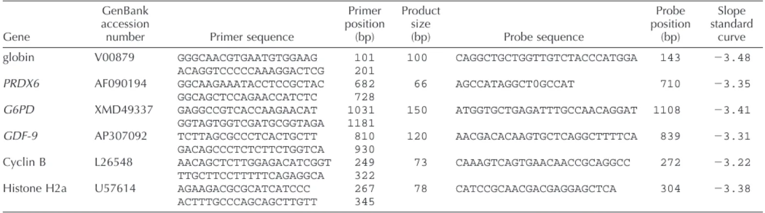
TABLE 1. Information on primers used for real-time PCR.
Gene
GenBank accession
number Primer sequence
Primer position (bp) Product size (bp) Probe sequence Probe position (bp) Slope standard curve globin PRDX6 G6PD GDF-9 Cyclin B Histone H2a V00879 AF090194 XMD49337 AP307092 L26548 U57614 GGGCAACGTGAATGTGGAAG ACAGGTCCCCCAAAGGACTCG GGCAAGAAATACCTCCGCTAC GGCAGCTCCAGAACCATCTC GAGGCCGTCACCAAGAACAT GGTAGTGGTCGATGCGGTAGA TCTTAGCGCCCTCACTGCTT GACAGCCCTCTCTTCTGGTCA AACAGCTCTTGGAGACATCGGT TTGCTTCCTTTTTCAGAGGCA AGAAGACGCGCATCATCCC ACTTTGCCCAGCAGCTTGTT 101 201 682 728 1031 1181 810 930 249 322 267 345 100 66 150 120 73 78 CAGGCTGCTGGTTGTCTACCCATGGA AGCCATAGGCT0GCCAT ATGGTGCTGAGATTTGCCAACAGGAT AACGACACAAGTGCTCAGGCTTTTCA CAAAGTCAGTGAACAACCGCAGGCC CATCCGCAACGACGAGGAGCTCA 143 710 1108 839 272 304 23.48 23.35 23.41 23.31 23.22 23.38
(Calbiochem, EMD Biosciences, San Diego, CA) and 12.5mM roscovitine (kindly provided by Dr. L. Meijer, Centre National de la Recherche Scien-tifique, Station Biologique de Roscoff, France): two were directly analyzed (premature), whereas the remaining two were further cultured for 24 h in a classic maturation medium (premature and mature). The two last pools of 50 oocytes were matured for 24 h (mature). This experiment was re-peated four times.
Experiment 3c: poly(A) RNA amount in oocytes maintained under meiotic arrest and in the presence of inhibitors of transcription or aden-ylation. In each experiment, five pools of 50 oocytes were used. One pool
of immature oocytes was directly analyzed. Three pools were cultured for 24 h in TCM-199 with 6.25mM BL-I and 12.5 mM roscovitine: 1) alone, or 2) with 500 mg/ml of 39-deoxyadenosine (cordycepin) an adenosine analogue that terminates poly(A) tail elongation and inhibits transcription, or 3) with 500mg/ml of 39-deoxyguanosine that inhibits transcription but not polyadenylation. These three pools were directly analyzed after 24 h of meiotic arrest. The last pool matured only after 24 h. This experiment was repeated four times.
Experiment 4: Real-Time PCR After RT with Hexamers
or oligo(dT)
Total RNA was extracted from two pools of 80 immature oocytes, and two pools of 80 in vitro-matured bovine oocytes as described above but with 5 pg of polyadenylated rabbit globin mRNA (Invitrogen) added to each pool. The RNA from each pool was divided into eight samples so that the RNA equivalent of 10 oocytes was reverse transcribed either with 250 ng of hexamers (Roche Applied Science) or with 200 ng of oligo(dT) (Amersham Biosciences). The reproducibility of the results obtained with each method was assessed by four equally treated replicates. All samples were denatured at 658C, flash-cooled to 48C, then reverse-transcribed for 1 h at 428C in a final volume of 15 ml containing 10 mM dithiothreitol, 1 mM dNTPs, 25 units of Expand RT (all from Roche Applied Science), and 10 units of RNA-guard (Amersham Biosciences). After RT, the vol-ume of each sample was extended to 65ml.
Six genes were quantified in each sample using real-time PCR and a specific molecular beacon for each gene (hybridization probe with a quencher [TAMRA] at the 59 end and a fluorescent dye [FAM] at the 39 end). The four genes of interest were two antioxidant enzymes, PRDX6 and G6PD, one transcript specific of the oocyte, GDF-9, and one member of the MPF complex, cyclin B1. Two reporter genes were amplified; the exogenous rabbit globin gene was spiked to account for the variations caused by manipulation of the samples (differences in pipetting or in the efficiency of reverse transcription between tubes), and the endogenous histone, H2a, to normalize different RNA amounts between pools. Accord-ing to a study on the quantification of several housekeepAccord-ing genes durAccord-ing maturation and early development in the bovine [33], only histone H2a mRNA levels appeared constant across the entire preimplantation period. The sequences of the different primers used are presented in Table 1.
PCRs were performed on an ABI Prism 7700 (Applied Biosystems, Foster City, CA). The amplification reaction used 5ml of the cDNA and the Platinum Quantitative PCR Super Mix-UDG (23) (from Invitrogen Life Technologies). This mix contains each dNTP, but dUTP instead of dTTP, 40 U/ml uracil-DNA-glycosylase (UDG), 60 U/ml Platinum Taq polymerase, and ROX as a passive fluorescent dye for well normalization; 400 nM of each primer (forward and reverse), and 200 nM of the Taqman probe were added to the reaction. The PCR protocol included a first step
at 508C (2 min) for the activity of UDG, preventing ‘‘carryover’’ contam-ination from previous PCR products, then 10 min at 958C to activate the
Taq polymerase, followed by 40 cycles for 15 sec at 958C, and 1 min at
608C. For each gene, a standard curve of amplification was established using five serial dilutions (in triplicate) of a reference cDNA. The same stock of cDNA was used in all experiments (cDNA from granulosa cells, except for GDF-9, for which we used a cDNA derived from a pool of oocytes).
Statistical Analysis
Total and poly(A) RNA content in immature and mature cow and calf oocytes were analyzed by two-way analysis of variance with the matura-tion status and the origin of the oocytes (cow or calf) as fixed factors. In experiments 3b and 3c, the poly(A) RNA content was analyzed with one-way analysis of variance. For analysis of mRNA expression assayed by quantitative RT-PCR, one-way repeated measures analysis of variance were used. Differences of P, 0.05 were considered significant.
RESULTS
Total RNA Quantification Before and After In Vitro
Maturation
Total RNA within one oocyte was about 1.9
6 0.2 ng
(mean
6 SEM) before maturation and 2.0 6 0.15 ng after
maturation, with a ratio OD
260:OD
280always
.1.85. No
significant change in total RNA occurred during
matura-tion.
Ribosomal RNA Before and After In Vitro Maturation
The intensity of the 18S ribosomal band hybridized to
Northern blots did not significantly change after maturation.
No ribosomal degradation appeared during meiotic
resump-tion of bovine oocyte (Fig. 1).
Poly(A) RNA Quantification
Experiment 3a: immature and matured cow and calf
oo-cytes. The amount of poly(A) RNA significantly decreased
during maturation (P
, 0.0001), but there was no effect of
the origin of the oocyte (cow or calf). Poly(A) RNA in a
cow oocyte was 53
6 4 pg (mean 6 SEM) before
matu-ration and declined to 24.5
6 2.5 pg at the end of 24 h of
maturation. In calf oocytes, the poly(A) RNA amount was
quite similar, but the variability appeared more pronounced.
The average amount was 57
6 8 pg before maturation and
declined significantly to 32
6 6.5 pg after maturation.
Experiment 3b: oocytes maintained 24 h under meiotic
arrest with CDK inhibitors. After 24 h of in vitro meiotic
arrest with BL-I and roscovitine, the poly(A) RNA amount
in bovine oocytes significantly increased to reach 91
6 12
FIG. 1. Comparison of the amount of ribosomal RNA in bovine imma-ture and maimma-ture oocytes by hybridization with an 18S rRNA probe to Northern blot. This experiment was repeated twice. Lane 1: RNA from 500 immature oocytes; lane 2: RNA from 500 matured oocytes.
FIG. 3. Poly(A)RNA amount per oocyte (in picograms) in the presence of CDK inhibitors and inhibitors of transcription and polyadenylation. Im-mature: oocytes directly analyzed after follicle extrusion; preIm-mature: oo-cytes analyzed after 24 h of meiotic arrest with a combination of two CDK inhibitors (BL-1 and roscovitine); premat1 dA: oocytes incubated for 24 h with CDK inhibitors and deoxyadenosine; premat1 dG: oocytes incubated for 24 h with CDK inhibitors and deoxyguanosine; mature: oocytes matured for 24 h. Columns with different superscripts are signif-icantly different,P, 0.05 (one-way ANOVA).
FIG. 2. Poly(A)RNA amount per oocyte (in picograms) before and after maturation, prematuration, or both. Immature: oocytes directly analyzed after follicle extrusion; premature: oocytes analyzed after 24 h of meiotic arrest with a combination of two CDK inhibitors (BL-1 and roscovitine). Premature1 mat: oocytes maintained 24 h under meiotic arrest with CDK inhibitors followed by 24 h maturation. Mature: oocytes matured for 24 h. Columns with different superscripts are significantly different,P, 0.05 (one-way ANOVA).
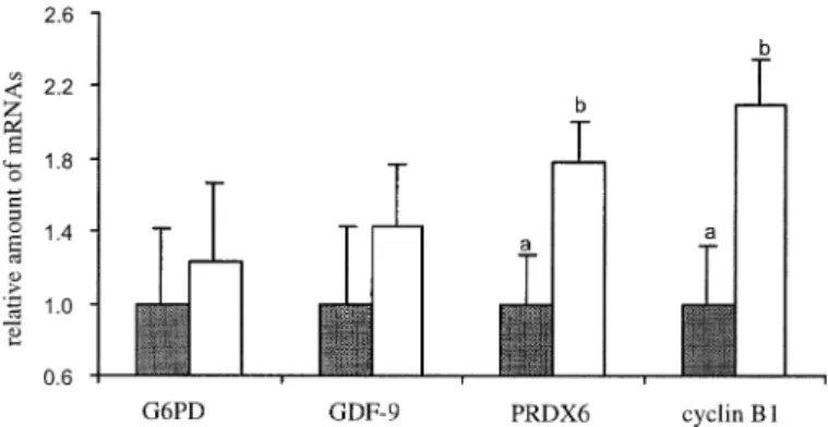
FIG. 4. Relative quantification (arbitrary units) of the expression of four transcripts reverse-transcribed with hexamers and normalized withH2a used as the reference gene. Gray bars: before maturation; open bars: after maturation. a±b, P , 0.05.
pg. Once these oocytes were allowed to mature after 24 h,
the poly(A) RNA significantly decreased to reach a value
that was not different from that measured in simply matured
oocytes (Fig. 2).
Experiment 3c: oocytes 24 h under meiotic arrest with
CDK inhibitors and in the presence of inhibitors of
tran-scription or adenylation. Under maintenance of meiotic
ar-rest with a combination of BL-I and roscovitine, the
poly(A) RNA amount again significantly increased (88.5
6
10.5 pg; P
, 0.05). If we simultaneously added
deoxygu-anosine, there was no significant increase. On the other
hand, in the presence of both deoxyadenosine and CDK
inhibitors, the level of poly(A) RNA sharply dropped to a
value as low as that observed in matured oocytes (14.3
6
7 pg/oocyte) (Fig. 3).
Real-Time PCR
For relative quantification, the efficiency of amplification
of the target (the four genes of interest) and of the reference
genes (globin or histone H2a) must be similar. This
effi-ciency is given by the slope obtained with standard curves
and was included between
23.2 (100% efficiency) and
23.5 for all studied genes (Table 1). According to the
glo-bin standard curve, the mean quantity of rabbit gloglo-bin
found in each replicate was 0.042
6 0.003 pg, as expected.
When the PCR results of endogenous genes were
normal-ized with the globin value, the variability between the
rep-licates did not exceed 10%. If RT was performed with
hex-amers, normalization with globin or histone H2a values
gave similar results, the intrinsic RNA quality of pools of
80 oocytes was apparently not quite different.
RT with Hexamers
As reported [43], the number of histone H2a transcripts
was not affected during in vitro maturation (data not
shown), thus relative quantification could be performed
with H2a used as the reference gene according to the
stan-dard curve method (ABI Prism 7700 Sequence Detection
System; User Bulletin #2, Applied Biosystems). The
rela-tive quantity of the four genes of interest obtained after
maturation is shown in Figure 4. The relative number of
G6PD and GDF-9 transcripts was not affected during
mat-uration, whereas there was an increase for PRDX6 and
cy-clin B1 messengers.
TABLE 2. Ratio of PCR signals obtained after RT with oligo(dT) versus hexamers.
Oocyte status Rabbit globin Histone H2a G6PD GDF-9 Cyclin B1 PRDX6 Immature Matured 9.856 1.05 11.056 1.55 0.080.036 0.016 0.0 2.950.516 0.636 0.11 5.486 1.08 a 0.696 0.13b 2.456 0.45 1.656 0.05 3.76 0.5 a 0.76 0.1b a,ba± b, P , 0.05.
RT with oligo(dT)
The amplification results were quite different if RT was
performed with oligo(dT) or with hexamers (Table 2). The
amplification of the histone H2a transcript, normally not
adenylated, was very low when using oligo(dT), and could
no longer be used as a reference gene. For the fully
poly-adenylated rabbit globin, amplification was much higher
using oligo(dT) instead of hexamers. For the other genes,
the efficiency of amplification after RT with oligo(dT)
changed, depending on the maturation status of the oocyte.
In these conditions, relative quantification was difficult to
realize. However, for each gene, we compared the ratio of
RT-PCR results with oligo(dT) on RT-PCR results with
hexamers for immature and matured oocytes. For GDF-9
and PRDX6, this ratio dropped significantly during
matu-ration. For G6PD and for H2a, the decrease was close to
significance (P
5 0.06 and P 5 0.09, respectively), whereas
for the exogenously added rabbit globin, the ratio was not
affected by in vitro maturation.
DISCUSSION
For the first time, variations in the amount of different
RNA species were analyzed during in vitro maturation of
bovine oocytes. Olszanska et al. [44], using ethidium
bro-mide-induced fluorescence, estimated the total RNA
con-tent of a bovine oocyte before meiosis resumption to be
close to 1 ng. Bilodeau-Goeseels and Schultz [18], using
Northern blot techniques, estimated the total RNA content
of a matured bovine oocyte to be around 2.4 ng. We found
no report of total RNA content before and after meiosis
resumption with the same detection technique. Here, with
a classic RNA detection system, we quantified total RNA
content to be close to 2 ng before and after maturation,
between the two values previously reported; more
specifi-cally, we demonstrated that total RNA content does not
decrease during in vitro meiotic resumption. In the mouse,
a 20% decrease of bulk RNA was observed during
matu-ration, including a 23% decrease in rRNA [45, 46]. Our
Northern blot analysis showed that in bovine oocyte,
ri-bosomal RNA was apparently not degraded during in vitro
maturation. Thus, rRNA appeared stable up to the 4-cell
stage in a bovine embryo [18]. The amount of poly(A)
RNA in a bovine oocyte was estimated to be around 55 pg
before meiosis resumption, consequently, it would represent
5% of total RNA, slightly lower than the 8% reported in a
mouse oocyte [6]. This amount of poly(A) RNA was
dra-matically affected during meiosis resumption, being
re-duced by half at the end of the process as described for
mouse [7] and Xenopus oocytes [9].
In bovine oocytes, a shortening of the poly(A) tail was
reported for 6 out of 10 transcripts analyzed before and
after maturation [47, 48]. Our results confirmed the
impor-tance of deadenylation during meiosis resumption. This
process could not be detected during the first 4 h of
mat-uration, but was completed after 18 h (data not shown). So,
according to the kinetics of meiosis resumption in the
bo-vine [49], deadenylation apparently occurs after GVBD as
reported in Xenopus [50]. Besides the deadenylation wave,
several transcripts can also be specifically polyadenylated
depending on the sequence of their 3
9 untranslated region
[51]. In the bovine oocyte, polyadenylation was described
for several transcripts as well [47, 48], and polyadenylation
activity was detected between 6 and 10 h after the
begin-ning of meiosis resumption [16].
Variable RT-PCR results using oligo(dT) versus
hexa-mers may indicate deadenylation [52]. Our data point out,
for three out of four mRNAs analyzed, the removal of
poly(A) tails during meiosis resumption, whereas cyclin B1
mRNA was not deadenylated. Cyclin B1 protein
heterodi-merizes with a cyclin-dependant kinase (CDK1) to form
MPF, a complex essential to meiosis resumption [27].
Dur-ing mouse oocyte maturation, the level of cyclin B1 protein
rises steadily [53] in correlation with a lengthening of the
poly(A) tail of the transcript [28]. We showed here that the
number of polyadenylated cyclin B1 transcripts was stable
during bovine oocyte maturation. It has been recently
re-ported that cyclin B1 mRNA would undergo cytoplasmic
polyadenylation before the beginning of in vitro maturation,
which had already occurred during the time when the
ova-ries were transported from the slaughterhouse to the
labo-ratory [54].
Although they were deadenylated, none of the transcripts
we analyzed was degraded during maturation. Recently,
transcriptome analysis of bovine oocytes using cDNA
ar-rays revealed that the relative abundance of most
messen-gers was effectively stable during maturation [55], with
10% of them showing a decrease, and 10% of them
show-ing an increase, as we observed here for cyclin B1 and
PRDX6 transcripts. This increase could be the result of the
low transcriptional activity detected in the GV at the
be-ginning of maturation [15, 16]. A higher level of cyclin B1
protein during bovine oocyte maturation may depend on
both an increase in transcription and translation.
The different amplification results obtained with one or
the other RT method clearly underline the importance of
adenylation for the regulation of expression during
matu-ration in bovine oocytes. Studies on purified poly(A) RNA
[56, 57] or on cDNA reverse transcribed only with
oli-go(dT) [58] do not take into account transcripts without a
poly(A) tail or with a too-short poly(A) tail. So the
differ-ences detected in those studies may reflect changes in the
number of transcripts during maturation, but also the loss
or addition of a poly(A) tail to transcripts.
Histone transcripts are unique because they usually lack
a poly(A) tail, ending instead in a conserved stem-loop
structure. However, some of the stored histone mRNAs in
amphibian oocytes have short oligo(A) tails added to the
stem-loop structure [59]. These oligo(A) tails are removed
at oocyte maturation [60]. Similarly, the low PCR signal
obtained for histone H2a reverse transcribed with oligo(dT)
could come from a small proportion of polyadenylated
tran-scripts present in immature bovine oocyte. This proportion
also appeared to decrease during maturation.
The lack of cytoplasmic competence of calf oocytes was
neither reflected by a reduced content of poly(A) RNA, nor
by a different processing during maturation. However, the
amount of poly(A) RNA was apparently more subject to
variations between pools of calf oocytes.
Unexpectedly, the poly(A) content of oocytes under
mei-otic arrest with a combination of BL-I and roscovitine
in-creased. Because there is still some transcription in the GV
[15, 16], this could be due to neotranscription, or to the
addition of poly(A) tails to transcripts not yet adenylated,
or both. To solve this, we used deoxyadenosine
(cordyce-pin) or deoxyguanosine in combination with CDK
inhibi-tors; the first one impedes transcription and adenylation,
whereas the second one interacts only with transcription.
As the rise in poly(A) content was no longer detectable in
the presence of deoxyguanosine, it probably resulted from
transcription. When polyadenylation was hampered, the
poly(A) RNA dropped to a very low level. Perhaps factors
that prevent deadenylation during meiotic arrest would
themselves depend on polyadenylation to be functional.
Poly(A) tails could also be the result of a dynamic process
[61] and could not be further elongated. Interactions of the
inhibitors with the different factors responsible for
trans-lational control should also be considered. Under meiotic
arrest of bovine oocytes with BL-I, the phosphorylation of
the cap-binding factor eIF4E, linked to the mRNA
recruit-ment to polyribosomes, is blocked [62], whereas
cordyce-pin prevents the dissociation of maskin from eIF4E [63].
Finally, as the rise of poly(A) RNA obtained in the
pres-ence of CDK inhibitors was no longer detectable after 24
h of maturation, the maintenance of meiotic arrest in vitro
could not improve the quantity of poly(A) RNA, but the
specific pattern of the transcripts affected is not known.
In conclusion, total RNA amount as well as ribosomal
RNA did not decrease during bovine oocyte maturation, but
half of the poly(A) RNA disappeared. Real-time PCR data
indicated that several mRNAs were deadenylated but not
degraded, whereas cyclin B1 transcript was not
deadeny-lated. The poly(A) RNA content within a calf or a cow
oocyte was not different. The poly(A) RNA content in
oo-cytes maintained under meiotic arrest with CDK inhibitors
increased. This increase, apparently due to transcription,
did not allow obtaining a higher poly(A) RNA amount once
the oocyte has finally achieved meiosis resumption. If
poly-adenylation was hampered during the maintenance of
mei-otic arrest, the poly(A) RNA amount decreased. These
sults clearly underline the importance of addition and
re-moval of poly(A) tails during meiosis resumption and
mei-otic arrest in bovine oocyte.
REFERENCES
1. Eichenlaub-Ritter U, Peschke M. Expression in in-vivo and in-vitro growing and maturing oocytes: focus on regulation of expression at the translational level. Review. Hum Reprod Update 2002; 8:21–41. 2. Wassarman PM, Liu C, Litscher ES. Constructing the mammalian egg
zona pellucida: some new pieces of an old puzzle. Review. J Cell Sci 1996; 109:2001–2004.
3. Gray NK, Wickens M. Control of translation initiation in animals. Review. Annu Rev Cell Dev Biol 1998; 14:399–458.
4. Groisman I, Huang YS, Mendez R, Cao Q, Richter JD. Translational control of embryonic cell division by CPEB and maskin. Review. Cold Spring Harb Symp Quant Biol 2001; 66:345–351.
5. Sternlicht AL, Schultz RM. Biochemical studies of mammalian oo-genesis: kinetics of accumulation of total and poly(A)-containing RNA during growth of the mouse oocyte. J Exp Zool 1981; 15:191– 200.
6. Picton H, Briggs D, Gosden R. The molecular basis of oocyte growth and development. Review. Mol Cell Endocrinol 1998; 145:27–37. 7. Bachvarova R, De Leon V. Polyadenylated RNA of mouse ova and
loss of maternal RNA in early development. Dev Biol 1980; 74:1–8.
8. Schultz RM, Wassarman PM. Specific changes in the pattern of pro-tein synthesis during meiotic maturation of mammalian oocytes in vitro. Proc Natl Acad Sci U S A 1977; 74:538–541.
9. Sagata N, Shiokawa K, Yamana K. Appearance of RNA with a spe-cific size class of poly(A) in oocytes and eggs in HCG-stimulated
Xenopus laevis females. J Exp Zool 1980; 13:117–122.
10. Crozet N, Kanka J, Motlik J, Fulka J. Nucleolar fine structure and RNA synthesis in bovine oocytes from antral follicles. Gamete Res 1986; 14:65–73.
11. Fair T, Hulshof S, Hyttel P, Greve T, Boland M. Nucleus ultrastructure and transcriptional activity of bovine oocytes in preantral and early antral follicles. Mol Reprod Dev 1997; 46:208–215.
12. Lonergan P, Monaghan P, Rizos D, Boland MP, Gordon I. Effect of follicle size on bovine oocyte quality and developmental competence following maturation, fertilization, and culture in vitro. Mol Reprod Dev 1994; 37:48–53.
13. Hendriksen P, Vos P, Steenweg W, Bevers M, Dieleman S. Bovine follicular development and its effect on the in vitro competence of oocytes. Review. Theriogenology 2000; 53:11–20.
14. Kastrop P, Hulshof S, Bevers M, Destree O, Kruip T. The effects of alpha-amanitin and cycloheximide on nuclear progression, protein synthesis, and phosphorylation during bovine oocyte maturation in vitro. Mol Reprod Dev 1991; 28:249–254.
15. Memili E, Dominko T, First NL. Onset of transcription in bovine oocytes and preimplantation embryos. Mol Reprod Dev 1998; 51:36– 41.
16. Tomek W, Torner H, Kanitz W. Comparative analysis of protein syn-thesis, transcription and cytoplasmic polyadenylation of mRNA during maturation of bovine oocytes in vitro. Reprod Domest Anim 2002; 37:86–91.
17. Hyttel P, Viuff D, Fair T, Laurincik J, Thomsen P, Callesen H, Vos P, Hendriksen P, Dieleman S, Schellander K, Besenfelder U, Greve T. Ribosomal RNA gene expression and chromosome aberrations in bo-vine oocytes and preimplantation embryos. Review. Reproduction 2001; 122:21–30.
18. Bilodeau-Goeseels S, Schultz GA. Changes in ribosomal ribonucleic acid content within in vitro-produced bovine embryos. Biol Reprod 1997; 56:1323–1329.
19. Clegg KB, Piko L. Poly(A) length, cytoplasmic adenylation and syn-thesis of poly(A)1 RNA in early mouse embryos. Dev Biol 1983; 95: 331–341.
20. Revel F, Mermillod P, Peynot N, Renard JP, Heyman Y. Low devel-opmental capacity of in vitro matured and fertilized oocytes from calves compared with that of cows. J Reprod Fertil 1995; 103:115– 120.
21. Damiani P, Fissore RA, Cibelli JB, Long CR, Balise JJ, Robl JM, Duby RT. Evaluation of developmental competence, nuclear and oo-plasmic maturation of calf oocytes. Mol Reprod Dev 1996; 45:521– 534.
22. Khatir H, Lonergan P, Carolan C, Mermillod P. Prepubertal bovine oocyte: a negative model for studying oocyte developmental compe-tence. Mol Reprod Dev 1996; 45:231–239.
23. Gandolfi F, Milanesi E, Pocar P, Luciano AM, Brevini TA, Acocella F, Lauria A, Armstrong DT. Comparative analysis of calf and cow oocytes during in vitro maturation. Mol Reprod Dev 1998; 49:168– 175.
24. Levesque JT, Sirard MA. Proteins in oocytes from calves and adult cows before maturation: relationship with their development capacity. Reprod Nutr Dev 1994; 34:133–139.
25. Khatir H, Lonergan P, Mermillod P. Kinetics of nuclear maturation and protein profiles of oocytes from prepubertal and adult cattle during in vitro maturation. Theriogenology 1998; 50:917–929.
26. Salamone DF, Damiani P, Fissore RA, Robl JM, Duby RT. Biochem-ical and developmental evidence that ooplasmic maturation of pre-pubertal bovine oocytes is compromised. Biol Reprod 2001; 64:1761– 1768.
27. Gautier J, Minshull J, Lohka M, Glotzer M, Hunt T, Maller JL. Cyclin is a component of maturation-promoting factor from Xenopus. Cell 1990; 60:487–494.
28. Tay J, Hodgman R, Richter JD. The control of cyclin B1 mRNA translation during mouse oocyte maturation. Dev Biol 2000; 221:1–9. 29. Knight PG, Glister C. Local roles of TGF-beta superfamily members in the control of ovarian follicle development. Review. Anim Reprod Sci 2003; 78:165–83.
30. Elvin J, Yan C, Matzuk M. GDF-9 stimulates progesterone synthesis in granulosa cells via a prostaglandin E2/EP2 receptor pathway. Proc Natl Acad Sci U S A 2000; 97:10288–10293.
31. Kletzien R, Harris P, Foellmi L. G6PDH: a housekeeping enzyme subject to tissue-specific regulation by hormones, nutrients and oxi-dant stress. FASEB J 1994; 8:174–181.
32. Chae HZ, Chung SJ, Rhee SG. Thioredoxin-dependent peroxide re-ductase from yeast. J Biol Chem 1994; 269:27670–27678.
33. Robert C, McGraw S, Massicotte L, Pravetoni M, Gandolfi F, Sirard MA. Quantification of housekeeping transcript levels during the de-velopment of bovine preimplantation embryos. Biol Reprod 2002; 67: 1465–1472.
34. Rizos D, Ward F, Duffy P, Boland MP, Lonergan P. Consequences of bovine oocyte maturation, fertilization or early embryo development in vitro versus in vivo: implications for blastocyst yield and blastocyst quality. Mol Reprod Dev 2002; 61:234–248.
35. Kubelka M, Motlik J, Schultz RM, Pavlok A. Butyrolactone I revers-ibly inhibits meiotic maturation of bovine oocytes, without influencing chromosome condensation activity. Biol Reprod 2000; 62:292–302. 36. Lonergan P, Dinnyes A, Fair T, Yang X, Boland M. Bovine oocyte
and embryo development following meiotic inhibition with butyrolac-tone I. Mol Reprod Dev 2000; 57:204–209.
37. Mermillod P, Tomanek M, Marchal R, Meijer L. High developmental competence of cattle oocytes maintained at the germinal vesicle stage for 24 hours in culture by specific inhibition of MPF kinase activity. Mol Reprod Dev 2000; 55:89–95.
38. Ponderato N, Lagutina I, Crotti G, Turini P, Galli C, Lazzari G. Bovine oocytes treated prior to in vitro maturation with a combination of butyrolactone I and roscovitine at low doses maintain a normal de-velopmental capacity. Mol Reprod Dev 2001; 60:579–585.
39. Dieleman S, Hendriksen PJ, Viuff D, Thomsen P, Hyttel P, Knijn H, Wrenzycki C, Kruip T, Niemann H, Gadella B, Bevers M, Vos P. Effects of in vivo prematuration and in vivo final maturation on de-velopmental capacity and quality of pre-implantation embryos. Re-view. Theriogenology 2002; 57:5–20.
40. Sankrithi N, Eskin A. Effects of cyclin-dependent kinase inhibitors on transcription and ocular circadian rhythm of Aplysia. J Neurochem 1999; 72:605–613.
41. Sirri V, Roussel P, Hernandez-Verdun D. In vivo release of mitotic silencing of ribosomal gene transcription does not give rise to pre-cursor ribosomal RNA processing. J Cell Biol 2000; 148:259–270. 42. Lonergan P, Faerge I, Hyttel PM, Boland M, Fair T. Ultrastructural
modifications in bovine oocytes maintained in meiotic arrest in vitro using roscovitine or butyrolactone. Mol Reprod Dev 2003; 64:369– 378.
43. Lonergan P, Carolan C, Van Langendonckt A, Donnay I, Khatir H, Mermillod P. Role of epidermal growth factor in bovine oocyte mat-uration and preimplantation embryo development. Biol Reprod 1996; 54:1412–1421.
44. Olszanska B, Borgul A. Maternal RNA content in oocytes of several mammalian and avian species. J Exp Zool 1993; 265:317–320. 45. Bachvarova R, De Leon V, Johnson A, Kaplan G, Paynton BV.
Changes in total RNA, polyadenylated RNA, and actin mRNA during meiotic maturation of mouse oocytes. Dev Biol 1985; 108:325–331. 46. Paynton BV, Rempel R, Bachvarova R. Changes in state of
adenyla-tion and time course of degradaadenyla-tion of maternal mRNAs during oocyte maturation and early embryonic development in the mouse. Dev Biol 1988; 129:304–314.
47. Brevini-Gandolfi TA, Favetta LA, Mauri L, Luciano AM, Cillo F, Gandolfi F. Changes in poly(A) tail length of maternal transcripts
dur-ing in vitro maturation of bovine oocytes and their relation with de-velopmental competence. Mol Reprod Dev 1999; 52:427–433. 48. Brevini TA, Lonergan P, Cillo F, Francisci C, Favetta LA, Fair T,
Gandolfi F. Evolution of mRNA polyadenylation between oocyte mat-uration and first embryonic cleavage in cattle and its relation with developmental competence. Mol Reprod Dev 2002; 63:510–517. 49. Sirard MA, Florman HM, Leibfried-Rutledge ML, Barnes FL, Sims
ML, First NL. Timing of nuclear progression and protein synthesis necessary for meiotic maturation of bovine oocytes. Biol Reprod 1989; 40:1257–1263.
50. Varnum SM, Hurney CA, Wormington WM. Maturation-specific deadenylation in Xenopus oocytes requires nuclear and cytoplasmic factors. Dev Biol 1992; 153:283–290.
51. Fox CA, Wickens M. Poly(A) removal during oocyte maturation: a default reaction selectively prevented by specific sequences in the 39 UTR of certain maternal mRNAs. Genes Dev 1990; 4:2287–2298. 52. El Mouatassim S, Guerin P, Menezo Y. Expression of genes encoding
antioxidant enzymes in human and mouse oocytes during the final stages of maturation. Mol Hum Reprod 1999; 5:720–725.
53. Chesnel F, Eppig J. Synthesis and accumulation of p34cdc2 and cyclin B in mouse oocytes during acquisition of competence to resume mei-osis. Mol Reprod Dev 1995; 40:503–508.
54. Tremblay K, Vigneault C, Bujold G, Sirard MA. Bovine oocyte cyclin B1 mRNA undergoes cytoplasmic polyadenylation before the begin-ning of in vitro maturation. In: Proceedings of the Annual Conference of the International Embryo Transfer Society; 2004; Portland, OR. Reprod Fertil Dev 16: 246–247.
55. Dalbies-Tran R, Mermillod P. Use of heterologous complementary DNA array screening to analyze bovine oocyte transcriptome and its evolution during in vitro maturation. Biol Reprod 2003; 68:252–261. 56. Wrenzycki C, Herrmann D, Carnwath JW, Niemann H. Alterations in the relative abundance of gene transcripts in preimplantation bovine embryos cultured in medium supplemented with either serum or PVA. Mol Reprod Dev 1999; 53:8–18.
57. Lonergan P, Gutierrez-Adan A, Rizos D, Pintado B, de la Fuente J, Boland MP. Relative messenger RNA abundance in bovine oocytes collected in vitro or in vivo before and 20 hr after the preovulatory luteinizing hormone surge. Mol Reprod Dev 2003; 66:297–305. 58. Robert C, Barnes FL, Hue I, Sirard MA. Subtractive hybridization
used to identify mRNA associated with the maturation of bovine oo-cytes. Mol Reprod Dev 2000; 57:167–175.
59. Ruderman JV, Pardue ML. A portion of all major classes of histone messenger RNA in amphibian oocytes is polyadenylated. J Biol Chem 1978; 253:2018–2025.
60. Ballantine JE, Woodland HR. Polyadenylation of histone mRNA in
Xenopus oocytes and embryos. FEBS Lett 1985; 180:224–228.
61. Groisman I, Jung MY, Sarkissian M, Cao Q, Richter JD. Translational control of the embryonic cell cycle. Cell 2002; 109:473–483. 62. Tomek W, Melo Sterza FA, Kubelka M, Wollenhaupt K, Torner H,
Anger M, Kanitz W. Regulation of translation during in vitro matu-ration of bovine oocytes: the role of MAP kinase, eIF4E (cap binding protein) phosphorylation, and eIF4E-BP1. Biol Reprod 2002; 66: 1274–1282.
63. Cao Q, Richter JD. Dissolution of the maskin-eIF4E complex by cy-toplasmic polyadenylation and poly(A)-binding protein controls cyclin B1 mRNA translation and oocyte maturation. EMBO J 2002; 21: 3852–3862.