AVIS
Ce document a été numérisé par la Division de la gestion des documents et des archives de l’Université de Montréal.
L’auteur a autorisé l’Université de Montréal à reproduire et diffuser, en totalité ou en partie, par quelque moyen que ce soit et sur quelque support que ce soit, et exclusivement à des fins non lucratives d’enseignement et de recherche, des copies de ce mémoire ou de cette thèse.
L’auteur et les coauteurs le cas échéant conservent la propriété du droit d’auteur et des droits moraux qui protègent ce document. Ni la thèse ou le mémoire, ni des extraits substantiels de ce document, ne doivent être imprimés ou autrement reproduits sans l’autorisation de l’auteur.
Afin de se conformer à la Loi canadienne sur la protection des renseignements personnels, quelques formulaires secondaires, coordonnées ou signatures intégrées au texte ont pu être enlevés de ce document. Bien que cela ait pu affecter la pagination, il n’y a aucun contenu manquant.
NOTICE
This document was digitized by the Records Management & Archives Division of Université de Montréal.
The author of this thesis or dissertation has granted a nonexclusive license allowing Université de Montréal to reproduce and publish the document, in part or in whole, and in any format, solely for noncommercial educational and research purposes.
The author and co-authors if applicable retain copyright ownership and moral rights in this document. Neither the whole thesis or dissertation, nor substantial extracts from it, may be printed or otherwise reproduced without the author’s permission.
In compliance with the Canadian Privacy Act some supporting forms, contact information or signatures may have been removed from the document. While this may affect the document page count, it does not represent any loss of content from the document.
IN VITRO AND IN VIVO VIRULENCE EVALUATION OF THE NEW
GENOTYPE OF PORCINE CIRCOVIRUS TYPE 2 AND
IDENTIFICATION OF A NEW CELL LINE PERMISSIVE TO VIRUS
REPLICATION
par
Nedzad Music, DMV
Département de pathologie et microbiologie Faculté de médecine vétérinaire
Mémoire présenté à la Faculté des études supérieures et postdoctorales en vue de l'obtention du grade de Maître ès sciences (M.Sc)
en sciences vétérinaires option microbiologie
Août, 2008
Ce mémoire intitulée
IN VITRO
AND IN VIVO VIRULENCE EVALUATION OF THE NEW
GENOTYPE OF PORCINE CIRCOVIRUS TYPE 2 AND
IDENTIFICATION OF A NEW CELL LINE PERMISSIVE TO VIRUS
REPLICATION
présentée par
NEDZAD MUSIC, DMV
a été évaluée par un jury composé des personnes suivantes: Dr Jacques Lussier, président-rapporteur Dr Carl A. Gagnon, directeur de recherche Dr Serge Messier, membre du jury
Vers la fin de l'année 2004, la population porc me canadienne a connu une recrudescence marquée du syndrome de dépérissement en post-severage (SDPS), problème que l'on avait associé à l'émergence d'un nouveau génotype de Circovirus porcin de type 2 (PCV -2b) jusque là inconnu en Amérique du Nord. Ainsi, il est devenu important de démontrer que la souche de PCV -2b circulant dans les élevages porcins canadiens est plus virulente que l'ancienne souche (PCV-2a). Des clones plasmidiques infectieux de l'ancien génotype (PCV-2a) et du nouveau génotype (PCV-2b) viral ont été conçus (pPCV-2a et pPCV-2b). L'efficacité de la lignée cellulaire épithéliale porcine de rein (PK-15)
tr~sfectée par pPCV -2a et par pPCV -2b a permis un cycle complet de réplication virale. Par la suite, la production de particules infec~ieuses de PCV -2 a été évaluée durant hùit passages cellulaires consécutifs. Ainsi, dix fois plus de particules virales infectieuses de PCV-2b ont été recueillies au 8e passage par rapport au PCV.;.2a. Plus tard la lignée cellulaire épithéliale trachéale de porcelets nouveau-nés (NPTr) s'est révélée comme étant permissive au PCV -2 et pouvant par conséquent permettre la production de particules virales infectieuses. Cette découverte est très importante car jusqu'à maintenant, il n'y avait, à notre connaissance, qu'un seul type de lignée cellulaire immortalisée (cellules épithéliales porcines de rein) qui permet la production de particules virales infectieuses, suite à l'infection virale.
Donc, pour corroborer les résultats précédents, les cinétiques de réplication virale de PCV-2 ont été évaluées chez les cellules PK-15 et NPTr: La cinétique de réplication virale de PCV-2b s'est montrée considérablement plus efficace que celle de PCV-2a dans les deux lignées cellulaires. Aucune différence significative par rapport à l'efficacité de la réplicati.on virale n'a été observée entre les deux lignées cellulaires. En conclusion, PCV -2b est approximativement 6 à 10 fois plus efficace que PCV -2a pour la synthèse de particules virales infectieuses' dans deux lignées cellulaires épithéliales porcines. De plus, la lignée cellulaire NPTr pourrait servir en guise de nouveau modèle in vitro I?our l'étude de la pathogénèse du PCV-2. Pai conséquent, l'efficacité de réplication virale de PCV .. 2b en comparaison à celle de PCV-2a pourrait, au moins en partie, justifier la recrudescence marquée du SDPS au Canada au cours des dernières années.
MOTS CLÉS: SDPS, cellules NPTr, réplication virale, génotypes de PCV -2, circovirus
By the end of 2004, the Canadian swine population had experienced a severe increase in the incidence of Porcine Circovirus-Associated Disease (PCV AD), a problem that was associated with the emergence of a new Porcine circovirus-2 genotype (PCV -2b) previously unknown in North America. Thus, it became important to demonstrate that the PCV -2b strain circulating in Canadian swine herds is more virulent than the older PCV -2a strain. ·Infectious DNA clones of PCV -2a and PCV -2b were constructed (pP CV -2a and pPCV-2b). The efficacy of pPCV-2a and pPCV-2b transfected porcine kidney epithelial cell line (PK-15) to allow a complete virus replication cycle and production of PCV-2 infectious particles was evaluated during eight consecutive cell passages. Ten times more PCV -2b infectious viral particles were recovered at the 8th cell passage compared to PCV-2a. Later on, a new porcine cellline, the newbom piglet tracheal(NPTr) epithelial cellline, was found to be permissive to PCV -2 and was consequently able to permit infectious viral particles production. This finding is interesting because until now, there was only one type of immortalized cell line (the porcine kidney epithelial ce Ils) known to permit infectious viral particles production.
Thus, to corroborate early results, the PCV -2a and PCV -2b replication kinetics were evaluated in PK-15 and NPTr cells. PCV-2b virus replication kinetic was significantly more efficient in both cell lines compared to PCV -2a. No differences in virus replication efficiency could be found between both cell lines. In conclusion, PCV -2b is about 6 to 10 times more efficient than the PCV -2a for the synthesis of infectious viral particles in two porcine epithelial celllines. Moreover, the NPTr ceIlline could be used as a new in vitro model for studying the PCV-2 pathogenesis. Consequently, the virus replication efficiency of PCV -2b compared to PCV -2a could, at least in part, explain the increase in the occurrence of PCV AD in Canada during the recent years.
TABLE OF CONTENTS
RÉSUMÉ ... i
ABSTRACT ... iii
TABLE OF CONTENTS ... ; ... v
LIST OF TABLES ... viii
LIST OF FIGURES ... ix
LIST OF ABBREVIATIONS ... xi
DEDICATION ... XIV ACKNOWLEDGEMENTS ... xv
1. INTRODUCTION ... 1
II. LITERATURE REVIEW ... : ... : ... 4
POSTWEANING MUL TISYSTEMIC WASTING SYNDROM - PMWS ... 5
1. History and definition ... 5
2. Disease tenninology ... 5
3. Postweaning Multisystemie Wasting Syndrome - PMWS ... 6
4. Po~cine Derniatitis and Nephropathy Syndrome - PDNS ... 7
PORCINE CIRCOVIRUS TYPE 2 ... : ... 9
1. History ... : ... 9
2. Taxonomy ... 9
3. Charaeterization ofPCV-2 ... 10
5. Replication and Mechanisms of Virus Entry into the Cells ... 12
6. PCV -2 isolates ... 13
6.1. PCV-2 genetic variation ... 13
6.2. The emergence of porcine circovirus 2b genotype (PCV -2b) in swine in Canada ... 15
PA THOGENESIS ... 17
1. Introduction ... ; ... 17
2. Transmission ofPCV-2 ... 17
3. Cellular distribution ofPCV-2 and cell-tissue tropisms ... ; ... 18
3.1. PCV-2 and Macrophages ... ; ... 18
3.2. PCV-2 and Porcine Kidp.ey epithelial cellline (PK-15) ... 19
3.3. Lymphoid tissues ... 20
3.4. Blood ... 20
4. Cytokine profiles in PMWS diseased pigs ... 21
5. Risk factors involved in PMWS development.. ... 23
DIAGNOSIS ... 26
INTERVENTION STRATEGIES ... 27
1. Good management practices ... 27
2. PCV -2 vaccines ... 27
EXPERIMENTAL INFECTION MODEL OF PMWS ... 30
III. MATERIAL, METHODS & RESUL TS ... 31
IDENTIFICATION OF A NEW CELL LINE PERMISSIVE TO PORCINE CIRCOVIRUS TYPE 2 REPLICATION: THE NEWBORN PIGLET TRACHEAL CELLS ... 32
. Introduction ... : ... : ... 34
Materials and Methods ...•... 37
Results ... 40
Discussion ... 42
Acknowledgments ... ; ... 44
References ... ' ... 48
DIFFERENTIAL VIRUS REPLICATION EFFICIENCY OF PORCINE CIRCOVIRUS 2A AND 2B IN PORCINE TRACHEA AND KIDNEY EPITHELIAL CELL LINES . 55 Abstract ... 56
Introduction ... ; ... 57
Materials and Methods ... 59
Results ... : ... , ... 63 Discussion ... 65 Acknowledgments ... : ... 68 References ... , ... 73 IV. DISCUSSION ... ; ... 79 V. CONCLUSION ... 86 VI. REFERENCES ... 89 APPEND IX 1 ... xvii APPENDIX 2 ... xx
LIST OF TABLES
Literature Review
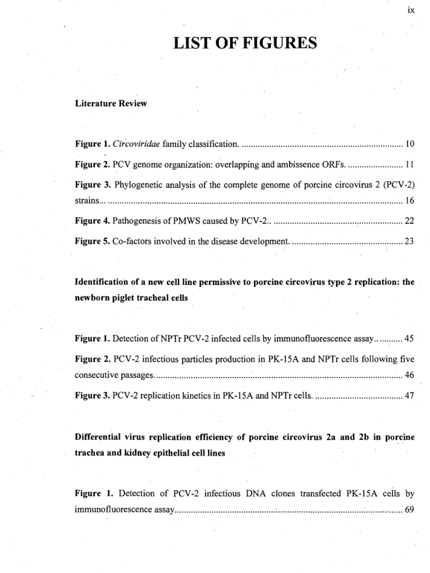
LIST OF FIGURES
Literature Review
Figure 1. Circoviridae family classification ... 10 Figure 2. PCV genome organization: overlapping and ambissence ORFs ... ~. Il
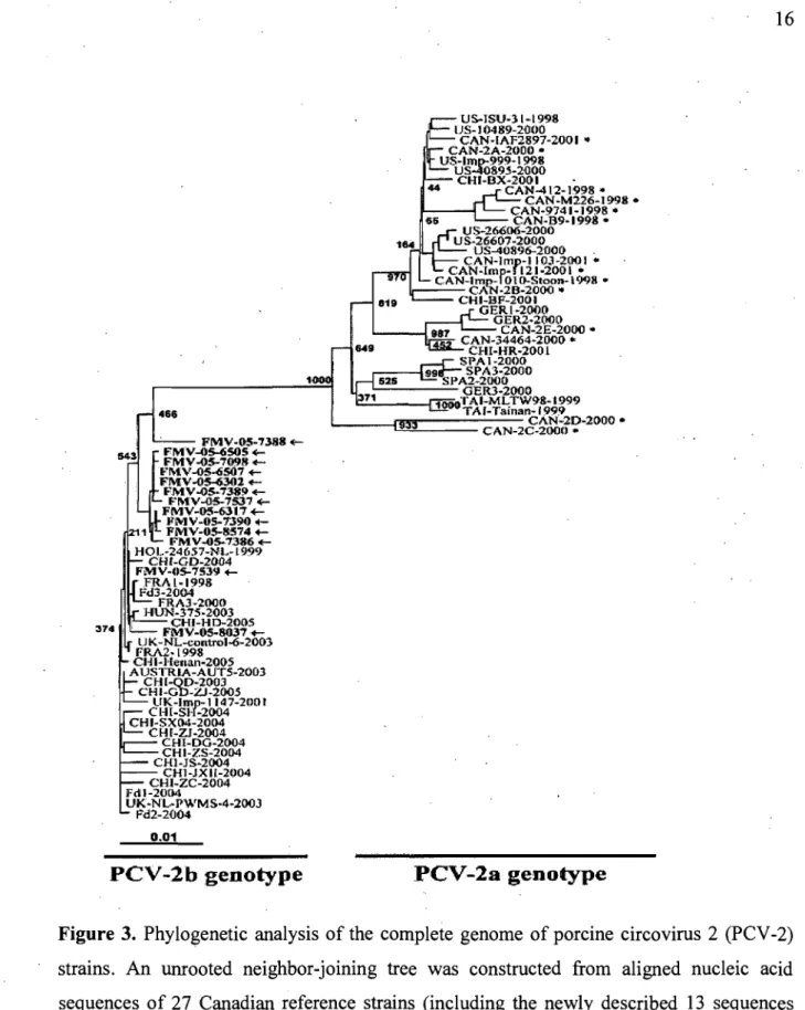
Figure 3. Phylogenetic analysis of the complete genome of porcine circovirus 2 (PCV -2)
strains ... ; ... 16
Figure 4. Pathogenesis ofPMWS caused by PCV-2 ... 22 ,. , Figure 5. Co-factors involved in the disease development. ... 23
Identification of a new cellline permissive to porcine circovirustype 2. replication: the newborn piglet trac he al cells
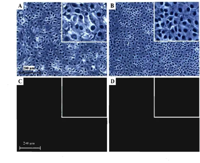
Figure 1. Detection of NPTr PCV-2 infected cells by immunofluorescence assay ... 45 Figure 2. PCV-2 infectious particles production i!1 PK-15A and NPTr cells following five
consecutive passages ... : ... : ... 46
Figure 3.'PCV-2 replication kinetics in PK-15A and NPTr cells ... 47
Differentiai virus replication efficiency of porcine circovirus 2a and 2b in po~cine trachea and kidney epithelial celllines
Figure 1. Detection of PCV-2 infectious DNA clones transfected PK-15A cells by
Figure 2. PCV-2 infectious viral partic1es production in PK-15A transfected cells ... 70
Figure 3. Detection ofPK-15A cells infected with the celllysates of the 8th cell passage. 71
LIST OF ABBREVIATIONS
aa
A. pleuropneumoniae BFDVCaCV
Cap
CAVCDCD
ConA CPEDNA
DuCV FBSFiCV
GoCV
GuCV
IFA IFN-y IHC ISHKHLIIFCA
Amino acids Actinobacillus pleuropneumoniaeBeak and feather disease virus Canary circovirus
Capsid prote in Chicken anemia virus
Cesarean-derived colostrum-deprived Concanavalin A
Cytopathic effect Deoxyribonudeic acid Duck circovirus Fetai bovine serum Finch circovirus Goose circovirus Gull circovirus ImmunofluQrescence assay Interferon gamma Immunohistochemistry In situ hybridization
Keyhole limpet haemocyanin in incomplete Freund' s adjuvant
LPS M. hyopneumoniae MOI mrtqPCR NPTr
OR
ORF PBMC PBS PCR PCV-2 PCV-2a PCV-2b PCVAD PDNS PERVPEP SCAN
PFA PiCV PK-lS PMWS pPCV-2a pPCV-2b Lipopolysaccharide Mycroplasma hyopneumoniae Multiplicity of infectionMultiplex real-time quantitative polymerase chain reaction
Newbom piglet tracheal epithelial cellline Odd ratio
Open reading frame
Peripheral blood mononuclear cells Phosphate buffer saline
Polymerase chain reaction Porcine circovirus - type 2
Porcine circovirus - type 2 (genotype 2a) Porcine circovirus - type 2 (genotype 2b) Porcine Circovirus Associated Disease .
Porcine Dermatitis and Nephropathy Syndrome Porcine endogenous retrovirus
Peptide scanning Paraformaldehyde Pigeon circovirus
Porcine kidney epithelial cellline
Postweaning Multisystemic Wasting Syndrome Infectious DNA clone ofPCV-2a
PPV
PRDCPRRSV
rcf Rep RFLP RNA S. suisSDPS
SIV
SKSPF
ss Porcine parvovirusPorcine Respiratory Disease Complex
Porcine Reproductive and Respiratory Syndrome Virus
Relative centrifugaI force Replicase
Restriction fragment length polymorphism Ribonucleic acid
Streptococcus suis
. Syndrome de dépérissement en post-severage Swine influenza virus
Swine kidney epithelial cellline Specific· pathogen free
1 , To my parents, my kids, and my wife' For believing in me and For supporting me
ACKNOWLEDGEMENTS
1 look back at these two past years of meticulous rèsearch and writing, years that mark an important period in my life, and 1 would like to take the time to hereby acknowledge thehelp 1 have received from many people that have contributed, directly or indirectly, to the achievement of the following thesis. First and foremost, 1 want to express my gratitude to my director, Dr Carl A. Gagnon, for his useful and helpful assistance, guidance, support, advice, experience, endless patience and the constructive criticism he has showed during the completion Of this work. 1 am also extremely grateful to the members of his laboratory and those of the diagnostic service for their input and interest in this research.
Furthermore, 1 would peculiarly like to give credit to sorne of my professors for important contributions to my project as well as their insightful comments, advice and encouragement. Among them figure Dr Josée Harel and Dr Jérome deI Castillo, who took charge of the epidemiologic diagnostic study, and Dr Pierre Hélie, who handled the histopathological lesions evaluation of the animaIs used in this study. A special thanks goes out to the members of my advising committee and the jury of this document for thoughtful remarks and general assistance: Dr Jacques Lussier, and Dr Serge Messier.
Likewise, 1 extend my appreciation to the foundations' that provided financial support to'me, therefore creating a pleasant atmosphere: Le Fond québecois de la recherche sur la nature et les technologies (FQRNT) and Centre de recherche en infectiologie porcine (CRIP). Moreover, several other organisations have provided valuable resources for this research: the Ministère de l'agriculture, des pêcheries et de l'alimentation du Québec (MAPAQ), the Fédération des Producteurs de Porc du Québec (FPPQ), the Conseil pour le développement de l'agriculture du Québec (CDAQ), and the Centre d'insémination porcine du Québec (CIPQ).
Most importantly, my deepest and warmest gratitude goes out to my family, for their everlasting love and support. Thanks to my wife, Biljana, who cared for me and encouraged me in difficult moments, to my daughter, Sanela, who believed in me and
helped me with linguistic matters, and to my son, Dino, who gave me confidence in my pursuit. Finally, thanks to my mother, Remzija Music, who has always been there for me and to which 1 hope 1 inspired pride, and to my father, Dzafer Music, whose memory gave me strength.
Postweaning Multisystemic Wasting Syndrome (PMWS) is a swine disease, that was first recognized in Canada in 1991 [39, Tl]. Afterwards, the syndrome was reported worldwide [8, 10,24, 128, 147, 151]. The primary etiological agent responsible for PMWS has been identified as a circovirus named Porcine circovirus-2 (PCV -2; family Circoviridae, genus Circovirus) [8, 52, 70, 106].
At the end of 2004, the swine industry In the prOVInce of Québec, Canada, experienced a significant increase in death rate related to PMWS [42]. It was hypothesized that this emerging problem was caused by the presence of a new type of circulating PCV-2 strain. This was confirmed by sequencing the entire viral genome of several PCV -2 strains originated from PMWS-affected herds [63]. Interestingly, this newly recovered PCV-2 genotype (named PCV -2b in comparison with the previously circulating genotype named PCV -2a) had, already been reported in Asia and Europe [46] and has also, been recently reported in 2007 in the United States [34, 79]. The appearance of the new PCV -2b genotype in Canada could explain the death rate increase related to PMWS, but this relationship has to be confirmed. Consequently, one of the main objectives of the swine industry and health scientists is to establish if there is a virulence variation between both PCV-2 genotypes (PCV-2a and PCV-2b).
The pathogenesis of2 infection and major cell types that support PCV-2 replication are poorly understood. The presence of a PCV -PCV-2 antigen and nuc1eic acids has ,been shown in different cell types in vivo by immunohistochemistry (IHC) or in situ hybridization (ISH). In vitro, the only immortalized ceIlline that support PCV -2 replication is porcine kidney epithelial cells [10, 106, 111, 142], and PCV-2 isolation and production is routinely performed using porcine kidney (PK-15) and swine kidney (SK) epithelial cell lines [10, 142]. Nonetheless, it is a good assumption to believe that kidney cells are not the first infected cells that favor propagation of the virus in theorganism. The oro-nasal route is considered the most likely and frequent route of PCV -2 transmission and experimental infection studies which have mainly used the intranasal route of inoculation support that idea [16, 53, 85, 128, 142].
Consequently, the goal of the present study is therefore: (l) to demonstrate that the PCV -2b strain that circulates inCanadian swine herds is more virulent in vitro and in vivo than the PCV -2a strain; (2) to establish if epithelial cells of the respiratory tract of swine
could support PCV -2 replication in vitro, and (3) to develop a diagnostic test that could rapidly and efficiently differentiate both genotypes.
POSTWEANING MULTISYSTEMIC WASTING SYNDROM - PMWS
1. History and definition
Postweaning Multisystemie Wasting Syndrome (PMWS) is a swine disease that was first reeognized in Canada in 1991 [39, 71]. Afterwards, the syndrome was reported worldwide [3, 10,24, 128, 147, 151]. The primary etiologieal agent résponsible for PMWS has been identified as a eireovirus named Porcine circovirus-2 (PCV -2; family
Circoviridae, genus Circovirus) [70].
PMWS is considered a multifactorial disease in pigs, in which a necessary factor is the presence of PCV -2. Since PCV -2 is ubiquitous in the pig population and infection doesn't equate to disease, a definition for PMWS was proposed [158]. Based on this definition, a diagnosis of PMWS requires (1) the presence of clinical signs such as wasting, weight loss and respiratory disease, (2) the presence of the hall mark PCV -2-associated microscopic les ions (lymphoid depletion and/or histiocytic replacement of follicles in lymphoid tissues or both), and (3) PCV -2 antigen or nucleic acids associated with the microscopie lesions as determined by immunohistoehemistry (IRC) or in situ
hybridization (ISR) [158].
2. Disease terminology
Today, the clinical expression of PCV-2 infection in swine is acknowledged to be . more eomplex than initially established because it ean play a pivotaI role in several syndromes, such as Porcine Dermatitis and Nephropathy Syndrome (PDNS), Porcine Respiratory Disease Complex (PRDC), granulomatous enteritis, reproductive failure and necrotizing lymphadenitis [25, 72]. In addition, PCV-2 has also been associated with· myocarditis and vasculitis in growing pigs, hepatitis, CNS disease, and exudative
, '
(PCV AD)" is now accepted to describe the syndromes in which PCV -2 plays a role. The extent of the involvement of PCV -2 in swine diseases other than PMWS is currently po orly understood. PCV -2 infection is widespread and essentially aIl pig herds are infected with PCV -2 but relatively few have PCVAD.
3. Postweaning Multisystemic Wasting Syndrome - PMWS
The disease usually affects 5 to 12 week-old piglets and is characterized in part by weight loss, dyspnea, jaundice, and enlarged lymph nodes, as weIl as by degeneration and necrosis of hepatocytes, multifocal lymphohistiocytic pneumonia, lymphocytic depletion andmultinucleated giant cell formation [71,·158]. Depletion of lymphocytes in the lymphoid follicles and their replacement by macrophages are the hallmark lesions observed
. .
in this syndrome [10, 106]. PMWS has six fundamental clinical signs: wasting, dyspnea, enlarged lymph nodes, diarrhea, paIlor, and jaundice [72]. While aIl the fundamental clinical signs may not be noted in a single pig, affected farms will present the majority of them over a period of time. Other clinical signs include coughing, fever,gastric ulceration, meningitis and sudden death [72]. Macroscopic lesions associated with PCV-2 infection include generalized lymphadenopathy, rubbery lungs with mottling and increased firmness, enlarged spleen and enlarged kidneys [39].· Characteristic microscopic lesions associated with PCV -2 infection and PMWS include lymphoid depletion and histiocytic replacement of follicles in lymphoid tissues. The lymphocellular depletion affects both lymphoid follicles and parafollicular zones [140]. Mild-to-severe granulomatous inflammation in lymphoid and other tissues also is commonly observed [11, 158]. Syncytial cells can be
,.
seen frequently, especially in lymph nodes, Peyer' s patches and lamina propria of the intestinal villi [140]. Macrophages in affected lymphoid tissues may contain sharply demarcated, spherical, basophilic cytoplasmic inclusion bodies [39, 140]. The inclusions are either large and single or smaller and multiple with groups ofup to 12 'inclusions [140].
PMWS has been reported from almost aIl swine producing countries. Morbidity due to PMWS reportedly varies from as little as 4% to as high as 30% and mortality in aÎfected . pigs is typically 70 to 80% [11, 39, 71, 99, 139]. There are two forms of PMWS: an
endemic and an epidemic form. The endemic form is seen in North America, where PMWS usually results in low grade but persistent death losses. Rarely, 3 to 4 fold increase in postweaning mortality can be observed. The epidemic form, which is characterized by persistent mortality, appeared to be observed primarily in Europe but based on recent evidence, has become increasingly observed in eastem Canada [22, 23, 42, 47, 63] and North Carolina. Pork pro duc ers in Canada and the U.S. have experienced a devastating disease in growing pigs which is manifested as wasting and pneumonia and resulting in persistent mortality of 20-40% of the pigs between 10 weeks and market age (25 weeks) [128].
4. Porcine Dermatitis and Nephropathy Syndrome - PDNS
PDNS is c1inically characterized by acute onset of skin lesions (raised purple skin lesions progressing to multifocal raised red scabs with black centers most prominent on the rear legs), fever and lethargy, and is almost always fatal [25]. Macroscopically, there are enlarged tan waxy kidneys with white foci and streaks [25]. Microscopically, there is systemic vasculitis with dermal and epidermal necrosis, and necrotizing and fibrinous glomerulonephritis [25]. The microscopic hallmark lesions of PDNS, generalized vasculitis and glomerulonephritis, are suggestive of a type III hypersensitivity reaction which is characterized by deposition of antigen-antibody aggregates or immune complexes on certain tissue sites [36, 141, 159].
There are two forms of PDNS described: the sporadic form and the epizootic form . . With the sporadic form, the mortality is rarely above 0.5%. The epizootic form was first observed in 1999 in England, when there was a sudden, marked increase of PDNS cases [160, 161]. The within-herd mortality was reported to range from 0.25-20% and similar observations were made in the Netherlands [51,171] . Investigations into this "outbreak" found that there was a c1ear temporal association of PMWS and PDNS; PDNS cases usually followed PMWS cases on the same farms. Studies have determined that the mean age of pigs affected by PMWS ranges from 6 to 14 weeks whereas the mean age of pigs affected by PDNS ranges from 12 to 16 weeks [69]. Similar observations were made in
Korea [37], and the authors of that study speculated that the presence of both PMWS and PDNS in the same herd but in different age groups was probably due to different strains of PCV -2 or varying susceptibility of the pigs [37]. A recent case:-control study irivestigating PDNS in the Netherlands found that there was a significant association of high antibody titers to PCV-2 and the development ofPDNS [171]. Thè authors were not able to show the PCV -2 antigen by IHC in aIl of the PDNS cases but they were a~le to confirm the presence of PCV -2 by polyrp.erase chain reaction (PCR) in aIl cases of PDNS. Importantly, the authors were able to show that the, porcine parvovirus or Porcine Reproductive and Respiratory Syndrome Virus (PRRSV) nucleic acids were not present in many of the PDNS cases as determined by PCR [171]. A study comparing PCV-2 serum load in PMWS and PDNS cases found that PDNS cases had significantly lower numbers of PCV -2 in serum compared to healthy, subclinical PCV-2-infected pigs [119].
PORCINE CIRCOVIRUS TYPE 2
1. History
Porcine circovirus (PCV) was first recognized as a contaminant of the continuous porcine kidney cell line (PK-15) (ATCC-CCL31) in 1974 in Germany and described as picomavirus-like virus [163]. Under experimental conditions, the PCV-PKI5-isolate did 'not induce dise~se in pigs [7, 164]. In the late 1990's, PCV was associated with a
newly-emerged disease syndrome in pigs, described as "postweaning multisystemic wasting syndrome" (PMWS) [3]. Sequence analysis of the PMWS-associated PCV revealed differences compared to the earlier described PCV [3, 8, 10, 52, 70, 106, 114]. In order to distinguish the pathogehic PMWS-associated PCV from the non-pathogenic PCV, the pathogenic type was designated porcine circovirus type 2 (PCV -2) and the non-pathogenic type as porcine circovirus type 1 (PCV -1).
2. Taxonomy
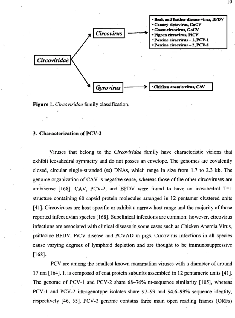
Both, PCV-l and PCV-2' are members of the Cireoviridae family [135, 168]. The Cireoviridae family is divided into the genera Cireovirus [Cireo indicates that the genome , of the virus has a circular conformation] and Gyrovirus [Gyro is a derivation from the Greek work "gyrus" meaning "ring" or "circuit"] (Fig. 1). The genus Cireovirus contains the following species: psittacine beak and feather disease virus (BFDV), canary circovirus (CaCV), goose circovirus (GoCV), pigeon circovirus (PiCV), PCV -1 and PCV -2 [77, 131, 137, 166-168]. The species tentatively placed in the genus Cireovirus are duck circovirus (DuCV), finch circovirus (FiCV), and gull circovirus (GuCV) (Fig. 1). The genus
ICircovirus
1Circoviridae
• Beakalid.'feather disease'virus, BFDV • CilBary cirtovirus, CaCV
) • Goo,se,circovirus,GoCV
--~ • Pigeon cïrcovirus, PiCV
• Porcine &:ÏJ'qJvÏruS -1, RCV4
• Porcine circovirus -2, PCV-2
1
Gyrovirus
I----)~
I·Chicken anemia virus, CAVFigure 1. Circoviridae family classification.
3. Characterization ·of PCV-2
Viruses that belong to the Circoviridae family have characteristic Vlflons that exhibit icosahedral symmetry and do not posses an envelope. The genomes are covalently closed, circularsingle-stranded (ss) DNAs, which range in size from 1.7 to 2.3 kb. The genome organization of CA V is negative sense, whereas those of the other circoviruses are ambisense [1681. CAV, PCV-2, and BFDV were found to have an icosahedral T=1 structure containing 60 capsid protein molecules arranged in 12 pentamer clustered units [41]. Circoviruses are host-specific or exhibit a narrow host range and the majority ofthose reported' infect avian species [168]. Subclinical infections are common; however, circovirus infections are associated with clinical disease insome cases such as Chicken Anemia Virus, psittacine BFDV, PiCV disease and PCVAD in pigs. Circovirus infections in all species cause varying degrees of lymphoid depletion and are thought to be immunosuppressive [168].
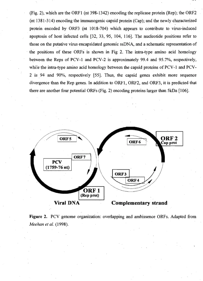
PCV are among the smallest known mammalian viruses with a diameter of around 17 nm [164]. It is composed of coat protein subunits assembled in 12 pentameric units [41]. The genome of PCV-l and PCV-2 share 68-76% nt-sequence similarity [105], whereas PCV-l and PCV-2 iI1tragenotype isolates share 97-99 and 94.6-99% sequence identity, respectively [46, 55]. PCV-2 genome contains three main open reading frames (ORFs)
(Fig. 2), whieh are the ORFI (nt 398-1342) eneoding the repliease protein (Rep); the ORF2 (nt 1381-314) encoding the immunogenic capsid protein (Cap); and the new1y characterized protein encoded by ORF3 (nt 1018-704) which appears to contribute tovi.rus-induced apoptosis of host infected eeUs [32, 33, 95, 104, 116]. The nucleotide positions refer to those on the putative virus encapsidated genomic ssDNA, and a schematic representation of .. the positions of the se ORFs is shown in Fig 2. The intra-type amino acid homology
between the Reps of PCV-I and PCV-2 is approximately 99.4 and 95.7%, respectively, while the intra-type amino acid homology between the capsid proteins of PCV -1 and PCV-2 is94 and 90%, respectively [55]. Thus, the capsid genes exhibit more sequence divergence than the Rep genes. In addition to ORF1, ORF2, and ORF3; it is predicted that there are another four potential ORFs (Fig. 2) encoding proteins larger than 5kDa [106].
~>
IORF61'
~<//
ORFI
~.~/
(Rep prot)
Viral DNA
'Complelllentaty strand
Figure 2. PCV genome organization: overlapping and ambissence ORFs. Adapted from
identity for the capsid protein. One strain identified in a herd without PMWS was found to be 100.0% homologous to a PCV -2 from a PMWS herd. [92]. In 2004, 31 pigs originating from 13 PMWS-affected herds and 25 pigs from 10 PMWS free herds were used for a comparative study, and 38 PCV-2 isolates were sequenced [46]. AlI the isolates shared 94.2-100% nucleotide identity. A wider nucleotide diversity was observed in the PCV-2 isolates originating from PMWS free herds compared with isolates from PMWS herds; however, residues found to be specific to non-PMWS strains were aiso found in PMWS strains and no molecular marker of virulence in PMWS strains could be identified [46]. In a . case control study done in 2002, PCV -2 was not only found in PMWS cases but also in 62.5% of the control cases. Sequencing and genetic comparison revealed no differences between 5 PMWS-associated PCV -2 isolates and 4 PCV -2 isol'l-tes recovered from cases not associated with PMWS [133].
In contrast, by comparing the ORF2 of Taiwanese PCV-2 isolates associated with PMWS, PDNS, nervous signs, abortion, a small number of residue difference associated with the different clinical conditions was found [170]. Research on the construction and characterization of 2 chimeric infectious DNA clones of PCV-l and PCV-2 has provided further insight into the virus virulence factors [59]. The chimeric PCV-I-2 DNA clone contained the PCV -2 capsid gene cloned in the backbone of the non-pathogenic PCV -1. The chimeric PCV -1-2 virus induced a strong and specific antibody response to the pathogenic PCV -2 capsid antigen and was attenuated (minimal to no lesions, low level and reduced length of viremia, low or nondetectable levels of viral antigen in lymphoid tissues) when inoculated into pigs [59]. In another study, two amino acid mutations occurred in the capsid prote in ofPCV-2 after 120 seriaI passages in cell culture and resulted in attenuation of the virus in vivo [58]. Significant differences were observed in the PCV -2 genomic copy numbers in serum, and the gross and microscopic lesions in pigs inoculated with the wild-type PCV isolate were more severe.than those inoculated with the passage 120 PCV-2 isolate [58]. This study confinrted that minimal changes in the genome of a PCV-2 isolate can markedly alter the virulence of PCV -2 viruses. Possible differences in virulence' among Midwestern US field isolates of PCV-2 with 98.9% nucleic acid and 96.7% amino acid sequence identities in ORF2 were investigated in an in vivo study reported in 2006 [127]. The in vivo study using SPF pigs confirmed that PCV -2 isolates with minimal genomic
4GG 543 FMV-OS-7388 E-FMV-OS-(iSOS E-FMV-05-7098 +-."M:V-05-<iS07 +-FMV-05-06301 +-FMV-05",7~9 E-374 , .,.°MV-05-7S37 E-FMV-05-6317 FMV-05-'iJ90 FMV-05-8:574 +-FMV-05-7386 +-HO 1..-24 6.$7-NL- L 999 CH'-GD-2004 FMV-05-7S39 +-FRAI-199B Fd3-2004 fRAJ·2000 HUN·37S-2003 CHI-HO-200S FMV -OS-8037+-UK-NL-cuntrol-6-2003 FRA2·1998 CHI·Henan-200S AUSTRIA-Alf'rS-20Cf1 CHI-QO-2003 CH I-CiO-ZJ -2005 trK-lmp-1147-2001 CHI-SH·2oo4 CHI-SX04~2004 CHI-ZJ-2004 CHI-DG-2004 CHI-ZS-2004 CH1·JS·2004 CHI-JXH-2004 CHI-ZC·2004 fdl-2004 UK-NL~PWMS-4-200J Pd2-2ù04 0001 PCV-2b genotype US-ISU-31 .. 1998 US-104B9-2fJOO CAN-IAF2897-2001 .. CAN-2A-20aO .. US-lmp.999-1998 US--40895-2000 CHI-BX-2001 CAN-412- 1998 '" CAN -M226-1998 • CAN-9741-1998 .. CAN·B9-199B .. US-26(>0(i-2000 US·26607-2OO0 US-40896-2:000 CAN-Imp-J 103-2001 .. CAN·lmp-J 121-2001 .. CAN-Imp-I OlO-Sloon-l991l .. I r - - -CAN·2B-2000 .. CHI-UF-lOOI GERI-2000 GER2·2000 CAN-2E-2000 .. CAN-34464-2000 .. CHI-HR·2ool SPAI-2000 ...----199 SPA3-2000 SPA2-2000 '---GERl-2oo0 "'""-' _ _ --I1000TAI-MLTW9S-1999 TAI-Tainan-1999 L---i~!C===ë;\-N_2 CAN-2D-2000 .. CAN-2C-2000 .. PCV -2a genotype
Figure 3. Phylogenetic analysis of the complete genome of porcine circovirus 2 (PCV -2)
strains. An unrooted neighbor-joining tree was constructed from aligned nucleic acid sequences of 27 Canadian reference strains (including the newly described 13 sequences identified with an arrow and the oIder sequences identified with an asterix) and
43
sequences found in GenBank. Taken from Gagnon et al. (2007).4. Biologie. and Physical Properties
PCV-l is stable at pH 3, at 56°C and at 70°C for 15 minutes, and is resistant to inactivation after exposure to chloroform [6]. The buoyant density of PCV-l in CsCI is 1.37 g per cm3 [163], and it is 1.36-1.37 g per ml CsCI [6]. The sedimentation coefficient (S) was determined to be 57S when compared with the sedimentation coefficient of a bovine enterovirus [6].
PCV -2 is readily isolated from tissue samples that have been stored at -70°C [52]. PCV-2 was shown to be resistant to sorne disinfectants (Nolvasan®, DC&R®, Weladol®,
s
or ethanol) but virus tÏiers were significantly reduced by sodium hydroxide, Virkon® S, and others [143].
5. Replication and Mechanisms of Virus Entry into the CeUs
AIl
viruses of the Circoviridae family are thought to have a similar replication strategy. A circular, double stranded replicative form of DNA intermediate is produced using host cell DNA polymerases during the S phase of cell division [165, 168]. The r~plicative form serves as template for generation of viral ssDNA using the rolling circle replication mechanism [168]. Viral DNA intermediates are generated in the nuclei and require host cell enzymes for completion of the replication cycle [165]. Virions are assembled in both nuclei and cytoplasm and released from infected cells in the absence of vir\il cytopathic effects.Other authors studied the binding characteristics of PCV -2. In the porcme monocytic line 3D4/31, PCV-2 enters predominantly via clathrin-mediated endocytosis and requires an acidic environrrient for infection [110]. The PCV -2 capsid protein is expressed between 6 to 12 hours post inoculation and nuclear relocation occurred around 12 to 24
hours post inoculation in PK-15 cells. In porcine alveolar macrophages or fetal
cardiomyocytes, nuclear localized antigens appeared approximately at 48 hours post inoculation and in fewer cells [108]. Treatment of PK -15 cell cultures with IFN -1 causes a 20 times higher production of PCV -2 progeny [109]. The enhancing effect of
interferon-gamma (IFN-y) on PCV-2 infeétion was found to be due to increased internalization of PCV -2 virion-like particles. Expressions of PCV -2 proteins in infected cells were not altered by IFN-y treatment [109].
During productive infection ofPK-15 cells, nine mRNA as capsid RNA, five Rep-associated RNAs (Rep, Rep', Rep3a, Rep3b, and Rep3c), and three NS-associated RNAs
(NS515, NS672, and NSO) are synthesized by PCV -2 [32]. It was demonstrated that Rep and Rep' are essential for PCV-2 replication [33]. PCV-l and PCV-2 were found to differ from
each other in expression levels ofNS and Rep3c-associated RNAs [31].
6. PCV -2 isolates
6.1. PCV -2 genetic variation
Molecular studies to determine the genetic variation of PCV -2 found that minor branches of PCV-2 were associated with geographic origin rather than with differences in virulence [55, 103]. The PCV-2 isolates fr<;>m cases of PDNS and abortions were closely related to a PMWS-associated PCV -2, further establishing the apparent genetic stability of PCV-2 [107]. Four dominant immunoreactive areas are identified by PEPSCAN analysis within ORF2 [101]. In 2002,34 Eastern Canadian PCV-2 isolates recovered from pigs with various clinical conditions such as PMWS, PRRS, generalized tremors, erysipelas, gastric ulcer, nervous signs, arthritis, and no clinical signs, were sequenced, and the obtained sequences compared to 36 published sequences. Sequence analysis indicated that all the isolates were closely related [91]. Three major regions of amino acid heterogeneity were identified among PCV -2 isolates, and two of the regions corresponded to two of the immunoreactive areas described before [101]. Comparison of three immunodominant regions, however, revealed no link. between the capsid protein variation and the pathogenicity of isolates [91]. In addition, PCV-2 from PMWS-affected herds was compared to PCV-2 from herds non-affected by PMWS (healthy pigs) [92]. It was found that closely related strains in 6 different herds (4 with PMWS and 2 without) sharing at least 99.4% of their nucleotide-sequence identity and more than 98.7% of their amino-acid
differences can differ significantly in virulence a~ measured by levèls of virus load in serum and tissues and severity ofPCV2-associated lesions [127].
6.2. The emergence of porcine circovirus 2b genotype (PCV-2b) in swine in Canada
Restriction fragment length polymorphism (RFLP) preliminary results suggest the appearance in 2004 of a new PCV -2 genotype in the province of Ontario (Canada) which seems to be related to PCV-2b genotype [21, 47]. Furthermore, it is interesting to obserVe that the appearance of a new type of circulating PCV-2 strains (seemed to co!ncide with the increases of death rate and PMWS across Canada and particularly in Québec's swine herds [42, 47]. Sequence arialysis of the entire viral genome of PCV-2 Canadian strains has permitted to establish the appearance of a nèw type of circulating PCV-2 strains in Canada [63]. These new strains have been classified in PCV-2b genotype (Fig. 3). Moreover, it is now obvious that the PCV -2b genotype straiiIs are more prevalent across Canada than the PCV-2a genotype strains [63]. The most variable prote in between PCV-2a and PCV-2b
~enotypes is the Cap prote in with an aa (amino acids) sequence identity varying between 88% to 94%. The two other known proteins to be expressed by PCV -2 were less variable than Cap protein with a aa sequence identity between the two genotypes varying from 96% to 99% for Rep protein and from92% to 98% for ORF3 protein [63].
Interestingly, this newly recovered PCV-2 genotype (namedPCV-2b in comparison' with the previously circulating genotype named PCV -2a) had already been reported in Asia and Europe [46] and has also been recentIy reported in 2007 in the United States [34, 81]. Now, an international consensus has been established in regards to the recognition of a least two major PCV-2 genotypes circulating worldwide [34, 50, ,63, 67, 120]. Recently, a nomenclature has been proposed, one that takes into account the classification of Gagnon et al (2007) and identified the two major genotypes (PCV -2a and PCV -2b) as well as a third one (PCV-2c) which inc1uded only three 80's isolates that had never been reported later on
PATHOGENESIS
1. Introduction
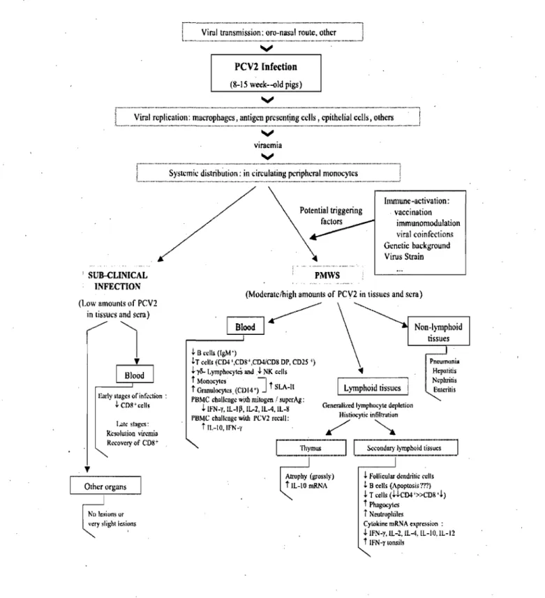
The pathogenesis ofPCV-2 infection and the major cell types that supportPCV-2 replication are poorly understood. The involvement of the immune system in the pathogenesis of PMWS seems central (Fig. 4). Two facts support this idea: 1) the existence of extensive lymphoid lesions [45] and 2) the association of PMWS with several secondary or opportunistic infections [49, 128]. Nevertheless,there are a number of questions that remain to be answered: What are the main virus producing ceUs? How does the immune response contribute to the development of the syndrome? Why, if most pigs<on a farm are infected, only sorne ofthem become ill?
2. Transmission of PCV-2
Transmission of PCV -2 is thought to occur through direct contact via oronasal (Fig. 4), fecal and urinary routes [17,100]. Direct contact with pigs inoculated with PCV-2 42 days previously resulted in virus transmission to 3. of 3 control cesarean-derived, colostrums-deprived (CDCD) pigs [17]. PCV-2 shedding in experimentally infected CDCD pigs was identified by polymerase chain reaction (PCR) in oropharyngeal swabs, nasal swabs, and feces [157]. In another study, the authors quantified PCV-2 DNA in tonsillar, nasal, tracheo-bronchial, urinary and fecal swabs of pigs with .and without PMWS. The authors were able to detect PCV -2 DNA in a high percentagè of the samples and concluded that PCV-2 iS,most likely excreted t~oughresp'iratory secretions, oral secretions, urine and feces of both PMWS':'affected and clinically-healthy pigs, with higher viral loads in the PMWS-affected pigs [153]. Vertical transmission has been demonstrated to occur in individual sows in the field [89, 118] and experimentally [80].
3. Cellular distribution of PCV -2 and cell-tissue tropisms
The target cells that support PCV -2 replication are poorly understood. The presence of PCV-2 antigen and nucleic acids has been shown in different cell types in vivo (Fig. 4) by IHC orJSH [11, 158]. A recent study found that antigen presenting cells in general, and not only macrophages stained positive by IHC for PCV-2antigen [35]. In contrast, PCV.:2 antigen in lymphocytes was only sporadically detected. In thymus, PCV -2 was only detected in few histiocytic cells in the medulla suggesting that thymocytes and T cells might be more resistant to PCV -2 infection [35]. Irnrnunophenotyping bf the target cells of PCV -2 replication in vivo has shown that the susceptible cell population in the pig depends on the stage of development of the host at the time of infection. In fetuses, infected cells were identified as cardiomyocytes, hepatocytes and macrophages during early gestation and mainly macrophages towards the end of gestation [144]. In the majority of unweaned piglets, low to moderate PCV-2-replication was demonstrated in macrophages [145]. A variety of cell types (Fig~ 4) have also been shown to contain PCV -2 antigens and/or nucleic acid such as: enterocytes, renal and alveolar epithelial celIs, vascular endothelial celIs, pancreatic acinar' and ductular celIs, lymphocytes, smooth muscle celIs, fibroblasts and germinal epithelial cells [45,126,173,174].
3.1. PCV -2 and Macrophages
Viruses that replicate in the monocyte/macrophage lineage such as porcine parvovirus (PPV) [9, 122] and porcine reproductive and respiratory virus (PRRSV) [12, 74, 142], have been shown to increase the replication of PCV -2 in coinfected pigs and increase the incidence of PMWS. Despite the presence of PCV -2 in macrophages and dendritic celIs, recent in vitro studies suggest that monocytic cells may not represent the primary target for PCV -2 replication [65]. Monocytes and macrophages were tested for the ability to support PCV -2-replication in vitro. PCV -2 replication in these cell types was not observed; however, PCV-2 was not degraded and was stored in the cytoplasm of the cells [65]. Similarly, no evidence of in vitro virus replication in dendritic cells was found [169];
however, PCV -2 did persist in dendritic cells without loss of infectivity or the induction of cell death. It has been speculated that because of their migratory capacity, dendriticcells can provide a potent vehicle for transport of the virus throughout the host without the need
. '
for replication [169]. In vitro studies showed that PCV-2 antigens could only be detected in the cytoplasm of PCV -2-inoculated monocyte/macrophage lineage cells, including swine alveolar macrophage without virus replication being detected [26, 27]. Nonetheless, two swine primary cell lines (alveolar macrophages and peripheral blood mononuclear cells -PBMC) have been demonstrated to allow PCV -2 infectious viral particles production when they were stimulated by bacteriallipopolysaccharide (LPS) or by concanavalin A (ConA)
[28, 94].
3.2. PCV-2 and Porcine Kidney epithelial cellline (PK~15)
Until now, swine kidney cells (like PK-15 cells) have beeri the only immortalized cells shown to be permissive to PCV -2 in vitro, meaning thatthey' allow the production of infectious viral particles fol1owing their infection with PCV -2 [106, 142]. On the other hand, other primary and immortalized cell lines were shown to be susceptible to PCV -2, meaning the cells could be infected and allow viral protein expression ,but without allowing infectious viral particles production [26, 28, 65, 110-112, 169]. PK-15 cells support PCV-2 replication in vitro; and these cells are routinely used for virus isolation and vaccine production., PCV -2-induced cytopathic effect is typically not observed, and to determine viral replication, immunofluorescent or immunoperoxidase staining has to be performed. Since PCV -2 does not encode its own polymerase, the replication of PCV -2, as for other circoviruses, depends on the cell polymerase present in the cells nucleus during the S phase of the cell cycle [165]. Consequently, glucosamine treatment of the PK-15 cells has shown to be effective in increasing PCV -2 replication [165];
3.3. Lymphoid tissues
Lymphocyte depletion together with histiocytic infiltration are the most
characteristi~ features of PMWS [8, 39, 45, 52]. The reduction of lymphocytes (Fig. 4) might be due to reduced production in the bone marrow, reduced proliferation in secondary lymphoid tissues, or due to increased loss of lymphocytes in the bone marrow or peripheral blood or secondary lymphoid tissues via necrosis or apoptosis. The severity of lymph node depletion can be graded as initial, intermediate, and end stage [146]. Studies on pigs with naturally-acquired PMWS revealed that the absence of follicles and depleÙon of lymphocytes was associa:ted with a reduction in the number of interfollicular dendritic cells and interdigitating ceUs as well as a reduction or absence Of B cells and CD4+ T lymphocytes [146]. By IHC characterization of PCV-2-associated lesions in naturally-PMWS-affected pigs, Chianini et al. (2003) [35] found increased numbers of macrophages. and partial loss and redistribution of antigen presenting ceUs throughout lymphoid tissues when compared to healthy control pigs. Decrease' of proliferation of both, . lymphoid and medulla-like tissues in the initial stage of PMWS, but not in the' intermediate or final stage, has been reported [35].
Depletion in B-cell associated areas of lymph nodes has been linked to apoptosis induced by the' virus [156]. In contrast, when inguinallymph nodes of pigs with naturally acquired PMWS were investigated, it was found that decreased cellproliferation (and not increased apoptosis) seemed to be the most important variable leading to Iymphoid depletion in PMWS[102]. The tissues from experimentally PCV-2 infected pigs were investigated by TUNEL staining and a positive signal was found only within the cytoplasm of virus-positive phagocytic mononuclear cells. It was concluded that apoptosis is not the primary mechanism oflymphoid depletion and hepatocyte loss in PMWS [87].
3.4. Blood
The hemogram ofpigs with PMWS (Fig. 4) shows' significant alterations [44, 148]. In wasted pigs, the number of lymphocytes is significantly decreased (especially of CD8+
and B cell subsets) and monocytes and neutrophils are clearly increased, with an inversion of the ratio lymphocyte/neutrophil [44, 148].
A
typical profile may be 60-70% neutrophils and 30-40% lymphocytes; while their theoretical numbers should be reverse [148, 149]. However, the total number of leukocytes is not altered. Pigs with PMWS usually have normocytic hypochromic anemia with a slight increase in the total number of red blood cells [44, 148].4. Cytokine profiles in PMWS diseased pigs
Pigs with PMWS have altered cytokines mRNA expression patterns (Fig .4.) in different lymphoid tissues [44]. These alterations consist of an over expression of IL-lO mRNA in thymus and INF-y mRNA in tonsil. The IL-lO mRNA over expression has been associated to thymic depletion and atrophy in the diseased animals [44]. A decrease in the mRNA expression was se en for 2 in the spleen, 4 in tonsils and lymph nodes, IL-12p40 in both spleen and inguinal lymph nodes and IFN-y and IL-lOin inguinal lymph nodes [44]. With regard to pro-inflammatory cytokines, it has been reported that IL-8 mRNA levels are high in tissues with slight or moderate lesions and with small amount of virus, while it is low in tissues with severe lesions [44].
On the other hand, in vitro PCV -2 alone modifies the cytokine responses of PBMC (Fig. 4) from healthy pigs when stimulated with mitogens such as phytohemaglutinin (PHA) or superantigens [43].
PCV2 Infection (8-15 week--old pigs) v
[,,'~~~a.~~~~i~~;i'~-~~;~:~~i~~~~;~~;~~~:~!.;~~;i~~";~;i~~~~~~l;~,~~~;~
___
=~'~~
V viracmia V rR_-'-'''~----'-'--'-~---'---''---''-'---'---.--.--.--.. -.---.-.. --.''1L __ ,_ ..
,~y.~c.,~i_~_~i~_t~~~~i~~_,i~ ~:~~~~i,~~_~~~~~:~~mo~,~c.~~~~_, ____ , __ ,_j , SUB-CLINICAL INFECTION (l.ow amounts of PCV2in tissues and sera)
~
1
~Harly slage~ oriht~clion :
~ CPR'ccU! 1.<1le sl.g"': Resolulion mmi. R~covcry of CD8~ Other organs
'"
PMWS Immune-activation: vacci nation immunomodulation viral c()infcction~ Genetie background Virus Strain(Moderatcihigh amounlS of PCV2 in tissues and sera)
BWodI~
~ El L'cil. «(gM')
h c~lIs (C04',CD8' ,c04/CDS DP, CD2S') ,l.yli-I,ympl\<>cyICÏf and.J. NK cclls t MonocV1cs 1 t
t Granul:XytcsjCllW) J SLA-ll
PllMe: challenge with mîtogcn l sUjX'rAg: ,l.IFN-"f, IL-I~, IL-l, IL-4, IL-8
l'BMC chalkngc widl l'CV2 rccall:
t IL-IO,IFN-'{
L)mphoid tissues
Gcnmli"ed Iymphocyle dcplction Hislio<:y1ie in@ntiun
/
~
Pneumonia Hcpalilis Ncphriris Enteritis~ FolJicular dondrilic relis
L B cells (Apoptosis 'Tm
~ T cells (HC04'»CD8 ,~)
t Phagocylos
i Neulrllphile~
Cywkinc mRNA e.pr.~$i"n :
.J.IF1Ii-y,lL-2,lL-4. [L-IO.IL-12
t IFN-YIODiiil5 .
Figure 4. Pathogenesis of PMWS caused by PCV -2. Proposed outline of the pathogenesis including immunologie aspects of PMWS in clinical and sub-clinical PCV -2 infections of piglets. Taken from Darwich et al. (2004).
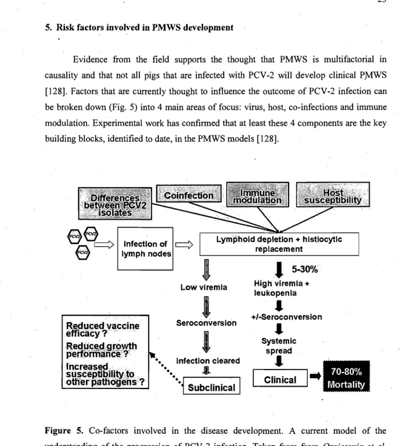
5. Risk factors involved in PMWS development
, '
Evidence from the field supports the thought that PMWS is multifactorial in causality and that not aH pigs that are infected with PCV -2 will develop clinical P.MWS [128]. Factors that are currently thought to influence the outcome of PCV-2 infection can be broken down (Fig. 5) into 4 main areas of focus: virus, host, co-infections and immune modulation. Experimental work has confirmed that at least these 4 components are the key building blocks, identified to date, in the PMWS models [128].
Infection of c::::)
Iymphnodes
Lym'phold depletlori +, h Istlocytlc replacement Reducedvaccihe éffiCàcyT" ,',' , .
~:~~~~'~~lmh
l'ncreased, , " su scé·tibilih~;;to otfi"erBattioffeijs:?'
1
Low vlrenila1
Seroconvers Ion1
.... Infection cleared....
,•.
••·~Subclinical
1
,1
5-30% High vlremla .. leukopenla,
"/-Seroconversion,
Systemic spread,
L...;C---,I---,in ... ic_a_1
---II".
Figure 5. Co-factors involved in the disease development. A current model of the understanding of the progression of PCV -2 infection. Taken from from Opriessnig et al.
Genetic analyses and sequence comparison of PCV -2 isohites to date have failed to fully explain differences in clinical manifestations. Then, one of the main objectives of the swine industry and health scientists is to establish if there is a virulence variation between both PCV-2 genotypes (PCV-2a and PCV-2b).
Host susceptibility and its role in PMWS development are po orly understood. Pigs
of aIl breeds seem to be susceptible to PCV -2 infection and clinical PMWS has been observed in a wide variety of purebred and crossbred pigs. Host susceptibility and its effect on the outcome of PCV -2 infection were recently investigated in a controlled pilot project [124]. Three breeds were compared in this study: Duroc, Landrace and Large White. The incidence of systemic PMWS based on gross and microscopic lesions was 0% (0/23) in Durocs, 15.8% (3/19) in Landrace, and 0% (0/21) in Large White [124]. The purebred Landrace pigs used in this experiment were clearly more susceptible to PMWS diseases as measured by the severity of clinical signs and microscopic lesions associated with PCV-2 [124]. A recent in vitro study investigating the replication patterns of PCV -2 in pulmonary· alveolai macrophages found clear differences between macrophages derived from different , conventional crossbred pigs suggestive of differences in susceptibility to PMWS [109].
The primary etiological agent responsible for PMWS has been identified as a PCV-2 [70]. The course of PCV -PCV-2 infection that will lead to the occurrence of the disease .has been associated with the presence of other pathogens such as Mycoplasma hyopneumoniae, Porcine parvovirus (PPV), Porcine reproductive and respiratory syndrome virus (PRRSV), and Swine influenza virus (SIV) [128]. Experimental coinfection ofpigs with PCV-2 and other virus es such a PPV [9, 14,81,85, 122], PRRSV [12, 74, 142] or bacteria such as Mycoplasma hyopneumoniae [123] has been shown to enhance the amount of PCV -2 viralload and PCV -2-associated lesions, and to increase the incidence of PMWS.
Studies have demonstrated that immunostimulation may trigger progression of PCV -2 infection to disease and characteristic lesions of PMWS. The PMWS was reproduced in gnotobiotic pigs stimulated with keyhole limpet hemocyanin in incomplete Freund adjuvant (KLHlICFA) and inoculated with PCV-2 [4, 86, 88, 121]. A recent study has determined that the timing of vaccination with a commercially available M hyopneumon~ae vaccine had an effect on PCV-2 replication and PCV-2-associated lesion
severity. It also confirmed differences in PCV-2-associated lesions among the treatment groups and conc1uded that no or minimal PCV -2-associated lesions were observed when pigs were vaccinated 2 to 4 weeks prior to expected PCV-2 exposure [125]. A study was conducted to determine if the adjuvants (as opposed to the antigen) in commercial swine vaccines increase replication of PCV -2 and incidence of PCV -2-associated disease. It also considered the possible difference between adjuvants in this regard [78]. Under the conditions of the study, it was found that by the later stages of infection (35 days postinoculation), the pigs vaccinated with the oil-in-water adjuvants had an increased length of PCV -2 viremia, increased amount of PCV -2 in serum and tissue and increased severity of lymphoid depletion comparedto pigs vaccinated with the aqueous and aluminum hydroxide products [78].
PMWS is most often observed in pigs between 5 and 12 weeks of age with most cases occurring bètween 6 and 10 weeks of age [128]. Reports of PMWS in adult pigs are rare. PCV-2 associated lesions are typically observed from 14 to 35 days post PCV-2 inoculation in experimental studies [45, 128].
DIAGNOSIS
Generally, diagnosis of viral diseases in swine is based on detection of the virus by culture, PCR, IHC or ISH, and/or detection of virus antibodies by serology. However, diagnosis of PMWS is different from this general diagnostic approach because PCV -2 can be detected in normal healthy pigs [11, 19, 82, 83]. The detection of PCV-2 alone does not necessarily confirm a diagnosis of PMWS. Therefore, the diagnosis of PMWS must meet three criteria: (i) the presence of compatible clinical signs, (ii) the presence of characterisÙc microscopic lesions, and (iii) the presence ofPCV-2 within these lesions ·[158]. These three criteria separately are not diagnostic of PMWS. Currently, IHC or ISH are considered the gold standard for detecting PCV -2 within characteristic microscopic lesions as part of the diagnosis ofPMWS [158].
Based on RFLP and gene sequence comparison "results, PCV -2b was suggested to be associated with the mortality rate increase observed in several Canadian swine herds [23]. Consequently, even if no experiment confirms yet if the PCV-2b strains found in Canada are morè virulent than previously circulating PCV -2a strains, it became important to develop a low cost (compared to entire" viral genome sequencing) diagnostic test that could rapidly and efficiently differentiate both genotypes.
INTERVENTION STRATEGIES
1. Good management practices
Prior to the availability of PCV-2 vaccines in 2006 in North America, successful treatment and control of PMWS had primarily focused on ensuring good production practices that minimize stress, eliminate co-infections or minimize their effect, and eliminate potential triggering factors that induce immune stimulation and trigger _ progression of PCV-2 infection to PMWS. A 20-point plan to control PCVAD on severely affected farms was proposed [98]. The main points of this plan have been summarized as the 4 golden rules (www.thepigsite.com. accessed April 10, 2007) and inc1ude 1) limiting pig-to-pig contact, 2) reduction of stress, 3) good hygiene and 4) good nutrition.
Use of disinfectants in buildings and transport vehic1es that have been demonstrated to be efficacious against PCV-2 transmission [143] is r~commended. For instance, with sodium hydroxide and Virkon S (Antec International, Sudbury, Suffolk), a reduction of in vitro virus titers was observed.
2. PCV -2 vaccines
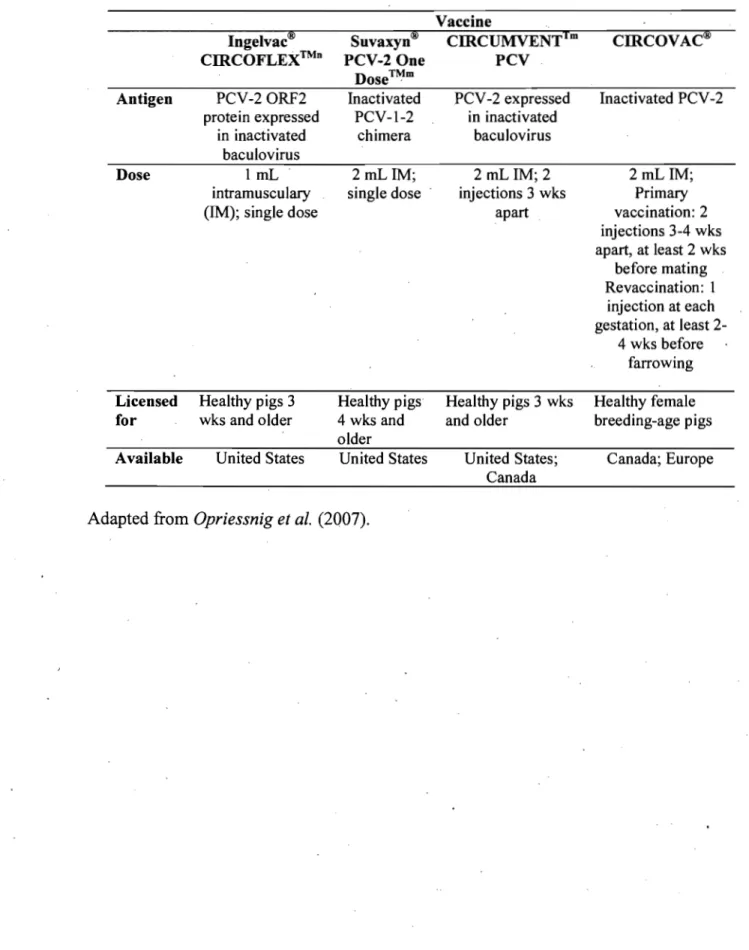
Evidence to date indicates that the commercial vaccines (Table 1) are a remarkably effective tool to reduce losses in herds and production systems experiencing PMWS in growing pigs. The inactivated, oil-adjuvanted PCV -2-vaccine (CIRCOVAC, Merial Corn, Lyon, France) licensed for use in breeding-age animaIs was the first vaccine on the market and has been used most extensively in Europe (Table 1). CIRCOV AC has been shown to be beneficial -in reducing PCV -2 circulation and shedding in the first weeks of life and improving pig health under experimental conditions [29]. During field efficacy studies in Germany and France, the use of CIRCOV AC resulted in a rise in PCV -2 antibody levels in the breeder herds and a concurrent decrease in PMWS rates in the growing pigs [29]. When
used in Canada in 2006, field trial~ demonstrated that CIRCOV AC significantly (P<0.05) decreased mortality (6.4-8.3% prior to vaccination) [132].
A conditionally licensed product (Circurnventtm PCV - Intervet Inc, USA) fo~ use in growing pigs became available to the North American market in April 2006 (Table 1). Investigations induding pigs, on 21 Canadian farms showed that the mortality rate In
vaccinated pigs was lowered by 77.5% when compared with nonvaccinated pigs [68]. SuvaxynH PCV-2 One Dose TM (Fort Dodge Animal Health, Fort Dodge, lA) is the first US Department of Agriculture-approved and fully licensed commercial PCV-2 vaccine in the United States (Table 1). This is the killed version of the live chimeric PCV-1-2 virus [57, 59]. Preliminary results froin severallarge fields studies in the United States demonstrated significantly decreased mortality (P<O.OO1) with the use of the killed chimeric vaccine, accompanied by reduced treatment costs compared to those encountered for nonvaccinated pigs [40].
Preliminary results from field trials using the baculovirus expressed PCV -2 vaccine Ingelvac CIRCOFLEX TM (Boehringer Ingelheim Vetmedica Inc., St. Joseph, MO) (Table 1) also demonstrated significant reduction in mortality (P<0.003) in vaccinated pigs compared to nonvaccinated pigs on four Canadian finishing sites [48].
Table J. Commercial PCV -2 vaccines available in North America.
Vaccine
Ingelvac® Suvaxyn® CIRCUMVENT1m CIRCOVAC®
CIRCOFLEX™n PCV-2 One PCV
Dose™m
Antigen PCV-20RF2 Inactivated PCV -2 expressed Inactivated PCV-2
protein expressed PCV-1-2 in inactivated
in inactivated chimera baculovirus
baculovirus
Dose 1 mL 2 mL lM; 2 mL lM; 2 2 mL lM;
intramusculary single dose injections 3 wks Primary
(lM); single dose apart vaccination: 2
injections 3-4 wks apart, at least 2 wks before mating Revaccination: 1 injection at each gestation, at least 2-4 wks before farrowing Licensed Healthy p igs 3 Healthy pigs Healthy pigs 3 wks Healthy female
for wks and older 4 wks and and older breeding-age pigs
older
Available United States United States United States; Canada; Europe Canada
EXPERIMENTAL INFECTION MODEL OF PMWS
Experimental PCV2-infection models indicate that PCV -2 is an opportunist, depending on immunostimulation [13, 15,86] or coinfecting agents like PPV [9, 14,53, 76, 81, 84, 85, 122, 130], PRRSV [12, 74, 142], or M hyopneumoniae [123] for PCV-2 infection to progress to clinical PMWS. While most research groups have not been able to reproduce clinical disease in pigs inoculated singularly with PCV -2, sorne groups have succeeded. In early experimental models, piglets inoculated with PCV -2 alone only developed a very mild clinical disease with slight histologicallesions characteristic to those ofPMWS [9, 16, 100].
A cofactor was needed for immune stimulation· to reproduce the disease. This hypothesis arose from experiments using days-old gnotobiotic piglets inoculated with PCV-2 and keyhole limpet haemocyanin in incomplete Freund's adjuvant (KLHlICFA) [86]. In that experiment, the piglets developed PMWS in higher proportion and with higher severity than the unstimulated animaIs, which developed a subclinical PCV -2 infection. This hypothesis was further explored under natural conditions. Others authors [88] showed that conventional on-farm pigs inoculated with a parapoxvirus immunomodulator combined with a vaccine against M hyopneumoniae developed more severe clinical signs than pigs to which none of the above was administered and were only naturally exposed to the virus.
Different type of animaIs models were used in experimental infection, such as gnotobiotic pigs [85], colostrums deprived cesarian derived (CDCD) pigs [17, 74, 134], colostrums deprived (CD) pigs [5,9, 12] andconventional pigs [1, 16,90].
Fenaux et al. (2002) investigated the infectivity of an US PCV-2 molecular infectious DNA clone. The infectious clone was directly inoculated into the liver or inguinal lymph nodes and mild PCV -2-associated lesions were. observed in 4-weeks-old conventional pigs confirming the role ofPCV-2 in PMWS [56].
•
Nedzad Musicl, Mario Jacquesl, Carl A. Gagnonl,.
IGroupe de Recherche sur les Maladies Infectieuses du Porc, Faculté de Médecine V étérinaire, Université de Montréal, Saint-Hyacinthe, Québec, Canada, J2S 7C6 .
Corresponding Author: Carl A. Gagnon, Faculté de Médecine Vétérinaire, Université de Montréal, 3200 rue Sicotte, Saint-Hyacinthe, Québec, Canada, J2S 7C6. Phone:
Fax: E-mail:
Short running title: NPTr cells are permissive to PCV -2 replication [information retirée / information withdrawn] [information retirée / information withdrawn] [information retirée / information withdrawn] [information retirée / information withdrawn]