HAL Id: tel-01143499
https://hal.archives-ouvertes.fr/tel-01143499
Submitted on 17 Apr 2015
HAL is a multi-disciplinary open access archive for the deposit and dissemination of sci-entific research documents, whether they are pub-lished or not. The documents may come from teaching and research institutions in France or abroad, or from public or private research centers.
L’archive ouverte pluridisciplinaire HAL, est destinée au dépôt et à la diffusion de documents scientifiques de niveau recherche, publiés ou non, émanant des établissements d’enseignement et de recherche français ou étrangers, des laboratoires publics ou privés.
Distributed under a Creative Commons Attribution - NonCommercial| 4.0 International License
Approche quantitative de la croissance microbienne avec
des colonies cylindriques de levures.
Clément Vulin
To cite this version:
Clément Vulin. Approche quantitative de la croissance microbienne avec des colonies cylindriques de levures.. Biotechnologies. Universite Paris Diderot-Paris VII, 2014. Français. �tel-01143499�
UNIVERSITE PARIS DIDEROT (PARIS 7) THÈSE
Pour obtenir le grade de DOCTEUR DE L’UNIVERSITÉ PARIS DIDEROT Spécialité: BIOPHYSIQUE
Ecole Doctorale Interdisciplinaire Européenne Frontière du Vivant (Ed 474) Laboratoire Matière et Systems Complexes (MSC)
A quantitative approach to
microbial population growth using
tailored cylindrical yeast colonies
Présentée par
Clément VULIN
dirigée par Pascal HERSEN
Soutenue le 23 septembre 2014 devant le jury compose de:
Timo BETZ Chargé de Recherche, CNRS Rapporteur
Matthieu PIEL Directeur de Recherche, CNRS Rapporteur
Arezki BOUDAOUD Professeur, ENS Examinateur
Stéphane DOUADY Directeur de recherche, CNRS Examinateur
Clément NIZAK Chargé de Recherche, CNRS Examinateur
Delphine SICARD Directrice de Recherche, INRA Examinateur
Au moment de me lancer dans cette aventure qu’est la thèse, je ne crois pas que j’avais bien
cerné ce qu’il en ressortirait. Pourquoi une thèse ? Outre me procurer le grade de docteur et
résoudre un petit bout des fascinantes questions sur la vie des unicellulaires et
multicellulaires, cette thèse est pour moi avant tout une formation humaine et scientifique
que je suis très heureux d’avoir suivie. Si la formation scientifique était pour moi une
évidence au moment de signer, je me doutais, mais sans en être bien sûr, que l’étude de la
vie en communauté des levures de bière se ferait de paire avec celle de la compréhension
d’une communauté humaine. Au final, je considère que ces quelques années sont pour moi
avant tout une formation la connaissance des autres et de moi-même. Le plus important de
ma formation n’aurait donc pas été possible sans les nombreuses personnes que j’ai
côtoyées pendant ces dernières années, et qui m’ont accompagné au travers de ce voyage.
Je souhaite donc les remercier ici. J’en oublierai surement. La coutume veut que l’on cherche
son nom dans cette partie d’une thèse plutôt que d’effectivement parcourir le document
scientifique, pardon donc d’avoir choisi de ne pas me borner à une liste de noms (ça aurait
été trop simple à écrire et trop digeste à lire, je crois). Se reconnaitront ceux qui veulent. J’ai
travaillé dans beaucoup de lieux aux ambiances différentes, et ces ambiances ne sont rien
sans leurs habitants.
Avant cela, un grand merci à tous ceux qui n’ont pas forcément fait de science avec moi,
amis et famille, grâce à qui j’ai pu m’entrainer sans relâche à expliquer ce que je faisais avec
mes colonies douteuses.
J’aimerai commencer par remercier les personnes présentes au début de l’aventure dans la
grande maison du CRI (centre de recherche Intedisciplinaire), où a émergé le projet. Il s’agit
d’un lieu où la créativité et l’excitation de la science est omniprésente. Mes « mercis ! » vont
donc à la fois aux encadrants qui sont les piliers du projet éducatif, en particulier durant mon
master; aux étudiants avec qui j’ai interagi pendant cette préparation, ma promotion de M2
en particulier; et à tous les élèves de l’école doctorale que j’ai pu rencontrer et avec qui j’ai
pu partager mes discussions. Je n’oublie pas bien sûr les bons anges administratifs, anciens
et nouveaux sans qui l’Administration serait pour nous tous un enfer. Un grand merci aussi à
ceux qui m’ont permis d’enseigner en M1 avec une totale liberté au Bootcamp, et en
particulier à celui qui montre toujours le plus d’enthousiasme, et ce malgré les difficultés
pour réunir l’ensemble de la faune professorale en un même lieu et temps. Parmi les
professeurs, j’aimerai remercier ceux qui prenaient le temps de passer avec nous quelques
soirées, et avec qui j’ai pu discuter de la qualité d’un bon houmous ou de l’importance de la
dance et de l’expression corporelle, ou bien du futur de l’éducation et même de la distance
optimale entre un urinoir et un groupe de buveurs des quais.
Avec le CRI, je pense en particulier aux Night Scientists des Marsouins avec lesquels j’ai
apprécié ces nombreuses bières, au moins autant que les conversations scientifiques sur le
développement embryonnaire des oursins ou les enseignements de Kant, sur la médecine
hospitalière ou la façon de faire un bon taboulé, sur la pharmacopée russophone ou
l’optimalité d’un bon kvas, le vieillissement des bactéries ou la vétusté des rapports humains
dans les squats, l’optimalité de la coopération en société ou comment apprendre à un singe
à faire de la voile, sur les champignons fourrageurs, l’escalade à Pasteur, la façon et l’utilité
de bien encadrer son directeur… Merci du fond du cœur pour ce groupe de soutient des
thésards anonymes, sur lequel je pourrais encore compter dans le futur.
Plus proche de mes travaux de thèse, je voudrais souligner l’ambiance merveilleuse qui
règne dans les locaux du laboratoire MSC. L’atmosphère d’entraide et créative y est
particulièrement développée, et je me sais chanceux d’avoir pu profiter de « ce monde de
bisounours ». Pourvu que ça dure ! Merci donc à ceux avec qui j’ai pu discuter de la façon
idéale de modéliser mes colonies, comme des mousses ou du sable ? Merci aux thésards
avec qui j’ai passé une grande partie de mes pauses cafés, oh combien importantes ! Merci
pour les discussions sur l’avenir de la recherche, les recherches de postes, l’avantage de la
vie à Edimbourg… Merci à toute la clique de SF. Merci aussi aux encadrants techniques que
nous avons la chance d’avoir et qui sont toujours ouvert aux discussions. Merci d’avoir
maintes fois sauvé mes montages électroniques archaïques, fabriqué les pièces parfaites
pour mes moulages, aidé à monter mon système de mesure laser, bref, à ceux qui ont eu la
patience d’enseigner a un débutant à la fois en mécanique et en électronique. Merci plus
généralement à ceux qui font la superbe ambiance autour de la sacrosainte machine à café.
En parlant d’ambiance à MSC, celle de la salle 756A est toute particulière, et j’aimerai
remercier tous ses hôtes, passés et présents, pour avoir fait vivre la bonne ambiance et les
PD du vendredi. Merci aux trois inconditionnels du vélo pour leur sagesse, leur utopisme et
leur humour grâce à qui j’ai découvert la chicorée, les règles de ponctuation et les méandres
de l’internet. Merci à la cliqueuse sous écharpe avec qui j’ai partagé le plus de temps dans ce
bureau à rédiger ma thèse et me battre contre les moustiques, et qui gagne toujours les
concours de pâtisseries. J’ai aussi apprécié le délicat accent du sud que distillai notre
post-doc attitré, ainsi que ses horaires matinaux, sorte d’objectif à atteindre. Merci au seul vrai
théoricien la salle, que je n’ai jamais pu aller voir en concerts, et que l’on finira par réussir à
mettre derrière une pipette remplie de vraies bactéries. Merci encore à notre mécanicien
des flux pour ces discussions sur les joies du journalisme scientifique.
Parmi les résidents de la 756A, j’ai eu aussi la chance de travailler au laboratoire 513 avec un
bon nombre d’entre eux pour avoir des discussions économico-politicardes au dessus d’un
point d’open-soudure, ou sur des séances d’escalades ponctuées de traitement du signal ou
de conseils Matlab, ou des débats intarissables sur dicty ou physarum ou des conseils sur la
tournure de mes discussions mondaines. J’ai évidement largement profité de l’expertise et
de l’esprit joyeux de tous les membres du laboratoire 513. Je voudrais donc remercier les
« permanents » comme les temporaires, les anciens comme les nouveaux, pour leur soutien
humoristique et scientifique, soit par la relecture à la virgule près de certaines de mes
productions, pour la discussion ultra professionnelle de l’optimalité de mes modèles, ou
pour leur conseils sur mon orientation et leur organisation de la vie de laboratoire. Pour
ceux qui sont partis du laboratoire, merci encore pour les séances microfluique et levure,
pour les discussions sur l’esthétique d’une belle présentation et l’invitation à voir Berlin (Wir
werden ein Bier zusammen trinken) ou Londres. Merci aussi à celui qui partage (presque)
toute ma formation, depuis l’agro et le master, puis au laboratoire à Boston et Paris, pour
toutes nos bêtises et les discussions sur la valeur réelle de l’entreprenariat. Merci surtout
aux stagiaires (puis thésards et amis), en particulier à ceux sans qui les projets de
microfluidique et du scan de colonie ne seraient pas aussi avancés.
I would also like to thank the great people I met during my stay in Boston, thanks to the
generous hosting of a great funny guy, that can invent a joke a minute (including baseball
references that I mostly never catch). Big thanks to everyone there then for your help,
especially to give a direction to my wanderings. Thanks also for your warm welcome and the
great evolution board games I discovered with you.
J’ai eu l’occasion aussi de me faire beaucoup d’autres choses pendant ma thèse, et je
remercie en particulier celle avec qui j’ai pu jouer à faire de beaux films sur la pourriture.
Je tiens évidement remercier tous les gens qui m’ont aidé matériellement où avec de
précieux conseils dans mes expériences pour le dosage de glucose, pour l’utilisation du
microscope électronique à balayage, et celui du cryotome, et surtout pour les conseils en
métabolisme du glucose et les dons généreux de souches fluorescentes. Puisqu’il s’agit
parfois des même personnes, merci à ceux qui ont bien voulu participer à mon comité de
thèse, et merci aussi pour avoir accepté d’être dans mon jury de thèse (même dans le cas où
cela ne s’est pas concrétisé). Merci enfin à tous les membres du jury pour leurs remarques
sur mon travail, et aux rapporteurs pour leur lecture très attentive de ma thèse. Au sujet de
cette thèse, merci à mon frère et à mon super relecteur agro qui ont bien voulu se glisser
dans la peaux de ma grand-mère pour m’aider à améliorer et traduire cette introduction, je
vous en suis très redevable.
Enfin, merci à mon directeur de thèse pour ces quelques années passées ensemble, qui ont
été un délice humainement et scientifiquement. Le plus important pour moi est d’avoir été
soutenu dans les activités variées que j’ai entamées pendant ma thèse, et pendant les
moments ou les résultats ne tombaient pas. Garde cet humour qui t’es caractéristique, ça
m’a beaucoup aidé.
Merci à celle qui me supporte depuis si longtemps et qui partage tout de ma vie, et comme
c’est le sujet ici, avec qui j’ai la très grande chance de pouvoir aussi parler de science.
11
Résumé ... 15
I.
Introduction for my grandmother ... 17
II.
Introduction... 29
1. Unicellulars as a model of multicellular development? ... 31
a. Blurring the lines of multicellularity ... 31
b. Colonies of microbes as multicellular organisms ... 35
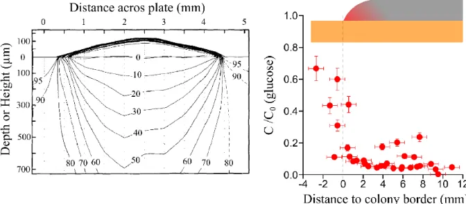
c. Nutrient variability inside dense microbial populations ... 36
2. Saccharomyces cerevisiae as a model organism ... 40
a. A short introduction to yeast metabolism ... 40
b. Thinking about S. cerevisiae as a multicellular organism ... 47
3. Models of multicellular colonial growth ... 52
III.
Setting up a confined zone for colony growth ... 61
1. Membrane preparation ... 64
a. Mask design and printing ... 65
b. Stamp fabrication ... 65
c. Membrane stamping ... 67
d. Resulting material analysis ... 68
e. Membranes for altered colony growth ... 69
2. Growth of yeast cylinders ... 72
3. Cells divide close to colony gel interface ... 76
4. Approximation of the steady state flux ... 82
5. Setting a numerical simulation of the problem ... 89
6. Growth rate variation with nutrient concentration ... 94
a. Experimental observations ... 94
b. Radial growth at low glucose concentrations is due to respiration. ... 95
7. A measure of colony yield ... 98
8. One dimensional model with fixed flux ... 101
9. Lessons from numerical simulations of growth ... 104
a. Effect of diffusion ... 104
b. Effect of uptake rate ... 106
10. Discussion on this chapter ... 109
IV.
A laser scanner for precise shape and growth patterns ... 113
1. Introduction ... 115
b. Measures of growth and fitness ... 116
2. Experimental Setup ... 118
3. Freely growing colonies ... 120
4. Vertical growth rate of a cylinder ... 123
5. Discussion about further work planned ... 125
V.
Towards real-time microscopy of a cell assembly:
microfluidics ... 127
1. Introduction ... 129
2. Principle of experiment ... 131
3. Protocol for chamber fabrication ... 133
a. Design and masks ... 133
b. Principle of lithography for one layer of 200 µm resist:... 134
c. Aligning multiple layers of different thickness ... 135
d. Loading cells in the microfluidic device ... 135
4. Estimation of local division rate ... 137
a. Particle image velocimetry (PIV) ... 137
b. Cell tracking ... 138
5. Discussion on future work ... 140
a. Notes on system optimization ... 140
b. Further work planned ... 141
VI.
Summary and openings ... 143
VII.
References ... 149
VIII.
Annexes ... i
1. Une introduction pour ma grand-mère ... iii
2. Cryosectioning of fluorescent pillars ... xv
3. Book chapter: Protocol for membrane patterning as published in “Methods for cell biology” ... xvii
13
Abbreviations and symbols used in this manuscript
Note that yeast, in this document, refers to Saccharomyces cerevisiae, unless otherwise specified
Abbreviations:
PDMS Polydimethylsiloxane, silicon polymer used for microfabrication
YPD Yeast extract, peptone, dextrose (= (D+)glucose), a rich growth media for yeast
SC Synthetic complete, a synthetic growth media for yeast
ATP Adenosine triphosphate, energetic molecule used by the cells
PIV Particle image velocimetry, method of analysis to follow movement of particles
Symbols:
a Radius of imposed pattern
C Limiting nutrient concentration (usually glucose). C* is concentration at interface between colony and gel, C0 is the initial gel concentration and concentration at infinite
D Diffusion coefficient of solute in media (Alternatively, Dilution rate in Figure I.2)
h Height of the cylindrical colony
H Height of cells that are dividing in the colony
I Flux of glucose the gel delivers to the colony
k Other constants obtained during mathematical resolution
n Number of replicates
N Number of cells dividing in the population
q Absorption rate of limiting nutrient
r Radius of colony (often r = a)
Rp Thickness of outer ring of colony
t Time
Y Colony yield: amount of cells (or volume) obtained per unit of nutrient consumed
γ Growth rate of cylinder, usually in height
15
Résumé
Microbes can form complex structures composed of millions to billions of cells. These assemblies contrast with the classical view we have of the “unicellulars” microbes. In fact, given their environment, they likely form heterogeneous connected structures. Our understanding of these assemblies is still scarce. The problem is that the models we develop suffer from the lack of experimental tools to understand these groups of cells. In this thesis, I propose to study they yeast Saccharomyces cerevisiae colonies by defining the flux of nutrients the colony receives. I use patterned filtration membranes intercalated between colonies and nutritive gel, leading to well controlled shapes. Using the cylindrical geometry resulting from a disc pattern, I first propose a quick study of the pillar organization, and then propose a simple model for colony growth in order to explain experimental growth in different environmental conditions, with respect to glucose levels, colony diameter and oxygen availability. I then discuss the biological relevance of this model with respect to cell division and nutrient absorption. To go further in investigations, I propose a automated measure of colony volume using a laser based measure with a 10 µm height precision. A microfluidic setup that mimics a two-dimensional colony growth is also proposed, where cells can be directly observed under a microscope.
Les microorganismes peuvent former des structures complexes composées de millions ou milliards de cellules. Ces assemblées de cellules contrastent avec la vue classique que nous avons des microbes « unicellulaires ». En fait, étant donné leur environnements, ces microbes forment certainement des structures hétérogènes et connectées. Notre compréhension de ces assemblées reste incomplète, et les modèles que nous développons souffrent du manque de méthodes expérimentales pour comprendre ces groupes de cellules. Dans cette thèse, je propose d’étudier les colonies de levure Saccharomyces cerevisiae en imposant le flux de nutriments que la colonie reçoit. Pour cela, j’utilise des membranes de filtration modifiées que j’intercale entre les colonies et le gel nutritif pour contrôler la forme de ces colonies. En utilisant la géométrie cylindrique que l’on obtient avec un motif en forme de disque, j’étudie d’abord rapidement l’organisation du pilier obtenu, puis je propose un modèle simple de la croissance pour expliquer les données expérimentales obtenues dans différents environnements, avec une variation de la concentration en glucose, du diamètre des colonies ou de la présence d’oxygène. Je discute ensuite de la signification biologique de ce modèle en rapport à la division cellulaire et l’absorption des nutriments. Pour aller plus loin, je propose un suivi automatisé du volume des colonies en utilisant une mesure laser avec une précision en hauteur de 10 µm. Un système microfluidique qui mime une croissance bidimensionnelle de colonies est aussi proposé pour observer les cellules directement sous un microscope.
I.
Introduction for my
grandmother
19
To my grandmother questions about my PhD, I usually answered that I worked on brewing yeast, the one incriminated in bread, wine, beer, kefir and other fermented human products. Surprisingly, this seemed to be a good enough explanation of my thesis subject, and a straightforward way to justify my being still a student at the age of 27. I only had few times so far to go further in explaining that, in fact, what I am interested in is not yeast in itself, rather how it would grow in communities. As described in many animal documentaries for lions or wolves, my thesis is asking the question of the fight for life: who would feed first, who would reproduce best, how could we predict anything on the pack of yeast growth and survival, and what kind of general laws can we understand in microbial packs? The following few pages of introduction are an attempt to clarify this further.
First, why am I not working on lions, wolves or parakeets? Moreover, what are microbes; who are they? The simplistic answer to the first question is that microbes are much easier to work with than big animals. Thus, more people have tried to understand them and our resulting knowledge makes them even easier to work with. In reality, and I will try to explain this, they are as important and interesting to study as lions. To answer the second
Figure I.1: Multicellularity: a few examples. A-E: common textbook examples of multicellulars: animals, land
plants, fungi, red algae, brown algae. F-H: The multicellular form of oomycetes, dictyostelids slime mold and green algae is either non-permanent or less developed. I-L: Multicellular bacteria? I: Freshwater bacteria Nostoc
pruniforme form centimeter scale multicellular structures, here washed up on the grassy shore. J: Bacterial slime
molds formed by Myxococcus Xanthus are transitory multicellular structures. K: Caulobacter crescentus clonal development shows a cellular differentiation between the cell stalk that stick to surfaces and the daughter cell with a flagella that can colonize new areas. L: A Bacillus subtilis colony with wrinkles provoked by localized cell death. We commonly consider these bacteria either as distinct individuals, or as society of individuals. Sometimes however, the whole group is considered as one meta-individual. Such colonies are what I have been studying during the past few years. Images from C-G,J are from Wikimedia, D from acquaportal.it, E from: noaa.gov F from: D. Blancard, INRA, H from: Wim van Egmond on microscopy-uk.org.uk, K from: Jeff Skerker, Berkeley and L from [133].
question, “what are they?” the classical definition of a microbe would be “an very small organism, of the microscopic scale. It is able, as we are, to feed, live and reproduce on its own”. This general definition encompasses all sorts of life, from complex mites or planktonic crustacean, to rather "simple" organisms, composed of only one cell.
A cell?
A simple definition of a cell could be a “living compartment” or said otherwise a well-delimited bag full of chemicals entities, and able to duplicate itself indefinitely. Cells were termed after their discovery in plants were they looked like squared compartments, but further microscopy observations taught us the variety of life at that scale, and with it the tremendously different and variable shapes cells can have. For instance, the size of a cell can range from under a thousandth of a millimeter to macroscopic objects such as eggs that are indeed just one cell!
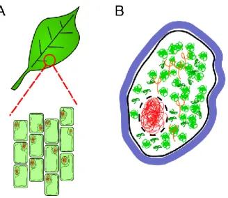
Nevertheless, cells all have in common that they contain similar molecules, sorts of bricks of life. Those molecules are repetitive chains of smaller molecules, and we call them polymers. One of them, the much famous DNA (deoxyribonucleic acid) is a long chain of sugars that form “characters” encoding information. The patterns and arrangement of these parts will define a code (like our alphabet) that the cells decipher and transmit from generation to generation. A simple way of looking at its usage is to consider two other important long molecules, RNA (ribonucleic acid) and proteins. The first one, RNA, is a support to copy and transfer the DNA code to machineries that will translate this RNA code and make proteins. Proteins are also long chains of molecules. The arrangement and folding of these chains allow the cell to build little machines (enzymes) or scaffolds, channels and linkers. In everyday life, we find proteins everywhere. Some proteins we use only to obtain energy, for instance, when we eat eggs or beans, but others we use to perform specific functions: the mechanical functioning of our muscles depends mostly on proteins. Others have biochemical functions: specific proteins in our body specifically degrade the alcohol we may drink when others focus on the sugar we eat. The cells perform those functions by using energy they get from the outside world. Plants harvest energy from light, but a lot of our energy comes from burning sugar, proteins or lipids that we eat. To this respect, there are many efficient ways to harvest energy. For instance, a sugar can be fermented, which is what yeast do in beer, or
Figure I.2: A: Cells are the small units of life. They can
be easily observed for instance on the surface of a leaf, where they are well arranged and approximately 1/20th of millimeter (you can discern them by eye). B: A schematic cell. The boundary made of lipids (here in black) defines the cell. This common boundary can be thin, and many cells have a thicker external boundary, the cell wall (in blue). Inside the cell, DNA (red) is a long compact and folded polymer. It can either be contained in an inner compartment, the nucleus (in our cells, plant cells and yeast cells for instance), or simply be in the bulk of the cell (in the case of bacteria). The cell transcribes it into RNA, a similar molecule (in orange) and then further translates RNA into proteins (green). The cell interior is crowded with such polymers, and many other proteins and molecules.
21
respired, which is what we usually do. The way we use those nutrients will affect the efficiency and the speed of our metabolism.
This entire little world of DNA, RNA and proteins is organized inside a bag, which is made of lipids (fat). Lipids are a convenient separator between inside and outside of the cell, because they do not mix well with water. The cell is in fact very densely crowded with all other sorts of small and big molecules, which the cell constantly burns, builds or moves around. One has to imagine a dense market, where everyone encounters every one, sometimes, although rarely, leading to effective interactions (talks or shopping). To make it more efficient, cells are organized in more or less defined zones, so that molecules that need to encounter each other have a higher chance to do so.
As we have seen earlier, most organisms we commonly see and name, plants and animals alike, are composed of many cells (tens of thousands of billions for human). Some however, the majority in fact, are composed of only one single cell. These are termed unicellulars.
Unicellular organisms
Unicellular organisms are, despite their small size, the most abundant life on the planet, both in number and mass. They are found in all groups of life, being bacteria, archae or eukaryotes (see glossary box). Because of their abundance and ubiquity, and despite their microscopic size, we can often see them with only our naked eyes. This is because as small as they are, they are many. A lot of them have the capacity to make groups comprising millions
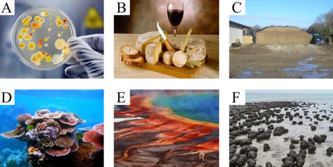
Figure I.3: Microorganisms are everywhere. The usual association to the word microbe is that of a medical
problem (A), but microbes are much more than this. They are for instance used by man to create food such as cheese, wine and bread (B), or at greater scale for silage (C, grass in can for cows) that is conserved because of a rapid change of acidity due to bacterial fermentation. For instance, they are necessary to digest many aliments we feed on. More generally, they are part of the whole ecosystem, from the soil to the symbiosis with corals (D). They are one of the main architects of the environment, sometimes with surprising results: in picture (E) is the Grand Prismatic Spring in USA, which various colors are due to microbial activity. They have been on earth for much longer than we have, and started to lay traces a long time ago (on F are stromatolithes, geological structures that were built long ago by microbes). Pictures: A from istockphoto.com; B from: Goodshoot via rfj.ch; C from grostracteurspassion.com; D, F from Wikimedia; E from Jim Urquhart / Reuters.
to billions of organisms forming centimeter-sized objects, alternatively termed clumps, flocks, mats, biofilms or colonies. Another way to notice their presence is that they usually can deeply and rapidly modify their environment by absorbing and releasing chemical compounds. Because of their rapid variations and high heterogeneity, these communities can in turn form complex assemblies leading to amazing shapes and properties. Those complex assemblies are hard to describe with the sole knowledge of the individuals properties of each microbe, and must be understood globally. This is even more true since in the last century researchers mostly studied isolated individuals in homogeneous environments.
Thus, when focusing on “unicellular” microorganisms, we can ask many questions about "multicellularity". The dilemma is a priori simple. Can we still talk about "unicellulars" when some organisms form complex structures for their reproduction and survival? This question can be regarded as philosophical and such unicellulars communities are usually regarded as different from typical multicellulars such as animals or plants. However, one of the reasons why these communities are interesting is because the multicellularity question is not so trivial to answer. Could the study of such colonies give us a grasp at the evolution of multicellularity, at its functioning and maintenance?
Cell culture
During my thesis, I choose to study one simple form of unicellulars communities, in which the multicellularity question is still little debated. The common reproduction mode of yeast, my preferred microbe, is indeed unicellular, one cell dividing to create two cells. However, yeasts often group and form complex communities with various properties. More generally,
Figure I.4: schematic of a life cycle of an unicellular organism: In a non sexual life cycle, the organism (the cell)
will grow, replicate its DNA by synthesizing a new copy, segregate the DNA in two equal parts, and physically divide in two (this is called mitosis). This process leads to two almost identical cells that can start dividing again. The top right scheme shows that if all goes well the increase in the number of cells will be faster and faster, here for example showing that within the time of only 3 divisions, 8 cells are produced. This process can be calculated mathematically, and follows an exponential law, shown at the bottom right.
23
common life examples of microbial populations include what we usually simply refer to as colonies. Those are groups of clonal yeast or bacteria on food (see glossary box). They can as well form microbial mats (thick layered, usually photosynthetic structures), flocks (cells aggregate resulting from flocculation in liquid), or more generally biofilms, which can be floating or attached to a surface.
We routinely grow yeast, bacteria and other unicellulars in our labs, where we would go back and forth from Petri dishes, containing gels with nutritive solutes on which microbes form colonies, to shaking liquid cultures where their growth is unicellular. Liquid cultures are preferred for most observations. Because it allows homogenous treatment of a population, liquid is the preferred media to induce cell changes and to observe them in the lab. Thus, most of our knowledge on of growth, behaviors, and responses to stimuli comes from this liquid state of life. Even among, we usually focus on cultures where every cell, old and young, is dividing, a state of the microbe that is rather rare in nature. In these cultures, termed "exponential" every cell sees the same environment at the same time.
More than this, lab cultures usually focus on quickly growing cells. This is for obvious reasons of rapidity. For instance, E-coli cells in appropriate conditions will divide every 15-20 minutes. This is fast! In our gut, E coli would divide only once every 12-24 hours [1]. In any case, a fast exponential growth cannot be the usual way of growth of microorganisms. Let us take an example. Consider we start with one single bacterium, dividing every 20 minutes. Let us assume all the cells will be dividing at the same time. After 20 minutes, the bacteria
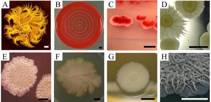
Figure I.5: Shapes of microbe colonies in the lab are variable. Even in controlled conditions, microbes can make
complex geometrical shapes of different nature. This is due to the interplay between growth, environment and cell-cell signaling. From top to bottom: Paenibacillus dendritiformis can form beautiful shapes (A) by moving around in groups. Also with movement, Proteus mirabilis swarming (B) on blood agar makes concentric rings.
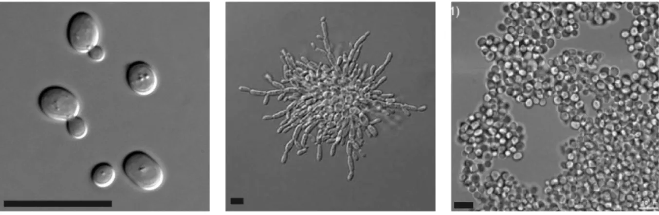
Serratia Marcasens (C) natural dual coloration is observed when grown as colonies. Candida albicans dual
morphotypes (D) are linked to their pathogenic potential. Bacillus subtilis (E) immobile fluffy state makes a wrinkled structure. Escherichia coli, the lab classical microbe, will start as a symmetrical colony, but mutations will rapidly emerge (F) and change its symmetry. Saccharomyces cerevisiae, can form simple smooth colonies in the lab (G), but wild type phenotypes can be much more complex (H). Scale bars: 2 mm. Images from: A: Wikimedia; B: pictures.life.ku.dk/atlas/microatlas/food; C: microbeworld.org from the Wistreich Collection; D: Felice Frankel, MIT; E: A.W. Rakosy/Encyclopedia Britannica
divided once, we have 2 cells. After 40 minutes, 4 cells. After 1 hour, we have 3 divisions, thus 2x2x2=8 cells. After 3h20, we have 1024 cells. Continuing this calculation, even if a cell weight is small (1 pg, one millionth of millionth of gram), we would have 100 kg of cells within only 18 hours, and the mass of Earth within 44 hours, less than 2 days ! This means that in real life cells die a lot, and spend most of their time not dividing or slowly dividing. In fact, predators eat and digest many cells. In addition, many cells are just sitting there waiting for food (in biofilms for example), and are thus not growing in a homogeneous environment.
Colonies
As I said, in the lab, we prefer liquid environments, but we still routinely use "solid state growth" on agar gels. In experiments where we mutate cells for instance, colonies have the tremendous advantage to be populations of cells that derive from a single and only clone. In this sense, colony acts as a handy sorting tool to identify mutations. Despite their common use, colonies are not well understood and not much studied by themselves.
There are two main barriers to the study of colony growth. First, colonies usually observed in nature are not a mere pile of cells. Their properties are usually to form fascinating and complex shapes that reveal all their beauty and geometry even when grown on defined media in the lab (Figure I.5). The shape variability is such that in medicine, one common way of recognizing microbes is still to use the colors and shapes of colonies. As shown in Figure I.6, with the same starting cells, the shapes of colonies will depend on growth
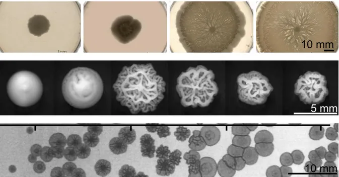
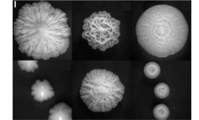
Figure I.6: Colony shape changes with growth condition. Top: Effect of gel hardness on Bacillus subtilis
swarming (movement on agar at gel surface) for agar concentration of 1.5, 1.25, 1 and 0.7% respectively, adapted from [4]. After 36h of growth, less pronounced movement of bacteria were observed when the agar concentration was higher. Middle: 5 days old S. cerevisiae colonies exposed to decreasing glucose concentration in the gel: from 2% to 1/16% in 2-fold steps show increased morphology complexity partly due to production of special sticky proteins [3]. Bottom: Salmonella colonies differ in morphologies depending on media acidity. This is linked to stress response and to their virulence for host [2]. Ticks on top mark initial pH of 4.6, 5.2, 6.4 and 6.7 respectively. Note that the pH changes with time: it becomes respectively 4.9, 7.1, 7.4 and 7.3 after 5 days (time of the picture). For all these colonies, I specify the date of the picture because the general shape is also maturing with time.
25
conditions, such as pH (acidity) [2], food availability or type of food [3], substrate softness [4] and more generally on the physical and chemical parameters of the environment.
Another barrier to the study of colony growth appears from the observation of these many shapes: in these assemblies, not all cells can be alike. Many factors, such as age, mutations and noise of cellular processes combine with the cells’ positions in the colony: cells that are close to the border of the colony will experience a drier, more oxygenated, and richer nutritive environment. Cells at the center of colony will only be able to feed on the eventual wastes the outer ones have produced. If on top of all of this, one considers the many physical and chemical communications cells tend to use, and the tremendous changes they can impose to their environment with time, it results that understanding the formation of colonies is a great challenge!
My questions about growth in community
Thus, the apparently naïve question “how does it grow” hides more than just our aim to predict a single colony growth. If focused on microbes, the main interests rely in the medical
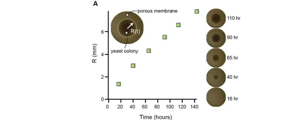
Figure I.7: Description of this manuscript. A. In this thesis, I develop a system to impose a geometrical shape to
yeast colonies. Any extruded geometry can be obtained, such as letters, circus, open patterns or closed discs. The latter leads to the formation of cylindrical colonies for which I try to build a model of growth (scale bar is 3 mm). This shape is strikingly different from usual conical growth (e.g. Figure I.5). B. I also developed a method to precisely measure the 3D morphology of yeast colonies and a microfluidic device (C) that allows the imaging of populations of growing cells with a fluorescent microscope at the single cell resolution.
applications of our answers. For instance, the antibiotic treatment of a biofilm developing on prosthesis or a tooth is made difficult because of the cells inner phenotypical variations [5]. We thus need a better knowledge of cells metabolism inside this biofilm, as well as their physical properties. In colonies of Candida albicans or Salmonella enteritisi (pathogenic
microbes), the passage from a smooth colony morphology to a complex morphology correlates with their stress and apparition of their virulence [6].
In addition, quite far from the microbial world, many studies focus on the embryo and its development, or on the special case of community organization of cancer cells. The mechanisms leading to proper organization within multicellular organisms are more than ever a hot research topic. In cancer for example, the passage from a primary tumor (benign) to a growing tumor (malign) correlates with the onset of collaboration between cells. In short, cells closer to blood vessels will share and recycle their food with cells that are deeper in the tissue [7]. This allows the cells deeply buried in tissue to grow, and thus the tumor grows too. Cellular differentiation and cross talks take place in such a process, but some of those complex mechanisms should result from general laws. In this case, the study of unicellular microbes may miss the complexity of multicellular organisms. However, as they are easier to work with than, say, mammalian cells, it may be easier to outline widely conserved biophysical mechanisms. This makes microbe communities a good tool to study the general laws of life within a community. These laws should also apply to wider communities.
Finally, the study of microbe community has its interest on its own, since many questions remain as to the inner ecology of such communities. Evolution of microbes is a fascinating question, as much as their abilities to colonize every place of the planet Earth. We still use only few of their capacities for our own good, and much more probably remains to be uncovered. In this thesis, I try to tackle the growth problem from a biophysics point of view. We use a system to constrain yeast colony growth to a quasi one-dimensional direction, in the form of cylinders. I created a method to strip the colonies from potential neighbors they could be talking to and interact with. I reduced their physical interactions with their environment to make the problem simpler to describe and to experiment on. I chose an organism that usually displays smooth colony shapes and has a well-known metabolism.
In the beautiful and simple case of cylinders, we can use the sub-question “what part of the colony grows?” to build a simple mathematical model. The idea is to explain experimental observations based on what we know of yeast metabolism. This was the focus of most of my thesis work. The question “how do cell divisions participate in colony growth?” is somewhat similar, but it involves understanding the interplay between cell divisions and the colony shape and expansion. Using a homemade high-resolution topography scanner, I will give hints as to what this interplay may be at the end of this document. To finish, I develop a microfluidic system to observe the cells under a microscope. Cells are in a "micro-fish tank" where I can constantly observe them. Hence, I organized the thesis manuscript as follows: an introduction to the various aspects of my work, then the study of the case of an ideal cylindrical colony. I then shortly describe the setting up of the two different methods that should allow us to go further in our studies. The first one is an attempt to precisely measure colony growth using a triangulation displacement laser and the other one is the development of a microfluidic setup that mimics colony growth.
27
i Candida albicans is a yeast pathogen that causes skin infections and can be lethal to
immuno-Glossary box:
- Archae/bacteria/eukaryotes: these are the 3 domains of life. Bacteria are well known for some of the
sickness they may cause, but are present everywhere. They are defined as cells with no inner compartments and are usually smaller than eukaryotes. Archae were long thought to be bacteria and are now considered different mostly because they have a far common ancestor and because of their weird way to harvest energy. They were first discovered in extreme environments, but are now discovered in many places. Eukaryote is the group we are part of, so is yeast, and is mostly defined by the presence of a nuclear sub-compartmentalization in cells. Eukaryotes cells are usually bigger than both bacteria and archae.
- Biofilm: a biofilm is a stack of cells that usually grows on a surface or that is floating. Cells stick to
each other and form a sort of rind with many different individuals.
- Clone: clones are individuals that are genetically identical to another. We refer to colonies as "clonal"
because one cell will divide into two genetically identical cells. This term is also found in multicellular organisms, for instance when potatoes are replanted without restarting from a seed, and contrasts with the genetic mixing that takes place in sexual reproduction.
- Colony: Used here the same way as the common life word “colonies”, such as would be a seal or
bird colony. A colony is an entity composed of several individuals, and that can be defined as a unity. Usually, it is the result of multiplication of organisms from a few starters.
- Culture media: this is the soup we are growing cells in, that contains all the food they need. It can be
“solid” if we add gelling agents, in which case it looks more like jam or custard.
- DNA, RNA, protein: these 3 molecule types are long chains of smaller bricks, otherwise termed polymers. DNA stands for deoxyribonucleic acid because its backbone chain is made of a sugar
(deoxy ribose) and is usually found in the nucleus of cells. Similarly, RNA means ribonucleic acid. - Genotype: ensemble of genes of one organism. By extension, it can be understood as the ensemble of
information contained in an organism DNA.
- Homogeneous/heterogeneous: An homogeneous environment means that every place of
environment is the same. Heterogeneous is the contrary.
- Metabolism: ensemble of the chemical reactions in a cell. This means all the activity of burning
energy (sugar, lipids, proteins…) and building new molecules with it (for moving, growing and reproducing…).
- Microbe: living organism that cannot be observed by eye, micrometers (µm) are 1/1000 of a
millimeter. A typical bacteria is 1 micrometer, a typical yeast 4-5 µm.
- Mutations: a mutation is an error in the DNA sequence, sometimes made during DNA copying for
division. It can also arise during the life of the cell. In any case, the "error" can be passed on to the next generation that will in turn duplicate it.
- Phenotype: all the traits that can be observed from outside. For humans, height, color of eyes or IQ
are phenotypes.
- Strain: for microbes, we term strain a cell line derived from a single cell. It can be seen as the
equivalent of animals “races” or plant “varieties”.
- Uni/multicellular: the cell being the smallest unit of life (capable of reproduction), some organisms
are made of one single cell. They are termed unicellulars. Organisms composed of more cells, such as animals or land plants are then termed multicellular. Note that the frontier between “uni” and “multi” cellulars is sometimes not well defined.
II.
Introduction
31
1. Unicellulars as a model of multicellular
development?
When talking about colony or population growth, it is hard not to linger a bit on multicellular structures, since a straightforward example of a cellular community is indeed a multicellular organism. Anthropocentric examples would be mammals, in which most of cells commit in their early life to perform specialized functions that will allow only a few of them (via gametes) to reproduce, and to create a new body. Although developed clonally, this whole cell community (the body) is a big, heterogeneous and physically well-organized group of cells. These cells are both physically connected and communicating. However, multicellularity is an evolutionary trait that is present everywhere across the different life groups (for a review, see [8]). In a strict sense, a multicellular organism is an organism composed of several cells. Those can be connected and/or differentiated.
a. Blurring the lines of multicellularity
First, it can be interesting to define what “an organism” is. Again, when we think about anthropocentric examples, an organism would be “a reproduction unit”. However, in an example such as land plants, vegetative reproduction can derive from many parts of the plant. Are all these parts a community of different organisms?
Thus, although the concept of “self” as in individual organism is natural in a human society, it is scientifically an evolving concept. Many other counter examples make “self” a vague notion. The immediate idea that comes to mind involves symbiosis, in which individuals cooperate so closely that the resulting society can intuitively be described as an individual in itself, for instance in lichen for the algae/fungus symbiosis. We can extend the idea to other systems, such as animal host/commensal flora. Indeed, would a cow still be a cow without its cellulose-digesting bacteria? Within this scope, it has been argued that the general idea of “individual” or “self” needs a rethinking in the broader sense of ecological systems [9].
This enlarged notion of “individual” then forces many different points of view for what a multicellular organism would be. A rapid look at a tree of life such as the one presented below (Figure II.1) would make the question even more apparent: in the scope of eukaryotes, definite branches of unicellulars, colonial unicellulars or multicellulars exist, even if the notion of multicellularity in some of these branches is blurry. One example is the case of slime molds, were cells can alternate between a unicellular state and an aggregated multicellular state. However, consensus work on bacteria or archae is even scarcer. We consider most bacteria as unicellulars, but bacteria are gregarious organisms that produce complex and organized communities. Moreover, some bacteria are commonly accepted as multicellular organisms. One example is the Nostoc species (Figure I.1) that produces filaments or balls of hundreds to millions of cells, with some cell-cell communication and differentiation to death. Another example, Myxococcus Xanthus bacteria (Figure I.1) is capable of creating assemblies resembling the slime molds ones.
The existence of such a wide prevalence of multicellular species raises questions as to the inner definition of multicellularity. In fact, the definition of a multicellular organism varies with the type of organism studied. I previously mentioned the two ideas of cell
differentiation and cell communication. I would like to review quickly their link to multicellularity.
Cell differentiation and multicellularity
First then, could it be that cell differentiation defines multicellularity? In multicellular land plants for instance, cells are specialized that can perform photosynthesis, transport sap or allow reproduction. The differentiation process follows repeatable body plans. In the green algae Volvoxii (Figure I.1), considered at the frontier of multicellularity, cell differentiation
exists, although less marked than in land plants. In this case, only specific cells will reproduce and will give rise to a new organism, thus creating a germ/soma differentiation. This would make differentiation a good argument for multicellularity. Cells of a land plants, however, can also dedifferentiate, when most animal cells cannot. Does that make a plant less multicellular than an animal? What can we say about transient differentiation?
The social amoeba Dictyostelium discoideum presents transient differentiation, where cell differentiation in the multicellular state disappears as soon as cells become unicellular again. However, even in colonies or biofilms of unicellular organisms, some cells express a stable “specialized” phenotype. In some cases, this phenotype is passed on for a few generations. Similarly to the case of land plant cells or social amoebas, it is however usually reversible with the apparition of new environmental conditions, and thus is rarely considered as a complete cellular differentiation.
Another point worth noting is that phenotypical differentiation does not necessarily implies programmed differentiation, and can instead be due to the emergence of a collective behavior. In this sense, cellular adaptation would emerge from cellular response to local rules that are not necessarily programmed in advance, such as nutritive gradients. This could create a global complex pattern (for a review on the bacterial biofilm side, see [10]). In a sense, differentiation is observed for multicellular species, but the levels of differentiations and its causes may vary greatly. It results that the link between differentiation and multicellular state needs not to be a direct one.
Multicellularity and cell communication
Multicellularity also brings the problem of cell behavior synchronization needed to perform complex functions. To synchronize cells, one way is to introduce cell-cell communication. Multicellulars such as animals, plants or fungi have several types of communication. Communication in long distance scale can be mediated for instance via hormones in animals, plants and fungi. Long distance communication is however not limited to the inside of an organism, and is performed across individuals and species, regardless whether individual are unicellular or not.
On the other end, direct local cell-to-cell communication needs proximity between cells, and thus could correspond more closely to the multicellular state. It can be mediated via exchange of cytosolic cellular contents via gap junctions in animals, plasmodesmata in plants or even wider pores in the case of fungi [11]. Filamentous multicellulars cyanobacteria such as Nostoc punctiforme are also thought to have direct cytosolic communications between cells [12], although observations of the responsible channels are scarce [13], [14]. However, direct exchange of intracellular content can also be done by unicellulars organisms, for
33
instance via the production of pili for DNA conjugation in bacteria. This in practice makes communication a necessary, but not sufficient argument for multicellularity.
Multicellularity and cell sacrifice
Another important point of multicellularity is the presence of a direct sacrifice of an individual towards population. This more general, social concept of multicellularity applies to all above-mentioned multicellular organisms. In all these organisms, some cells will
Figure II.1: A non exhaustive, simplified "evolution tree of life", from sequenced organisms from the 3 domains
of life that are archae, bacteria and eukaryotes (branch length are not representing genetic distances, data from [134]. Eukaryote group was enlarged in order to show the occurrence of multicellularity in this group, as depicted by different colors, adapted from [8], [135]. Multicellular states are also known to exist in archebacteria and eubacteria, through simpler forms. These multicellularity states are indicated instead by arrows. A yellow arrow points toward prokaryotes groups that contain species that have already specifically been described as “multicellular” in the literature [136]–[141]. It can be due to transient phenotypes in stress conditions or various interpretations of biofilm/colonial growth. Orange arrows denote groups in which at least one species is commonly regarded to as multicellular. Nostoc punctiforme, Streptomyces coelicolor, Myxococcus Xanthus are examples respectively for cyanobacteria, actinobacteria and proteobacteria. For prokaryotes, most subgroups are unicellulars, with a few of them being colonial or multicellulars (more interdependent group). However, the general capacity of prokaryotes to for organized complex communities such as biofilms blurs the line between unicellulars and multicellulars. Multicellularity, in itself, has appeared several times in history of evolution [8]. It may be worth noting first that the term multicellular has been applied to bacteria in general [21], and that the mere notion of “tree of life” is, in itself, a disputed notion. This is especially true for bacteria in which a big amount of genetic exchanges occurs in these complex communities.
benefit other cells at fitness expand for them. This altruistic behavior goes from small sacrifices such as the production of a molecule for communication to the total absence of reproduction, in the case of somatic lines. This is studied in coevolutionary games, in which the user compares reproductive success of cooperators and cheater. However, the notion of cooperation is, in itself, more societal than purely linked to a multicellular behavior: we cooperate all the time with other humans, although we usually do not consider our society as an individual organism. (for a review, see e.g. [15]).
Multicellularity is common
In the end, a population of cells can be considered as a multicellular organism in many ways, and this makes multicellularity a common trait. In fact, evolutionary studies agree that multicellularity has appeared several times in the course of evolution, in the different kingdoms of life. Easy examples are the apparition of multicellular land plants, red algae, animals or fungi, with some of those proven to have emerged several times in the same branch. In the end, as many as 25 occurrences have been documented [8]. Some reversions to unicellularity have been shown in bacteria [16]; they are debated in the case of ascomycota fungi (yeast form versus filamentous form); or have been provocatively suggested in mammals, in the case of some unicellular parasitic cancers [17]. Evolutionarily speaking, this makes multicellularity a convergent and versatile trait. Well-described examples of multicellularity emergence include eukaryotes, with land plants, animals or fungi. Eukaryotic traces of multicellularity are however more recent (1 billion years ago [18]) than the fossil record of a pluricellular structure of a filamentous prokaryote (dating back up to 3 to 3.5 billion years ago). These structures resemble the filamentous structures produced by Nostoc cyanobacteria [19], although signs of cell differentiation are younger and date back to 2 billion years ago with prokaryotes [20].
Advantages to the multicellular state are numerous. They include larger size for better foraging, predation and nutrient storage, as well as functional and metabolic specialization. However, several models can explain evolutionary maintenance of the multicellular state. Indeed, collaboration between the different cells at the early stage of multicellular life is not straightforward [8]. Depending on the model, the initial grouping of population can develop either by accretion of several individual cells (e.g. slime molds), syncytial divisions, or clonal growth. These formation models will set the factors on which multicellularity success will depend (Figure II.2). They imply a specific mode of reproduction and population bottlenecks. For instance, in animals or land plants, only a few cells will reproduce (in the case of mammals, only gametes). Collaboration is easier to maintain because eventual cheaters mutations are less likely to hit reproductive lines. Nevertheless, this means many cells will die in the process, and the reason exactly why they commit at first is no straightforwardiii. In the accretion case of Dictyostelids slime molds however, a large proportion of the population can access reproduction and this thus puts incentive to cooperationiv. However, this maintains a possibility for cheaters to emerge and spread in the population.
Because multicellularity is a generally occurring trait, many model organisms have been chosen to study the apparition of multicellularity. To cite but a few, these include already settled multicellulars such as green algae (Volvox sp.) or multicellular bacteria (Nostoc sp.). Many studies focus on slime molds (Dictyostelium sp.) that are transient multicellulars. Even
35
organisms commonly defined as unicellulars such as yeast (Saccharomyces cerevisiae) or bacteria (Bacillus subtilis) have been suggested.
b. Colonies of microbes as multicellular organisms
The idea of bacteria or other unicellulars as multicellular organisms is first intended as a thought experiment by Shapiro in 1988, under the title “Bacteria as Multicellular Organisms”. The aim was to underline that we should understand a colony of unicellulars as a whole, and that cell-cell communication, nutrient gradients or physical interaction necessarily shape the overall population growth. To illustrate his idea, Shapiro took several examples of accepted multicellulars bacteria and concluded by proposing that all colonies of bacteria could be regarded as multicellular entities. Although the notion of interactions in a unicellular group is far from new [22], we keep discovering more social traits of bacteria. So far, no satisfying model of the formation and development of such complex population exist. Indeed, colonies ask for a more global understanding of the population that the usual liquid cultures, where average cell behavior gives a decent picture of population.
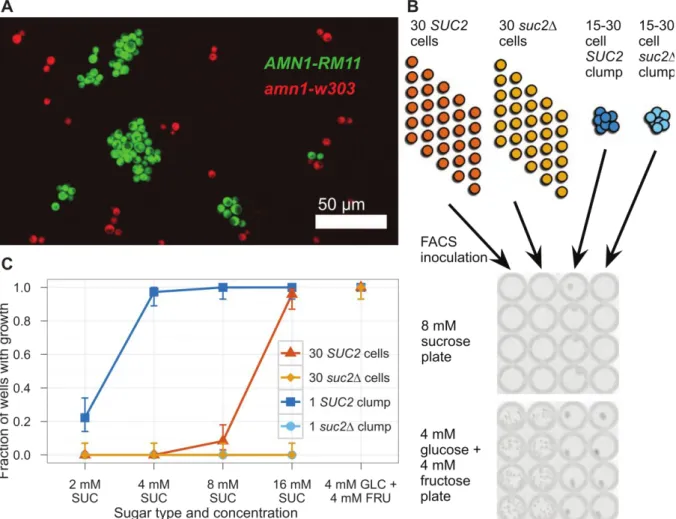
Unicellular populations of microorganisms are used as a model to understand the emergence of multicellularity because both phenotypical variations and cell-cell communication are observed at the scale of the colony. In many cases, cell-cell adhesions make the group of cells a physical unit, for example in yeast flocks or biofilms-embedded bacteria. Unicellulars have long developed the ability to stick to surfaces and/or to themselves, and the usual portrait we have of them, as isolated cells, is not accurate. Bacteria are most often found inside
Figure II.2 : Multicellular life cycles and their consequences on cooperation maintenance. a. Development from
a clonal population. In this case, mutant non-cooperator phenotypes (in red) can limit population growth, but will be diluted out when the population is split to start from single cells. b. If the multicellular assembly develops by accretion and leads to a reproductive subset (circled in black), defectors can stabilize at a constant proportion in the population (right) unless they are actively excluded from reproductive population (left). Reproduced from [8].
communities of millions to billions individuals, regrouping several species in interaction. The size of such population imposes constraints on its development that we will discuss.
Cell grouping and sticking is usually linked to environmental stress, and known to correlate with virulence increase [2], [6]. In the lab, one usually obtains a “grouped” population either from clonal development, from induced grouping via the production of sticking agentsv or from artificial experimental methods where cells are mechanically regrouped in compartments [23].
In such populations, interaction range is usually small. Even if external resources are unlimited, the local resource density is limited. While an infinite resource for each cell would promote competition, a grouped setup is theoretically enough to promote cooperation. For example, in a structured environment such as a biofilm, local yield loss will allow more of the local cells to divide (food penetrates further in biofilm). Since neighbors are likely to be genetically close cells, this favors yield clusters [24], [25]. In another example, cooperative secretion allows local population to reach and stay at the outside of biofilm, therefore allowing the group to access more oxygen and develop faster [26].
As mentioned earlier, multicellular group behavior can also be enhanced by communication between individuals of the population. In bacteria, cell-cell communication has long been studied since the observation of quorum sensing [22]. In common quorum sensing, bacteria can produce and release in the environment small chemical molecules such as AHL (Acylated Homoserine Lactones), auto inducers or small peptides [27]. The cells sense population local density [28] by sensing this signal. This allows relevant phenotypical adaptation: swarming optimization, entrance to dormancy or other types of bet hedgingvi (see the work of P.S. Stewart, for instance [5], [29], [30]). In yeast, sensing between populations can also rely on dosing secondary metabolites such as ammonia in S. cerevisiae [31], or tyrosol and farnesol in Candida Albicans [32]. The existence communication is considered in itself as a type of cooperative behavior since it is costly for cells to produce and release chemicals in the environment. It can lead to the apparition of non-communicant cheater populations that will not pay the cost of production but still be able to access the signals and their benefits [33].
The common points of colonies and biofilms with established multicellulars are often pointed out, and they are even being regularly proposed as models for cancer [34], [35] or a good proxy for the understanding of key evolutionary points toward multicellularity [36], [37]. It is important however to keep in mind that colonies, biofilms or flocks still display a fully distinct type of multicellularity than the well-defined one of a land plant, animal or fungi.