HAL Id: tel-01875895
https://tel.archives-ouvertes.fr/tel-01875895
Submitted on 18 Sep 2018HAL is a multi-disciplinary open access archive for the deposit and dissemination of sci-entific research documents, whether they are pub-lished or not. The documents may come from teaching and research institutions in France or abroad, or from public or private research centers.
L’archive ouverte pluridisciplinaire HAL, est destinée au dépôt et à la diffusion de documents scientifiques de niveau recherche, publiés ou non, émanant des établissements d’enseignement et de recherche français ou étrangers, des laboratoires publics ou privés.
The inhibitory microcircuit in mouse presubiculum :
from interneuron properties to input-output connectivity
Mérie Nassar
To cite this version:
Mérie Nassar. The inhibitory microcircuit in mouse presubiculum : from interneuron properties to input-output connectivity. Neurons and Cognition [q-bio.NC]. Université Pierre et Marie Curie - Paris VI, 2016. English. �NNT : 2016PA066722�. �tel-01875895�
Spécialité Neurosciences
cole doctorale Cerveau-Cognition-Comportement
Présentée par:
Mérie Nassar
Pour obtenir le grade de
DOCTE R DE L NI ERSIT PIERRE ET MARIE C RIE
Su et de la th se:
Soutenue le 16.09.2016
devant le ury composé de:
Dr Jean Christophe Poncer
Président du ury
Pr Matthew Nolan
Rapporteur
Pr Marlene Bartos
Rapportrice
Pr Dietmar Schmitz
Examinateur
Abstract
Spatial orientation and navigation are controlled by specific neuronal circuits and elements. The presubiculum, a transitional cortical area of the parahippocampal formation, is located between the hippocampus and the entorhinal cortex, and it participates in spatial navigation in animals and humans. More than half of presubicular neurons are head direction cells that fire as a function of the directional heading. The presubiculum is thought to be a crucial node for transferring directional heading information to the entorhinal-hippocampal network, and feed-ing back visual landmark information to upstream regions of the head directional circuit. De-spite its functional importance, information processing within the 6-layered presubicular mi-crocircuit remains not completely understood.
During my PhD, I studied inhibitory neurons of the presubicular microcircuit in the slice preparation using patch-clamp recordings. I characterized their anatomo-physiological proper-ties as well as their functional connectivity with local principal neurons.
In the first part, I examined the diversity of two major populations of GABAergic neurons, the parvalbumin (PV) and somatostatin (SOM) expressing interneurons in mouse presubicu-lum. Using transgenic mouse strains Pvalb-Cre, Sst-Cre and X98, where interneurons were fluorescently labeled, I showed the existence of typical PV fast-spiking basket-like interneu-rons mainly in the Pvalb-Cre line and SOM low-threshold spiking Martinotti cell-like inter-neurons in the X98 and Sst-Cre line. Unsupervised cluster analysis based on electrophysiolog-ical parameters further revealed a transitional group containing interneurons from either Pvalb-Cre or Sst-cre lines with quasi-fast-spiking properties and heterogeneous morphologies. A small subpopulation of ~6% of interneurons co-expressed PV and SOM in mouse presubic-ulum. The presubiculum appears to share the whole complexity of other cortical areas in term of inhibition.
In the second part, I investigated the integration of thalamic inputs by principal neurons as well as PV and SST interneurons in the presubiculum using double patch-clamp recordings. I found that thalamic axons selectively innervated superficial layers and made direct synaptic contacts with pyramidal neurons that project to medial entorhinal cortex and also with PV interneurons in superficial layer 3. In contrast, SST interneurons were indirectly recruited by presubicular pyramidal cells in a facilitating and frequency dependent manner. They may me-diate lateral inhibition onto nearby principal cells, and at the same time, preserve sustained firing of principal neurons. In paired recording experiments, I found that PV cells inhibit neighboring pyramidal neurons with a high connection probability. PV interneurons are rapid-ly recruited by thalamic excitation and mediate feed-forward inhibition in presubicular py-ramidal neurons.
My PhD work brought fundamental knowledge about the presubicular inhibitory microcircuit. It has unraveled different populations of GABAergic interneurons and revealed canonical feedforward and feedback inhibitory motifs that are likely to be recruited at different times during head direction signaling.
Acknowledgments
First of all, I would like to thank Jean Christophe Poncer, Matthew Nolan, Marlene Bartos and Dietmar Schmitz for being part of my thesis jury.
Je remercie tout naturellement Desdemona de m’avoir permise de travailler avec elle au cours de ces trois dernières années. J’ai eu beaucoup de chance d’avoir une directrice de thèse toujours présente pour moi à n’importe quel moment. Sans ta présence, je n’aurais pas pu avancer dans mes projets aussi vite que j’ai ainsi pu le faire. Tu m’as laissée une certaine li-berté de réflexion tout en sachant à la fois me guider dans ma démarche quand je te le deman-dais. Face à mes doutes concernant les bonnes expériences à réaliser, les protocoles et les ana-lyses à faire, j’ai eu souvent besoin de conseils et tu as su être là. Ton optimisme face à mon découragement m’a toujours rassurée et réconfortée dans la poursuite de mes projets. Je te remercie pour ta compréhension par rapport à mes projets futurs et pour ton soutien perma-nent. Enfin, tu as été d’une grande aide et disponibilité pour la rédaction de ce manuscrit : tes conseils, corrections et relectures ont été très utiles. Encore une fois, je te remercie pour tout cela.
Je remercie Richard qui m’a accueillie dans son laboratoire thèse. Merci pour tous les pré-cieux conseils que vous m’avez donnés tout au long de ces années. Je tiens à vous remercier pour votre présence et votre soutien inconditionnels. Vous avez toujours su déceler mes hu-meurs changeantes et me remonter le moral en fin de journée après des manips décevantes. J’ai également apprécié nos conversations scientifiques et extra-scientifiques. Enfin, vous avez su être un vrai médiateur par rapport aux problèmes administratifs qui ont concerné le déménagement.
Je remercie Mathieu et toute son équipe de nous avoir récemment accueilli dans son labora-toire. Je te remercie pour le soutien, la disponibilité et la compréhension dont tu as fait preuve dès le début. Le meilleur est à venir.
Je remercie Jean qui m’a formée au cours de mon stage de Master 2 et les premières années de la thèse. Travailler avec toi n’a pas souvent été très facile, je l’avoue, mais j’ai eu la chance d’avoir une personne qui a enduré tous les problèmes de la thèse avant moi. Je te remercie d’avoir partagé avec moi ton expérience de la recherche en me prodiguant tes conseils et les aides nécessaires pour avancer au mieux dans mes projets. Je te remercie pour ton aide avec les analyses: tes routines Matlab m’ont permis de gagner un temps considérable. Enfin, merci
pour l’exigence scientifique que tu as su me transmettre et qui a certainement influencé ma façon de travailler.
Merci à tous les membres de l’équipe Miles : Farah, Maja, Ivan, Bertrand, Giampaolo, Caroline, Etienne, Mélanie, Juliane et Katia. Merci pour les échanges, les réunions, déjeuners et les sorties et que l’on a pu avoir tous ensemble. Merci à Farah, mon amie et confidente de l’équipe, tu m’as beaucoup apportée tant sur le plan scientifique qu’humain. Je te remercie pour ta présence et ta gentillesse. Merci également à Maja avec qui j’ai pu partager de très bons moments au labo. Merci à Bertrand pour son travail et sa disponibilité, sans qui je n’aurais pas pu maîtriser aussi bien les injections stéréotaxiques. Merci à Giampaolo dont j’ai fait la connaissance que très récemment mais avec qui j’ai passé des moments agréables en cette dernière période d’écriture.
Merci à l’équipe Bacci avec qui j’ai énormément interagi et qui m’ont très vite intégrée dans leur équipe. Merci à Giulia avec qui j’ai passé les meilleurs moments de ma thèse que ce soit lors des déjeuners ou des pauses café. Nos discussions diverses et variées, et surtout nos fous rires m’ont permis de décompresser au cours de ses années. J’ai eu la chance d’avoir à ma disposition tes choco princes pour me remonter le moral. Enfin, merci à Charlotte avec qui j’ai beaucoup discuté de mes problèmes de souris, de tranches mais aussi des soucis de la vie. Tu as été toujours présente pour m’écouter et me soutenir à n’importe quel moment.
Merci à l’équipe des Charpier. Merci à Tristan, Mark et Adrien ainsi qu’à mes autres collègues avec qui j’ai passé de très bons moments au cours des divers évènements organisés à l’ICM.
Merci à mes amis du master et de la thèse. Merci à Marie, Pauline, Léa mais aussi Virginie, Céline et plein d’autres que j’oublie surement…Je tiens à remercier Marie qui a été d’une aide précieuse et qui avait souvent les solutions à mes problèmes. J’ai beaucoup apprécié nos dis-cussions ainsi que le partage de tes découvertes et connaissances scientifiques avec moi. Je pense, par exemple, à ta chambre à interface qui, a considérablement facilité mes enregistre-ments.
Merci à mon ami Romain. Tu as su être présent pour moi dans les bons comme dans les mau-vais moments malgré la distance qui nous sépare. Merci pour ton soutien.
J’aimerais remercier mes amis proches qui ont vécu indirectement cette thèse avec moi et qui ont su m’écouter, m’encourager et parfois me supporter. Merci à Léa, Shirin, Philippine, Sze-mung, Elise, Thomas, et Anthony.
Enfin, je tiens à remercier ma famille et notamment mes parents, mon frère et ma sœur. Merci de m’avoir encouragée durant toute cette thèse. Merci à ma petite sœur Léa qui a toujours été là pour moi. Cette thèse n’aurait pas vu le jour sans votre soutien.
Table of Contents
LIST OF ABBREVIATIONS 1
I INTRODUCTION 4 1- The Hippocampal-Parahippocampal system: Anatomy, Connectivity and Function .. 5
1.1 Gross anatomy of the hippocampal-parahippocampal region ... 5
1.2 Hippocampal-Entorhinal connectivity ... 5
1.3 Spatial navigation and its neuronal schemes ... 7
2- The Presubiculum: Anatomy, Function and Connectivity ... 9
2.1 Anatomy of the presubicular cortex ... 9
2.1.1 Anatomical delineation ... 9
2.1.2 Laminar organization ... 10
2.1.3 Modular structures ... 12
2.2 Presubiculum and spatial orientation ... 12
2.2.1 Sense of orientation ... 12
2.2.2 Properties of presubicular head direction neurons ... 13
2.2.3 Head Direction Circuit ... 15
2.2.4 Subcortical source of head direction signal ... 17
2.2.5 Functional importance of presubiculum in spatial coding ... 19
2.3 Neuronal components of the presubiculum ... 22
2.3.1 Intrinsic excitability of presubicular neurons ... 22
2.3.2 Input and output regions of the presubicular microcircuit ... 24
3- GABAergic neurons of a cortical network ... 25
3.1 GABAergic neuron types ... 27
3.1.1 Morphological properties and postsynaptic targets ... 28
3.1.2 Molecular markers and gene expression ... 30
3.1.3 Physiology: firing patterns and intrinsic properties ... 35
3.2 Other characteristics of interneuron populations ... 37
3.2.1. Interneuron input connectivity ... 37
3.2.2. Synaptic excitation of interneurons ... .39
3.2.3. Interneuron outputs ... 40
3.3. Toward a classification of interneuron diversity ... 41
4-How inhibition shapes cortical information processing: functional importance of PV and SOM interneurons ... 43
4.1 Building blocks of the inhibitory circuit ... 44
4.2 The hippocampal-entorhinal circuit ... 47
4.2.1 Behavioral states and oscillations ... 47
4.2.2 Long-range GABAergic neurons ... ....49
4.2.3 Importance of interneurons in grid cell activity ... 52
4.4 Somato-sensation ... 57
4.5 VIP interneurons - Influence of disinhibition ... 59
5-Novels methods for investigating the functional connectivity ... 63
5.1 Neuroanatomical tracers ... 63 5.1.1 Retrobeads ... 64 5.1.2 Cholera toxin B ... 64 5.2 Optogenetics ... 65 5.2.1 Adeno-associated virus ... 66 5.2.2 Microbial opsins ... 67
6-Aims of the thesis……….70
II METHODS 72 III RESULTS 86 Article 1. Diversity and overlap of parvalbumin and somatostatin expressing interneurons in mouse presubiculum 88
Article 2. Anterior thalamic nuclei provide excitation and PV mediated feed-forward inhibition onto presubicular layer 3 neurons 108
Article 3. Activity dependent feedback inhibition supports head direction coding in the presubiculum 149
Article 4. Different intrinsic properties of presubicular projection neurons: pathway-specific transmission of head direction related information 199
Article 5. Direct excitatory inputs from retrosplenial cortex onto pyramidal neurons and PV interneurons in layer 3 of the presubiculum 218
Article 6. Presubicular principal neurons and Parvalbumin expressing neurons send long-range projections to the anterior thalamic nuclei 226
Article 7. Increasing the effectiveness of intracerebral injections in adult and neonatal mice: a neurosurgical point of view 238
IV DISCUSSION 252
1-Interneuron diversity in the presubicular microcircuit………...254
2-From interneuron diversity to functional implications ………...258
V GENERAL CONCLUSION 264
LIST OF ABBREVIATIONS
Anatomy
ADN anterodorsal thalamus AVN anteroventral thalamus ATN anterior thalamic nuclei AM anteromedial thalamus AV anteroventral thalamus CA corpus ammonis DG dentate gyrus HF hippocampal formation HD head direction
DTN dorso tegmental nucleus LDN laterodorsal thalamus LMN lateral mammillary nucleus MEC medial entorhinal cortex
MS medial septum
LEC lateral entorhinal cortex PHR parahippocampal region PaS parasubiculum
PrS presubiculum PER perirhinal cortex POR postrhinal cortex
PoS postsubiculum = dorsal part of PrS RSC retrosplenial cortex
Sub subiculum
Physiology
EPSP/EPSC excitatory post-synaptic potential/current FS fast spiking
IPSP/IPSC inhibitory post-synaptic potential/current LT late spiking
LTS low threshold spiking RS regular spiking BS burst spiking PPR paired-pulse ratio
Others
AAV adeno-associated virus
AMPA alpha-amino-3-hydroxy-5-methyl-4-isoxazolepropionic acid Arch archaerhodopsin
CCK cholecystokinin
ChAT choline acetyltransferase ChR2 channelrhodopsin-2 CR calretinin
CTB Cholera toxin B DTX dendrotoxin
GABA gamma aminobutyric acid HIPP hilar perforant path associated
IN interneuron
LBC large basket cell MC martinotti cell NBC nest basket cell
NMDA n-Methyl-D-aspartic acid
NpHR natronomonas pharaonic halorhodopsin NPY neuropeptide
O-LM oriens lacunosum moleculare PCR polymerase chain reaction
PN pyramidal neuron
PV parvalbumin
SBC small basket cell
scRT-PCR single cell reverse transcription SOM somatostatin
TTX tetrodotoxin
1- The Hippocampal-Parahippocampal system: Anatomy,
Connectivity and Function
1.1 Gross anatomy of the hippocampal-parahippocampal region
The cortex that forms the hippocampal formation (HF) has a three-layered appearance also called Allocortex. In the HF, three distinct subregions can be distinguished: the subiculum, the dentate gyrus (DG), the hippocampus proper (consisting of Ammon’s horn divided in CA3, CA2 and CA1) (Van Strien et al., 2009). The parahippocampal region (PHR) is general-ly described as having 6 layers, classified as Periallocortex, a transitional zone between the Allo- and Neocortex. Lying adjacent to the HF, it is divided in 5 subregions which are the presubiculum (PrS), the parasubiculum (PaS), the entorhinal cortex (EC) consisting of medial (MEC) and lateral (LEC) cortices, peri- (PER) and postrhinal (POS) cortices (Witter et al., 2000; Van Strien et al., 2009) (Fig. 1).
1.2 Hippocampal-Entorhinal connectivity
A complex picture of the rodent connectivity within and between the HF and the PHR has emerged over the years and has provided new elements for understanding its functions. Over-all, the entorhinal cortex is commonly perceived as a major input and output structure of the hippocampal formation, entertaining the role of the nodal point of cortico-hippocampal cir-cuits (Fig. 2A). The perforant pathway provides a connectional route from the entorhinal cor-tex to all fields of the HF including the DG, all CA fields and the subiculum. Indeed, entorhi-nal layer 2 neurons project to the dentate gyrus and also directly to CA3. EC layers 3, 5 and 6 also contribute to this projection, although to a lesser extent (Canto et al., 2008). In the mo-lecular layer of the DG and the stratum lacunosum-momo-leculare of CA3, projections from the EC converge onto the apical dendrites of dentate principal cells and interneurons (Canto et al., 2008). Entorhinal layer 3 projects to CA1 and the subiculum (Amaral and Witter, 1989). CA2 region is considered as the convergence point of direct projection from EC layer 2 and 3 (Chevaleyre and Siegelbaum, 2010; Rowland et al., 2013) even though a lack of direct projec-tions from entorhinal layer 3 projecprojec-tions to CA2 pyramidal cells has been recently described (Kohara et al., 2014).
Figure 1: iews of the hippocampal region of the rat brain. . 3D oblique frontal view em-bedded in a transparent rat brain. . Oblique occipital view. . Coronal sections. . Sagittal sections. E. orizontal sections. Schematic color coded delineation of divisions of the hip-pocampal region. Color code as presented in the lower panel. NeuN-stained sections. P -stained sections. CB-stained sections. Abbreviations: CA1–3, Cornu ammonis 1–3 CB, Calbindin DG, Dentate Gyrus EC, Entorhinal Cortex F, ippocampal Formation LEC, Lateral Entorhinal Cortex MEC, Medial Entorhinal Cortex PaS, Parasubiculum PER, Perirhinal Cortex P R, Parahippocampal Region POR, Postrhinal Cortex PrS, Presubicu-lum P , Parvalbumin Sub, SubicuPresubicu-lum 35, Perirhinal Area 35 36
More generally, there are reciprocal connections between the EC and CA1/the subiculum (Amaral and Witter, 1989). Projections from the PaS and PrS convey monosynaptic inputs in all layers of the MEC (Canto et al., 2012). The HF output to the PHR arises from CA1 and the subiculum and, according to the standard view, terminates primarily in the deep layers of the EC (Canto et al., 2008) and to a much lesser extent in the superficial layers of the EC (Van Groen et al., 2003). Other PHR subregions have also been observed to project to the HF di-rectly, although less strongly than the EC. CA1 projections to the dorsal part of the presubicu-lum have been described (Van Groen and Wyss, 1990c), but countered thereafter by another study (Cenquizca and Swanson, 2007).
1.3 Spatial navigation and its neuronal schemes
Given its fundamental role in memory processing and formation, spatial navigation, emotion-al processing and learning, many studies have been conducted to understand the flow of in-formation into, through, and out of the various fields that comprises the hippocampal-parahippocampal region. On the neuronal level, various types of spatially tuned cells have been identified in the HF-PHR (Fig. 2B): place cells (O’Keefe and Dostrovsky, 1971), grid cells (Hafting et al., 2005), border cells (Solstad et al., 2008) and head direction cells (Taube et al., 1990 a,b).
Place cells are principal neurons in the hippocampal formation that discharge for discrete lo-cations (place field). Place cells have been recorded throughout the hippocampus (CA1, CA3), the subiculum and the entorhinal cortex, but whether these recordings represented ac-tual place cells or individual nodes of a periodic grid such as found in grid cells, remains to be verified (Boccara et al., 2010).
The grid cell system (Hafting et al., 2005) has been first described in the entorhinal cortex and then in pre- and parasubiculum of the rat (Boccara et al., 2010). Grid cells have periodically spaced firing fields that span the entire environment in a grid-like fashion. The vertices of the firing fields define a triangular or hexagonal array.
Border cells, which are active only close to the environmental borders, were identified in the entorhinal cortex (Solstad et al., 2008) and the presubiculum (Boccara et al., 2010). Some cells encode a conjunctive representation of position, direction, and velocity in the entorhinal
cortex (Sargolini et al., 2006) and presubiculum (Boccara et al., 2010). Border cell activity is often co-localised with grid- and head directional-activity (Solstad et al., 2008).
Figure 2: Connectivity of the hippocampal-parahippocampal region and its spatially tuned cells. . Subcortical structures-including the medial septum, mammillary bodies, and anterior thalamus–pro ect to all subfields of the hippocampal-parahippocampal formation. Post- and perirhinal cortices provide neocortical input to medial entorhinal cortex (MEC) and lateral entorhinal cortex (LEC), respectively. The presubiculum pro ects to MEC, whereas the para-subiculum pro ects to MEC, LEC, and dentate gyrus (DG). Both MEC and LEC provide input to the DG, CA3, and CA1 subfields of the hippocampus proper via the perforant path. Within the hippocampus proper, DG sends mossy fiber pro ections to CA3, CA3 sends Schaffer col-lateral pro ections to CA1, and CA1 sends output pro ections to the deep layers of MEC and LEC both directly and via the subiculum, adapted from Bush et al. (2014). . Typical firing patterns of the ma or cell types in the cognitive map. Color-coded rate maps for a hippocam-pal place cell a medial entorhinal grid cell and a medial entorhinal border cell (Red, maximum firing rate blue, minimum firing rate), adapted from Clark and Taube. (2012). A head direction cell with a preferred firing direction to the Southwest of the enclosure, adapted from Marozzi and Jeffery. (2012).
Head direction cells fire when the animal’s head points to a specific direction regardless of the animal’s position in an environment and its ongoing behaviour. They have first been de-scribed in the dorsal part of the presubiculum (Ranck, 1984; Taube et al., 1990 a,b) of the rat, also known as the postsubiculum,where they are particularly abundant, but have been ob-served in a number of cortical and subcortical regions since.
Since the discovery of grid cells in the MEC, grid cells are considered substrate for path inte-gration-based navigation, particularly for place cell formation (McNaughton et al., 2006; Moser and Moser, 2013, Fyhn et al., 2004). A recent study, showing entorhinal grid cells pro-jecting directly onto the hippocampus supports this theory showing a possible direct influence onto place cells (Zhang et al., 2013). Conversely, place signals may also be necessary for grid cells to emerge. Excitatory drive from CA1 hippocampus is crucial for grid pattern in the MEC: 1) loss of firing rate and of grid structure itself was observed when the hippocampal region was silenced with muscimol, 2) without hippocampal excitation, entorhinal grid cells turn into head direction cells (Bonnevie et al., 2013).
2-The Presubiculum: Anatomy, Function and Connectivity
2.1 Anatomy of the presubicular cortex2.1.1 Anatomical delineation
The presubiculum, as part of the parahippocampal formation, is located in the brain’s tem-poral lobe. The presubiculum (PrS) corresponds to Brodmann’s area 27 and 48 (Brodmann, 1909) and is in direct continuation with the septo-temporal axis in rodents. PrS has been di-vided into a dorsal and a ventral portion referring to slight differences in connectivity. The dorsal portion of the PrS corresponding to Brodmann’s area 48 is also known as postsubicu-lum (Van Groen and Wyss, 1990c). In the proximo-distal axis, PrS borders anteriorly the su-biculum (Sub) and posteriorly, follows the parasusu-biculum. Susu-biculum, pre- and parasubicu-lum form the so called “subicular complex”. At the dorsal-lateral extreme, PrS borders the retrosplenial cortex (Ding and Rockland, 2001). PrS architecture based mainly on connection-al patterns, gene expression and neurochemicconnection-al markers is overconnection-all conserved across rodents, monkeys and humans (Ding, 2013).
2.1.2 Laminar organization
The presubicular Periallocortex, which consists of inner and outer lamina, separated by lami-na dissecans (Witter et al., 1989) shares classical features of cortical lamilami-nar organization described earlier by Ramon y Cajal, in 1899 in terms of neuronal content and cellular density (Fig. 3A). The molecular layer 1 is a relatively cell-free plexiform layer that contains some putative interneurons and glial cells. PrS is distinguishable by its layer 2, easily well marked in Nissl or NeuN stained sections as a densely and small packed pyramidal cell layer. Layer 2 combines to layer 3 to form superficial layers of the PrS. Layer 3 is broader than layer 2 with a much lower neuronal density and is composed of pyramidal neurons, presenting a looser cell arrangement. A cell-sparse lamina dissecans or layer 4 separates the superficial from the deep layers. The main cytoarchitectonic characteristic differentiating deep layers from each other is that layer 5 contains one or two rows of large pyramidal cells whereas layer 6 harbors smaller fusiform pyramidal cells (Van Groen and Wyss, 1990c). The presubicular deep layers are in continuum with the pyramidal cell layer of the subiculum and the deep layers of the parasubiculum and entorhinal cortex (Amaral and Witter, 1989).
Presubiculum exhibits both interlaminar and intralaminar connections (Funahashi and Stew-art, 1997b). Excitatory neurons in deep layers are highly interconnected with each other, gen-erating bursting behavior which distinguishes them from superficial layer neurons (Funahashi and Stewart, 1997b). Neuroanatomical tracing experiments showed that interlaminar connec-tions are almost exclusively in the direction of superficial to deep in contrast with a weak connectivity from deep to superficial layers (Honda et al., 2008). Projections from layer 2 cells are bilateral and confined to layers 2 and 5 whereas projections from layer 5 cells are ipsilateral with fewer projections mostly to layer 2 (Fig. 8D; Honda et al., 2008). Deep layer 5 excitatory neurons consist of at least six types of cortical projection neurons with various pat-terns of collateralization distributing output signals to different regions such as the subiculum (Sub), parasubiculum (PaS), retrosplenial cortex (RSC), medial entorhinal cortex (MEC) and recurrently to the PrS, (Honda et al., 2011) and thus, are thought to coordinate networks of brain areas involved in spatial signaling.
Generally, the presubiculum is easily distinguishable from neighboring regions by the charac-teristics of its cytoarchitecture ( ). Along most of the long axis of Sub, the subicular cell sheet is distally continuous with the deep layers of PrS. The precise border between Sub and the deep layer of PrS is generally crescent-shaped and characterized by a subtle change from a homogeneous layer of cells in Sub to a more radially organized cell layer in PrS. Bor-ders of PrS and PaS can be defined on the basis of the larger size of the parasubicular neu-rons. Also, parasubiculum is lacking the densely packed layer 2 of presubiculum and there is no clear laminar separation between superficial layers 2 and 3. Parvalbumin immunoreactivity
may contribute to establishing the PaS/PrS borders with a quite homogeneous neuronal distri-bution in superficial layers of PrS contrasting with a higher number and uniformly bigger PV in the superficial layers of PaS (Boccara et al., 2015). The calcium binding protein calbindin shows a strong reactivity along most of the extent of the presubicular layer 2 contrasting with the absence of reactivity for calbindin in Sub and PaS (Boccara et al., 2010).
2.1.3 Modular structures
During early post-natal stage, superficial layers of the presubiculum transiently exhibit corti-cal columns (Nishikawa et al., 2002). The development of functional modules has been at-tributed to reelin secreted by Cajal–Retzius cells, seemingly cooperating with the influences of early serotonergic projections (Nishikawa et al., 2002; Janusonis et al., 2004). At the adult stage, the presubiculum is characterized by pronounced cell islands confined to the upper lay-er 2, that co-localize with anatomical modules or « patches ». This patchy organization is re-vealed by immunostaining with several markers, such as the calcium binding proteins calretinin, calbindin, parvalbumin or markers for myelin and cytochrome oxidase (Preston-Ferrer et al., 2016; Ding and Rowland, 2001). These labeling patterns are also convenient marker for the boundary between the presubiculum and the retrosplenial cortex. In the most dorsal part of mouse presubiculum, layer 2 cell bodies were unstained with AchE staining and tend to form clusters separated by fiber stripes (Slomianka and Geneser, 1991). More recent-ly, these modules have been linked to the grid like arrangement of calbindin positive theta-modulated layer 2 pyramidal neurons found in the entorhinal cortex (Ray et al., 2014) con-served in size and periodicity in five mammalian species including human entorhinal cortex (Naumann et al., 2015).
2.2 Presubiculum and spatial orientation 2.2.1 Sense of orientation
Spatial orientation and its related function, navigation, represent fundamental cognitive func-tions that mammals depend on for survival. To navigate an environment successfully requires the integration of multimodal sensory information, the maintenance of an accurate world model, and the ability to localize oneself and recover from mistakes. Mammalian brains show a remarkable ability to navigate through their environment. For many species, they cover large regions of the local terrain in search of a goal (food, mates, etc.) and are able to return
immediately and safely to their nesting spot. The neuronal representation of space enables two fundamental processes: path integration and landmark navigation. Path integration (often re-ferred to as “dead-reckoning”) uses internally self-movement generated information to keep track of current position and directional heading. This information is referred to as “idiothet-ic”: idiothetic cues include vestibular, proprioceptive and motor inputs. By contrast, landmark navigation (often referred to as piloting) involves the use of environmental/external cues (landmarks) relying on visual, auditory and olfactory modalities to establish current position relative to familiar terrain.
2.2.2 Properties of presubicular head direction neurons
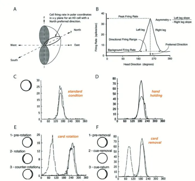
A head direction (HD) cell fires whenever the animal’s head is pointed in a particular direc-tion in the horizontal plane irrespective of the animal’s locadirec-tion (Fig. 4A; Taube et al., 1990a). Cell firing will continue whether the animal is moving or still and is largely inde-pendent of the animal’s ongoing behavior: discharge will persist without adaptation as long as the preferred head orientation is maintained. Within the range of the cell’s preferred direction, the firing rate follows a triangular or Gaussian tuning curve so that the firing rate is the high-est in the middle of the range, and falls off symmetrically around that center (Blair and Sharp 1995, Taube. 1998) (Fig. 4B). The direction at which the cell fires maximally (the « peak fir-ing rate ») is referred to as the cell’s preferred firfir-ing direction. When the animal’s head is not pointing in a cell’s preferred firing direction, basal firing rate is at or close to zero. The range of head directions in which the firing rate is above the cell’s baseline firing rate ranges from 60° to 150° (average 90°) around the preferred direction (Blair and Sharp, 1995; Taube, 1995). Different HD cells have different preferred directions and all directional headings are represented within a population of HD cells. The overall set of HD cells, therefore, acts as a compass that signals head orientation. Peak firing rates vary for different HD cells in the same or different brain areas ranging from 5 to 120 Hz (Taube, 2007).
HD cells receive and utilize information from both external landmark cues and internal cues. Visual landmarks dominantly control the preferred direction of the HD cell in order to stabi-lize, correct the signal and avoid drifts (Zugaro et al., 2003). Visual cue card rotations cause equal rotations of firing fields corresponding to deviations of the firing fields (Fig. 4E). Inter-estingly, card removal leaves firing fields and the peak firing rate intact but can drift over time (Fig. 4F). In addition to cue card shifts, changing the shape of the testing enclosure from a cylinder to a square or rectangle also caused changes in the preferred direction (Taube et al., 1990b). Goodridge et al., (1998) looked at the role of sensory modalities other than vision on
the firing of head direction cells. Whereas a simple auditory cue, such as a localized series of clicks or bursts of noise, was ineffective, a localized smell did exert a small but significant control over the preferred direction.
Figure 4: Basic features of presubicular head direction cells. . Three-dimensional model of D cell firing. The surface of the hemi-torus-shaped figure represents the maximum firing rate of the cell as a function of azimuth and height. Note that there are abrupt transitions from high firing rates to directions where the cell ceases responding. . Firing rate vs head direc-tion tuning curve for a hypothetical D cell. Five parameters are measured to study the firing properties of the cell: (1) preferred firing direction, (2) peak firing rate, (3) directional firing range, (4) background firing rate, (5) asymmetry score, from Taube. (1998). . Stability of head direction cell firing across two recording sessions, one (dashed line) recorded 15 days after the other (solid line). In standard condition, a prominent cue card is disposed as a polar-izing cue on one side of the open field wall. . Carrying the animal by hand and moving it around in the arena (dashed line) only decreased peak firing rate compared to standard condi-tion (solid line). . Cue card rotacondi-tion causes a corresponding shift in preferred direccondi-tion. The same head direction was recorded in standard condition (1, solid line), after a 180 clockwise
rotation of the cue card (2, dash-dot line) and after the equivalent counter rotation putting the card in its initial position (3, line with 2 short dashes). F. Drift of preferred direction follow-ing card removal. The same head direction was recorded in standard condition (1, solid line), after cue card removal (2, dash-dot line) and after cue card return to its initial position (3, line with 2 short dashes), adapted from Taube et al. (1990a,b).
While the HD signal appears to be generated from the self-movement information that arises from the vestibular system, proprioceptive and/or motor efference cues also play a major role in updating the signal during movement. Motor activity is likely to improve signal quality without affecting its generation: preventing an animal from moving reduces peak firing rate with no changes in preferred direction and directional firing (Fig. 4C-D) (Taube et al., 1990b).
2.2.3 Head Direction Circuit
With the discovery of head direction cells in the postsubiculum, a succession of following studies explored the existence and characteristics of HD cells in several brains areas that are part of the Papez circuit (1937), connected directly or indirectly with the postsubiculum, forming a head direction macrocircuit.
Head directional activity has been described in numerous brain regions: the anterodorsal tha-lamic nucleus (ADN), the anteroventral thatha-lamic nucleus (AVN) together forming the anterior thalamic nuclei (ATN) (Taube, 1995), the nucleus reuniens of thalamus (Jankowski et al., 2014), the lateral mammillary nucleus (LMN) (Stackman and Taube, 1998), the medial ento-rhinal cortex (MEC) (Sargolini et al., 2006), the retrosplenial cortex (both granular and agran-ular regions) (Chen et al., 1994; Cho and Sharp, 2001), the lateral dorsal thalamus (LDN) (Mizumori and Williams, 1993) and the dorsal striatum (Wiener, 1993). Smaller numbers of head direction cells have been described in the dorsal tegmental nucleus (DTN) (Basset and Taube, 2001) and the hippocampal area CA1 (Leutgeb et al., 2000). These areas are hierarchi-cally interconnected, starting from vestibular system to DTN, LMN, ADN, PrS and finally MEC (Fig. 5A), and contain head direction cells that contrast in their specific tuning proper-ties. One striking parameter is the directional range that is narrower for presubiculum and retrosplenial cortex compared to ADN, LMN and DTN (Fig. 5B). Along the dorsoventral axis, no topographical organization of directional tuning was observed in the presubiculum, contrasting with a loss of sharply tuned cells from dorsal to ventral position in the MEC (Gio-como et al., 2014). Interestingly, head direction cells in the ADN and LMN were found to
exhibit anticipatory firing in that they predicted the animal s future head direction few (25) msec in advance (Blair and Sharp, 1995).
Figure 5: The head direction circuit. . Circuit diagram showing the principle connections between brain regions containing place cells (yellow), grid cells (red), border cells (blue), D cells (green), and angular velocity cells (orange). Arrows indicate excitatory pro ections, and lines that end with a bar represent inhibitory pro ections. ADN, anterodorsal thalamus A N, anteroventral thalamus DTN, dorsal tegmental nucleus PC, hippocampus LDN, laterodor-sal thalamus LMN, lateral mammillary nuclei MEC, medial entorhinal cortex M N, medial vestibular nuclei NP , nucleus prepositus hypoglossi PaS, parasubiculum PoS, postsubicu-lum RSP, retrosplenial cortex SGN, supragenual nucleus, from Clark and Taube. (2012). . Typical tuning curves of head direction cells in presubiculum (postsubiculum), retrosplenial cortex, ADN, left LMN and DTN. Solid lines represent tuning curves during clockwise, dashed lines during counterclockwise head turn, adapted from Wiener and Taube. (2005).
2.2.4 Subcortical source of head direction signal
Generation of the head direction signal takes place in subcortical regions from information provided by the vestibular system composed of the semicircular canals and the otolith organs that detect angular and linear acceleration respectively. Semicircular canals are necessary for head direction cell activity in ATN (Muir et al., 2009; Clark and Taube, 2012; Valerio and Taube, 2016) whereas the otolith organs provide robustness and stability for HD signal (Yoder and Taube, 2009). All along the integrative pathway, from the vestibular organ to the DTN, vestibular information is carried by angular head velocity cells that fire as a function of head rotation speed and direction (Bassett and Taube, 2001; Sharp et al., 2001b). These cells are tonically active when the head is still, but increase their firing rate during one kind of turn (clockwise versus counterclockwise) and decrease their firing rate during the opposite kind of turn. The angular velocity signal is received by the DTN from the vestibular nuclei via the supragenual nucleus and the nucleus prepositus. The generation of the HD signal is thought to occur within the reciprocal connections between the dorsal tegmental nuclei (DTN) and lat-eral mammillary nuclei (LMN) transforming angular head velocity signals into HD signals (Bassett et al., 2007; Clark and Taube, 2012).
Several theoretical models have been developed to simulate the spatial firing properties of HD cells and show how such cells might be combined to form a compass-like directional sys-tem (Skaggs et al., 1995; Redish et al., 1996; McNaughton et al., 2006). Particularly, attractor network-based models are a class of neural networks that are considered to be a good approx-imation to those in the HD system (Fig. 6). In these models, HD cells are arrayed in a circle with the location of each cell on the circle representing the preferred direction of that cell. An estimate of current HD is accomplished by a vector summation of the angular displacement and the animal’s previous HD. Nearby HD cells with similar preferred directions are connect-ed with strong excitatory synapses and fire together within a temporally correlatconnect-ed group of HD neurons (activity packet). The activity packet is believed to move on a virtual ring as the animal turns its head. Cells that are far apart on the ring with greatly different preferred direc-tions are suppressed by lateral inhibitory synapses. This symmetric connectivity creates a sin-gle « bump » of excitation (a “hill” of activity) on the ring whose shape is determined by the connections weight, and position guided by the external inputs (Knierem et Zhang, 2012). Recent studies of Peyrache et al., (2015) revealed that the postulated attractor network was preserved in both ADN and presubiculum during sleep and wake states.
Figure 6: A continuous attractor network model of head direction signal generation. . In this model, D cells are arranged in a circle or ring with each D cell (colored circles) positioned according to their corresponding preferred tuning direction. Each D cell sends strong excita-tory axons to nearby neurons, and weaker excitaexcita-tory inputs to more distant neurons. Inhibito-ry pro ections (not shown) within the network limit net activity resulting in a focused point, or a hill , of high activity (warm colors). Movement of the activity hill corresponding to an animal s head movements is achieved by two additional neural signals: one that is sensitive to changes in an animal s angular head velocity (A ) (gray circle), and another that con unc-tively encodes current D A (black circle). . Following a head turn, con unctive D A cells drive the activity hill in the appropriate D. A right head turn would engage D A neurons that are specifically sensitive to clockwise head turns (solid arrows). These neurons would in turn activate D cells to the right of the hill and drive activity to the ani-mal s current D, adapted from Clark and Taube. (2012).
Thus, the coordinated activity in the HD system is not only shaped by incoming sensory signals, but is also strongly influenced by internal self-organized mechanisms.
Combined lesion and recording studies have critically elucidated how the head direction sig-nal is transmitted from one region to the next. Bilateral lesions of the LMN abolish the HD signal in the ATN (Bassett et al., 2007). HD cell activity is conveyed in both directions be-tween the ATN and the PrS. But only the ATN is a crucial and necessary upstream element in the head direction circuit since its lesion abolished head direction cells activity in presubicu-lum (Goodridge and Taube, 1997) and other areas including parasubicupresubicu-lum and superficial layers of the medial entorhinal cortex (Clark and Taube, 2012; Winter et al., 2015). In con-trast, lesions of the PrS had very subtle effect on direction-specificity of ADN neurons but rather influences ADN HD neurons by increasing the extent of anticipation in ADN and dis-rupt visual landmarks based cue control in the ADN (Goodridge and Taube, 1997).
2.2.5 Functional importance of presubiculum in spatial coding
Spatial navigation engages a wide brain circuit and coding for the animal’s head direction is considered a pivotal component of the brain’s navigation system. Presubiculum appears to be a crucial node where directional heading information and visual landmarks are transferred to the entorhinal-hippocampal network that participates in spatial cognition (Yoder and Taube, 2011).
The dorsal part of the presubiculum is a crucial entry point for integrating visual landmark information in the HD circuit. PoS receives direct input from primary and secondary visual cortices (Vogt and Miller, 1983) and projections from retrosplenial cortex, relaying infor-mation from visual cortex (Vogt and Miller, 1983; Van Groen and Wyss, 1990a; Jones and Witter, 2007), from LDN (Bett et al., 2013; Clark and Harvey, 2016) and associative visual cortical areas, such as posterior parietal and postrhinal cortices (Yoder et al., 2011). The im-portance of the PrS in processing landmark information into subcortical generators of the HD signal is demonstrated in PrS lesioned rats that severely altered landmark control of HD cell’s in both ADN (Yoder and Taube, 2011; Goodridge and Taube, 1997) and LMN (Yoder and Taube, 2011; Yoder et al., 2015). Thus, the PoS is well suited to exert feedback control on its efferent destinations ADN and LMN but also the retrosplenial cortex (Wyss and Van Groen, 1992), LDN (Van Groen and Wyss, 1990b, c) or medial entorhinal cortex (Honda et al., 2008)
by refining the local HD signal with visual information (Goodridge and Taube, 1997). Recent work has shown that the PrS is an important component for providing direct landmark control to the HD signal in the LMN and also contributes to its path integration maintenance when familiar visual landmarks are unavailable (Yoder et al., 2015).
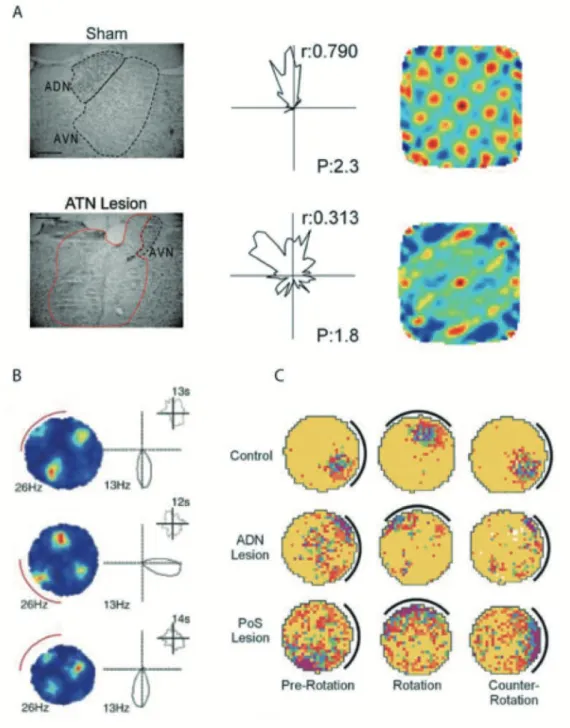
Compared to grid and place signals, head direction signal maturation occurs earlier during post-natal development (Bjerknes et al., 2015; Langston et al., 2010; Wills et al., 2010). Thus, head direction cells do not require place and grid cell information of the hippocampal-entorhinal circuit. The early maturation of the head-direction signal is thought to guide the development of spatial representations in entorhinal cortex and hippocampus later on. Follow-ing hippocampal silencFollow-ing of place cell activity and dissolution of the grid map, former grid cells developed a preference to spike when the animal pointed its head in a certain direction (Bonnevie et al., 2013) indicating that the grid cell signal somehow carries a head direction signal. Inactivation or lesion of ATN significantly reduced the spatial periodicity of grid cells with a decrease in direction-specific firing properties and number of HD cells in the parahip-pocampal region (Fig. 7A; Winter et al., 2015). Taken together, these findings suggest that there are multiple excitatory inputs to grid cells, from both the hippocampus and regions that carry head-direction signals. The plausible source of a directional signal for grid cells is the presubicular head direction cells. Indeed, many studies have hypothesized that the PrS (and PaS) by providing strong bilateral inputs to MEC may be required for the generation of grid and place cells (Van Groen and Wyss, 1990c; Caballero-Bleda and Witter, 1994; Honda and Ishizuka, 2004). Particularly, ipsi- and contralateral projections from presubiculum reach lay-er 2/3 of entorhinal cortex (Honda et al., 2008), whlay-ere grid cells are found.
Visual landmarks influence head direction cells, grid cells and place cells because rotation of a visual landmark produces an equivalent rotation of grid, place and preferred direction fields (Fig. 7B; Knierim et al., 1995; Sargolini et al., 2006). Particularly, the presubiculum was shown to be important for the landmark control of place cells in the hippocampus: place fields from PrS-lesioned were not controlled by the cue and shifted randomly between sessions (Fig.
7C). Visual landmarks no longer influenced place fields when PrS, but not ADN, was
le-sioned (Calton et al., 2003). Behaviorally, PoS lesions impaired performance on spatial memory tasks, such as the water and radial arm mazes, where the use of landmark cues is necessary for task completion (Taube et al., 1992).
Figure 7: Presubiculum as a relay for generation and landmark control of D circuit. . ATN region from sham (top, left) and ATN large lesion (bottom left) animals. Only the right hemi-sphere is illustrated however, this example is representative of bilateral damage in animals with 85 damage. Scale bars, 0.5 mm. Middle, Examples of D cells from sham (top) and ATN large lesion (bottom) animals. D cell from ATN lesion animals has less robust polar plots, with lower r and P. Right, two grid cells from a sham animal (top) and from an ATN large-lesion animal (bottom), adapted from Winter et al. (2015). . Rate map for a grid cell (left) and polar plot for a head-direction cell (right) recorded simultaneously in layer of MEC after rotation of a polarizing cue card on the wall of the circular environment (red arc). Top and bottom, cue card in original position. Middle panel, cue card rotated 90 . Insets show distribution of time across head directions, from Sargolini et al. (2006). . Place/rate plots showing the response of representative place cells from each condition during the three ses-sions of the cue-rotation experiment, from Calton et al. (2003).
Thus, PoS is likely to play a role in selecting which cues to use as landmarks that would require the use of spatial memory.
2.3 Neuronal components of the presubiculum
2.3.1 Intrinsic excitability of presubicular neurons
Despite anatomical characterization that highlights functional uniqueness of this region, basic electrophysiological properties of cells in the PrS, relative to neighboring MEC and hippo-campus, have been addressed in only a handful of studies (Fricker et al., 2009; Funahashi and Stewart, 1997a; Menendez de la Prida et al., 2003; Simonnet et al., 2013, Abbassi. 2014, 2015; Tukker et al., 2015; Preston-Ferrer et al., 2016).
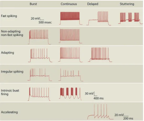
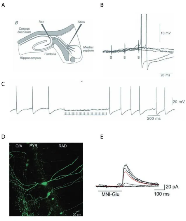
Funahashi and Stewart, (1997a) were the first to examine the presubicular neurons physiology and morphology. They found that pyramidal neurons in layer 3 and 5, as well as stellate cells in layer 2 and 5 were regular spiking neurons (Funahashi and Stewart, 1997a). Menendez de la Prida et al., (2003) found a large variability of cellular types classified according to their response to current injection in the subicular complex such as bursting (involving both Ca2+ and persistent Na+ components), regular-spiking and fast-spiking cells. In our laboratory, Jean Simonnet provided a full description of principal neurons in all 6 layers of presubiculum using unsupervised cluster analysis based on the somatic location, morphology and firing pattern (Fig. 8A; Simonnet et al., 2013). Superficial cells that fired regularly (layers 2 and 3, cluster 1) were separated from deep cells (layers 5 and 6; cluster 3), burst firing pyramidal neurons of layer 4 emerged as a distinct third group. Burst firing layer 4 neurons had depolarized resting potential, hyperpolarized firing thresholds, expressed the Ih current strongly consistent with previous work (Yoshida and Hasselmo, 2009), and their apical dendrites extended to layer 1. Deep layer neurons fired regularly and had little frequency adaptation consistent with a role in maintained signaling of head position. Soon afterwards, another group published a similar study focused specifically on superficial layers (Abbasi and Kumar, 2013). The description of the electrophysiological properties of superficial layers covered much of the existing diversity and showed that superficial layers 2 and 3 contain a neuronal population more diverse than previously reported, even though it was not systematically correlated with morphology. The same group showed that in the context of temporal lobe epilepsy, only a subset of superficial layer 2 and 3 neurons undergoes alterations in their firing profiles and synaptic drive (Abbasi and Kumar, 2014).
Figure 8: Physiological properties and connectivity of presubicular pyramidal neurons. . Cluster neurons with somata in presubicular layers 2, 3, 4 and 5/6. ( ) Reconstructions of bio-cytin-filled neurons in superficial and deep layers of the presubiculum. Axons in red, den-drites in blue, with layer limits and the pial surface in green. ( ) Current-clamp records of responses to 800 ms hyperpolarizing and depolarizing current steps. ( ) Regular or burst firing induced by 100-150 pA step current in ections. ( ) A waveforms. Adapted from Simonnet et al. (2013). . A TT -insensitive sodium current with slow
vating and inactivating kinetics in presubicular principal neurons, from Fricker et al. (2009). C. Persistent firing was induced by the current injection (2 s, 50 pA) in the presence of carbachol (10 µM) in a postsubicular neuron. Bottom trace shows frequency of persistent firing. From Yoshida and Hasselmo, (2009). D. Summary of interlaminar
and intralaminar connections of the presubiculum. Projections from layer 2 cells are bilateral and confined to layers 2 and 5, whereas those from layer 5 cells are ipsilateral and confined largely to layer V, with fewer projections to layer 2, from Honda and Ishizuka, (2004).
The presubiculum appears to carry the HD information via excitatory pyramidal cells, possi-bly also involving FS interneurons, without using a theta-rhythmic temporal code (Boccara et al., 2010; Tukker et al., 2015), at least not in layer 3 (Preston-Ferrer et al., 2016). The under-lying mechanisms for the non-adapting HD signal still remain unknown. Intrinsic cellular properties may support persistent firing, such as a tetrodotoxin (TTX)-insensitive sodium cur-rent with slow kinetics in superficial layer presubicular neurons (Fig. 8B; Fricker et al., 2009), or a calcium-sensitive nonselective cation current (Fig. 8C; Yoshida and Hasselmo, 2009). Persistent activity may also depend on network properties, recurrent synapses and facilitating synapse dynamics (Goldman-Rakic, 1995; Wang, 2001; Major and Tank, 2004).
2.3.2 Input- and output regions of the presubicular microcircuit
Information processing in the presubiculum must be determined by the physiological and ana-tomical features of its local connections as well as the integrative properties of these neurons in response to afferent inputs. Combining the neuronal description studies with the anatomical and functional connectivity with incoming and outgoing pathways, a clearer picture of the presubicular organization will emerge. Here, I will review the major input/output pathways of the presubiculum that I have focused on during my thesis (i.e ATN, RSC, LMN and MEC).
The ATN and the retrosplenial cortex are considered to provide major inputs to presubiculum (Yoder et al., 2011). The projections from retrosplenial cortex to the entire dorsoventral axis of both superficial and deep layers of PrS have been described (Van Groen et al., 1990a; Van Groen et al., 1992a; Van Groen and Wyss, 2003; Jones et al., 2007; Sugar et al., 2011; Sugar and Witter, 2016). Projections depend on the parts of the retrosplenial cortex since it is divid-ed into agranular (Van Groen et al., 1992a) and granular (a and b) regions (Van Groen et al., 1990a; Van Groen and Wyss, 2003). More specifically, direct inputs from retrosplenial cortex have been shown to project onto superficial layer 3 presubicular neurons (Kononenko and Witter, 2012).
In turn, deep layers of the presubiculum project to the retrosplenial cortex (Vogt and Miller, 1983; Wyss and Van Groen, 1992). The ATN projects to layers 1, 3 and 4 in the presubiculum (Van Groen and Wyss, 1990b, c; Van Groen and Wyss, 1995). In turn, the presubiculum-to-ATN projections originate from the deep layers of the ipsilateral presubiculum (Van Groen and Wyss, 1990b, c).
The presubiculum-to-LMN projection emanates only from the thin layer 4 (Fig. 9; Yoder and Taube, 2011) containing pyramidal cells with intrinsic bursting properties (Simonnet et al., 2013). Presubicular superficial layer neurons constitute the major output toward ipsilateral and contralateral medial entorhinal cortices (MEC), mainly targeting their superficial layers (Honda and Ishizuka, 2004; Canto et al., 2012; Tukker et al., 2015; Preston-Ferrer et al., 2016). However, some projections originate from deep layer neurons of the presubiculum and target deep layers and layer 1 of the ipsilateral MEC (Honda and Ishizuka, 2004). Projections from MEC back to PrS are sparse: Wyss (1981) described projections from MEC to layer I of the presubiculum.
Because most studies of inputs to presubiculum are anatomical, the identity of the main postsynaptic target types within presubiculum has remained uncertain. In particular, superfi-cial layer neurons (mostly superfisuperfi-cial layer 3 neurons) of the presubiculum are likely to relay the visual and vestibular information directly to the entorhinal cortex. A part of my thesis work has addressed the functional connectivity of thalamic-to-presubicular layer 3 MEC pro-jecting neurons and interneurons (Nassar et al., in prep). We also examined whether projec-tion-specific neurons possess distinct biophysical and anatomical properties (Huang et al., in prep.). These data are presented in the results section.
3-GABAergic neurons of a cortical network
The cortex is a complex, but relatively stereotypic organized network, composed of multiple cell types forming micro- to large-scale neuronal circuits (DeFelipe and Fariñas, 1992; Sil-berberg et al., 2002; Varela et al., 2001). Information flows through synapses in a finely orga-nized network composed of afferent fibers and local cell bodies of diverse neurons. The cor-tex is composed of two main neuronal groups, glutamatergic pyramidal neurons (PN) and GABAergic interneurons (IN), which are distributed across different cortical layers. The
lam-inar organization of the cortex, largely conserved across mammalian species, is based on cy-toarchitectonic criteria that define six layers, horizontally to the cortical surface (Douglas and Martin, 2004 Nieuwenhuys, 1994). PNs and INs from a specific layer can interact within their home layer and across layers. Inter-laminar connections are the anatomical framework of the cortical columns that form functional entities of interconnected neurons (Adesnik and Scanziani, 2010 ubel and Wiesel, 1962 Mountcastle, 1957).
Figure 9: Pro ection specific neurons in layer 4 and deep layers (5/6). . Cholera toxin-Alexa Fluor 488 (CTB-488) in ection sites in LMN. The left LMN is partially filled with CTB-488, whereas the right LMN is completely filled with CTB-488. Dashed lines indicate LMN, cor-responding to LMN (shaded areas in rectangle) in diagram above. 3, third ventricle 3m, third ventricle, mammillary recess. . CTB-594 in ection sites in the ADN. CTB-594 is pre-sent in the ADN and surrounding areas including the anteroventral (A N) and laterodorsal thalamic (LDN) nuclei. Dashed lines indicate ADN and surrounding nuclei, corresponding to the rectangle in the diagram above. . Tracers migrated retrogradely in non-overlapping
neu-ronal populations in the presubiculum. Somata of layer 4 neurons show green labeling (green) as injected tracer in LMN. Somata of layer 5-6 neurons show labeling following tracer injec-tion (red) into the right ADN. ADN, Anterodorsal thalamus; AVN, Anteroventral thalamus; LMN, Lateral mammillary nucleus; sm, stria medullaris; V3, third ventricle; V3m, third ven-tricle, mammillary recess. Adapted from Yoder and Taube, (2011).
To understand the functions of a microcircuit one also needs to understand its structural frame. What kind of neurons form a cortical circuit? How do these elements participate in information processing? How do individual neurons integrate one or multiple inputs to pro-duce their firing? During my PhD, I aimed to answer these questions, focusing on the inhibi-tory microcircuit of the presubciulum. I examined the diversity of the multiple inhibiinhibi-tory neu-ronal components, their anatomical and functional connectivity with local principal neurons and incoming inputs from the thalamus. In this chapter, I will review the characteristics and the specificity of the different INs that constitute hippocampal and cortical circuits.
3.1 GABAergic neuron types
GABAergic neurons represent about 15-30% of cortical neurons located in all cortical layers (DeFelipe, 2002). They were first classified by Ramon y Cajal as ‘short axon cells’ in Golgi studies of human visual cortex (Ramón y Cajal, 1899) because they are almost exclusively involved in local neuronal networks due to their restricted axonal and dendritic arborizations. They were therefore called interneurons (INs). Some features distinguish them from pyrami-dal shaped principal neurons (PN); indeed, most mature inhibitory interneurons have aspiny dendrites and receive both excitatory and inhibitory synapses onto their somata. Their axon terminals form symmetrical synapses, corresponding to Gray’s type 2, as opposed to glutama-tergic, asymmetrical synapses of PNs, corresponding to Gray’s type 1 (Gray, 1959). Cortical inhibitory INs are highly diverse and comprise many types according to their anatomy, elec-trophysiology and molecular diversity (Ascoli et al., 2008; DeFelipe et al., 2013; Gupta et al., 2000; Markram et al., 2004; Kubota, 2014; Kubota et al., 2016). Remarkably, individual INs selectively target distinct compartments of PNs (dendrites, soma or axon initial segment) or other IN types. Some INs are electrically coupled through Gap junctions (Beierlein et al., 2000; Bartos et al., 2002; Hestrin and Galarreta, 2005; Ma et al., 2011; Hu et al., 2011). Im-portantly, none of the anatomical, neurochemical or electrophysiological criteria alone can reliably classify cortical inhibitory INs. Therefore, many studies have applied multidimen-sional analyses to detect associations between these features. (Cauli et al., 1997; Markram et
al., 2004). More recently, the developmental origin and genetic makeup has been investigated (Kepecs and Fishell, 2014; Taniguchi, 2014; Tasic et al., 2016), and ultimately, we wish to further consider complexity across different brain structures and define interneuron types by their circuit specific function (Hangya et al., 2014).
3.1.1 Morphological properties and postsynaptic targets
Using Golgi based-impregnation methods, Cajal showed that cortical INs greatly vary in their somatic, dendritic and axonal morphologies. With his colleague Lorento de No, they demon-strated that dendritic arbors are a variable element and cannot define the interneuron types but rather predict the possible sources of afferent inputs. However, the pattern of axon arboriza-tion of an interneuron can provide strong evidence of its postsynaptic targets. In the hippo-campus and the neocortex, some INs preferentially innervate perisomatic regions, proximal dendrites or axon initial segments, whereas others target more distal dendrites (Fig. 10; Freund, 2003; Freund and Katona. 2007; Markram et al., 2004; Kubota, 2014; Kubota et al., 2016). Some INs have been described as specialized in targeting other cortical INs (Acsády et al., 1996; Gulyas et al., 1996, Freund and Buzsáki, 1996; Somogyi et al., 1998; Staiger et al., 1997). The specialization of IN connectivity is thought to contribute largely to their functional specificities (Gentet et al., 2012; Isaacson and Scanziani, 2011; Jiang et al., 2015).
Basket cells innervate the somata and proximal dendrites of PNs, representing the largest populations of interneurons (about 50%). In the hippocampus, their bitufted dendritic tree extends towards all layers and their axon is confined to the stratum pyramidale (Freund and Buzsáki, 1996). Basket cells can be further subdivided into several classes based on their so-ma size, frequency of axonal branching, axo-dendritic morphology and firing properties (i.e., large (LBC) or small basket cells (SBC) and nest basket cells (NBC)) (Karube et al., 2004; Krimer et al., 2005; Markram et al., 2004; Uematsu et al., 2008; Kubota, 2014; Kubota et al., 2016). Basket cells are mutually interconnected via chemical and electrical synapses (Cobb et al., 1995; Gibson et al., 1999; Fukuda, 2007; Somogyi et al., 1998, Bartos et al., 2002; Hestrin and Gallaretta, 2005; Baude et al., 2007). The axo-axonic cells (Somogyi, 1977) are also called chandelier cells because their axon terminals form multiple climbing-fiber-like contacts resembling candlesticks on a chandelier. Originally described in neocortex, chandelier cells have also been found in the CA3, CA1 and dentate gyrus regions of the hippocampus. They target specifically the axon initial segment of PNs (Karube et al., 2004; Krimer et al., 2005; Markram et al., 2004; Kubota, 2014; Kubota et al., 2016). Thus, they are positioned at a
cru-cial site to override both dendritic integration and somatic gain, updating the action poten-tial output.
Figure 10: Morphologies of neocortical interneurons. Exemplar 3D reconstructions of 55 morphological types. Morphologies in L2 and L3 are not separated. Axon in blue, dendrites in red. Full morphologies are not always shown. From Markram et al. (2015).
The dendrite-targeting cells can be subdivided into several cell types: bipolar, double bouquet, bitufted and neurogliaform cells. The bipolar, double bouquet and bitufted cells present bipo-lar and bitufted dendrites but are differentiated by their axonal orientation or morphology (Markram et al., 2004). The neurogliaform somata can be found in all cortical layers and in the stratum lacunosum moleculare (Muralidhar et al., 2014; Capogna, 2011). Neurogliaform cells present a spherical dendritic field and thin and dense intertwined axonal arbors, forming electrical synapses with INs from the same or different type (Markram et al., 2004).
The dendritic and tuft-targeting cells are divided in Martinotti (MC) and layer 1 cells. The MCs have often bitufted dendrites with ascending axons composed of two parts, one near the cell body and the other projecting at variable distance innervating PNs tuft-dendrites in layer 1 (Markram et al., 2004). Oriens lacunosum moleculare (O-LM) cells and SOM hilar perfo-rant path associated (HIPP) cells found in the hippocampus (stratum oriens and hilus) have axons ascending to stratum lacunosum moleculare or innervating dendritic tree of granule cells in the molecular layer respectively (Freund and Buzsáki, 1996; Houser, 2007). The layer 1 cells are confined to layer 1 and are divided in two groups. The first one corresponds to Ca-jal-Retzius multipolar cells (Druga, 2009) mostly present during development whose axons are confined to layer I making horizontal collaterals. The second class comprises the hetero-geneous group of small, multipolar interneurons with diverse axonal arbors (Muralidhar et al., 2014).
3.1.2 Molecular markers and gene expression
Interneurons contain GABA-synthesizing enzymes GAD65 and GAD67 (Martin and Rimwall, 1993), and different neurochemical markers allow to divide the interneuron popula-tions into subgroups. Several populapopula-tions of interneurons contain different Ca2+ binding pro-teins or peptides. The most widely used interneuron markers are calcium-binding propro-teins such as parvalbumin (PV), calretinin (CR) and calbindin (CB) and neuropeptides such as so-matostatin (SOM), vasoactive intestinal peptide (VIP), cholecystokinin (CCK), and neuropep-tide Y (NPY). Besides, molecules such as the ionotropic serotonin receptor 5HT3a, reelin, nitritic oxide synthase or choline acetyltransferase (ChAT) can be used. Morphologically- or physiologically-defined interneuron types can co-exist and overlap in a single neurochemical-ly identified subgroup (Fig. 11). Some interneurons co-express up to seven different molecu-lar markers and patterns of co-expression differ between regions and species.
Figure 11: Multiple dimensions of interneuron diversity. Interneuron cell types are usually defined using a combination of criteria based on morphology, connectivity pattern, synaptic properties, marker expression and intrinsic firing properties. The highlighted connections de-fine fast-spiking cortical basket cells, from Kepecs and Fishell. 2014.
P is expressed in all compartments of basket and axo-axonic cells in the rat cerebral cortex and in the hippocampus (Celio, 1986 Kosaka et al., 1987). P immunopositive neurons ac-count for 40-50 of GABAergic neurons in rodents. A property of P neurons is that they do not stain with antibodies against CR, SOM, IP, CCK, NPY or ChAT (Gonchar et al., 2008
ematsu et al., 2008) even though the overlap between P mRNA and others from CB and peptides is much more extensive meaning that P mRNA is not translated in many neurons (Cauli et al., 1997 Two types of basket cells are classically recognized: those expressing both P and CB ( artwich et al., 2009) and those expressing CCK (Freund et al., 1986 Ku-bota and Kawaguchi, 1997 Somogyi et al., 2004).