Faculdade de Ciências e Tecnologia da Universidade de Coimbra
Effect of Purines in the Developing Hippocampus -
Consequences for the Establishment of Circuits
Related to Learning and Memory
Carla Sofia Gomes da Silva
Coimbra-Portugal
Carla Sofia Gomes da Silva
Effect of Purines in the Developing Hippocampus - Consequences for
the Establishment of Circuits Related to Learning and Memory
Dissertação apresentada à Faculdade de Ciências e Tecnologia para prestação de provas de Doutoramento em Biologia, na especialidade de Fisiologia
Agradecimentos/Acknowledgements
Gostaria de agradecer em primeiro lugar ao meu orientador Rodrigo Cunha por aceitar supervisionar o trabalho experimental conducente à realização da minha tese de doutoramento e pela confiança dada ao permitir-me escolher um tema que aborda o desenvolvimento do sistema nervoso, o qual que não faz parte do seu domínio directo de investigação. Independentemente deste aspecto, sempre se mostrou disponível para discutir esta temática de uma forma construtiva, o que contribuiu grandemente para a evolução do trabalho. A sua capacidade de integração do conhecimento proveniente de diversas áreas e a facilidade com que é capaz de elaborar um esquema geral para a interpretação de resultados tornaram o meu doutoramento sob a sua supervisão uma experiência muito positiva e de grande valor para o meu crescimento científico. Agradeço ainda aos seniores do grupo, Paula Agostinho, Ângelo Tomé e Henrique Silva pela ajuda prestada em vários momentos.
Agradeço também à Doutora Catarina Oliveira, por me ter proporcionado realizar parte do trabalho apresentado nesta tese no Centro de Neurociências e Biologia Celular onde desempenha o cargo de presidente.
Aos meus colegas do grupo ―Purines at CNC‖ com quem tive o prazer de me cruzar e partilhar a paixão pela ciência e a vontade de saber e descobrir. Em especial à minha querida amiga Patrícia, a pessoa com quem mais interagi e com a qual passei noites fascinantes a fazer experiências, à Paula Canas que se encarregou de integrar os ―mais novos‖ no grupo, ao João Duarte, Atilla Köfalvi e Rui Sanches e Nélio Gonçalves pelo intercâmbio científico. Aos agora pertencentes à categoria de ―mais novos‖ Samira Ferreira, Elisabete Augusto, Pedro Garção e Marco Matos que sempre interagiram comigo de uma forma positiva e construtiva, mantendo um diálogo científico aceso. Um especial obrigado ao Nuno Machado que aceitou colaborar num dos projectos aqui apresentados ainda à Catarina Gomes com quem é um prazer contactatar tanto pela sua integridade pessoal e científica.
Um obrigado àqueles que não pertencendo ao ―Purines in CNC‖ passaram pelo meu percurso e contribuíram para o trabalho aqui apresentado e para a minha formação como aluna de doutoramento. Cito o Alexandre Rodrigues que passou um ano no Centro de Neurociências e Biologia Celular e me ensinou muito sobre astrócitos e transportadores de adenosina, à Inês Araújo pela ajuda com experiências biologia molecular, à Fabienne Agasse um agradecimento especial pela ajuda espontânea que prestou na elaboração de experiências de biologia celular e pela sua determinação.
I am grateful to Dr. Christophe Bernard and Dr. Patrick Chauvel for receiving me so openly in their laboratory Epilepsie et Cognition Unité 751 of INSERM in Marseille, France. The positive side of Dr. Christophe Bernard and the fact that he always believed in the work I developed under his supervision were of extreme importance for the final result. His efforts in making colaborations with
other laboratories in France, namely with the group of Dr. Daniel Choquet from Bordeaux with whom I studied the internalization of GABAA and AMPA receptor subunits with the help of Leandro Royer and with Dr. Sabine Levy and Dr. Christine Métin from Paris who are investigating the effect of the antagonism of adenosine receptors in the neuronal migration and the movement of GABAA and AMPA receptors in the plasma membrane by Quantum dot analysis. These collaborations were also important to increase the quality of the work. I am also grateful to Dr. Monique Esclapez for her interest, patience, honesty, humanity and sensibility. She was a real teacher and the knowledge I gained from working with her about science in general and about technical aspects of the technique of immunohistochemistry was valuable. J’ai eu aussi le plaisir de travailler avec ―Plume‖, la technicienne de Dr. Esclapez et ça a été une experience magnifique. Je remercie l’effort qu’elle a fait pour m’aider à finir le travail, en passant des heures supplementaires au laboratoire. Elle a été aussi très positive et elle a bien montré la valeur du rôle du technician dans un laboratoire de recherche scientifique. Je remercie aussi les autres techniciens at animaliers de l’Unité 751 INSERM à Marseille qui mon vraiment aidé, notament Antoine Ghestem et Jean-Marc, Nicholas et Mélanie. The interactions I had with Anton Ivanov, Yuri Zilberter and Mohit Adhikari were also notable. Anton helped me immensely with the electrophysiological recordings becoming my personal Jesus in Marseille and Yuri, with all his experience and knowledge, his sensibility and care helped me to improve the quality of the recordings I was perfoming. I am gratefull to Mohit Adhikari for the collaboration with the analysis of the in vivo recordings.
O trabalho devesenvolvido no âmbito desta tese de doutoramento foi financiado (SFRH/BD/31783 / 2006) pela Fundação para a Ciência e Tecnologia, Portugal.
Index
Abbreviations ………...……...Pag. 1 Resumo/Abstract ………...…………..Pag. 6 Introduction………... ………...………...Pag.10
1. The Purinergic System - Overview………....Pag.10
2. Neural Development ……….Pag. 15 2.1. Embryogenesis, Neural induction, Neurulation and Regionalization ………...Pag.15 2.2. Cell Death ……… Pag. 18 2.3. Hippocampal Formation and Migration of Neurons to this Structure …………..…Pag. 23 2.4. Synaptogenesis ……….…Pag. 26 2.4.1. The GABAergic Synapse ………....Pag. 29
2.4.1.1. The Fundamental Role of GABA and GABA Receptors in the Neural
Development ……….…Pag. 31 2.4.2. The Glutamatergic Synapse ……….…....Pag.36 2.4.3. The Network Activity in the Immature in vivo Hippocampal Slice and Neural
Circuit Refinement ………..…Pag. 38
3. The Purinergic System in the Context of the Nervous System Development – Why to Study It? ……….….. Pag. 39 3.1. Adenosine Receptors ………..……..Pag. 40
3.1.1. A1 Receptor ……….………Pag. 40
3.1.2. A2A Receptor ………..…………. Pag. 44
3.1.3. A2B Receptor ………..…………..Pag. 46
3.1.4. A3 Receptor ………..…………...Pag. 47
3.2. ATP Receptors ………..………Pag. 48 3.2.1. P2Y Receptors ……….…………Pag. 48 3.2.2. P2X Receptors ……….…………Pag. 52 3.3. Enzymes from the Purinergic System ………..………Pag. 53 3.3.1. Adenosine kinase (AK) ………..……….Pag. 53 3.3.2. Ecto-nucleoside triphosphate diphosphohydrolases (e-NTPDases) …...….Pag. 53 3.3.3. 5’-Nucleotidase ………....Pag. 54
CHAPTER 1 – Evaluation of the Presence of Different Components of the Purinergic System in the Immature Hippocampus ……….Pag. 56 CHAPTER 1 –Material and Methods ………Pag. 57 CHAPTER 1 – Results ………...…Pag. 65
1.1. Adenosine Receptors ………..Pag. 65 1.2. ATP Receptors ………Pag. 72 1.2.1. P2X Receptor Subunits ………Pag. 71 1.2.2. P2Y Receptors ……….Pag. 75 1.3. Ectonucleotidases ………...………Pag. 78 1.4. Nucleoside Transporters ……….Pag. 83 1.5. Other Synaptic Structures ……….….Pag. 83 CHAPTER 1 – Conclusions ………..….Pag. 89
CHAPTER 2 – Modulation by Adenosine A2A Receptor of Apoptotic Cell Death of ―Young‖ Cultured Hippocampal Neurons ………Pag. 91 CHAPTER 2 – Material and Methods ………...…Pag. 93 CHAPTER 2 – Results ………...…Pag. 97 CHAPTER 2 – Conclusions ……….……Pag. 102
CHAPTER 3 – Modulation by Adenosine A1 and A2A Receptors of the Spontaneous Activity in the
Hippocampal Slice ……….…..Pag. 103 CHAPTER 3 – Material and Methods ……….……Pag. 105 CHAPTER 3 – Results ……….………Pag. 111 3.1. Modulation Afforded by A1 and A2A Receptor Antagonists in Hippocampal Slices ….…..Pag. 111
3.2. Modulation of AMPA and GABAA Receptor-Mediated Synaptic Events by Antagonists of A1
and A2A Receptor Subtypes ………...…Pag. 118 3.3. GABAA and AMPA Receptor Internalization by Exposure to A1 and A2A Receptor
Antagonists ………..Pag. 135 3.4. Effect of A1 and A2A Receptor Antagonism on Synaptic Activity in a More Integrated
Model – The Cortico-Hippocampal Preparation……….………...……Pag. 153 CHAPTER 3 – Conclusions ………...……..Pag. 158
CHAPTER 4 – Short- and Long-Term Consequences of A1 and A2A Receptor Blockade During Brain
Development ………...Pag. 160 CHAPTER 4 – Material and Methods ………....Pag. 163 CHAPTER 4 – Results ………Pag. 166 CHAPTER 4 – Conclusions ………....Pag. 214
General Conclusions and Future Perspectives ……….Pag. 216
1
Abbreviations
AC: Adenylyl cyclase
aCSF:Artificial cerebrospinal fluid ADA: Adenosine deaminase ADK: Adenosine kinase ADP: Adenosine 5’-diphosphate AMP: Adenosine 5’-monophosphate
AMPA:(2-amino-3-(5-methyl-3-oxo-1,2- oxazol-4-yl)propanoic acid)
ANOVA: Analysis of variance AP: Alkaline phosphatase AP-2: Adaptor Protein-2
APAF1: Apoptotic protease activating factor 1
ARNO: ARF (ADP-ribosylation factor) nucleotide-binding site opener ATP: Adenosine 5’-triphosphate
Bak: Bcl2-antagonist/killer 1 Bax: Bcl2-associated X protein Bcl-2: B-cell lymphoma 2
Bcl-xL: B-cell lymphoma-extra large BDNF: Brain-derived neurotrophic factor bFGF: basic fibroblast growth factor BH3: Bcl-3 homology domain 3
Bid: BH3 interacting domain death agonist
BIG2: Brefeldin A-inhibited guanine nucleotide-exchange protein 2 Ca2+: Calcium
CamKII: Ca2+/calmodulin-dependent protein kinase
cAMP: Cyclic adenosine monophosphate CGE: Caudal ganglionic eminence
CHAPS: 3-[(3-Cholamidopropyl)dimethylammonio]propanesulfonic acid CHP: Cortico-hippocampal preparation
2
Cm: Membrane capacitance
CMF-HBSS: Ca2+- Mg2+-freeHank’s solution
CNS: Central nervous system
cGMP: Cyclic guanosine monophosphate CP: Cortical plate
CPA: N6-cyclopentyladenosine
CRE: cAMP response element
CREB: cAMP response element-binding Cx-43: Connexin 43
DAB: 3,3'-diaminobenzidine
DAG: Diacylglicerol
D-APV: D-2-amino-5-phosphonovaleric acid DCC: Deleted in colorectal cancer
DG: Dentate gyrus
DIABLO: Direct IAP binding protein with low pI DISC: Death-inducing signaling complexes DIV: Days in vitro
DMSO: Dimethylsulfoxide DNA: Deoxyribonucleic acid
EDTA: Ethylenediamide tetraacetic acid
EGTA: Ethylene-bis(oxyethylenenitrilo)tetraacetic acid EGFR: Epidermal growth factor receptor
E-NPP: Ecto-nucleotide pyrophosphatase/phosphodiesterases ENT: Equilibrative nucleoside transporter
e-5NT: Ecto-5’-nucleotidase
E-NTPDase: Ecto-nucleotide triphosphate diphosphohydrolase EPSCs: Excitatory postsynaptic currents
ER: Endoplasmic reticulum
3
ERNI: Early response to neural induction FADD: Fas-associated protein with death domain FGF: Fibroblast growth factor
GABA: Gamma-aminobutyric acid
GABARAP: GABA receptor-associated protein GAD: Glutamate decarboxylase
GAT-1: GABA transporter-1 GD: Gestational day
GDNF: Glial-derived neurotrophic factor GFAP: Glial fibrillary acidic protein GDP(s): Giant depolarizing potential(s)
GRIP1/ABP: Glutamate Receptor Interacting Protein 1/AMPA binding protein GRK-2: G protein-coupled receptor kinase 2
GSK3β: Glycogen synthase kinase 3
3
H-CHA: Tritiated cyclohexyldenosine
HEPES: 4-(2-hydroxyethyl)-1-piperazineethanesulfonic acid HtrpA2: High temperature requirement protein A2
IAP: Inhibitor of apoptosis proteins ILE(s): Interictal-like event(s) IP3: Inositol triphosphate
IPSC(s): Inhibitory postsynaptic current(s) IZ: Intermediate zone
JAK: Janus kinase K+: Potassium
KCC2: K+Cl- cotransporter 2
KPBS: potassium-phosphate solution LGE: Lateral ganglionic eminence LM: Lacunosum moleculare LRD(s): Late recurrent discharge(s)
4
LTP: Long-term potentiation
MAP-2: Microtubule-associated protein 2 MAP kinase: Mitogen-activated protein kinase MAPKAP: MAPK activated protein kinase MEM: Minimal essential medium
mEPSC(s): Miniature excitatory postsynaptic current(s) Mg2+: Magnesium
MGE: Medial ganglionic eminence
mIPSC(s): Miniature postsynaptic current(s) mRNA: messenger ribonucleic acid
MTT: 3-(4,5-dimethylthiazol-2-yl)-2,5-diphenyltetrazolium bromide MZ: Marginal zone
Na+: Sodium
NBQX: 2,3-dihydroxy-6-nitro-7-sulfamoylbenzo[f]quinoxaline NECA: 5’-N-ethylcarboxamido adenosine
NF-κB: Nuclear factor –κB NGF: Nerve growth factor NMDA: N-methyl-D-aspartate NMP: Neuronall plating medium NGS: Normal goat serum NHS: Normal horse serum
NSF: N-ethylmaleimide-sensitive factor NRSF: Neuron-restrictive silencer factor O-LM: Oriens-lacunosum moleculare PAF: Paraformaldehyde
PB: Phosphate buffer
PBS: Phosphate buffered saline PD: Postnatal day
5
PI: Propidium iodide
PICK1: Protein interacting with PRKCA PI3K: Phosphatidylinositol-3’-kinase Pir Cx: Piriform cortex
PKA: Protein kinase A PKB: Protein kinase B PKD: Protein kinase D PLC: Phospholipase C
PMSF: Phenylmethanesulfonylfluoride POA: Preoptic Area
PSD-93: Postsynaptic density 93 PSD-95: Postsynaptic density 95 RA: Retinoic acid
REST: RE1-silencing transcription factor Rm: Membrane resistance
RRP: Readily releasable pool
SAP97: Synapse-associated protein 97 SAP102: Synapse-associated protein 102 SDS: Sodium dodecyl sulfate
SEM: Standard error of the mean SEP: Super ecliptic phluorin
SMAC: Second mitochondria-derived activator of caspases SNAP-25: Synaptosomal-associated protein-25
SPL: Subplate
STAT: Signal transducer and activator of transcription STS: Staurosporine
SVZ: Subventricular zone TBS: Tris buffered saline TE: Tris-EDTA
6
TEMED: Tetramethylethylenediamine TGN: Trans golgi network
TNF: Tumor Necrosis Factor
TRAF-2: TNF receptor-associated factor 2 TrkA: Tyrosine receptor kinase A
TrkB: Tyrosine receptor kinase B UDP: Uridine diphosphate
VGLUT1: Vesicular glutamate transporte 1 VZ: Ventricular zone
7
Resumo/Abstract
O sistema nervoso desenvolve-se seguindo uma série precisa de etapas que fazem parte de um programa de desenvolvimento. O sistema purinérgico - que compreende os neuromoduladores/neurotransmissores denominados purinas (dos quais se destacam a adenosina e o ATP), respectivos receptores, enzimas e transportadores que promovem a sua produção ou remoção – está presente no sistema nervoso central desde fases muito iniciais do desenvolvimento. A versatilidade deste sistema manifesta-se pela sua capacidade em modificar a expressão e localização dos seus receptores, enzimas e transportadores de acordo com o nível de maturação do animal. O aperfeiçoamento de métodos de isolamento de fracções membranares permitiu observar que o receptor A2A para a adenosina, cuja expressão é muito reduzida no hipocampo adulto onde é
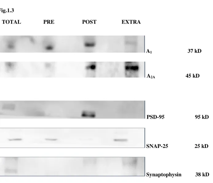
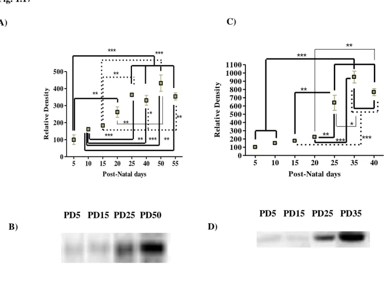
conhecido controlar a libertação pré-sináptica de neurotransmissores, tem uma expressão abundante em domínios pós- e extrasinápticos no hipocampo de ratinhos com 5 a 7 dias pós-natais. Em fases mais tardias do desenvolvimento, parece haver uma diminuição progressiva da sua expressão. A activação deste receptor no hipocampo em desenvolvimento parece depender já nesta fase da libertação pré-sináptica de ATP como sugere a presença do enzima ecto-5’-nucleotidase envolvida na produção de adenosina a partir do ATP em regiões igualmente pós-sinápticas.
Durante a primeira semana pós-natal, a adenosina endógena parece ser necessária para a estabilidade de receptores do tipo AMPA/kainato e GABAA uma vez que o bloqueio de receptores A1
e A2A, os dois principais subtipos de receptores de adenosina presentes no hipocampo, conduz ao
“enfraquecimento” da transmissão sináptica GABAérgica e glutamatérgica. Técnicas de biologia celular e molecular confirmaram a endocitose dependente de clatrina dos receptores ionotrópicos para o GABA e glutamato após bloqueio de receptores A1 e A2A. Este efeito modulador sobre a
actividade sináptica parece ser reversível em sinapses glutamatérgicas mas de difícil recuperação em sinapses GABAérgicas. Embora se desconheçam os mecanismos subjacentes, estas observações sugerem um papel relevante dos receptores de adenosina durante a sinaptogénese.
A disrupção do sistema de modulação mediado pelos receptores de adenosina revelou-se prejudicial ao desenvolvimento de circuitos de aprendizagem e memória no hipocampo, uma vez que o bloqueio crónico de receptores de adenosina pela cafeína (que bloqueia não especificamente receptores A1 e A2A a baixas concentrações) induziu uma série de modificações morfológicas e
8
interneurónios. Algumas destas modificações são stresse celular, síntese anormal de GAD 67 e aglomeração de proteínas do sistema GABAérgico bem como uma desregulação do balanço entre a transmissão sináptica GABAérgica e glutamatérgica. Os animais adultos expostos a antagonistas de receptores de adenosina durante o desenvolvimento revelaram défices cognitivos quando sujeitos a testes comportamentais que avaliam a memória espacial e não espacial, o que mostra que houve uma violação do programa de desenvolvimento desencadeada pelos tratamentos. Se no animal imaturo as principais modificações após exposição crónica a cafeína foram observadas no sistema GABAérgico, no adulto o sistema glutamatérgico parece ser morfológica e fisologicamente mais afectado.
O GABA e o glutamato foram descritos como moléculas importantes agindo como factores tróficos durante o desenvolvimento. A adenosina surge então, como um poderoso modulador que parece alterar alterar a sinalização mediada por receptors ionotrópicos para o GABA e glutamato, podendo interferir em processos como diferenciação, migração e morte celular, sinaptogénese e neurotransmissão. Uma violação das leis que governam estes processos poderá estar na base das modificações observadas a longo-termo e ao estabelecimento de circuitos de aprendizagem e memória no hipocampo que funciona de forma aberrante.
The nervous system develops following a series of steps respecting a developmental programme. The purinergic system - comprising the neuromodulators/neurotransmitters called purines (the most famous being ATP and adenosine), their receptors, enzymes and transporters that promote their production or removal – is present in the central nervous system since initial stages of development.
The versatility of this system relies on its ability to modify the expression and localization of their receptors enzymes and transporters according to the stage of the development. Improvement of methods for isolation of membrane fractions allowed the observation that A2A receptors, whose
expression is very low in the adult hippocampus where they control the presynaptic release of neurotransmitters, has an abundant expression in post and extrasynaptic fractions of hippocampal synaptic membranes from 5-7 days-old mice.In later stages of development, A2A receptors suffer a
progressive down-regulation . The activation of this receptor subtype in the developing hippocampus seems to depend on the presynaptic release of ATP, suggested by the presence of the enzyme ecto-5 '-nucleotidase, involved in the production of adenosine from ATP, in postsynaptic membranes.
9
During the first postnatal week, endogenous adenosine seems to be necessary for the stability of AMPA/kainate and GABAA receptors, since the blockade of A1 and A2A receptors, the two main
subtypes of adenosine receptors present in the hippocampus induced the weakening of the GABAergic and glutamatergic synaptic transmission. Techniques of cellular and molecular biology confirmed the clathrin-dependent endocytosis of ionotropic GABA and glutamate receptors after A1
and A2A receptor blockade.This effect on synaptic activity seems to be reversible in glutamatergic
synapses and long-lasting in GABAergic synapses. Although the intracellular mechanisms linking adenosine receptor blockade to the internalization of GABAA and AMPA/kainate receptors are not
understood, these observations suggest a role of adenosine during synaptogenesis.
The disruption of the modulation afforded by adenosine receptors seems to have detrimental effects in the establishment of hippocampal circuits related to learning and memory, since the chronic blockade of adenosine receptors by caffeine (which is a non-selective antagonist of A1 and
A2A receptors at low concentrations) changed several physiological and morphological parameters
in hippocampus of 6 days-old mice, particularly evident in interneurons. Some of these modifications are cellular stress, abnormal synthesis of GAD 67 and agglomeration of proteins belonging to the GABAergic system, as well as an imbalance between the GABAergic and glutamatergic neurotransmission. Adult animals exposed to adenosine receptor antagonists during development showed cognitive deficits when subjected to behavioral tests that assess spatial and non spatial memory, suggesting that there was a violation of the normal developmental program. If in immature animals the main changes observed after chronic exposure to caffeine were observed in the GABAergic system, in the adult, the glutamatergic system seems to be the most affected.
GABA and glutamate were identified as capable of exerting trophic actions during brain development.Adenosine, having a powerful control over signaling cascades activated/inactivated by ionotropic receptors for GABA and glutamate can interfere with fundamental stages of neural development such as the differentiation of progenitor cells, migration and cell death, synaptogenesis and neurotransmission, which can be the cause or contribute to the genesis of the long-term modifcations observed in the adult hippocampus.
10
INTRODUCTION
1. The Purinergic System - Overview
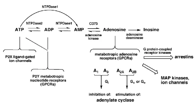
The purinergic system corresponds to the assembly of several types of ligands, generally called purines and responsive receptors as well as proteins (enzymes and transporters) responsible for production and/or reuptake of purines that mediate intra/intercellular physiological events (see Fig.1). Adenosine triphosphate (ATP) is one of the best known molecules belonging to this system (Burnstock, 1972). ATP binds to and activates two subfamilies of receptors, P2X and P2Y. P2X receptors comprise ligand-gated channels, permeable to sodium (Na+), potassium (K+) and calcium (Ca2+). Until now, seven receptor subtypes have been characterized - P2X1 through P2X7 (Ralevic and Burnstock, 1998; North, 2002). P2X receptors are composed by transmembranar subunits containing an extracellular site for binding to the ligand and intracellular domains involved in the modulation of the channel. P2Y receptors are G-protein-coupled receptors. Depending on their type of coupling, they can be subdivided into two groups, Gq preferring receptors - P2Y1, P2Y2, P2Y4, P2Y6, P2Y11 - and Gi preferring receptors -P2Y11, P2Y12, P2Y13, P2Y14 (Abbracchio et al., 2006). P2 receptors can be activated by other purines such as adenosine diphosphate (ADP), uridine triphosphate (UTP), uridine diphosphate (UDP), UDP glucose and other nucleotide sugars (Neary and Zimmerman, 2009). Some P2 receptors can have greater affinity for other purines than for ATP itself (Neary and Zimmerman, 2009).
ATP can reach the extracellular space using ATP release channels – hemichannels, anion channels including P2X7 receptor, ATP-bindig cassette transporters - (Sabirov and Okada, 2005), facilitated diffusion through nucleotide transporters such as ADP/ATP exchange carrier and vesicular exocytosis (Pankratov et al., 2006). Recently, the protein responsible for the accumulation of ATP into synaptic vesicles released by exocytosis, the vesicular nucleotide transporter (VNUT), was identified and characterized in human and mouse tissues (Sawada et al., 2008). Once outside the cell, ATP can be metabolized into adenosine which is another signaling molecule belonging to the purinergic system. Several enzymes called generically ecto-nucleotidases are involved in this process – ecto-nucleoside triphosphate diphosphohydrolases (E-NTPDases) (Robson et al., 2006), ecto-nucleotide pyrophosphatase/phosphodiesterases (E-NPPs) (Goding et al., 2003; Stefan et al., 2006), ecto-5’-nucleotidase (Hunsucker et al., 2005; Colgan et al., 2006) and alkaline phosphatases (APs) (Millan, 2006).
11
Adenosine can bind to and activate four metabotropic subtypes of receptors, A1, A2A, A2B and A3
-A1 and A3 receptors inhibiting via Gi/0 and Gi/Gq respectively and A2A receptors activating adenylate cyclase via Gs/Golf –A2A receptors- or Gs/Gq –A2B receptors- (Jacobson and Gao, 2006; Ryzhov et al., 2006). All adenosine receptors share a similar structure, a polypeptide chain forming seven transmembrane helices, with the N-terminus placed outside the cell and the C-terminus inside the cell (Constanzi et al., 2007). Adenosine receptors display different affinities for the ligand: A2B receptor subtype having the lowest affinity for adenosine and A1 and A2A receptor subtypes having higher affinity for the ligand. A3 receptor has intermediate affinity for adenosine comparing with the other adenosine receptor subtypes (Jacobson et al., 1996; Dunwiddie and Masino, 2001), although it is a high affinity subtype of adenosine receptor in human tissue (Ribeiro and Sebastião, 2010). They desensitize also after agonist binding and it can occur rapidly in the case of A3 receptor subtype and more slowly in the case of the A2A receptor subtype, being associated to receptor down-regulation, internalization and degradation (Klaase et al., 2008).
Adenosine actions are terminated when extracellular adenosine is transported into the cell via nucleoside transporters (ENTs). Since adenosine is neither stored nor released as a classical neurotransmitter since it does not use synaptic vesicles for storage and release, the direct release of cytosolic adenosine can occur via the same transporters. It constitutes an alternative way of adenosine release independently of the exocytosis of ATP. This adenosine that can be directly released by transporters, is formed by the action of an AMP selective 5’-nucleotidase and the rate of production is dependent on the amount of available AMP (Schubert et al., 1979; Zimmermann et al., 1998) or by the hydrolysis of adenosyl-homocysteine (Broch and Ueland, 1980) by the action of S-adenosyl-homocysteine hydrolase being L-homocysteine the limiting reagent of the reaction (Reddington and Pusch, 1983). An alternative source of adenosine is the release of cyclic AMP (cAMP) (Rosenberg and Li, 1995).
The direction of the transport of adenosine in or out of the cells is dependent upon the concentration gradient across biological membranes (Gu et al., 1995). Two groups of transporter proteins were identified, equilibrative – ENT1-4 - that use facilitated diffusion driven from the concentration gradient and concentrative nucleoside transporters – CNT1-3 - which use proton gradients to drive concentrative fluxes of nucleosides (Podgorska et al., 2005). Their widespread distribution includes the central nervous system (Jennings et al., 2001).
12
Adenosine is more concentrated inside the cells (Fredholm et al., 2005). Extracellular adenosine concentration in the brain was estimated to be 20 nM (Fredholm et al., 2005). Two enzymes constitute the major pathways for adenosine removal – adenosine kinase (ADK) and adenosine deaminase (ADA). ADK phosphorylates adenosine into AMP intracellularly, decreasing the available pool of free adenosine and adenosine deaminase cleaves adenosine into inosine (Arch and Newsholme, 1978; Lloyd and Fredholm, 1995). Adenosine deaminase seems to be relevant when very high amounts of adenosine have to be cleared (Fredholm et al., 2005).
Once adenosine binds to responsive receptors in the plasma membrane, an intricate network of intracellular signaling pathways is recruited. The modulation of cAMP levels is one of the most important pathways triggered by adenosine receptors. cAMP-dependent protein kinase A (PKA) which will be activated and able to phosphorylate cytoplasmic and nuclear targets, including ion channels and transcription factors. A major target of PKA is cAMP response element binding protein (CREB) that binds to the cAMP response element CRE (Mayr and Montminy, 2001), regulating gene transcription. The phospholipase C (PLC)/inositol triphosphate (IP3) pathway can also be activated by adenosine receptors. The action of phospholipase C produces molecules that will act as second messengers such as inositol triphosphates (IP3) and diacylglicerol (DAG). IP3 binding to ryanodine receptors, located in the endoplasmic reticulum (ER), increasing cytoplasmic calcium concentration. This signal increases the activity of dependent protein kinases (PKC) or other calcium-binding proteins, including calmodulin and even adenylyl ciclase (AC). PKC can be recruited to the plasma membrane after binding to DAG. In PC12 cells, upon stimulation of A2A receptor, PKC seems to be involved in a negative loop that regulates the cAMP signal triggered by A2A receptors (Lai et al., 1997). Also, PKC activation facilitates activities of adenosine transporters in the hippocampus, reducing the adenosine available for A1 receptor activation (Pinto-Duarte et al., 2005). Phosphatidylinositol-3’-kinase (PI3K) can also be recruited if βγ subunits of G-protein are activated (Yart et al., 2002). These kinases are involved in the production of phosphatidylinositolphosphates that activate several signaling proteins, such as protein kinase B (PKB)/Akt pathway (Vanhaesebroeck and Alessi, 2000). PKB/Akt activation leads to phosphorylation of glucogen synthase 3β (GSK-3β), nuclear factor-κB (NF-κB), B-cell lymphoma 2 (Bcl-2) protein (involved in apoptotic mechanisms and S6 kinase associated to ribosome. Adenosine can also activate mitogen-activated kinases (MAP kinases) (Schulte and Fredholm, 2003). In the classical model proposed for the activation of MAP kinases, some of the final targets are transcription factors (Turjanski et al., 2007) and other proteins including the Signal Transducer and Activator of Transcription (STAT),
13
tyrosine hydroxylase, MAPK activated protein kinase (MAPKAP) and S6 kinase, relevant in cell differentiation, proliferation and apoptosis (Jacobson and Gao, 2006; Che et al., 2007). For example, A3 receptor activates Akt to inhibit apoptosis. A2A receptor can also recruit β-arrestin via GRK-2 (Khoa et al., 2006) and the transduction pathways are independent on G-proteins. The arrestin pathway can be involved in signal transmission and internalization of the receptor (Klaasse et al., 2008). The A2A receptor subtype was also found also to be able to bind proteins such as α-actinin, ARF nucleotide-binding site opener (ARNO) and translin-associated protein-X (Zezula and Freissmuth, 2008).
Even P2XRs which main feature in to increase intracellular Ca2+ levels can be coupled to signaling molecules not directly related to Ca2+. They can activate PKC (Heo and Han, 2006), PKB, PKD (Bradford and Soltoff, 2002), extracellular signal regulated kinase 1 and 2 (ERK1/2) (Amstrup and Novak, 2003), p38 MAPK, caspases (Bulanova et al., 2005), PI3-K and phospholipases (Alzola et al., 1998; Pérez-Andrés et al., 2002; Andrei et al., 2004;. Some of these signaling pathways are equally triggered by P2Y subtype of receptors (see Abbracchio et al., 2006).
A characteristic that increases even more the complexity and versatility of the purinergic system is the ability of both adenosine and ATP receptors to establish interactions with other proteins. Adenosine receptors can be assembled in homo/heterodimers such as A1R/D1R and A2AR/D2R, with dopamine receptors, A1R/P2Y1R and A1R/P2Y2R (Franco et al., 2006). P2X receptors can form homo- and hetero-oligomers (Torres et al., 1999), metabotropic ATP receptors can also associate with NMDA, dopamine and β-adrenergic receptors (Volonté et al., 2006). Interactions between metabotropic and ionotropic ATP receptors were also reported however classified as indirect interactions (Gerevich et al., 2007). It also seems that ecto-nucleotidases can establish direct interactions between them and with metabotropic and ionotropic ATP receptors (Schicker et al., 2008).
In the middle of this complex network of molecules and pathways, the correct coupling of the signaling in response to purines may be achieved by compartmentation in lipid rafts at the level of biological membranes (Garcia-Marcos et al., 2008). In fact there is evidence that purine receptors may exist in restricted domains of biological membranes (Lasley et al., 2000; Vacca et al., 2004; Barth and Volonté, 2009). Also, since both adenosine and ATP receptors may be present together and some of the effects of different signaling pathways may oppose, the rapid conversion of ATP into adenosine may determine which effect will prevail.
14 Fig.1
Fig. 1 – Purinergic System – Summary of the principal components of the purinergic system: ATP,
adenosine, ATP and adenosine receptors, ecto-nucleotidases (nucleoside transporters are not depicted in the scheme) and relation between them as well as some of important signaling pathways, such as MAP kinases and their targets (e.g. ion channels and adenylate cyclase). From Introduction to Adenosine Receptors as
15
2.
Neural Development
In this section, the steps involved in the generation, shaping and reshaping of the maturating nervous system will be briefly described. The most important steps acting in brain development will be presented with the purpose of making familiar some concepts and facilitating the understanding and interpretation of some results that will be presented later on. This presentation will emphasise the rodent hippocampal formation which is the ―brain target structure‖ explored in this thesis.
The succession of stages in which the nervous system develops from embryogenesis throughout life is called neural development. It includes neural induction, neurulation, regionalization, patterning, differentiation, cell migration, synapse formation and elimination, functional (morphological and physiological) network formation, network refinement. Each of these steps will be presented in the following paragraphs.
2.1. Embryogenesis, Neural Induction, Neurulation and Regionalization
After fertilization, successive steps of cell division without significant cell growth produce successive clusters of cells, zygote, morula, blastula and gastrula which has two or three layers, ectoderm and endoderm or ectoderm, mesoderm and endoderm, respectively. Ectoderm folding is the basis of the formation of the neural groove that deepens and forms the neural tube (this step is known as neurulation). The neural tube assumes the form of three vesicles that correspond to the future forebrain, midbrain and hindbrain. This stage is known as regionalization. The remainder of the neural tube will generate the spinal cord. This step involves neural inducers which are molecules responsible for the turning on and off genes that lead to the specialization of ectoderm in neural tissue, the process known as neural induction. Spemann and Mangold (Spemman and Mangold, 1924) proposed that neural induction probably occurs at the gastrula stage. Examples of inducers are fibroblast growth factor 8 (FGF8) that induce the sequential expression of ERNI, Sox 3 and Churchill (pre-neural markers – see review of Stern, 2007). These three factors are needed to the formation of neural plate. As soon as neural tube is formed, it acquires a dorso-ventral organization and the region between the top and bottom of this structure is occupied by neural precursors (Politis et al., 2008).
Patterning corresponds to the formation of axes. The ventral part of the neural plate is controlled by the notochord generated by the endoderm and the dorsal axis is controlled by the ectoderm. The
16
rostrocaudal axis generation is dependent on molecules such as FGF and retinoic acid (RA) (Diez del Corral and Storey, 2004). Patterning leads to segmentation of the neuroepithelium into progenitor domains for neurons and glia (Rowitch and Kriegstein, 2010). Each domain has a distinct developmental potential and differentiation programs for neurons, which leads to the generation of different neuronal subtypes. The decision of maintaining their proliferative capacity or differentiation in post-mitotic neurons seems to be related with the control of their cell cycle (Ohnuma and Harris, 2003). Neurons and microglia generate from the neuroepithelial cells that line the ventricles and the spinal canal, a process named neurogenesis. Radial glial cells are the first progenitors appearing (Malatesta et al., 2000; Myiata et al., 2001; Noctor et al., 2001). They also line the ventricles and their asymmetrical cell division produces neurons, oligodendrocytes and intermediate progenitor cells (Haubensak et al., 2004; Miyata et al., 2004; Noctor et al., 2004; Noctor et al., 2008), which are the main source of neurons and glia in the telencephalon (Haubensak et al., 2004; Noctor et al., 2004).
Several molecules and signaling pathways are implicated in the self-renewal, quiescence or differentiation of stem cells (see Fig. 2). For example, Notch signaling pathway and Hes proteins (effectors) are involved in keeping progenitors undifferentiated (Hatakeyama et al., 2004; Mizutani and Saito, 2005). Endothelial cells are proposed to be the cells that secrete soluble factors to trigger Notch signaling pathway (Shen et al., 2004). The FGF pathway also seems to be involved in stem cells renewal (Yoon et al., 2004). In the cortex, Wnt/β-catatenin pathway is both implicated in self-renewal (Chenn and Walsh, 2002) and neuronal differentiation (Hirabayashi et al., 2004), however, the type of response seems to be dependent on the stage of cortical development (Hirabayashi and Gotoh, 2005). Cross-talk between FGF and Wnt/β-catatenin pathways can also determine if cells undergo (in the absence of FGF) or not (in the presence of FGF) differentiation (Israsena et al., 2004). The signaling mediated by of Notch, FGF and Wnt involve transcription activators (Hirabayashi et al., 2004; Israena et al., 2004; Miyata et al., 2004) as well as transcription repressors, such as REST/NRSF that represses neuronal genes in non-neuronal cells (Ballas et al., 2005). When stem cells differentiate in neurons there is a decrease of the binding of REST to neuronal promoters (Ballas et al., 2005).The generation of astrocytes occurs after neuronal generation and it also requires instructive signals and activation of signaling pathways such as bone morphogenic protein BMP/ Sma and Mad related proteins (Smad), JAK/STAT and Notch (Kamakura et al., 2004; Hirabayashi and Gotoh, 2005).
17
Some genes involved in neurogenesis are also implicated in neuronal progenitor specification to a particular identity. One example is Neurogenin 1 and 2, apart from its role in neurogenesis, are also involved in the specification of the glutamatergic transmission phenotype and dendritic morphology of telencephalic neurons (Schuurmans et al., 2004).
Fig. 2
Fig.2 - Signaling pathways shown to promote self-renewal of telencephalic stem cells such as FGF and Notch
pathways or FGF and Wnt/β-catatenin. Glial-derived neurotrophic factor (GDNF) seems to be required for cortical interneuron differentiation and Gsh2/RA pathway is necessary for striatal interneuron differentiation. From Cellular and Molecular Control of Neurogenesis in the Mammalian Telencephalon by Guillemont, F. Current Opinion in Cell Biology 17: 639-647 (2005)
18 2.2. Cell Death
Both undifferentiated and differentiating cells, including neurons and glia need to be eliminated during pre- and post-natal development. In fact, cell death during nervous system development seems to be important for the formation or elimination of structures, in the control of the cell numbers and in the elimination of abnormal cells (Glücksmann, 1951; Jacobson, 1997).
In conformity with the nomenclature committee on cell death, a cell should be considered death when a point-of-no-return is achieved; this may correspond to a loss of plasma membrane integrity, formation of apoptotic bodies resultant from deoxyribonucleic acid (DNA) condensation or engulfment by neighboring cells (Kroemer et al., 2009). The ―death signals‖ may arise intrinsically from the cell that will undergo death (autonomous specification) or from other cells (conditional specification) and they can be cell-lineage information, extracellular survival factors, steroid hormones, membrane-bound receptors and DNA-damaging agents. Furthermore, newly generated neurons compete for neurotrophic molecules, avoiding cell death (Hamburger and Levi-Montalcini, 1949).
Cells may die by apoptosis, a regulated process, also known as programmed cell death. Several morphologic changes characterize this type of cell death, such as cytoplasmic and nucleus condensation, followed by the fragmentation of the nuclear content. These fragments are encapsulated and give origin to the apoptotic bodies that are eliminated by adjacent cells (Kerr et al., 1972). This type of remodeling is regulated and interacts with the cytoskeletal proteins such as actin, lamins and tubulin – (Cryns and Yuan, 1998). Also, phosphatidylserine migrates to the external layer of the plasma membrane and it may serve as a death signal. Indeed, a phosphatidylserine receptor is present in the membrane of phagocytes and recognizes phosphatidylserine at the membrane of dying cells (Fadok et al., 2000). In mammals, there are at least two major pathways of apoptosis, called extrinsic and intrinsic. The first one is triggered by death receptors, like Fas, Tumor Necrosis Factor Receptor - 1 (TNFR-1) and TNF-related apoptosis inducing ligand(TRAIL), expressed in the plasma membrane in response to external signals (Lavrik et al., 2005). Protein interaction assemble these receptors with intracellular proteins involved in signaling pathways that lead to death, forming death-inducing signaling complexes (DISC) into which are recruited and activated regulatory enzymes including pro-caspases that will give origin to the death executioners called caspases. Fas-associated protein with death domain (FADD) and Apoptotic protease activating factor 1(Apaf-1) are two examples of regulatory proteins responsible for the aggregation of procaspase-8 and procaspase-10
19
and conversion into active caspases by auto- or trans-activation (Martin et al., 1998; Muzio et al., 1998). Caspases are a class of cysteine proteases constitutively expressed in virtually all cells in an inactive form. When an inactive caspase is cleaved, two subunits of different molecular weight are produced. The association of two light and two heavy subunits gives rise to the tetrameric structure of active caspases.
When cell death is triggered by factors belonging to the cell that will die such as inadequate cytokine support or intracellular damage, cytochrome c and death-promoting proteins are released from mitochondria as a consequence of mitochondrial membrane permeabilization (Newmeyer and Ferguson-Miller, 2003). Several regulatory proteins play a role in cytochrome c release, as explained latter in this chapter. Cytochrome c induces a conformational change in Apaf-1 which is then able to recruit procaspase-9 (Acehan et al., 2002). Altogether, procaspase-9 and Apaf-1 form a giant structure (~ 1 megaDalton) called apoptosome. It is in the apoptosome that caspase-9 is activated by allosteric change and dimerization (Rodriguez and Lazebnik, 1999; Boatright et al., 2003). In many cells the apoptosome seems not to be required for apoptosis (Hoppe et al., 2002; Kilic et al., 2002). The observation that in the absence of the apoptosome the activity of caspases is very low, led to the proposal that the apoptosome is also an amplifier of the caspase cascade (Fumarola and Guidotti, 2004). Caspases- 8, -9 and -10 are the proposed initiators that activate the effector caspases-3 and -7. They may act upstream or independently of mitochondria (Marsden et al., 2002).
Several substrates may be cleaved by caspases during the process of cell death (Cryns and Yuan, 1998) and there is biochemical and functional consequences, like the activation of dormant pro-apoptotic factors, elimination of endogenous death antagonists and disruption of the cell apparatus as well as structural dismantling (Cryns and Yuan, 1998) – see also Table II.
Several reports suggest that caspase activation may not be a crucial step for the ongoing process of apoptosis. For example, the second mitochondria-derived activator of caspases /Direct IAP Binding Protein with low pI (Smac/Diablo) and Omi/High temperature requirement protein A2 (HtrA2) are capable of blocking the activity of proteins involved in the inhibition of active caspases activity but it seems to be ineffective in the arrest of apoptosis (Okada et al., 2002) and as well, the inhibition of endogenous inhibitors of caspase activity does not trigger apoptosis per se (Verhagen et al., 2000).
A caspase-independent mechanism was proposed in an attempt to solve the inconsistency of this caspase-dependent model for the process of cell death. According to this mechanism, the genetic
20
material of cells is affected - chromatin condensation and DNA degradation – (Penninger and Kroemer, 2003) and protein Apoptosis Inducing Factor (AIF) and endonuclease G which promote DNA condensation seem to contribute to this type of cell death.
Table II
Table II – Summary of proposed functions for different caspases.
Caspase
Proposed Funtion
Caspase 1
Processing the precursors of Interleukin- 1β (IL-1β) and IL-18 (Kuida et al., 1995); processing executioner caspases in vitro (Van de Craen et al., 1999); promotes death of neutrophils in culture (Rowe et al., 2002) and of macrophages (Hilbi et al., 1998)
Caspase 2
Contributes to death of thymocytes and neuronal cells triggered by several stimuli (O’Reilly et al., 2002); is required for Bcl2-associated X protein (Bax) translocation to mitochondria and cytochrome c release (Lassus et al., 2002) can cleave BH3 interacting domain death agonist (Bid) and initiate mitochondrial disruption through truncated Bid or Bax translocation (Guo et al., 2002); Locates into the nucleus and Golgi apparatus (O’Reilly et al., 2002) and it has been argued that it can trigger apoptosis from the nucleus cleaving Bid which is small enough to pass through nuclear pores and translocate into the cytoplasm (Paroni et al., 2002)
Caspase 3 Cleavage of most apoptotic substrates Caspase 6 Lamin cleavage
Caspase 7 Cleavage of most apoptotic substrates
Caspase 8
Required for the apoptosis triggered by death receptors (Strasser et al., 2000; Ashkenazi et al., 2002); it can directly process caspase-3 and in hepatocytes it can also cleaves Bid, which is related to mitochondria permeabilization (Yin et al., 1999) and capase-9 activation; a possible function in stress-induced death was also proposed (Breckenridge et al., 2003; Jimbo et al., 2003)
Caspase 9 Caspase-3/7 activation after mitochondrial stress
Caspase 11 Activation of caspase-3, caspase-7 and caspase-1 (Kang et al., 2002); in oligodendrocytes avoids their refractoriness to cytotoxic cytokines (Hisahara et al., 2001)
Caspase 12
It is placed on the cytoplasmic side of ER and is activated by ER stress (Nakagawa and Yuan, 2000; Nakagawa et al., 2000); it may activate caspase-9 that leads to caspase-3 activation (Rao et al., 2002); TRAF2, a factor associated to death receptors, may interact with caspase-12 promoting it self-processing (Yoneda et al., 2001)
21
Other players in the cell death process are the Bcl-2 family of proteins. The members of this family interact with cellular membranes of healthy cells, including in the outer mitochondrial membrane, ER and nuclear membrane (Janiak et al., 1994). Other members of this family, like Bcl2-antagonist/killer 1 (Bak), Bax, Bcl-w and B-cell lymphoma extra large (Bcl-XL) move to one or all of these compartments during the process of cell death (Hsu et al., 1997; Kaufmann et al., 2003). Bax and Bak are bound to membranes and can undergo conformational changes and oligomerize, forming large complexes which can permeabilize mithocondria allowing the passage of several cytosolic ―poisons‖, including cytochrome c. Interestingly proteins of the Bcl-2 family seems also to have protective roles. For example, embryonic stem cells that lack Apaf-1 are protected by Bcl-2 from stress-induced death (Haraguchi et al., 2000).
Beyond mitochondria, other organelles play a role in apoptosis. The ER may play a role both in apoptosis as well as in necrosis (described latter). In normal conditions, ER verifies the folding of proteins. The continuous stress, induced by the misfolding and perturbed glycosylation of proteins and deficits in calcium homeostasis, are factors that affect ER function (Ferri and Kroemer, 2001; Kaufman, 2002). ER stress can promote calcium release and this process is mediated by Bax and Bak – (Scorrano et al., 2003; Zong et al., 2003) that is channeled, for example, into mitochondria, inducing ―mitochondrial stress‖. Blockade of Bcl-2 located in ER may interrupt the cross-talk between the two organelles (Annis et al., 2001; Rudner et al., 2001). Bax and Bak have also direct actions on ER, promoting activation of caspase-12 (Marsden et al., 2002). Although there is a link between ER and mitochondria, apoptotic pathways involving ER and not mitochondria were proposed, which involve caspase-12 activation; in this model, caspase-12 activates caspase-9, which can then cleave caspase-3 (Rao et al., 2002).
Necrosis is other type of cell death that can also occur during development and is characterized by morphological and ultra-structural features that contrast to those characterizing apoptosis. Swelling of the cytoplasm, distension of cellular organelles, random DNA degradation, extensive plasma membrane endocytosis and autophagy (Ferri and Kroemer, 2001) are some of them. Intracellular components may be spilled out the cell and initiate inflammatory and immune responses in the extracellular space. The most remarkable characteristic that distinguishes apoptosis from necrosis is the fact that, during apoptosis, there is a regulated removal of cells in the absence of inflammation. This necrotic death does not require new protein synthesis, the energy requirements are minimal and there are no homeostatic processe to regulate it. Necrosis can be triggered by physiological ligands, which implies that specific signal transduction pathways are connected to its
22
induction. Thus, necrosis may be regulated genetically or epigenetically (several strains of mice are more sensitive to cell death by necrosis). Necrosis seems to be triggered by extreme conditions like toxins and reactive oxygen species, hypoglycemia and nutrient deprivation, hypoxia, ischemia and high temperature (Walker et al., 1988; Nicotera et al., 1999).
Fig.3
Fig.3 - Overview of apoptotic signaling in mammalian cells. Fas recruits FADD and caspase-8, which
activates caspases-3, -6 and -7. Those effector caspases are responsible for surface alterations, chromatin condensation and DNA fragmentation. Bid, Bcl-2 Homology Domain 3 (BH3) and Bim may induce mitochondrial stress leading to the release of cytochrome c, which will form the apoptosome with Apaf-1 and caspase-9. AIF induces caspase-independent apoptosis. Smac/DIABLO inactivates the inhibitor of apoptosis proteins (IAP) derepressing caspases. ER stress leads to caspase-12 activation that may also have a role in mitochondrial damage. From Genetic Analysis of the Mammalian Cell Death Machinery by Joza, N; Kroemer, G. and Penninger, J.M. Trends in Genetics 18: 142-149 (2002)
23
There is evidence that Fas and TNF family of death receptors are required for necrosis induction (Henriquez et al., 2008). Normally, the death receptors bind to FADD and caspase-8 and from this assembly results active caspase-8 which in turn activates downstream caspases like caspase-3 (Cryns and Yuan, 1998), in apoptosis. However, in the absence of caspases, the same death receptors may trigger a type of cell death with necrotic features - swelling of the mitochondria and ER, intracellular vacuolization and dilation of the nuclear membrane, loss of mitochondrial transmembrane potential without loss of cytochrome c (Van Cruchten and Van den Broeck, 2002).
Developing cells may die by processes whose characteristics are mixed between apoptosis and necrosis (Nelson and White, 2004). For example, in some cells the diameter of the cytoplasm increases due to mitochondrial swelling with little nuclear alterations which suggests the occurrence of necrosis; other cells die tipically by apoptosis (with nuclear chromatin condensation) or may die later, exhibiting cytoplasmic features of necrotic cell death when caspases have been inhibited or deleted.
2.3. Hippocampal Formation and Migration of Neurons to this Structure
A representation of the rodent hippocampal formation is shown below in Fig. 4 and a brief description of this brain structure is required to later understanding of how newly generated neurons migrate from proliferative regions to occupy and form this structure. The term hippocampus refers only to the portion comprising CA1, CA2 and CA3 fields (Lorente de Nó, 1934). The hippocampal formation includes the hippocampus and the adjacent regions, dentate gyrus (DG), subiculum, presubiculum, parasubiculum (the three regions forming the called ―subicular complex‖) and the entorhinal cortex.
The pyramidal cell layer, also named Stratum pyramidale, contains principal or pyramidal glutamatergic cells, the most numerous cell types in the hippocampus. The narrow and cell-free layer located above the pyramidal cell layer is called Stratum oriens. The basal dendrites from pyramidal cells are located in this layer. Alveus is a thin fiber-containing layer and the most extreme layer of the hippocampal formation. Below the pyramidal cell layer and in the region of CA3, is present the layer containing mossy fibers originating from the DG. It is called Stratum lucidum. The Stratum radiatum is located more internally to the Stratum lucidum in CA3 and immediately below the
24
pyramidal cell layer in CA1 and CA2. The most internal layer of the hippocampus is called Stratum lacunosum-moleculare.
Fig. 4
Fig.4 – Main axonal pathways of the hippocampal formation. Perforant (pp) and alvear path (ap) are shown in
brown. Ipsilateral afferents originating from layer II and III of the entorhinal cortex (EC) innervate the outer molecular layers (oml) of the DG and the Stratum lacunosum moleculare (lm) of Ammon’s horn. The commissural/associational projection (C/A) originating from mossy cells of ipsilateral and contralateral polymorphic layer (po) and terminating in the inner molecular layer (iml) of DG and Stratum radiatum (r) of Ammon’s horn is shown in yellow. The septal projection (Sept) is shown in green, consisting of fibers from the medial septum and diagonal band of Brocca terminating underneath granular cell layer (g) and in both molecular layers of DG, r and Stratum oriens (o) of Ammon’s horn. The mossy fiber projection (Mf) is shown in blue. These fibers connect the DG with CA3. Schaffer collaterals (Sch) are shown in pink. These fibers connect CA3 with CA1. Abbreviations: a, alveus; p, Stratum pyramidale; SUB, subiculum. From New Molecules for Hippocampal Development by Skutella, T. and Nitsch, R. Trends in Neurociences. 24: 107-113 (2001).
Interneurons or gamma aminobutyric (GABA)-releasing cells can be found in all layers (Freund and Buzsaki, 1996) and display a high heterogeneity of types. For example, basket cells are located close to the pyramidal cell layer and the dendrites locate in Stratum oriens, radiatum and lacunosum-moleculare (Lorente de Nó, 1934). They receive projections mostly from pyramidal cells but they also receive GABAergic projections (Jones, 2002). They innervate mostly dendrites from pyramidal cells. Axo-axonic or chandelier interneurons are also located close to the pyramidal cell layer and
25
their dendrites, similarly to basket cells, can be found in all layers, inervating the initial segment of pyramidal cells axons (Woodruff et al., 2010). Oriens lacunosum-moleculare cells (O-LM) cells have a dense axonal arborization restricted to the Stratum lacunosum-moleculare and in CA3, the cell body and dendrites of this type of interneuron can be in the Stratum oriens or radiatum whereas in CA1 the cell body is located specifically in the Stratum oriens (Tort et al., 2007). Bistratificated cells have the cell body located close to pyramidal cell layer and the dendrites never reach the Stratum lacunosum-moleculare. The axon projects in the Stratum oriens or radiatum. Some interneurons from the Stratum radiatum have the dendrites confined to this layer and the axon ramify there and terminate on the dendrites of pyramidal cells (Freund and Buszaki, 1996). Interneurons located in the Stratum lacunosum-moleculare (LM neurons) or in the border between Stratum radiatum and lacunosum-moleculare have dendrites oriented horizontally and some branches extend into the pyramidal cell layer and the axon projections and ramifications are also restricted to Stratum radiatum and lacunosum-moleculare (Kunkel et al., 1988). The interneuron-selective cells are located in all layers and their dendrites form bundles with dendrites of other IS neurons and their axons terminate in the axons of other interneurons (Freund and Gulyás, 1997).
In the hippocampus, pyramidal neurons are generated between GD 14 and 16 in the mouse and between GD 16 and 19 in the rat brain (Soriano et al., 1986). The generation of pyramidal cells that will later occupy CA3 region precede the generation of future CA1 pyramidal cells (Altman and Bayer, 1990). Stem cells of pyramidal neurons originate from the ammonic neuroepithelium located in the ventricular zone of the dorsal telencephalon (Altman and Bayer, 1990). Pyramidal cells start migrating one day after their generation and after 48 hours they start migrating towards the future pyramidal cell layer forming the hippocampal plate (Altman and Bayer, 1990). It takes 4 days to reach CA1 region and even more days to reach CA3 (Altman and Bayer, 1990). The pyramidal cell layer is formed at GD 20 for CA1 and GD 22 for CA3. Some pyramidal cells are still migrating at birth (Nakahira and Yuasa, 2005).
Granule cells are mainly generated during the post-natal period (Bayer, 1980a;) from the dentate neuroepithelium. The structure of DG can be recognized at around GD 21-22 (Altman and Bayer, 1990c).
Interneurons are generated prenatally in rodents (Amaral and Kurz, 1985; Lübbers et al., 1985; Soriano et al., 1986, 1989a,b). They are produced from both dentate and ammonic neuroepithelia located in the subpallial telencephalon generated from medial ganglionic eminence (MGE) at GD
26
13.5 – 14.5 (Pleasure et al., 2000). There is also a heterogeneity in the day of birth of different types of interneurons, the ones belonging to the S. radiatum and S. oriens being generated before than the ones that will occupy the S. pyramidale and in this region, the interneurons that occupy the inferior portion of the stratum are the latest to be produced (Bayer, 1980a; Soriano et al., 1989a). MGE also gives rise to cortical (Lavdas et al., 1999; Wichterle et al., 1999) and striatal (Marin et al., 2000; Wichterle et al., 2001) interneurons. The caudal ganglionic eminence (CGE) will generate interneurons that will be placed in DG, however some interneurons that have as destination the Ammon’s horn can also be generated in this region (Nery et al., 2002).
Newly generated neurons experience migration and generate different brain structures. Fibers of radial glia serve as scaffolding for migrating cells or differentiate itself into astrocytes or neurons (Rakic, 1972; Myiata et al., 2001). Rakic (Rakic, 1988a) proposed a hypothesis supporting that post-mitotic cells migrate from the neuroepithelium toward the cortical surface along the same radial glial fascicle – radial migration. Other studies revealed that some neurons can migrate using the known tangential migration (Fishell et al., 1993; O’Rourke et al., 1997), not remaining associated to a radial glial fascicle. In rodents, 20% to 35% of all the cortical neurons are produced in the ganglionic eminence and arrive to the cortex by tangential migration independently of radial glial cells. In primates, virtually all the interneurons use this type of migration to reach the cortex (Anderson et al., 1997; Tamamaki et al.,1997). Migration depends on the expression of genes that mediate chemoattraction/repulsion, cell adhesion, motility and cytoskeletal dynamics (Métin et al., 2008). Environmental factor may also have an impact in neuronal migration (Ang et al., 2006).
Pyramidal cells and interneurons from the hippocampus seem to originate from different precursors and they adopt different types of migration. Glutamatergic cells move by radial migration and GABAergic cells adopt tangential migration (Danglot et al., 2006) – see Fig. 5.
2.4. S ynaptogenesis
The formation of the presynaptic terminal precedes the postsynaptic maturation and it happens earlier in CA3 region of the hippocampus, comparing with CA1 region (Altman and Bayer, 1990a) – see Fig. 6, where is presented the staged of formation of a glutamatergic synapse.including the contact between the pre and postsynaptic sites, the recruitment of pre and postsynaptic specializations, the increase of synaptic size and accumulation of receptors in the postsynaptic sites.
27 Fig. 5
Fig. 5 – Types of migration of interneurons and pyramidal cells from the telencephalon. Pyramidal cells
originate in the neuroepithelium and migrate orthogonally toward the pial surface (right part of the figure, red plain arrows). They can adopt four different modes of migration: somal translocation during early corticogenesis, glia-guided locomotion, multipolar migration at the intermediate zone/subventricular zone (IZ/SVZ) border and ventricule-directed migration. Most of all interneurons are believed to come from the ganglionic eminence by tangential migration (left part of the figure — violet plain arrows). Interneurons from the medial ganglionic eminence (MGE) migrate to the piriform cortex, the neocortex, or the striatum. Interneurons migrating tangentially follow two different streams: one in the SVZ/IZ and another one in the marginal zone (MZ). Interneurons in the MZ migrate tangentially to the plane of the cortex. They can adopt various directions and thus spread all over the cortex. Some neurons in the IZ can switch to radial migration and reach the MZ. Conversely MZ neurons can move radially toward the cortical plate (CP) but display a prolonged pause at the MZ/CP interface. Some interneurons can also adopt ventricule-directed migration (yellow arrows): they migrate toward the ventricular zone (VZ) (1), pause in the VZ, and then turn back toward the pial surface to their final destination. Interneurons can reach the hippocampus either by the SVZ/IZ or by the MZ stream.
Abbreviations: CGE, caudal ganglionic eminence; CP, cortical plate; IZ, intermediate zone; LGE, lateral ganglionic eminence; MGE, medial ganglionic eminence; MZ, marginal zone; Pir Cx, piriform cortex; POA, preoptic area; SPL, subplate; SVZ, subventricular zone; VZ, ventricular zone. Adapted from Development of Hippocampal Interneurons in Rodents by Danglot, L., Triller, A, Marty, S. Hippocampus. 16: 1032-1060 (2006)