Université de Montréal
THE ROLE OF INTERFERON BETA (IFN-β) IN THE
PATHOGENESIS OF INFECTION CAUSED BY
STREPTOCOCCUS SUIS SEROTYPE 2
Par
AGUSTINA XOANA SANTINÓN
Département de pathologie et microbiologie Faculté de médecine vétérinaire
Mémoire présenté à la Faculté de médecine vétérinaire en vue de l’obtention du grade de maître ès sciences (M. Sc.)
en sciences vétérinaires option microbiologie
Avril 2017
RÉSUMÉ
Streptococcus suis serotype 2, est un agent pathogène porcin important et un agent
zoonotique émergent de septicémie et de méningite. La connaissance des réponses immunitaires de l'hôte envers S. suis, et les stratégies utilisées par ce pathogène pour subversion de ces réponses sont rares. L'augmentation de la gravité des infections à
S. suis chez l'homme souligne le besoin critique de mieux comprendre les interactions
entre S. suis et le système immunitaire pour générer une réponse immunitaire efficace contre ce pathogène. Les cellules dendritiques (DC) et les macrophages (Mθ) sont de puissantes cellules présentant les antigènes. Une fois activés, ils produisent des médiateurs inflammatoires, à leur intérieur, l'interféron-β (IFN-β), l'un des membres les plus importants de la famille d'interféron de type I (IFN-I) qui, bien qu'également est associé à un rôle antiviral, les dernières études ont montré un rôle de confusion dans les infections bactériennes. Ainsi, l'objectif principal du projet était d'étudier le rôle de l'IFN-β dans la pathogenèse de l'infection causée par S. suis serotype 2.
Pour obtenir une meilleure connaissance de la source d'IFN-β induite par S. suis serotype 2, ainsi que des récepteurs cellulaires et des voies responsables de l'activation cellulaire et de la production ultérieure de cette cytokine, nous avons étudié l'induction
in vitro d'IFN-β par les DC dérivés de la moelle osseuse murine et Mθ activés par des
souches de sérotype 2 de S. suis de milieux différents et de virulence, et nous avons évalué le rôle de différents récepteurs et voies cellulaires dans l'induction de l'IFN-β par l'agent pathogène. Les DC ont résulté en une source beaucoup plus importante d'IFN-β que Mθ et la souche moins virulente testée était la plus internalisée et la principale souche inductrice d'IFN-β, ce qui suggère une production d'IFN-β dépendante de l'internalisation et un rôle protecteur de cette cytokine. La production d'IFN-β a montré qu'il dépendait fortement des voies de signalisation dépendantes de MyD88, et les récepteurs les plus impliqués sont le récepteur endossomal Toll-like (TLR) 7 et TLR9. Enfin, l'infection in vivo a démontré que l'IFN-β joue un rôle protecteur dans la maladie causée par S. suis.
Dans l'ensemble, ces résultats fournissent une connaissance plus approfondie des mécanismes et de la capacité d'induction de l'IFN-β par différentes souches de S. suis serotype 2, ainsi que le rôle de cette cytokine lors de l'infection par ce pathogène.
Mots-clés: Streptococcus suis serotype 2; Interféron de type I; Cellules dendritiques;
SUMMARY
Streptococcus suis serotype 2 is an important swine pathogen and an emerging
zoonotic agent of septicemia and meningitis. Knowledge of host immune responses towards S. suis, and strategies used by this pathogen for subversion of these responses is scarce. An increased severity of S. suis infections in humans underscores the critical need for a better understanding of the interactions between S. suis and the immune system in order to propose more effective therapeutic strategies against this pathogen. Dendritic cells (DCs) and macrophages (Mθ) are powerful antigen-presenting cells. Once activated, they produce inflammatory mediators, within them interferon-β (IFN-β), one of the most important member of the type I interferon (IFN-I) family which although is normally associated with an anti-viral role, latest studies have indicated a confounding role of IFN-β in bacterial infections. Thus, the main objective of the project was to study the role of IFN-β in the pathogenesis of the infection caused by S. suis serotype 2.
To obtain a better knowledge about the source of IFN-β induced by S. suis serotype 2, as well as the cellular receptors and pathways responsible for cell activation and subsequent production of this cytokine, we studied the in vitro induction of IFN-β by murine bone marrow-derived DCs and Mθ activated by strains of S. suis serotype 2 from different backgrounds and virulence, and we assessed the role of different cell receptors and pathways in the induction of IFN-β by the pathogen. DCs resulted to be a much more important source of IFN-β than Mθ, and the less virulent strain tested was the most internalized and the main IFN-β inducing strain, which suggests an internalization-dependent IFN-β production and a protective role of this cytokine. IFN-β production has shown to strongly rely on MyD88-dependent signaling pathways, and the most implicated receptors are the endosomal Toll-like receptor (TLR) 7 and TLR9. Finally, the in vivo infection has demonstrated that IFN-β plays a protective role in the disease caused by S. suis.
Overall, these results provide a deeper knowledge of the mechanisms and the capacity of induction of IFN-β by different strains of S. suis serotype 2, as well as the role of this cytokine during infection with this pathogen.
Keywords: Streptococcus suis serotype 2; type I interferon; dendritic cells;
TABLE OF CONTENTS
RÉSUMÉ ii
SUMMARY iv
TABLE OF CONTENTS vi
LIST OF TABLES viii
LIST OF FIGURES viii
LIST OF ACRONYMS AND ABBREVIATIONS x
ACKNOWLEDGMENTS xiii
I - INTRODUCTION 1
II - SCIENTIFIC LITTERATURE REVIEW 5
1. S. suis 6
1.1 Introduction: History and epidemiology 6
1.2 Disease and clinical manifestations 7
1.2.1 Pigs 7
1.2.2 Humans 7
1.3 Pathogen characteristics 8
1.3.1 Characteristics of S .suis, including serotype, serotype distribution 8 1.3.2 Data obtained by multilocus sequence typing (MLST): Sequence type
(ST), distribution of serotype 2 10
1.3.3 Virulence 12
1.3.3.1 Virulence factors 12
1.3.3.2 Difference in virulence of different ST and geographical origin 16
1.4 Steps of the pathogenesis 17
1.4.1 Adherence to, colonization and invasion of epithelial host cells 17 1.4.2 Surviving in the bloodstream: innate immune response 18 1.4.2.1 Phagocytosis: monocytes/macrophages, PMN, dendritic cells (DCs) 18
1.4.3 Meningitis 19
1.4.3.1 Blood brain barrier (BBB) penetration 19
1.4.4 Inflammation as a hallmark of S. suis infection 20
1.4.4.1 Systemic 20
1.4.4.2 CNS 23
1.4.5 Receptors involved in inflammation 24
1.4.5.1 Pattern recognition receptors (PRRs) 24
1.4.5.2 PRRs implicated in the recognition of S. suis 26
2. Type I Interferon (IFN-I) 27
2.1 General characteristics 27
2.2.1 Role in viral infections 29
2.2.2 New described role in bacterial infections 31
2.2.2.1 Extracellular bacteria 31
2.2.2.2 Intracellular bacteria 33
2.2.3 Alternative induction pathways 35
3. Problematic, Hypothesis and Objectives of Master’s project 40
III - MATERIALS, METHODS AND RESULTS 41
Type I Interferon Induced by Streptococcus suis Serotype 2 is Strain-Dependent and
May Be Beneficial for Host Survival 42
IV- DISCUSSION 93
V- GENERAL CONCLUSION AND PERSPECTIVES 100
LIST OF TABLES
SCIENTIFIC LITERATURE REVIEW
Table 1. PRRs that recognize conserved microbial structures of pathogens Table 2. IFN-I induction by different extracellular pathogens, under distinct
experimental conditions.
MATERIALS, METHODS AND RESULTS
Supplementary Table
Table S1. Primer sequences used for qPCR
LIST OF FIGURES
SCIENTIFIC LITERATURE REVIEW
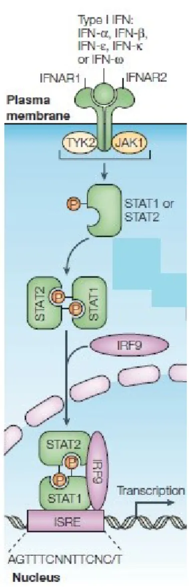
Figure A. Interferon receptors and activation of classical JAK-STAT pathways by type
I interferon.
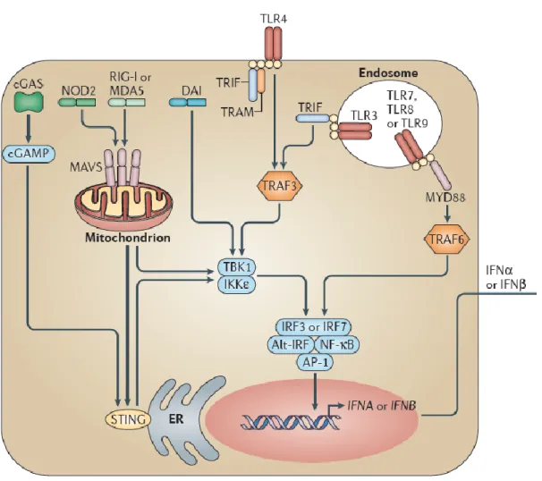
Figure B. Pathways of IFN-I induction.
MATERIALS, METHODS AND RESULTS
Figure 1. Dendritic cells (DCs) produce higher levels of IFN-β than macrophages
following infection with S. suis serotype 2.
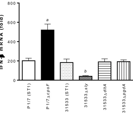
Figure 2. The presence of the capsular polysaccharide interferes with S. suis-induced
IFN-β expression by dendritic cells, while the suilysin is partially responsible for its activation.
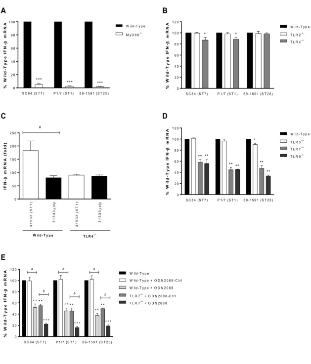
Figure 3. Recognition of S. suis by the Toll-like receptor (TLR) pathway is required for
induction of IFN-β expression by dendritic cells (DCs).
Figure 4. The interferon regulatory factors (IRFs) 1, 3, and 7 are involved in IFN-β
expression by dendritic cells (DCs) following infection with S. suis.
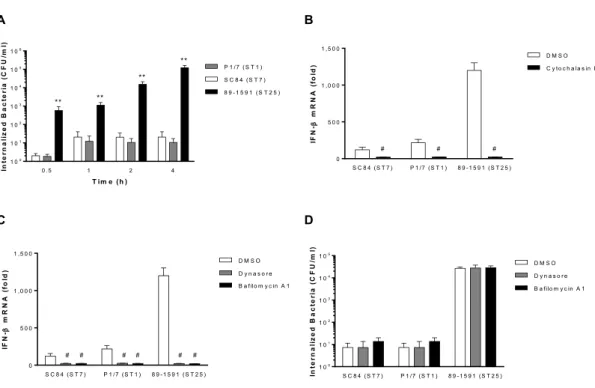
Figure 5. S. suis-induced IFN-β expression by dendritic cells requires internalization
and phagosome maturation.
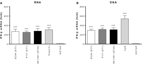
Figure 6. The S. suis nucleic acids are responsible for inducing IFN-β expression by
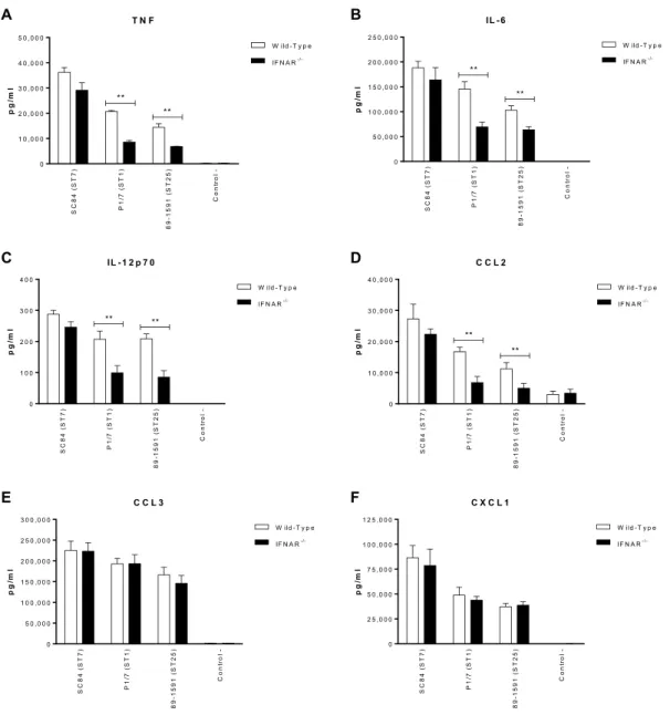
Figure 7. S. suis-induced type I interferon produced by dendritic cells modulates
autocrine cytokine production.
Figure 8. Type I interferon is beneficial for host survival following infection with
intermediate virulent and virulent S. suis serotype 2 strains.
Figure 9. Type I interferon modulates plasma pro-inflammatory cytokines involved in
S. suis-induced systemic inflammation.
Figure 10. Type I interferon is required for the control of blood bacterial burden
following infection with intermediate virulent and virulent S. suis serotype 2 strains.
Supplementary Figures
Figure S1. Intermediate virulent S. suis ST25 strains induce high levels of IFN-β
expression by dendritic cells.
Figure S2. Ligands of the different Toll-like receptors evaluated in this study, with the
exception of TLR2, induce IFN-β by dendritic cells.
Figure S3. IL-6 and CXCL1 induced by S. suis and bacterial TLR2 ligands partially
requires TLR2 expression by dendritic cells.
Figure S4. The intermediate virulent S. suis strain 89-1591 is highly encapsulated. Figure S5. S. suis-induced IL-6 and CXCL1 expression by dendritic cells is partially
internalization-dependent.
DISCUSSION
LIST OF ACRONYMS AND ABBREVIATIONS
APC: Antigen presenting cell BBB: Blood brain barrier
BMEC: Brain microvascular endothelial cells CARD: Caspase activation recruitment domain CCL: Chemokine (C-C motif) ligand
CD: Cluster of differentiation CFU: Colony-forming unit CLR: C-type lectin receptor CNS: Central nervous system
CPEC: Choroid plexus epithelial cells CpG: Cytosine-phosphate-Guanosine CPS: Capsular polysaccharide
CSF: Cerebrospinal fluid
CXCL: Chemokine (C-X-C motif) ligand CytD: Cytochalasin D
DC: Bone marrow-derived Dendritic Cell DNA: Deoxyribonucleic acid
Dpp: Dipeptidylpeptidase ECM: Extracellular matrix EF: Extracellular factor
Fbp: Fibronectin-binding protein
GAPDH: Glyceraldehyde-3-phosphate dehydrogenase GAS: Group A Streptococcus
GBS: Group B Streptococcus
GM-CSF: Granulocyte macrophage colony-stimulating factor IFN: Interferon
IFNAR: Interferon-α/β receptor IFNγR: Interferon-γ receptor Ig: Immunoglobulin
IKK: IκB kinase IL: Interleukin
iNOS: Inductible Nitric Oxide Synthase IRF: Interferon Regulatory Factor ISGF: Interferon-stimulated gene factor JAK: Janus kinase
KO: Knock-out LLO: Listeriolysin O LTA: Lipoteichoic acid
MAVS: Mitochondrial antiviral-signaling protein MCP-1: Monocyte Chemoattractant protein-1 M-CSF: Macrophage colony-stimulating factor MDAs: Melanoma differentiation antigens MLST: Multilocus sequence typing
Mθ: Macrophages
MOI: Multiplicity of infection MRP: Muramidase-released protein
MyD88: Myeloid Differentation Primary Response Protein-88 NF-κB: Nuclear factor-κβ
NK: Natural killer cell NLR: NOD-like receptor NO: Nitric oxide
PAMP: Pathogen-associated molecular pattern PBMC: Peripheral blood mononuclear cell PGN: Peptidoglycan
p.i.: Post-infection
PLP: Myelin proteolipid protein PMN: Polymorphonuclear cell PRR: Pattern recognition receptor RecN: Recombination/repair protein
RLR: Retinoic acid-inducible gene (RIG)-I like receptor RNA: Ribonucleic acid
RT-qPCR: Quantitative Reverse Transcription Polymerase Chain Reaction SLSS: Septic-like shock syndrome
SLY: Suilysin
SspA: Surface-associated subtilisine-like protease ST: Sequence type
STAT: Signal transducer and activator of transcription STING: Stimulator of interferon genes
STSLS: Streptococcal toxic shock-like syndrome TBK1: TANK-binding kinase 1
TGF-β: Transforming growth factor-β THA: Todd Hewitt broth agar
THB: Todd Hewitt broth Th1: T helper cell type 1
TICAM: TIR-domain containing adaptor molecule TIRAP: TIR-containing adaptor protein
TLR: Toll-like receptor TNF: Tumoral necrosis factor
TRIF: TIR-domain-containing adaptor-inducing interferon-β TyK: Tyrosine kinase
ACKNOWLEDGMENTS
First of all, I would like to remark that for me, this experience was more than an academic experience, since it has allowed me to confirm that there are no limits in life. As Walt Disney said: « IF YOU CAN DREAM IT, YOU CAN DO IT ». In Canada I have discovered a new lifestyle, new people, a new language, a different culture, and everything have been awesome. I will be forever thankful to all the people that have been part of this "adventure", supporting me from close distance or miles away.
Thus, I would like to thank all the people and institutions that have participated in the achievement of this master project, particularly:
- My research supervisor, Dr. Marcelo Gottschalk for having provided me with the opportunity of working within his laboratory and for his constant support, guidance, and endless help with the writing of this memoire and article.
- Dr. Mariela Segura, my co-director, for having opened the doors of her laboratory to me. Also, I would like to thank her for her input in the presentations I have given throughout this master’s Project.
- All members of Dr. Gottschalk and Dr. Segura laboratories, past and present, for their help with my project and for making this an awesome experience.
- Jean-Philippe Auger for his unconditional help with all technical aspects related to my project, and also, for teaching me the technical basis needed for functioning in a laboratory during my research project, which greatly served me during completion of this master.
- Institutions like GREMIP, CRIPA, NSERC – CRSNG and University of Montreal, and their members for making this a very enriching experience, and for the financial support they have provided me during the course of my M.Sc. study.
- Members of my committee and jury: Dr. Marcelo Gottschalk, Dr. Mariela Segura, Dr. Levon Abrahamyan, Dr. Jean-Pierre Lavoie and Dr. Carl Gagnon.
Thanks to all the professors and members of GREMIP. And finally, last but not least, thanks to my family and friends for their constant moral support.
S. suis, an encapsulated Gram-positive bacterium, is an important porcine bacterial
pathogen and zoonotic agent responsible for sepsis and meningitis. In fact, S. suis has become the leading cause of adult meningitis in Vietnam, the second leading cause in Thailand, and the third leading cause in Hong Kong. The clinical characteristics typical of acute meningitis in humans caused by S. suis changed after the 2005 outbreak in the Chinese province of Sichuan. That was characterized as streptococcal toxic shock-like syndrome (STSLS), similar to that usually associated with Streptococcus pyogenes (125).
To date, 35 serotypes based on the capsular polysaccharide (CPS) composition have been described, the serotype 2 being the most commonly isolated from diseased animals and humans in many countries. Data obtained by multilocus sequence typing (MLST) have showed that different serotype 2 STs predominate in different regions of the world. The ST1 is mostly associated with disease in pigs and humans in Europe, Asia and Argentina. The ST7, responsible for the 1998 and 2005 epidemics, is mostly endemic to mainland China. North American cases vary from those in Eurasia, with most strains being either ST25 or ST28 (125).
In recent years, an increasing number of studies were performed to understand the pathogenesis of the S. suis infection, however, further investigation is needed to clearly elucidate the molecular mechanisms of its pathogenesis. Similarly, the mechanisms involved in the host immune response to S. suis as well as those used by S. suis to subvert this response remains poorly characterized. However, several virulence factors have been proposed to be involved in the pathogenesis of S. suis infection. Among them, the CPS, which presents an anti-phagocytic role, has been demonstrated as a critical virulence factor and its structure has been described (149). In addition, a hemolysin (suilysin - SLY) has been characterized and described to be involved in the modulation of S. suis interactions with host cells, such as endothelial cells, epithelial cells, neutrophils, dendritic cells (DCs) and macrophages (Mθ) (149). Regarding the survival in blood and dissemination, the resistance to phagocytosis require modification of the cell wall peptidoglycan by means of N-deacetylation. A well encapsulated mutant strain devoid of the deacetylase peptidoglycanN–acetylglucosamine (PgdA), responsible for this modification, showed impaired resistance to neutrophil killing and was severely attenuated in murine and porcine infection models (149, 170). Similarly, d-alanylation of the S. suis lipoteichoic acid (LTA) plays a major role in survival of this pathogen
Inflammation is a protective response of the body to ensure removal of detrimental stimuli, as well as a healing process for repairing damaged tissue (239). Germline-encoded pattern recognition receptors (PRR) are responsible for sensing the presence of microorganisms by recognizing structures conserved among microbial species, which are called pathogen-associated molecular patterns (PAMPs). Different classes of PRR families have been identified, such as transmembrane and endosomal proteins: the Toll-Like Receptors (TLRs), as well as cytoplasmic proteins such as the Retinoic acid-inducible gene (RIG)-I-like receptors (RLRs) and NOD-like receptors (NLRs). These PRRs are expressed in macrophages, neutrophiles and dendritic cells but also in various nonprofessional immune cells. The sensing of PAMPs by PRRs upregulates the transcription of genes involved in inflammatory responses. These genes encode pro-inflammatory cytokines, type I IFNs, chemokines and antimicrobial proteins, proteins involved in the modulation of PRR signaling, and many uncharacterized proteins (240). Focusing on interferons, these are a family of cytokines which act early in the innate immune response and are very well known for inducing an antiviral activity in infected cells. In addition to this antiviral activity, they play a role in regulating the immune response (12). Among the three distinct interferon families, the type I IFN (IFN-I) family is a multi-gene cytokine family, being IFNα and IFNβ (IFNα/β) the best-defined and most broadly expressed ones. IFN-I have numerous additional functions not only during the viral, but also in bacterial infections. The outcome of its response during infectious diseases is highly context-dependent. Different conditions induced during specific infections modulate when and where IFN-I signals are delivered, as well as the signalling pathways that are triggered downstream of the type I IFN receptor (IFNAR) (19). Recents in vivo studies with extracellular bacteria have shown contradictory results, since in some cases type I interferon have played a protective role, but in other cases the role was detrimental (44-52, 255). In relation with the source of IFN-I, particularly IFN-β, previous studies have compared the role of antigen presenting cells, such as DCs and Mθ, after the infection with different streptococci (83, 85).
Not much data are available about the interplay between S. suis infection and IFN-β. A study has shown that after an in vivo infection with strains of S. suis serotype 2 of different backgrounds and virulence, the least virulent strain tested resulted to be the main IFN-β inducing strain (247).
Thus, the scatter and lack of data regarding the ability of different strains of S. suis serotype 2 to induce IFN-β have motivated this work. Moreover, the host source of IFN-β induced by S. suis is unknown. Furthermore, the cellular receptors and pathways responsible for cell activation and subsequent production of IFN-β by S. suis have barely been studied, and the role of this cytokine in infections caused by S. suis, is not known.
Based on the observations mentioned above, we hypothesized that IFN-β plays a protective role in infections caused by S. suis serotype 2. Based on the aforementioned effect of North American strains, it is further hypothesized that the increased production of this type I interferon by them would be responsible, at least in part, of their lower virulence.
The general objective of this thesis was to study the role of IFN-β in infections caused by S. suis serotype 2. Moreover, three specific objectives were proposed:
1. Study the in vitro induction of IFN-β in murine bone marrow-derived dendritic cells and macrophages activated by different well-encapsulated S. suis serotype 2 strains.
2. Assess the role of different cell receptors and pathways in the induction of IFN-β by S. suis serotype 2.
3. Study the role of IFN-β during in vivo infections with S. suis serotype 2.
In this research, we have demonstrated that DCs are an important source of S. suis-induced IFN-β, which occurs mainly through a MyD88 pathway and requires bacterial internalization. With the less virulent strain, a greater internalization and higher IFN-β production was observed. In addition, a protective role of type I interferon was described during an in vivo infection by S. suis. Thus, our findings have provided a novel knowledge of the mechanisms and the capacity of induction of IFN-β by different strains of Streptococcus suis serotype 2, and shed a light on the role of this cytokine during the infection with this pathogen.
II - SCIENTIFIC LITTERATURE
REVIEW
1. S. suis
1.1 Introduction: History and epidemiology
Streptococcus suis is a pathogen in pigs that can cause severe systemic infection in
humans and was first reported by veterinarians in 1954, after outbreaks of meningitis, septicemia, and purulent arthritis occured among piglets (1). Among the 35 serotypes based on capsular antigens that have been described, the type 2 is the most frequently isolated from diseased pigs in most countries (2, 3) and is also considered a zoonotic agent. First reported in Denmark in 1968 (4), human infections have been documented in several European and Asian countries as well as in North and South America, Australia and New Zealand (5,6). In western countries, S. suis infections in humans have most often been restricted to workers in close contact with pigs or swine byproducts. However, in southeast and east Asia, also affects the general population and it represents a significant public health concern (5).
In 2005, a human and pig outbreak (molecularly demonstrated as being caused by the same S. suis strain) in the Sichuan province of China resulted in 204 human cases and 38 deaths, a mortality rate that was higher than expected; the outbreak was characterized by the unexpectedly high percentage of patients developing streptococcal toxic shock-like syndrome (STSLS - unexpected because S. suis was not considered to be cause of that until then), shorter incubation period, rapid disease progression, and high mortality rate (1,5). A subsequent report from Thailand indicated that S. suis meningitis was far from being a rare sporadic disease in the area, with a series of 66 cases in the period 2005–2007 showing the traditional disease characteristics (86). Even more surprisingly, this pathogen was convincingly demonstrated to be the commonest cause of bacterial meningitis in humans older than 14 years of age in two different cohort studies from Vietnam in 2011 and 2012 (87, 88), and the third most common cause of community-acquired bacterial meningitis in Hong Kong, indicating that, at least for Southeast Asia, the disease is not as sporadic as was previously considered (7-11).
S. suis can be isolated from other animals, such as ruminants, cats, dogs, deer, and
horses, and is believed to be a commensal in the intestinal flora (89). Healthy pigs can carry multiple serotypes in their nasal cavities, tonsils, and upper respiratory, genital, and alimentary tracts (89–91).
1.2 Disease and clinical manifestations
1.2.1 Pigs
With almost 100% of pig farms worldwide having carrier animals, S. suis is one of the most important bacterial pig pathogens. Transmission among animals is considered to be mainly through the respiratory route (92). There are many descriptions of the pathological and histopathological lesions in infected pigs (89). The most common gross lesions are the congestion of the meninges, lymph nodes and lungs, and the most common histopathological findings are located within the choroidal plexus. Evidence of encephalitis, oedema, and congestion of the brain may be present. In the central nervous system (CNS), lesions associated with meningitis and choroiditis may be observed, including oedema of the leptomeninges and the dura mater, hyperaemic meningeal blood vessels, and an increased quantity of cerebrospinal fluid (CSF). The most characteristic histopathological lesion of acute S. suis meningitis is a diffuse neutrophilic infiltrate (94, 95). Nevertheless, in peracute cases of infection, pigs are often found dead with no premonitory signs of disease (93). Presumptive diagnosis in pigs is usually based on clinical signs and macroscopic lesions. Confirmation of the infection is a mandatory requirement and must be achieved by isolation and characterization of the pathogen (89).
1.2.2 Humans
Human infections with S. suis are most frequently manifested as purulent meningitis, but reports of septic shock with multiple organ failure, endocarditis, pneumonia, arthritis, and peritonitis have also been reported. Differences in clinical signs among patients infected have been observed. In the acute form of meningitis, symptoms include high fever, headache, chills, nausea, vomiting, and vertigo, followed by one or more of the following: hearing loss, walking ataxia, coma, neck stiffness, petechia, articular pain, peripheral and facial paralysis, severe myalgia, ecchymosis, rashes, and rhabdomyolysis (94, 96-99). In the acute form of toxic septic shock, besides high fever, chills, headache, vomiting, vertigo, and abdominal pain, other clinical signs were also observed, such as hypotension, tachycardia, liver dysfunction, subcutaneous haemorrhage, disseminated intravascular coagulation, acute renal failure, and acute
respiratory distress syndrome (95, 97, 98, 100). Hearing loss is the most common sequela after recovery from purulent meningitis, whereas death often follows septic shock.
An increased amount of CSF has also been reported in human meningitis cases by lumbar puncture (94, 95). Obtaining this fluid is important because it is an important element in the diagnosis of neurological diseases, such as meningeal syndromes, subarachnoid hemorrhages, cerebrospinal tumors, etc.
1.3 Pathogen characteristics
1.3.1 Characteristics of S .suis, including serotype, serotype distribution
S. suis is a Gram-positive facultative anaerobe, coccoid or ovoid, and occurs as
single cells, in pairs, or in short chains (101). Based on the CPS, 35 serotypes have been identified (types 1–34 and 1/2) (125) but serotypes 32 and 34 have since been proven to be Streptococcus orisratti (102). Recent analyses of genes encoding manganese-dependent superoxide dismutase (sodA) and the recombination/repair protein (recN) indicated that the serotypes 20, 22, 26, and 33 need to be taxonomically removed from S. suis (249), and serotypes 20, 22, and 26 were proposed to be a novel species,
Streptococcus parasuis (250). Serotype 2 is most commonly associated with diseases in
pigs and human beings, and is the most frequently reported serotype worldwide (3, 103, 104). The natural habitat is the upper respiratory tract, specially tonsils and nasal cavities, as well as the genital and alimentary tracts of pigs (105, 106). It colonises the palatine tonsils of clinically ill and apparently healthy pigs, and is usually transmitted nasally or orally (107). In relation with the resistence to enviromental conditions, it can survive for 10 min at 60°C, 2 h at 50°C, and 6 weeks in carcasses at 10°C. At 0°C, can survive for 1 month in dust and for over 3 months in faeces, whereas at 25°C, it can survive for 24 h in dust and for 8 days in faeces. However, it can be killed easily with 5% bleach at 1:799 dilution (108).
S. suis is sensitive to antibiotics, including penicillin, ceftriaxone, cephalosporin,
ampicillin, and amoxicillin. Penicillin G is commonly used to treat or control infections but penicillin-resistant strains have been isolated (109,110), and strains highly resistant to other commonly used antibiotics have also been reported (111).
The genome, which has been completely sequenced, contains 20074917 bp (112). Although the functions of 20–30% of the genes are unknown, many genes that may play a part in the pathogenesis of the infection have been studied, including polysaccharide production, capsular transport, iron-restriction factors, suilysin, virulence-associated proteins, various enzymes, arginine deiminase system, and IgG binding proteins (113-118).
S. suis is able to grow in anaerobic or aerobic conditions, but not in 6,5% NaCl
solution (119). Colonies are small (0,5–1,0 mm diameter), greyish or transparent, and slightly mucoid. They produce narrow zones of α-haemolysis on sheep blood agar plates and β- haemolysis on horse blood agar plates (89). Presumptive identification based on four biochemical tests (Voges-Proskauer, salicin, trehalose, and 6,5% NaCl) can be successful for almost all capsular types (124). Recently, Ishida et al. developed a novel PCR method targeting the recombination/repair protein (recN) gene of S. suis, which corresponds to the current reclassification of this bacterium. recN PCR using S. suis reference strains could discriminate S. suis from those that should not been included in S. suis (251). Regarding the serotyping, molecular assays by PCR amplification of serotype specific cps genes does not require antisera and is an attractive alternative to the current agglutination and co-agglutination tests. However, it is known that the cps gene clusters of serotype 1 and serotype 14, and of serotype 2 and serotype 1/2 are very similar with the nucleotide sequence of the wzy genes being nearly identical in these two pairs of serotypes. Therefore, the mPCR assays cannot discriminate these two pairs of serotypes. Because of that serotype 1 and 14, and serotype 2 and 1/2 will require the use of serotype specific antisera (252). Recently, was developed a pipeline which permits in silico serotype determination from whole-genome sequencing (WGS) short-read data that can readily identify S. suis serotypes. Indeed, it can discriminate between serotype 1 and 14, and between serotype 1 and 1/2, which solves a three-decade longstanding S. suis typing issue (272).
Globally, the predominant serotypes isolated from clinical cases in pigs are, in decreasing order, serotypes 2, 9, 3, 1/2 and 7. However, there is a clear geographical effect on the distribution of serotypes and these figures are influenced by the number of published studies. Almost 70% of studies on worldwide isolates recovered from diseased pigs are from North America. In fact, 97% of North American data are from Canada and the rest from the United States, with no data from Mexico. Serotype 2 is the most prevalent in Canada, while in the United States, it is serotype 3. Both serotypes are
the two most prevalent serotypes isolated from clinical pig cases in North America with 24.3% and 21.0% of prevalence, respectively, followed by serotypes 1/2, 8 and 7. Similar distributions may be explained by a fluid movement of animals between the two countries (125).
In South America, only two studies have been published, both from Brazil, which report serotype 2 as being the most prevalent with a mean of 57.6% of cases, followed by, in decreasing order of prevalence, serotypes 1/2, 14, 7 and 9. In Asia, the most prevalent serotypes in infected pigs are, in decreasing order, serotypes 2, 3, 4, 7 and 8 (125).
Important pig producing European countries, such as Denmark, Belgium, France, Germany, Italy and the United Kingdom, have not recently reported the distribution of serotypes recovered from clinical cases in pigs (3, 127). Before the year 2000, serotype 2 was the most common serotype recovered in Italy, France and Spain, whereas serotype 9 was more frequently found in the Netherlands, Germany and Belgium (128). The only two countries with more recent data are Spain and the Netherlands. In the first one, serotype 2 is no longer the most prevalent, but the second behind serotype 9, followed by serotypes 7, 8 and 3 (129-131). In the Netherlands, between 2002 and 2007, serotype 9 was still the most prevalent, followed by serotypes 2, 7, 1 and 4 (132). Moreover, in 2013, was reported the first case of S. suis serotype 9 human infection in Thailand (273). As such, there is an urgent need to evaluate fresh data on the prevalence of S. suis in clinical isolates from pigs in Europe where many countries are among the most important pig producers in the world (125).
1.3.2 Data obtained by multilocus sequence typing (MLST): Sequence type (ST), distribution of serotype 2
Different serotype 2 sequence types predominate in different regions of the world. The ST1 is mostly associated with disease in pigs and humans in Europe (though the ST20 is important in the Netherlands), Asia (Cambodia, mainland China, Hong Kong, Japan, Thailand and Vietnam) and Argentina. The ST7, responsible for the 1998 and 2005 epidemics, is mostly endemic to mainland China. North American cases vary from those in Eurasia, with most strains being either ST25 or ST28, which were also recovered in Thailand and Japan, respectively. Finally, the ST101 to ST104 are endemic
ST104. So, we can observe that the current distribution of the S. suis serotype 2 STs greatly varies throughout the world, though data have only been available for a little over a decade, from only a few countries and mostly only for the serotype 2 (133). In North America, the majority of MLST studies conducted on S. suis strains isolated from diseased pigs have been serotype 2. It was determined that 44% of North American strains are ST25, 51% ST28 and 5% are ST1. In Canada, the proportions of ST25 and ST28 are 54% and 46%, respectively, but in the United States, 75% of strains were shown to be ST28 and only 10% ST25, while the remaining 15% are ST1 (133). As with North American strains, the majority of European studies have been done using serotype 2. Most of these have demonstrated that ST1 is predominately isolated from diseased pigs in the Netherlands, Spain and the United Kingdom (132, 134, 135). King
et al. had already associated the serotype 2 ST1 strains with invasive infections (134).
Nevertheless, many strains of serotype 9, have also been recovered from diseased pigs and typed (132, 135). In the Netherlands and Spain, serotype 9 isolates were identified as belonging to the ST16, where they represent 43% of strains in the Netherlands (132). Unlike some countries in Europe, where the serotype 9 is as important as the serotype 2, most strains isolated from diseased pigs in Asia are serotype 2, and it represents 90% of cases in mainland China (136). However, there are relatively few reports of isolation from diseased pigs in Asia. Of these serotype 2 cases, the predominant STs are ST1, ST7 and ST28. In mainland China, Chen et al. demonstrated that 22% of serotype 2 strains are ST1 and 77% are ST7. The few ST28 strains recovered in that country were mostly associated with cases of pneumonia (137). With respect to Japan, ST1 and ST28 strains isolated from cases of endocarditis in diseased pigs account for 8% and 76% of serotype 2 strains, respectively (138).
Regarding human cases, globally, ST1 strains have been described as mostly responsible for S. suis serotype 2 infections, particularly in South America, Europe and Asia, but also one case in North America (10, 132, 139-142). Nevertheless, multiple other STs have been described worldwide, though these appear to be endemic to certain geographical regions. For example, the ST20 is important in the Netherlands and France but not in the rest of Europe (132). ST7 strains, the ones responsible for the 1998 and 2005 Chinese epidemics were isolated from human patients only in mainland China and Hong Kong (141, 142). ST25 and ST28 strains have been particularly associated with human cases in North America and Japan, respectively (141, 143, 144). In Thailand, ST1 and ST104 strains are predominant, causing mainly meningitis and non-meningitis
cases, respectively (140, 143, 145). A few cases of ST25, ST28, ST101, ST102 and ST103 have also been described (140).
Although S. suis serotype 14 infections are less frequent in humans than serotype 2 cases (2% of all the serotype-confirmed cases) the number appears to be increasing. The ST105 is prevalent in Southeast Asia, especially in Vietnam and Thailand. Only three human S. suis cases, other than those caused by serotypes 2 and 14, have been typed by MLST, and were described as newly identified STs. Those are serotypes 5, 16 and 24, identified as ST181, ST106 and ST221, respectively (146, 147), but no data have yet been published on the possible presence of these three newly identified STs for human isolates in diseased pigs.
1.3.3 Virulence
1.3.3.1 Virulence factors
Identification of S. suis virulence factors was limited from the lack of clear definition of ‘virulence’. Different studies have defined field strains as virulent or avirulent based on the clinical condition of the animal (or human being) from which the strains were isolated, or based on the presence of previously described and nonuniversal virulence-associated proteins or, using different experimental infection models. For instance, if strains were isolated from the clinically healthy animals they were arbitrarily considered avirulent whereas the ones isolated from diseased animals/human beings gives strains arbitrarily considered as virulent. However, it is known that the presence of some proposed virulence factors does not necessarily define the strain virulence potential, because its absence is not a sufficient condition for classifying the strain as avirulent. Moreover, the virulence of serotype 2 strains recovered in Europe and Asia from diseased piglets seems to be higher than that observed in strains from North America (133, 148).
Although the actual early mechanisms used by S. suis to colonize the host are poorly known, it was reported that the pathogen may survive in swine tonsils for long periods of time. However, it is still unknown how S. suis manages to cross the first natural line of the host defense and initiates the disease. The most accepted hypothesis is that it breaches the mucosal epithelium in the upper respiratory tract of pigs (106). In humans,
(5, 6, 146). Adhesins at the surface of S. suis appear to be hindered by the CPS, as suggested by the fact that S. suis CPS deficient mutants adhered better than the encapsulated parental strain to porcine (LLC-PK1 and PK15), canine (MDCK) and human (A549 and HeLa) cell lines. Thus, we can hypothesize that S. suis downregulates expression of CPS during the early steps of the infection in response to signals from the environment, resulting in a better interplay between bacterial adhesins and host receptors (149, 274).
S. suis interacts with components of the extracellular matrix (ECM) such as
fibronectin and plasminogen (150). The fibronectin-binding protein Fbps was shown to bind human fibronectin and fibrinogen in vitro. Infection of pigs with a fbps mutant strain showed that Fbps is not required for colonization of the tonsils but it may play a role in colonization of specific organs, including CNS (151, 190). S. suis enolase which binds plasminogen and fibronectin, is highly expressed in vivo, inducing the production of antibodies in infected pigs, although its potential being used as a protective antigen remains controversial (152, 153). In addition, antibodies against enolase decrease adhesion and invasion of porcine brain microvascular endothelial cells (BMEC) (191). A dipeptidylpeptidase DppIV was also shown to interact with human fibronectin, and the virulence of a dppIV-deficient mutant was greatly attenuated (154). Binding of
S. suis to collagen has also been reported (150), and a mutant strain defective in a
putative collagenase was impaired in survival after experimental inoculation of pigs (126). Similarly, a mutant strain devoid of the housekeeping sortase SrtA showed weaker adherence to ECM proteins, suggesting that peptidoglycan- anchored, LPXTG motif-containing adhesins are also important for interactions of this pathogen with ECM proteins. The same was observed with BMEC (155).
Other proteins were identified as adhesins, including a 39-kDa glyceraldehyde- 3-phosphate dehydrogenase (GAPDH) (156,157). Two studies showed reduced adherence of S. suis to porcine tracheal rings and HEp-2 cells when cells were preincubated with recombinant S. suis GAPDH (157,158). The pathogen upregulates the expression of the gene encoding this protein in vivo in different porcine organs (159). An enzyme with 6-phosphogluconate- dehydrogenase activity, a bifunctional amylopullulanase, as well as a glutamine synthetase, were shown to contribute to S. suis adherence to epithelial cells (160,161). Mutants devoid of the two-component regulatory system CiaRH or the orphan transcriptional regulators RevSC21 and CovR were also impaired in adherence to epithelial cells (162-164), but the mechanisms have not yet been deciphered.
Epithelial cells invasion may also represent the beginning of systemic dissemination and disease. As in the case of adhesion, only unencapsulated strains seem to be able to invade these cells (165). The disruption, including the disruption to cross the blood– brain barrier (BBB) and the blood– CSF barrier of Choroid plexus epithelial cell (CPEC), is also possible since suilysin (SLY)-positive strains are highly toxic for the cells (106, 193). This 54-kDa hemolysin is a thiol-activated toxin that targets cholesterol in the membrane of eukaryotic cells (166, 167). However, strains not producing SLY are also able to do that (2, 192). But this concern remains still controversial.
Regarding the survival in blood and dissemination of the pathogen, the resistance to
phagocytosis require modification of the cell wall peptidoglycan by means of
N-deacetylation. A well encapsulated mutant strain devoid of the deacetylase PgdA,
responsible for this modification, showed impaired resistance to neutrophil killing and was severely attenuated in murine and porcine infection models (170). Similarly, d-alanylation of the S. suis lipoteichoic acid (LTA) plays a major role in survival of this pathogen (171), and was shown that mutants impaired in LTA d-alanylation adhered and invaded porcine BMEC lesser than the wildtype strain. Although SLY-negative strains can be virulent and survive in blood, SLY-positive strains, additionally, reduce phagocytosis and killing of S. suis. This pathogen is also able to affect neutrophil recruitment by degrading IL-8, presumably by the production of a serine protease (172). A cell wall-anchored DNAse, specific for single and double-stranded linear DNA is expressed by S. suis (173) and it is believed that it plays a role in disruption of neutrophil extracellular traps (174). S. suis requires nutrients like trace metals, whose availability within the infected host is relatively low. AdcR, a streptococcal transcription factor homologous to the zinc-uptake regulator Zur, and the ferric uptake regulator Fur, one of the most important transcription factors controlling iron metabolism, are both important for S. suis survival in vivo (175, 176). Eighteen unique iron restriction induced genes have been identified, including the cpsA gene, encoding a putative regulator of CPS biosynthesis and iri-7, homolog of Streptococcus mutans
rpgG, a gene involved in capsule biosynthesis. It was proposed that because the CPS of S. suis becomes thicker after growth in vivo, where free iron is scarce, an upregulated
expression of cps2A and rpgG under iron starvation might be expected (116). Despite these findings, it has been reported that S. suis, which does not secrete siderophores,
manganese or magnesium (177, 178). Interestingly, the lipoprotein TroA, which is required for S. suis growth in environments low in manganese, is crucial for bacterial survival in vivo (179). Recently, it was shown that deletion of a lipoprotein involved in zinc uptake - SSU0308 (Lipoprotein 103) - resulted in a mutant strain that was 50-times less virulent than the parental one (180). As mentioned earlier, the CPS constitutes a physical barrier against phagocytosis. However, if internalized, S. suis possesses factors that might contribute to the resistance to intracellular killing machinery of phagocytic cells (165). It is worth mentioning the presence of an active superoxide dismutase (SodA), although it is unlikely that SodA produced by S. suis type 2 mediates intracellular survival of pathogenic isolates in macrophages (181). In addition, survival of S. suis under acidic conditions has been linked to the presence of an arginine deiminase system catalyzing the conversion of arginine to ornithine, ammonia and carbon dioxide (117, 118).
Although rare, septic shock in some cases can be a result of excessive or poorly regulated immune response to the offending organism (182). Are the virulence factors responsible? Lipoproteins present in the cell wall could be, in part, responsible for cell receptor(s) recognition (183). Recently, it has been shown that a putative prolipoprotein diacylglyceryl transferase present in S. suis cell wall is required for innate immune activation (184), while suilysin was shown to activate phagocytes and to induce the release of pro-inflammatory cytokines (185,186). In addition, suilysin may cause a release of hemoglobin from red blood cells, which will raise the levels of pro-inflammatory mediators by acting in synergy with S. suis cell wall components (187), while a surface-associated subtilisinlike protease (SspA) induces the secretion of different pro-inflammatory cytokines and chemokines by macrophages (188).
Finally, there are some putative virulence factors like a glutamine synthetase (194), a serum opacity-like factor (195), a protein of unknown function encoded by virA (196) and a trag factor (197), but the mechanisms by which these factors affect S. suis virulence remain obscure. Also, there are proteins whose deletion affect virulence, including the response regulator RevS (198), the autoinducer LuxS (199), the sugar regulator CcpA (200), Rgg-like regulators (201), the orphan transcriptional regulators RevSC21 (162) and CovR (164), as well as the SalK/SalR (176) and CiaRH two-component systems (163). Again, how these regulators influence virulence (and the extent of their regulons) has not yet been elucidated. In addition, other proteins have been suspected to be ‘virulence factors’ since antibodies against them confer protection.
They are HP0245, HP0272 and HP0197, of unknown function (202-205), the surface antigen One (206), and HtpS, a histidine triad protein (207).
1.3.3.2 Difference in virulence of different ST and geographical origin
The virulence markers used in association with STs are the SLY, the Muramidase Released Protein - MRP and the Extracellular Factor - EF. Serotype 2 ST1 strains, mostly associated with disease in pigs and humans in Europe, Asia (Cambodia, mainland China, Hong Kong, Japan, Thailand and Vietnam) and Argentina, have for the most part been genotyped/phenotyped as sly+ mrp+ epf+ / SLY+ MRP+ EF+, regardless of the geographic origin, which is identical to the serotype 2 ST7 strains isolated from diseased pigs and humans in mainland China (133, 135, 137, 138, 141, 235). Nevertheless, important genetic differences vary between ST1 and ST7 strains including the presence of a 89K pathogenicity island in ST7 strains (236). These ST1 complex strains differ from the human serotype 2 ST104 strains of Thailand which are
sly+ mrp- epf- and from the human serotype 2 ST20 strains recovered in the
Netherlands that were epf- (132, 140, 141). Interestingly, a human case from Spain whit the serotype 2 strain was typed as being a ST3, and presented a mrp variant (mrp*), which has a higher molecular weight, though being sly+ epf+ (237, 238).
In Europe, it was determined that strains isolated from diseased pigs belonging to the ST61 (serotype 9) complex differ from the ST1 (serotype 2) complex strains in being
mrp* rather than mrp. Particularly in Spain, the endemic ST123 and ST125 (serotype 9)
are both mrp- and epf- (135).
In North America, ST25 strains isolated from diseased pigs were identified as sly-
mrp- epf-, while the ST28 isolated from North America, mainland China and Japan are sly- mrp+ epf- or SLY- MRP+ EF- (133, 137, 138).
Although not as widely used, different pili (srtB, srtC, srtD, srtF and srtG) have also been associated with different STs. It was identified that ST1 strains isolated from both diseased pigs and human cases of serotype 2 infections in Japan and Thailand are
srtBCD+ and srtF+, but srtG- (143). North American ST25 strains isolated from
diseased pigs and human cases are srtF- and srtG+ and ST28 strains isolated from diseased pigs and human cases from North America and Japan are srtF+ and srtG+ (133, 138).
An experimental study performed by Lachance et al. compares the innate immune response of the host following an acute infection by three different strains of S. suis with different virulence potentials: an intermediately pathogenic ST25 North American strain (89-1591), a highly pathogenic ST1 European strain (P1/7) and the epidemic ST7 Chinese (SC84) strain responsible from outbreaks in China. Authors observed that, during the acute infection, the survival of mice infected with the North American 89-1591 strain was not significantly affected and was similar to that of mock-infected mice. Contrarily, mice infected with the highly virulent Chinese strain had a 50% survival rate at 17 h p.i., while mice infected with the virulent European strain had a 50% survival rate at 44 h p.i. They also observed that cytokines levels, but not bacterial burden, correlate with the degrees of virulence of ST7, ST1 and ST25 strains (247).
1.4 Steps of the pathogenesis
1.4.1 Adherence to, colonization and invasion of epithelial host cells
As mentioned above, adhesins present at the surface of S. suis appear to be hindered by the CPS and, it is believed that S .suis down regulates their expression during the early steps of the infection in response to signals from the environment, resulting in a better interplay between bacterial adhesins and host receptors (149). Moreover, S. suis interacts with components of the ECM such as fibronectin and plasminogen (150). In addition, it is already known that IgA-mediated immunity plays a major role in defense against recurrent mucosal pathogens. It has been reported that S. suis produces an IgA1 protease capable of cleaving human IgA1 (168). This protease is highly immunoreactive to convalescent sera, and an isogenic mutant defective in the production of this enzyme showed significantly decreased lethality in pigs (169).
It is believed that nonvirulent strains of S. suis can probably adhere and colonize pigs without causing disease. However, since the disease caused by this pathogen is mainly systemic, virulent strains should, after adhesion, either directly invade and translocate, and/or reduce cell viability and increase mucosal barrier permeability (274). Although cell invasion is one of the expected outcomes of bacterial adhesion to cells, the invasion of mucosal epithelial cells by well-encapsulated S. suis is still controversial. As in the case of adhesion, only poorly encapsulated strains seem to be able to clearly invade these cells (274). The disruption of epithelial cell is possible, since SLY-positive strains
are toxic for these cells (106) because they attack cholesterol in the membrane of eukaryotic cells (166, 167). However, SLY-negatives are also able to reach the bloodstream and disseminate (2).
1.4.2 Surviving in the bloodstream: innate immune response
It was proposed that S. suis may gain entry to the systemic circulation primarily through the palatine tonsils, after adhesion and invasion of epithelial cells and later, through the interaction with cells of the myeloid lineage (222, 223). Once pathogen reaches deep tissues and/or the bloodstream, it is subjected to the action of phagocytic cells of the innate immune system.
1.4.2.1 Phagocytosis: monocytes/macrophages, PMN, dendritic cells (DCs)
Bacterial survival depends on the production of CPS, since it was documented by in
vitro and in vivo experiments thatvCPS protects S. suis from neutrophil and
monocyte/macrophage-mediated phagocytosis and killing (2). The fine structure of the
S. suis serotype 2 CPS has recently been solved, indicating the presence of
N-acetyl-neuraminic acid (sialic acid) residues. It is worth noting that the capsules of the two most important serotypes that cause disease in humans (2 and 14) possess sialic acid (224). Sialic acid has also been implicated in the adherence (without phagocytosis) of
S. suis to monocytes, suggesting a ‘modified Trojan horse’ hypothesis, in which the
pathogen would travel in the bloodstream externally associated with these phagocytic cells (106). In addition, an effect of molecular mimicry has been suggested (2), based on the fact that the conserved 2–6 linked sialic acid terminal capsular moiety found in serotypes 2 and 14 (225) is similar to sugar epitopes displayed on the surface of all mammalian cells (5). This molecular mimicry could be the reason of the absence of antigen recognition by the immune system of the host (123, 226). Despite the critical role played by the CPS in S. suis virulence, some avirulent serotype 2 field strains are well encapsulated (227), indicating that the survival in blood does not rely solely on encapsulation.
Resistance to phagocytosis is multifactorial and requires modification of the cell wall peptidoglycan - N-deacetylation- by a deacetylase PgdA (170). A well encapsulated
Similarly, d-alanylation of the S. suis lipoteichoic acid (LTA) plays a role in survival of this pathogen (171). A mutant strain producing LTA devoid of d-alanine residues was more susceptible than the parental strain to the action of cationic antimicrobial peptides and killing by porcine neutrophils. Apart from these major cell wall structures, many surface proteins can induce antibodies that increase the killing of S. suis by phagocytes (152, 207, 228, 229). However, the mechanisms of action of these proteins or their role at the bacterial-phagocyte interface are still unknown.
Although SLY-negative strains can be virulent and survive in blood, SLY-positive strains seem to benefit additionally from their toxic effects on monocytes and neutrophils (230, 231).
S. suis is also able to affect neutrophil recruitment by degrading IL-8, because of the
production of a serine protease (172). The pathogen requires nutrients including trace metals like zinc and iron, whose availability within the infected host is relatively low, and are important for the survival in vivo (175, 176). It has been reported that S. suis, is able to adapt to iron-restricted conditions by a change in its metabolism, through replacing iron by manganese or magnesium (177, 178).
As mentioned before, the CPS constitutes a physical barrier against phagocytosis. However, if internalized, S. suis possesses factors that might contribute to resist the intracellular killing machinery of phagocytic cells (165), like the arginine deiminase system which catalyzes the conversion of arginine to ornithine, ammonia and carbon dioxide under acidic conditions (117, 118).
1.4.3 Meningitis
1.4.3.1 Blood brain barrier (BBB) penetration
If death from sepsis or toxic shock-like syndrome does not occur and bacteremia remains high, S. suis may cause meningitis (148, 212). As a blood-borne pathogen,
S. suis must cross the BBB and/or the blood–CSF barrier in order to cause CNS
infections. The BBB is an anatomical and functional barrier that separates the brain from the intravascular compartment and maintains the homeostasis of the CNS environment (189); its main cellular type is BMEC. S. suis survived up to 7 h within porcine BMEC (192), which is a very interesting finding, because a crucial element for
the development of meningitis is the ability of pathogens to cross the BBB as live bacteria (213).
As was mentioned, the CPS of S. suis interferes with the process of adhesion/invasion. Although serum components may participate in the interactions between the pathogen and porcine BMECs (192, 214), only fibronectin was shown to play an important role in this process (190). SLY positive strains may also disrupt the BBB through cytotoxic effects, however it was reported that the negative mutants can successfully invade these cells (192).
Another CNS entry portal for S. suis can be the blood–CSF barrier. This invasion route is supposed to involve three steps: invasion of porcine CPEC from the basolateral side; transport within membrane-bound endocytic vacuoles to the apical side and exocytosis onto the apical membrane of the blood–CSF barrier (193). The translocation across the blood–CSF barrier activates neutrophils (215) and affects the barrier´s function and integrity, further facilitating trafficking of bacteria and leukocytes. It has been shown that S. suis induces CPEC necrosis and the release of pro-inflammatory cytokines and chemokines by human and porcine BMEC, murine microglia and astrocytes (216-219). However, the specific bacterial components responsible for exaggerated inflamatory reactions are not accurately known. In addition to cell wall components, bacterial CPS induces human macrophages to secrete prostaglandin E2 and matrix metalloproteinase 9, which may also be involved in disruption of the BBB (220). Purified SLY has been shown to induce the release of several pro-inflammatory cytokines by human and porcine BMEC (172, 192, 218) and the upregulation of adhesion molecules on human monocytes (221).
1.4.4 Inflammation as a hallmark of S. suis infection
1.4.4.1 Systemic
Although activation of the immune system during microbial infection is generally protective, septic shock may result as a consequence of excessive or poorly regulated immune response against the microorganism (182).
1.4.4.1.1 In vitro (monocytes/macrophages, PMN, dendritic cells)
In vitro experiments have shown that some pattern recognition receptors (PRR), such
as CD14 and Toll like receptor 2 (TLR2), could be responsible for cell activation by
S. suis, which would lead to release inflammatory compounds (185, 231-233).
However, macrophages isolated from TLR2 knockout mice have showed a highly reduced, but not completely abrogated cytokine production. That suggests the involvement of other TLRs in cytokine production (232).
Suilysin was shown to activate phagocytes, induce the release of pro-inflammatory cytokines IL-1β, IL-6 and chemokine IL-8 (185, 186), and it might release hemoglobin from red blood cells, contributing to the increased levels of the pro-inflammatory mediators by acting in synergy with S. suis cell wall components (187). Bi et al. showed that SLY also induced a strong TNF-α release from human peripheral blood mononuclear cells (PBMCs) and primary peritoneal murine macrophages. Moreover, a long-terme release of that cytokine from PBMCs was observed. In adittion, the authors demonstrated for the first time that SLY activates human PBMCs via TLR4, a receptor that has been reported to recognize the lipopolysaccharides from Gram-negative bacteria (253). In addition, Sspa induces the secretion of different pro-inflamatory cytokines and chemokines by macrophages (188). S. suis may modulate the response, and improve its survival, by actively degrading the chemokines and thus delaying recruitment of neutrophils to the site of inflammation (172).
1.4.4.1.2 In vivo
It is known that inflammation plays an important role in the pathogenesis of S .suis-induced septicemia and meningitis, and that TLRs are critical sensors in detecting infections and initially activate the innate immune system, specially TLR2 which has been implicated as the major PRR for ligands derived from gram-positive bacteria (258). Data from Lachance et al. indicated that in vivo TLR2-mediated recognition contributes to the severe outcome following infection with S. suis ST1 European strain. A signifincantly lower induction of pro-inflammatory cytokines and chemokines in TLR2-/- mice, was confirmed too, but interestingly, inflammatory mediators were significantly reduced but not abolished. These data suggest the contribution of other receptors. The same study showed that TLR2 does not seem to be mainly implicated in
the severe symptoms and lethality associated with the ST7 Chinese strain, since bacterial loads and levels of pro-inflammatory cytokines and chemokines were independent of TLR2. Levels of mRNA and protein of interferon (IFN)- γ were significantly higher in ST7 than ST1-infected mice, confirming previous observations about that the higher mortality observed with the ST7 strain compared to ST1 strain was mainly due to a massive secretion of IFN- γ by Natural killer (NK) cells. Based on in
vitro studies results, they hypothesized that different bacterial components present in
typical S. suis strains and those from the epidemic ST7 strain may vary and play a distinct role on cell activation and in the pathogenesis of the systemic inflammatory disease caused by this pathogen (260).
Another in vivo study, performed using a S. suis serotype 2 ST7 05ZYH33 strain, have showed that the cytokine levels may represent a prognostic indicator of the disease outcome. Massive amounts of cytokines TNF-α, IL-1β, IL-6, IL-10, IFN-γ and TGF-β levels were found in sera of piglets with septic-like shock syndrome (SLSS). Conversely, lower and irregular cytokine expression levels were found in the pigs with prostration syndrome, indicating that a strong cytokine storm caused the SLSS. By this way, cytokine over-expression breaks the immunologic balance between pro and anti-inflammatory actions which can protect hosts against pathogens (211) and is necessary for disease recovery. Moreover, a systemic cytokine storm can produce systemic sepsis and septic shock. In addition, the destructive effects of virulence factors, in the infected hosts ultimately manifest as persistent hypotension, leukocytosis or leucopenia, multiple organ dysfunction or lesions, STSLS or SLSS, coma, and even rapid death (261).
Lachance et al. have compared cytokines levels following an in vivo infection with different STs of S. suis serotype 2, such as the intermediately pathogenic ST25 North American Strain, the highly pathogenic ST1 European strain and the epidemic Chinese ST7 strain (247). They found that cytokine and chemokines levels, but not bacterial burden, correlate with the degrees of virulence of the strains, being that plasma levels of CXCL1, CXCL2, CCL2, CCL3, CCL4, IL-1β, IL-6, TNF and IFN- γ were higher in mice infected with the epidemic Chinese strain of S. suis. Other proteins, such as CXCL9 and CXCL10, were produced at high levels following the induction by ST7, and ST25 strains of S. suis. It was because the latter strain induces high levels of IFN-β, which can also contribute to the CXCL9 and CXCL10 induction (247).
1.4.4.2 CNS
Meningitis-associated brain injury and neuronal death is also associated with a host reaction to bacterial components (234).
1.4.4.2.1 In vitro (astrocytes, glial cells)
Studies have shown that S. suis is able to induce the release of pro-inflammatory cytokines and chemokines by human and porcine BMEC, murine microglia and astrocytes. However, the specific bacterial components responsible for the exaggerated inflammatory reactions are not accurately reported. Specifically in microglia cells,
S. suis induces the release of high levels of TNF-α and the chemoattractant MCP-1, but
low levels of IL-β1 even in absence of CPS. In the case of IL-6, it is released after infection only in absence of the CPS. This observation is interesting because it is known that this cytokine is highly secreted in the bloodstream during the septicaemic phase, after infection with wild type encapsulated strain of S. suis. Under the same condition, with an unencapsulated strain, was also observed a high activation of inducible nitric oxide synthase (iNOS), and accordingly with this a high nitric oxide (NO) production. Regarding the TLRs expressed in microglia, it is known that S. suis induces a high microglial TLR2 upregulation, and in agreement with the cytokines results, the upregulation is higher with the CPS mutant. Once in contact with microglia, virulent encapsulated S. suis can modulate intracellular signaling events, such as avoiding the phagocytosis and delaying the activation of the inflammatory response. These findings denote the relevance of CPS in the regulation of pro-inflammatory events (216-219).
1.4.4.2.2 In vivo
It has been shown that after 6 days post-injection (intraperitoneal injection), the most important changes in the brain of mice with clinical nervous symptoms comprised hemorrhagic foci and malacia mainly at the somatosensory cortex, striatum, hippocampus, thalamus, and hypothalamus, together with gliosis and the presence of inflammatory foci composed primarily of neutrophils (216). In several mice, the meninges were thickened and severely infiltrated by a mixture of neutrophils, macrophages, and lymphocytes. The expression levels of immune mediators were