Page(s) not included in the original manuscript are
unavailable from the author or university. The
manuscript was microfilmed as received.
9
This reproduction is the best copy available.
Dynamique laryngee lors de la ventilation nasale chez
I'agneau nouveau-ne
Par
FRANQOIS MOREAU-BUSSIERE M.D. Unite de recherche respiratoire neonatale Departement de physiologie et biophysique
Memoire presente a la Faculte de medecine en vue de I'obtention du grade de mattre es sciences (M.Sc.) en physiologie
Published Heritage Branch 395 Wellington Street Ottawa ON K1A0N4 Canada Direction du Patrimoine de I'edition 395, rue Wellington Ottawa ON K1A0N4 Canada
Your file Votre reference ISBN: 978-0-494-49548-3 Our file Notre reference ISBN: 978-0-494-49548-3
NOTICE:
The author has granted a non-exclusive license allowing Library and Archives Canada to reproduce, publish, archive, preserve, conserve, communicate to the public by
telecommunication or on the Internet, loan, distribute and sell theses
worldwide, for commercial or non-commercial purposes, in microform, paper, electronic and/or any other formats.
AVIS:
L'auteur a accorde une licence non exclusive permettant a la Bibliotheque et Archives Canada de reproduire, publier, archiver,
sauvegarder, conserver, transmettre au public par telecommunication ou par Plntemet, prefer, distribuer et vendre des theses partout dans le monde, a des fins commerciales ou autres, sur support microforme, papier, electronique et/ou autres formats.
The author retains copyright ownership and moral rights in this thesis. Neither the thesis nor substantial extracts from it may be printed or otherwise reproduced without the author's permission.
L'auteur conserve la propriete du droit d'auteur et des droits moraux qui protege cette these. Ni la these ni des extraits substantiels de celle-ci ne doivent etre imprimes ou autrement reproduits sans son autorisation.
In compliance with the Canadian Privacy Act some supporting forms may have been removed from this thesis.
Conformement a la loi canadienne sur la protection de la vie privee, quelques formulaires secondaires ont ete enleves de cette these. While these forms may be included
in the document page count, their removal does not represent any loss of content from the thesis.
Canada
Bien que ces formulaires
aient inclus dans la pagination, il n'y aura aucun contenu manquant.
Table des matieres 11 Liste des abrogations Ill
Resume IV Introduction 6 Article 8 Discussion 47 Perspectives 55 Conclusion 56 Remerciements 57 Liste des references 58
CRF = Capacite rediduelle foncitonnelle CT = Cricothyroidien Dia = Diaphragme EV = Eveil EMG = Electromyogramme PS = Pression de support
PTVAS = Pression a travers les voies aeriennes superieures SA = Sommeil agite
SC - Sommeil calme TA = ThyroarytenoTdien VC = Volume contr6le
VPPln = Ventilation a pression positive intermittente par voie nasale
Mise en contexte: Contrairement a la ventilation endotracheal, la ventilation a
pression positive intermittente par voie nasale (VPPIn) doit faire progresser la colonne d'air a travers les voies aeriennes superieures. Alors que des etudes endoscopiques chez I'humain adulte suggerent qu'une fermeture de la glotte peut limiter la ventilation alveolaire en VPPIn, aucune donnee analysant le comportement des muscles larynges n'est encore publiee. De plus, la dynamique laryngee en VPPIn n'a pas encore ete etudiee chez les nouveau-nes.
But du projet: Le but de ce travail est done de d§crire la reponse des muscles
larynges a la VPPIn chez I'agneau nouveau-ne sans sedation et leur impact sur la ventilation pulmonaire.
Meihodes: A cette fin, neuf agneaux nouveau-nes ont ete instruments pour
Studier les stades de conscience, I'activite electromyographique (EMG) d'un muscle constricteur (thyroarytenoTdien, TA) et dilatateur (cricothyro'idien, CT) de la glotte, PEMG du diaphragme (Dia) ainsi que pour mesurer les pressions au niveau de la trachee et du masque. La ventilation nasale etait delivree via un masque nasal en mode volume control^ (VC) et pression de support (PS).
Resultats : Nos resultats demontrent qu'une augmentation du niveau de VPPIn en eveil et en sommeil calme entratne a I'inspiration une disparition progressive de I'EMG du Dia et du CT et I'apparition de I'EMG du TA ainsi qu'une
Cette demiere est plus marquee en mode VC que PS. De plus, ('augmentation de I'activite du muscle TA est associee a une limitation de la ventilation pulmonaire. Moins frequemment, la transmission des pressions a travers les voies aeriennes est totalement bloquee par une fermeture active et complete de la glotte. Ce dernier phenomene se retrouve habituellement en sommeil agite, caracterise par de frequentes et irregulieres bouffees d'EMG du TA.
Conclusion: La VPPIn s'accompagne d'une fermeture glottique active
chez I'agneau nouveau-ne non sedationne, particulierement en mode VC, causant une augmentation de la resistance glottique. Ces observations suggerent que les cordes vocales peuvent limiter activement la ventilation pulmonaire en VPPIn chez les nouveau-nes humains.
Mots cles : muscle thyroarytenoidien, muscle cricothyro'idien, periode
Introduction
La ventilation a pression positive intermittente par voie nasaie (VPPIn) est maintenant couramment utilisee en periode neonatale (GOLDBART et GOZAL, 2004). Des etudes ont demontre son efficacite pour traiter le syndrome de detresse respiratoire (KIEFFER et al., 1997), I'apnee du premature (BARRINGTON et at., 2001, LEMYRE et al., 2002), et pour accelerer le sevrage de la ventilation par tube endotracheal (DAVIS et al., 2001, KHALAF et al., 2001). De precedentes etudes utilisant une visualisation glottique directe par endoscopie chez des humains adultes ont demontre une fermeture laryngee lors de la VPPIn, particulierement en mode volume controle (VC) (JOUNIEAUX et al., 1995(1),
JOUNIEAUX et al., 1995(2), PARREIRA et al., 1996a, PARREIRA et al., 1996b,
PARREIRA et al., 1997). Cette fermeture des cordes vocales augmente progressivement avec I'augmentation du support ventilatoire plus important et entratne une diminution de la ventilation alveolaire (RODENSTEIN, 2001). La publication d'episodes de diminution de la saturation arterielle en oxygene lors de la VPPIn chez des humains adultes endormis souligne Pinteret clinique d'elucider les mecanismes physiologiques entourant cette fermeture glottique (DELGUSTE et al., 1991). En periode neonatale, une telle fermeture glottique pourrait entrainer une fuite de la colonne d'air vers I'oesophage et ainsi causer des distensions gastro-intestinales et des perforations gastriques (GARLAND et al., 1985).
Bien que la fermeture glottique observee endoscopiquement en VPPIn suggere un mecanisme actif par la contraction des muscles adducteurs des cordes vocales (constricteurs glottiques), aucune etude n'a encore analyse I'activite
electromyographique (EMG) des muscles intrinseques du larynx lofs de ce type de ventilation. Finalement, aucune donnee n'est disponible sur la dynamique laryngee lors de la VPPIn en periode neonatale.
Le laboratoire de recherche respiratoire neonatale a done entrepris d'elucider les differents impacts physiologiques de la ventilation nasale en periode neonatale. Le but de I'etude etait de tester les deux hypotheses suivantes: 1) Que la fermeture glottique observee chez I'agneau en VPPIn est aussi presente en periode neonatale; 2) Que cette fermeture glottique est causee par une augmentation de I'activite EMG du muscle thyroaryteno'idien (TA, un constricteur de la glotte) et une diminution de I'activite EMG du muscle cricothyroTdien (CT, un dilatateur glottique). Les experiences de cette etude ont ete conduites en mode volume contrdle (VC) et pression de support (PS) lors des differents stades de conscience. Le modele experimental et les donnees obtenues lors de cette etude permettront au laboratoire de poursuive les recherche sur les mecanismes centraux entratnant les changements de la dynamique laryngee observes.
Article
LARYNGEAL RESPONSE TO NASAL VENTILATION IN NON-SEDATED NEWBORN LAMBS
Frangois Moreau-Bussiere* Nathalie Samson*, Marie St-Hilaire, Philippe Reix, Joelle Rouillard Lafond, £lise Nsegbe et Jean-Paul Praud
*Co-premiers auteurs
Publie en juin 2007 dans le Journal of Applied Physiology
ABSTRACT
Although endoscopic studies in adult humans have suggested that laryngeal closure can limit alveolar ventilation during nasal intermittent positive ventilation (nIPPV), there are no available data regarding glottal muscle activity during nIPPV. In addition, laryngeal behavior during nIPPV has not been investigated in neonates. The aim of the present study was to assess laryngeal muscle response to nIPPV in non-sedated newborn lambs. Nine newborn lambs were instrumented for recording states of alertness, electrical activity (EMG) of glottal constrictor (thyroarytenoid, TA) and dilator (cricothyroid, CT) muscles, EMG of the diaphragm (Dia), and mask and tracheal pressures. Nasal intermittent positive ventilation in pressure support (PS) and volume control (VC) modes was delivered to the lambs via a nasal mask. Results show that increasing nIPPV during wakefulness and quiet sleep led to a progressive disappearance of Dia and CT EMG and to the appearance and subsequent increase in TA EMG during inspiration, together with an increase in trans-upper airway pressure (TUAP). On rare occasions, transmission of nIPPV through the glottis was prevented by complete, active glottal closure, a phenomenon more frequent during active sleep epochs, when irregular bursts of TA EMG were observed. In conclusion, results of the present study suggest that active glottal closure develops with nIPPV in non-sedated lambs, especially in the VC mode. Our observations further suggest that such closure can limit lung ventilation when raising nIPPV in neonates.
Keywords: thyroarytenoid muscle, cricothyroid muscle, diaphragm, states of
INTRODUCTION
Nasal intermittent positive pressure ventilation (nIPPV) is increasingly used in the neonatal period (12) as treatment for respiratory distress syndrome (22), apneas of prematurity (3, 27) and as a bridge between endotracheal tube ventilation and spontaneous ventilation (6,19). Previous studies using endoscopic observations in adult humans have shown that laryngeal closure can occur during nIPPV, especially in the volume control (VC) mode (17, 18, 34-36). In addition, laryngeal closure appears to increase with increasing ventilatory support, together with decreasing sub-glottal (alveolar) ventilation (40). Such laryngeal behavior is of high clinical importance since it has been linked to falls in oxygen saturation when increasing nIPPV during sleep in adult humans (7), and could divert positive pressure from the airways, leading to increased gastric distension (11). However, although the glottal closure observed endoscopically during nIPPV suggests an active contraction of glottal constrictor muscles, there are, to our knowledge, no data on glottal muscle EMG during nIPPV. Moreover, there are no currently available studies on laryngeal dynamics during nIPPV in the neonatal period. Thus, the aim of the present study was to test the hypotheses that 1) glottal narrowing during nIPPV is also present in the neonatal period, especially in the VC mode; 2) glottal narrowing during nIPPV is due to both an increase in thyroarytenoid (TA, a glottal constrictor) and a decrease in cricothyroid (CT, a glottal dilator) muscle electrical activity (EMG). The experiments were conducted in the VC and pressure support (PS) modes throughout the different states of alertness.
MATERIALS AND METHODS
Experiments were conducted in 9 mixed-bred term lambs aged from 3 to 5 days and weighing 4.2 kg (standard deviation SD 1.2; range 3.1 to 7) on the day of the experiment. All lambs were born at term by spontaneous vaginal delivery and housed with their mother in our animal quarters. The study protocol was approved by the ethics committee of the Universite de Sherbrooke for animal care and experimentation.
Surgical preparation
Surgery was performed 1-3 days after birth under general anaesthesia (1 - 2% Isoflurane + 30 - 50% N20 + 48 - 68% 02). Intramuscular atropine sulphate (0.1 mg/kg)
and ketamine (10 mg/kg) were injected prior to endotracheal intubation. Vital sign monitoring included electrocardiogram, rectal temperature, pulsed oximetry, end tidal CO2, venous pH and glycemia. A mixture of dextrose 5%, NaCI 3 mEq/kg/day, KCI 1.5 mEq/kg/day and Ca2+ 2 mEq/kg/day was systematically infused per-operatively. Bipolar
enamelled chrome wire electrodes were inserted into the thyroarytenoid (TA, a glottal constrictor), cricothyroid (CT, a glottal dilator) and diaphragm (Dia) muscles for recording electromyographic (EMG) activity (20). Custom-designed electrodes for electro-encephalogram (EEG), electrooculogram (EOG) and ECG recordings were also implanted as previously described (39). A custom catheter was inserted between the third and fourth tracheal rings to record sub-glottal pressure (26). Leads from each electrode were subcutaneously tunnelled to exit on the back of the lambs. Finally, an
arterial catheter (Insyte, 18GA, Infusion Therapy Systems, Sandy, UT) was inserted into a radial artery for blood sampling and gas analysis.
Post-operative care included daily intramuscular injection of penicillin G (Duplocillin 0.05 ml/kg/day) and gentamicin (5 mg/kg/day) until the end of the experimentation. The arterial catheter was flushed daily with heparin solution. Lambs were euthanized at the end of experiments by pentobarbital overdose. Correct electrode positioning was systematically verified at autopsy.
Experimental equipment
Ventilatory equipment. Nasal ventilation was performed with a Siemens Servo 300
ventilator and Servo Screen (Siemens Corporation, New York, NY) with heated (33°C) and humidified air. A custom-made nasal mask was built from a plaster shell filled with dental paste to fit the muzzle of each lamb as previously described (42). Briefly, the mask included a double nasal canula, a naso-gastric tube and a plastic catheter for end tidal C 02( P E T C 02) sampling.
Recording Equipment. Polysomnographic recordings were obtained by using our
custom-designed radiotelemetry system with channels for EEG, electrooculogram (EOG), electrocardiogram (ECG) and 4 EMGs, as previously described (28, 29). The raw EMG signals were sampled at 500Hz, rectified and moving-time averaged on 100 ms. Mask pressure (a measure of the level of ventilatory support) and sub-glottal pressure (a measure of the ventilatory support reaching the lower airways) were recorded using two calibrated pressure transducers (MP 45-30-871, Validyne, Northridge, CA). Thoracic and abdominal volume variations were qualitatively assessed with their sum using respiratory inductance plethysmography (Respitrace, NIMS, Miami
Beach, FL). Arterial hemoglobin O2 saturation (SPC>2) was monitored with a probe
attached at the base of the tail (38). PETC02 was continuously recorded using a CO2
analyzer (Capnomac II, Datex-Ohmeda Canada Inc., Mississauga, ON, Canada), with a 50 ml/min flow sampling rate. Arterial blood gases and pH were also measured (I11306; Instrumentation Laboratory, Lexington, MA) and corrected for rectal temperature of the lamb (1). All signals were recorded on a Power Macintosh 7300 using the Acknowledge 3.2 acquisition software (Biopac Systems, Santa Barbara, CA).
Design of the study
The study was performed without sedation and at least 48 hours after surgery. The lambs were comfortably positioned in a sling with loose restraints. The study was designed to allow for simultaneous recording of EEG, EOG, ECG and EMG activity, variations of sub-glottal and mask pressure, respiratory movements, PETC02 and Sp02
while using different levels of ventilation in the three different states of alertness. Arterial blood gases (Pa02, PaCCk, pHa) were measured at each level of ventilation.
Following a first recording with the nasal mask only (no CPAP, i.e., no connection to the ventilator), ventilatory support was initiated via the nasal mask at CPAP 4 cmFfeO. Two ventilatory modes, i.e. pressure support (PS) and volume control (VC), were used in all lambs in a random order. In the PS mode, three different levels of positive inspiratory pressure (PIP) were studied, namely 10, 15 and 20 cmH20, while maintaining positive end expiratory pressure (PEEP) at 4 cmhfeO, as used in a previous study in adult humans (34, 36) The trigger was adjusted in flow mode at the lowest (easiest) stable setting. In the VC mode, respiratory rate (RR) and tidal volume (VT) were initially set at
baseline). Minute ventilation was then successively increased three times (VC #1, VC #2 and VC #3). Following preliminary tests, VC#1 was associated with an increase in RR to 40 or 50 breaths per minute (mean 42, SD 4.2, range 40 to 50) to avoid both auto-PEEP and rebreathing. VC#2 and VC#3 corresponded to an increase in VT with 15 or 20
ml increments (depending on the lamb's weight) to a maximum of 23 ml/kg (SD 3.2, range 18 to 27). Positive end expiratory pressure was maintained at 4 cmH20 throughout the VC mode experiments. Since stable ventilation has been shown to be difficult to obtain in the pressure control mode in a previous study (36) and during our preliminary tests in lambs, this mode was not tested in the present study. Every effort was made to obtain recordings in wakefulness (W), quiet sleep (QS) and active sleep (AS) at each level of ventilation. At any given time during experiments, ventilation was stopped if the following criteria were met: 1) iamb discomfort or agitation; 2) obvious abdominal distension or presence of liquid reflux via the nasogastric tube; 3) sub-glottal pressure over 30 cmhbO; 4) presence of auto-PEEP; 5) inability to obtain the three states of alertness after one hour of continuous recording.
Data analysis
States of alertness. Standard electrophysiological and behavioural criteria were used
to define W, QS and AS, from EEG, EOG and continuous observation (39). Arousal from QS was characterized by sudden disappearance of high-amplitude, low-frequency waves on the EEG trace, together with sudden appearance of any EMG activity and increase in heart rate, whereas arousal from AS was recognized by direct observation of the lamb and disappearance of intense EOG activity.
Respiratory parameters. For each state of alertness and every ventilatory level, an
observer blinded to the goal and hypothesis of the study selected 20 consecutive breaths, which had to be preceded and followed by 20 seconds of stable respiratory pattern. Thereafter, respiratory parameters (inspiratory moving time average amplitude of CT, TA and Dia EMG, RR, mask and sub-glottal pressures and PETC02) were
quantified, analyzed and averaged on the 20 selected breaths, using the Acknowledge (Version 3.7.0, Biopac Systems, Santa Barbara, CA) and Microsoft Excel software. In the present study, the qualifier "inspiratory" was used for Dia, CT and TA muscle EMG activities during nIPPV, when they occurred simultaneously with lung inflations, even when there was no evidence of central inspiratory drive i.e., no visible Dia EMG activity. For both Dia and CT muscles, the inspiratory EMG maximal amplitude measured during W with no CPAP was averaged and used as reference value (100%) for subsequent calculations in the different ventilatory modes and states of alertness in each lamb. Since no TA EMG was recorded during inspiration in spontaneous, baseline breathing, the averaged EMG maximal amplitude recorded during 5 swallows was chosen as the reference value (100%). In addition, the pressure difference between mask and sub-glottal pressures, i.e., the trans-upper airway pressure (TUAP), was calculated and analyzed on the same 20 breaths during baseline breathing. Analysis of the relationship between TUAP and TA EMG was conducted in each lamb as follows. Both TA EMG and TUAP were measured at 2 discrete time points during each lung inflation in the VC mode, at the highest level of ventilation (VC#3), when TA EMG was present. As airflow, by definition, is constant in the VC mode, any increase in TUAP indicated an increase in trans-upper airway resistance. Finally, one additional lamb was further instrumented with a chronic catheter positioned just above the glottis to directly measure transglottal
pressure (TGP) (10). The latter parameter, together with measurement of airflow (Hewlett-Packard 21070-60040 pneumotachograph interposed between the ventilator and nasal mask) enabled us to study the relationship between trans-glottal resistance
TGP
(TGR) (TGR = ) and TA EMG in VC#3.
Airflow
Statistical analysis. Statistical analyses were performed using the SAS software
version 9.1 (SAS Institute Inc., Gary, IL). Results were first averaged in each lamb, then in all lambs as a whole, and expressed as mean and standard deviation (SD). Normality was first tested using the Shapiro-Wilks test. Blood gases, which assumed a normal distribution, were analyzed using ANOVA with repeated measures. All the other analyses (CT, TA and Dia EMG, TUAP and respiratory rate) were performed using the Poisson regression model with repeated measures (GENMOD procedure). Power analysis was performed for each variable using the Nquery 4.0 software. Unless specified, all non-statistically significant results given in this report have been tested beforehand for at least 80% power. Finally, regression analysis (REG and MIXED procedures) were also performed for testing the relationship between trans-upper airway pressure and TA EMG in the VC#3 mode. All results with p value < 0.05 were considered as statistically significant.
RESULTS
Of the nine Iambs that underwent surgery, CT and TA muscles could be analyzed In 8 lambs only, due to displacement of the electrodes observed at autopsy in one lamb. Total duration of polysomnographic recordings analyzed was 2151 min, with a mean recording time of 239 min (SD 60; range 149 to 369). Mean duration of states of alertness in each lamb was 187 min (SD 63; range 103 to 330) in W, 47 min (SD 20; range 18 to 74) in QS and 4 min (SD 5; range 0 to 13) in AS.
Baseline breathing with no CPAP in wakefulness
Regular phasic inspiratory Dia and CT EMG were consistently observed in all lambs during baseline recording with no CPAP, i.e., with the nasal mask in place but without the ventilator. In addition, phasic CT EMG was observed during the second part of expiration (Ez) in 4 out of 8 lambs, while consistently absent in the first part of expiration (Ei or post-inspiratory period). No tonic CT EMG was present during baseline breathing with no CPAP. While phasic expiratory TA EMG was observed in Et in 4 out of the 8 lambs studied, TA EMG was consistently absent during both inspiration and E2 in all lambs. Values for the various respiratory parameters measured during baseline breathing and during different ventilatory support modes are given in table 1.
Breathing with CPAP 4 during wakefulness
A small but statistically significant decrease in RR was observed with CPAP 4, as compared to no CPAP (p < 0.0001). No changes m inspiratory Dia EMG was observed
from breathing with no CPAP to CPAP 4 (p = 0.32). In contrast, a significant decrease in inspiratory CT EMG was observed from no CPAP to CPAP 4 (p = 0.03). Inspiratory CT EMG even disappeared in two of the 8 lambs when breathing with CPAP 4. Small amplitude, phasic inspiratory TA EMG was observed in one of the 8 lambs with CPAP 4. A significant decrease in expiratory TA EMG was observed with CPAP 4, as compared to no CPAP (p = 0.03). Finally, a small but significant increase in inspiratory TUAP was observed from CPAP 0 to CPAP 4 (p < 0.0001) (table 1).
Pressure support mode in wakefulness
A progressive decrease in RR was observed with each step increase in ventilatory
support. Overall, a 58% decrease in respiratory rate was observed with PS 20/4 as compared to no CPAP (p < 0.0001). A progressive decrease in Dia EMG was observed with increasing PS, dropping to half of the values recorded with no CPAP (p < 0.0001). Similarly, inspiratory CT EMG decreased steadily with every increase in PS and eventually disappeared in 5 lambs (p < 0.0001). Phasic inspiratory TA EMG appeared with PS 10/4 (in one lamb) and was eventually observed in 7 of the 8 lambs with PS 20/4 (p < 0.0001) (see figure 1 as an example of EMG changes between no CPAP and PS 20/4). Meanwhile, expiratory (Ei) TA EMG decreased and disappeared, except in 3 lambs where both inspiratory and expiratory (Ei) TA EMG were present. Finally, a significant increase in TUAP was observed from CPAP 0 to PS 20/4 (p = 0.01) (see table 1 and figure 2 for graphic illustration).
While RR was maintained virtually constant in the VC mode throughout the experiment, VT was progressively increased from 56 ml in VC baseline to 89 ml in VC #3. As noted previously in PS mode, both Dia (p = 0.0004) and CT (p < 0.0001) EMG decreased from no CPAP and from VC baseline to VC #3. Also, inspiratory TA EMG appeared in 7 out of 8 lambs and increased progressively from VC baseline to VC #3 (p = 0.006). Expiratory (Et) TA EMG was still present in 4 lambs in VC #3 {3 of which already had Et TA EMG
activity with no CPAP). A major increase in TUAP was progressively observed with increasing VC, culminating at 17.5 cmH20 on average in VC #3 (p < 0.0001) (see table
1). Interestingly, the pattern of inspiratory TA EMG was different in VC, when compared to PS. Indeed, the slope of the increase in TA at the onset of inspiration was less abrupt in VC than in PS. Also, during PS mode, following the early peak of activity at onset of lung inflation, a decrescendo in inspiratory TA EMG was observed, as opposed to a more sudden decrease in VC mode (figure 3).
Further analysis showed that the increase in trans-upper airway pressure was significantly correlated with TA EMG in VC #3 in each lamb, indicating that trans-upper airway resistance increased simultaneously with TA EMG (p < 0 001) in the VC mode in each lamb (figure 4A).
End tidal CO? and arterial blood gases
A slight but statistically significant decrease in PCO2 and PETC02 was observed when
increasing nasal ventilation in both PS and VC (table 2). While averaged values remained within normal physiological ranges, PCO2 was outside the normal range in some lambs. One hypercapnic lamb during no CPAP (PCO2 = 50 Torr) decreased its
PC02 to 45 Torr in VC#3. Two other lambs went from normocapnia to PCO2 = 32 Torr. A
fourth lamb remained hypercapnic throughout the entire experiments (maximal PCO2 = 49 Torr). However, neither TA nor CT EMG evolved differently in lambs with PCO2 values out of the normal range.
Apneas
Twenty-nine central apneas were recorded during the experiments (0.8 apnea/h), with a mean duration of 8.7 sec (SD 2.9; range 3 to 14.9). Most apneas occurred in W (25/29) in the PS mode (15/29) and were preceded by a sigh (24/29). Seven apneas occurred during no CPAP, five during CPAP 4 and finally two in VC mode. No episodes of periodic breathing were observed in any of the lambs or ventifatory modes.
Influence of the states of alertness
Overall, the majority of results obtained in PS and VC modes in QS were identical to those obtained in W, including a significant decrease in inspiratory Dia and CT EMG and a significant increase in both inspiratory TA EMG and TUAP (see table 3>. However, low statistical power precluded any possible comparison of glottal muscle EMG response between QS and W for the same level of nIPPV in a given ventilatory mode.
No statistical analysis could be performed in AS, due to a lack of sufficient data, including 4 out of 9 lambs, who did not sleep in AS. Semi-quantitative observations suggested that Dia EMG was increased in CPAP 4, PS and especially in the VC mode. In addition, white CT EMG was clearly increased in both inspiration and expiration in all five lambs in AS, TA EMG did not appear to change, aside from bursts of TA EMG resembling bursts of non-nutritive swallows. Interestingly, the latter occasionally
occurred simultaneously with ventilator insufflations, leading to a total glottal blockade of ventilation, with a marked increase in trans-glottal pressure and an absence of inspiratory volume variation on the respiratory plethysmography in VC mode (see figure 5). Complete glottal closure in such cases induced a marked elevation in mask pressure in the VC mode, from 15 to 20 cmhhO in normal breaths to as high as 55 cmH20. The
VC mode was also associated with asynchronism between respiration and the ventilator in 5 out of 8 AS episodes in the VC mode, due to irregular respiratory pattern of the
DISCUSSION
The present results in lambs indicate that raising nIPPV progressively inhibits phasic laryngeal dilator EMG and triggers the onset of phasic laryngeal constrictor EMG during lung inflations in wakefulness and quiet sleep. In addition, our observations further suggest that modifications in laryngeal muscle EMG are responsible for active glottal narrowing, which limits lung inflation during nIPPV. The observation that nIPPV is accompanied by modifications of laryngeal muscle activity is a unique finding and furthers previous endoscopic reports of glottal narrowing in adult humans. We believe that such findings are of high physiological interest, and may bear significant consequences for neonatal respiratory care.
Inspiratory glottal muscle electrical activity during nIPPV
Thyroarytenoid muscle inspiratory activity. When present, phasic respiratory
contraction of the TA muscle normally occurs in early expiration (13, 23, 31, 32, 41, 47). This has been shown to be especially important in the neonatal period as a braking mechanism for expiratory lung emptying, ultimately helping the newborn to maintain a sufficiently high residual functional capacity, thus preventing atelectasis and enhancing oxygenation (4, 8, 13). Conversely, phasic inspiratory TA EMG is rare with eupneic breathing (37) and has been observed only in limited experimental or pathological conditions such as anoxic gasping (15, 30, 46), hypoxia (9), C fiber stimulation by capsaicin (31), upper airway occlusion (21) or in patients with amyotrophic lateral
sclerosis (16). Consequences and mechanisms of phasic inspiratory TA EMG in the above conditions are unclear.
To our knowledge, the activation of TA EMG with lung inflation during nIPPV is a unique observation, whose causal mechanisms are likely related to stimulation of extra- and/or intrathoracic airway receptors by increased transmural pressure. Accordingly, the earlier onset of TA EMG during lung inflation in the PS vs. the VC mode (see figures 1 and 3) may be related to the fact that airway pressure peaks earlier in the PS mode. As recently reviewed (2), available but scarce data suggest that, while stimulation of slow adapting receptors inhibits TA EMG (5, 13), stimulation of rapidly adapting receptors or C fibers could enhance phasic TA EMG, although in the expiration phase (14, 45). Alternatively, increased afferent activity from positive pressure receptors in the upper airways (pharynx and/or larynx) (33, 43) could be involved in the activation of inspiratory TA EMG, as suggested by recent results from experiments on isolated larynx in piglets (44). Involvement of other laryngeal receptors such as "flow" (thermal) receptors (43, 44) during nIPPV is less likely, since insufflated air utilized in the present study was heated and humidified. Finally, while passive hyperventilation to hypocapnia using nIPPV has been shown to activate TA EMG during expiration (24), involvement of this mechanism would at best be marginal. Indeed, even if PCOz was slightly decreased in a few lambs, the increase in TA EMG was similar in lambs with or without any change in PC02.
Cricothyroid muscle inspiratory activity. As recently shown in both humans and
lambs, CT muscle acts as a glottal dilator in phase with the posterior cricoarytenoid muscle during inspiration (25, 32, 41). The present results showing disappearance of inspiratory CT EMG in newborn lambs in both PS and VC modes extend similar
observations in adult humans during nIPPV in the PS mode (24). Disappearance of phasic inspiratory Dia and CT EMG is likely, or at least partly, related to the increase in vagal afferent activity from bronchopulmonary stretch receptors (2). Reflexes originating from the upper airways can also be at play, secondary to loss of negative inspiratory pressure. Indeed, this negative inspiratory pressure normally increases posterior cricoarytenoid muscle activity in eupneic breathing (44). Finally, while passive hypocapnia reduces inspiratory CT EMG (24), this mechanism is most likely not involved in the present study, as explained previously for TA EMG.
Active inspiratory glottal narrowing during nIPPV
The present observations of both enhanced TA and decreased CT EMG during lung inflations strongly suggest that the glottis is actively narrowed in inspiration during nIPPV, as previously hypothesized from endoscopic observations during nIPPV in the VC mode in adult humans (40). The simultaneous increase in TA EMG and trans-upper airway pressure (TUAP) observed in all lambs at constant airflow in the VC mode indicates that upper airway resistance is increased with TA EMG during lung inflation. This increase in upper airway resistance could theoretically be related to active contraction of pharyngeal constrictor muscles, e.g., at the velopharyngeal level, or to passive mechanisms such as narrowing of the pliable laryngeal inlet due to the Venturi effect. However, several evidences strongly suggest that active glottal closure is at least partly responsible for the increase in upper airway resistance. Firstly, this study provides one example in which that trans-glottal resistance increases with TA EMG during inflation in the VC mode (see figure 4B). Secondly, previous endoscopic observations in adult humans have shown that the glottis narrows in inspiration during nIPPV in tne VC
mode (17, 18). Finally, the existence of a complete stoppage in pressure transmission throughout the upper airways during the burst in TA EMG activity in AS, as shown in figure 5, strongly suggests an active mechanism.
Increased laryngeal resistance during lung inflation in nIPPV may consequently limit lung ventilation with increasing levels of nIPPV, especially in the VC mode. This is readily apparent in the one lamb illustrated in figure 4B during wakefulness, with lower tidal volume when both TA EMG and trans-glottal pressures are higher. In addition, bursts of TA EMG during AS were at times strong enough to totally prevent transmission of ventilator insufflations to the trachea (Figure 5). Though such bursts of TA EMG have already been reported in adult humans during eupnea (23) and in newborn lambs during nIPPV (42), their relation with effective ventilation has not previously been discussed. Furthermore, such limitation of pressure transmission across the glottis may further increase the risk of gastric dilation and digestive perforations in newborns (11).
While we were not able to compare the PS and VC mode with regards to glottal resistance during lung inflations, some observations are nonetheless noteworthy. Indeed, as previously described, TA EMG in PS mode in all lambs was maximal in early inspiration and progressively decreased to zero before the end of inspiration (see figure 1), allowing prolonged transmission of constant ventilator pressure through the open glottis. Conversely, TA EMG in VC mode increased in parallel with ventilator pressure throughout inspiration, which suggests that glottal resistance was maximal when ventilator pressure peaked at the end of inspiration. In addition, pressure transmission across the glottis was further impeded in VC mode by a much shorter inspiratory duration, comparatively to the PS mode (see figure 3). While these observations could explain the important differences in TUAP between the PS and VC modes (2.3 and 17.5
cm H2O respectively, see table 1), it is clear that a definitive assessment regarding the superiority of one nIPPV mode vs. the other in achieving lung ventilation in the newborn cannot conclusively be reached from the present results.
In conclusion, the present study shows that nIPPV, in either the PS or VC mode,
induces both an inspiratory increase in glottal constrictor EMG and a decrease in glottal dilator EMG in lambs. Presence of this active glottal narrowing significantly limits lung ventilation, especially in the VC mode.
ACKNOWLEDGEMENTS
The authors wish to acknowledge Christophe Grenier and Christine Mayrand-Charette for their technical assistance as well as Robert Black (PhD in Biostatistics) and Marie-Pierre Garant (MSc in Biostatistics) for their invaluable help for the statistical analyses. Francois Moreau-Bussiere was a MD-MSc scholar of the Fonds de /a recherche en
sante du Quebec at the time of the study. Jean-Paul Praud is a national scholar of the
Fonds de la recherche en sante du Quebec. This work was supported by the Canadian
Institutes for Health Research (FRN 15558) and the Foundation for Research into Children's Diseases.
REFERENCES
1. Andritsch RF, Muravchick S and Gold Ml. Temperature correction of arterial
blood-gas parameters: A comparative review of methodology. Anesthesiology 55: 3: 311-316, 1981.
2. Bailey EF and Fregosi RF. Modulation of Upper Airway Muscle Activities by Broncho-Pulmonary Afferents. J.Appl.Physiol. 2006.
3. Barrington KJ, Bull D and Finer NN. Randomized trial of nasal synchronized intermittent mandatory ventilation compared with continuous positive airway pressure after extubation of very low birth weight infants. Pediatrics 107: 4: 638-641, 2001.
4. Bartlett D,Jr. Respiratory functions of the larynx. Physiol.Rev. 69:1: 33-57,1989.
5. Bartlett D,Jr, Remmers JE and Gautier H. Laryngeal regulation of respiratory airflow. Respir.Physiol. 18: 2:194-204,1973.
6. Davis PG, Lemyre B and de Paoli AG. Nasal intermittent positive pressure ventilation (NIPPV) versus nasal continuous positive airway pressure (NCPAP) for preterm neonates after extubation. Cochrane Database Syst.Rev. 3: 003212, 2001.
7. Oelguste P, Aubert-Tulkens G and Rodenstein DO. Upper airway obstruction during nasal intermittent positive-pressure hyperventilation in sleep. Lancet 338: 8778: 1295-1297,1991.
8. Diaz V, Kianicka I, Letourneau P and Praud JP. Inferior pharyngeal constrictor electromyographic activity during permeability pulmonary edema in lambs.
J.Appl.Physiol. 81: 4:1598-1604,1996.
9. Dutschmann M and Paton JF. Trigeminal reflex regulation of the glottis depends on central glycinergic inhibition in the rat. Am.J.Physiol.Regul.lntegr.Comp.PhysioL 282: 4:
R999-R1005, 2002.
10. Fortier PH, Reix P, Arsenault J, Dorion D and Praud JP. Active upper airway closure during induced central apneas in lambs is complete at the laryngeal level only.
J.Appl.Physiol. 95: 1: 97-103, 2003.
11. Garland JS, Nelson DB, Rice T and Neu J. Increased risk of gastrointestinal perforations in neonates mechanically ventilated with either face mask or nasal prongs.
Pediatrics 76: 3: 406-410,1985.
12. Goldbart AD and Gozal D. Non-invasive ventilation in preterm infants.
Pediatr.Pulmonol.Suppf. 26:158-161, 2004.
13. Harding R, England SJ, Stradling JR, Kozar LF and Phillipson EA. Respiratory activity of laryngeal muscles in awake and sleeping dogs. Respir.Physiol. 66: 3: 315-326, 1986.
14. Holmes HR and Remmers JE. Stimulation of vagal C-fibers alters timing and distribution of respiratory motor output in cats. J.Appl.Physiol. 67: 6: 2249-2256,1989.
15. Hutchison AA, Burchfield DJ, Wozniak JA and Mohrman SJ. Laryngeal muscle activities with cerebral hypoxia-ischemia in newborn lambs. Am.J.Respir.Crit.Care Med. 166: 1:85-91,2002.
16. Isozaki E, Osanai R, Horiguchi S, Hayashida T, Hirose K and Tanabe H. Laryngeal electromyography with separated surface electrodes in patients with multiple system atrophy presenting with vocal cord paralysis. J.Neurol. 241:9:551-556,1994.
17. Jounieaux V, Aubert G, Dury M, Delguste P and Rodenstein DO. Effects of nasal positive-pressure hyperventilation on the glottis in normal awake subjects.
J.AppL Physiol. 79: 1: 176-185, 1995.
18. Jounieaux V, Aubert G, Dury M, Delguste P and Rodenstein DO. Effects of nasal positive-pressure hyperventilation on the glottis in normal sleeping subjects.
J.Appl.Physiol. 79: 1:186-193, 1995.
19. Khalaf MN, Brodsky N, Hurley J and Bhandari V. A prospective randomized, controlled trial comparing synchronized nasal intermittent positive pressure ventilation versus nasal continuous positive airway pressure as modes of extubation. Pediatrics 108:1:13-17,2001.
20. Kianicka I, Leroux JF and Praud JP. Thyroarytenoid muscle activity during hypocapnic central apneas in awake nonsedated lambs. J.Appl.Physiol. 76; 3: 1262-1268, 1994.
21. Kianicka I and Praud JP. Influence of sleep states on laryngeal and abdominal muscle response to upper airway occlusion in lambs. Pediatr.Res. 41:6: 862-871,1997.
22. Kieffer F, Magny J- and Voyer M. Ventilation nasale chez le nouveau-ne. In:
Ventilation artificielle chez le nouveau-ne' et I'enfant, edited by Devictor D, Hubert P and
Moriette G. Paris: Arnette Blackwell, 1997, p. 149-155.
23. Kuna ST, Insalaco G and Woodson GE. Thyroarytenoid muscle activity during wakefulness and sleep in normal adults. J.Appl.Physiol. 65: 3:1332-1339,1988.
24. Kuna ST, McCarthy MP and Smickley JS. Laryngeal response to passively induced hypocapnia during NREM sleep in normal adult humans. J.Appl.Physiol. 75: 3: 1088-1096, 1993.
25. Kuna ST, Smickley JS, Vanoye CR and McMillan TH. Cricothyroid muscle activity during sleep in normal adult humans. J.Appl.Physiol. 76: 6: 2326-2332,1994.
26. Lemaire D, Letourneau P, Dorion D and Praud JP. Complete glottic closure during central apnea in lambs. J.Otolaryngol. 28:1:13-19,1999.
27. Lemyre B, Davis PG and de Paoli AG. Nasal intermittent positive pressure ventilation (NIPPV) versus nasal continuous positive airway pressure (NCPAP) for apnea of prematurity. Cochrane Database Syst.Rev. 1: 002272, 2002.
28. Letourneau P, Dumont S, Kianicka I, Diaz V, Dorion D, Droiet R and Praud JP. Radiotelemetry system for apnea study in lambs. Respir. Physiol. 116:1: 85-93,1999.
29. Letourneau P and Praud JP. A radiotelemetry system for polysomnographic recordings in lambs. Methods 30: 2: 115-121, 2003.
30. Lieske SP, Thoby-Brisson M, Telgkamp P and Ramirez JM. Reconfiguration of the neural network controlling multiple breathing patterns: eupnea, sighs and gasps.
Nat.Neurosci. 3: 6: 600-607, 2000.
31. Lu IJ, Lee KZ, Lin JT and Hwang JC. Capsaicin administration inhibits the abducent branch but excites the thyroarytenoid branch of the recurrent laryngeal nerves in the rat. J.Appl.Physiol. 98: 5:1646-1652, 2005.
32. Ludlow CL. Central nervous system control of the laryngeal muscles in humans.
Respir.Physiolo Neurobiol. 147: 2-3; 205-222, 2005.
33. Mathew OP, Sant'Ambrogio G, Fisher JT and Sant'Ambrogio FB. Laryngeal pressure receptors. Respir.Physiol. 57:1:113-122,1984.
34. Parreira VF, Delguste P, Jounieaux V, Aubert G, Dury M and Rodenstein DO. Effectiveness of controlled and spontaneous modes in nasal two-level positive pressure ventilation in awake and asleep normal subjects. Chest 112: 5:1267-1277,1997.
35. Parreira VF, Delguste P, Jounieaux V, Aubert G, Dury M and Rodenstein DO. Glottic aperture and effective minute ventilation during nasal two-level positive pressure ventilation in spontaneous mode. Am.J.Respir.Crit.Care Med. 154: 6 Pt 1: 1857-1863, 1996.
36. Parreira VF, Jounieaux V, Aubert G, Dury M, Delguste PE and Rodenstein DO. Nasal two-level positive-pressure ventilation in normal subjects. Effects of the glottis and ventilation. Am.J.Respir.Crit.Care Med. 153: 5:1616-1623,1996.
37. Praud JP, Kianicka I, Leroux JF and Dalle D. Laryngeal response to hypoxia in awake lambs during the first postnatal days. Pediatr.Res. 37: 4 Pt 1:482-488,1995.
38. Reix P, Dumont S, Duvareille C, Cyr J, Moreau-Bussiere F, Arsenault J and
Praud JP. Monitoring pulse oximetry via radiotelemetry in freely-moving lambs.
Respir.Physiolo Neurobiol. 147:1: 65-72, 2005.
39. Renolleau S, Letourneau P, Niyonsenga T, Praud JP and Gagne B. Thyroarytenoid muscle electrical activity during spontaneous apneas in preterm lambs.
Am.J.Respir.Crit.Care Med. 159: 5 Pt 1:1396-1404,1999.
40. Rodenstein DO. The Upper Airway in Noninvasive Ventilation. In: Long-Term
Mechanical Ventilation, edited by Hill NS. New York: Marcel Dekker, 2001, chapt. 5, p.
87-103.
41. Samson N, Lafond JR, Moreau-Bussiere F, Reix P and Praud JP. Cricothyroid muscle electrical activity during respiration and apneas in lambs.
Respir.Physiol.Neurobiol. 2006.
42. Samson N, St-Hilaire M, Nsegbe E, Reix P, Moreau-Bussiere F and Praud JP. Effect of nasal continuous or intermittent positive airway pressure on nonnutritive swallowing in the newborn lamb. J.Appl. Physiol. 99: 5:1636-1642, 2005.
43. Sanf Ambrogio G, Mathew OP, Fisher JT and Sanf Ambrogio FB. Laryngeal receptors responding to transmural pressure, airflow and local muscle activity.
44. Stella MH and England SJ. Modulation of laryngeal and respiratory pump muscle
activities with upper airway pressure and flow. J.Appl.Physiol. 91: 2: 897-904, 2001.
45. Stransky A, Szereda-Przestaszewska M and Widdicombe JG. The effects of lung reflexes on laryngeal resistance and motoneurone discharge. J.Physiol.(Lond) 231: 3: 417-438, 1973.
46. Thuot F, Lemaire D, Dorion D, Letourneau P and Praud JP. Active glottal closure during anoxic gasping in lambs. Respir.Physiol. 128: 2: 205-218, 2001.
47. Zhou D, Huang Q, St John WM and Bartlett D,Jr. Respiratory activities of intralaryngeal branches of the recurrent laryngeal nerve. J.Appl.Physiol. 67: 3: 1171-1178,1989.
TABLE
S
Tabl e 1 : Respirator y parameter s durin g n o CPAP , CPA P 4 cmH 2 0 an d nasa l intermitten t positiv e pressur e ventilatio n i n wakefulnes s N o CPA P CPA P 4 P S 10/ 4 P S 15/ 4 P S 2 0 / 4 V C bas e VC# 1 VC# 2 VC# 3 C T insp i EM G 1 0.5 8 (0.44 ; 0.18-1.54 ) ad e 0.3 4 (0.23 ; 0.11-0.78 ) ad e 0.2 7 (0.18 ; 0.12-0.68) a 0.2 5 (0.09 ; 0.17-0.46 ) a 0.5 9 (0.45 ; 0.10-1.40 ) "• ft W 0.3 8 (0.41 ; 0.11-1.22 ) ah i 0.1 8 (0.10 ; 0.08-0.37 ) a 0.2 1 (0.08 ; 0.08-0.35 ) a T A insp i EM G 0 0 0.0 7 (0.05 ; 0.03-0.20 ) ad e 0.1 4 (0.09 ; 0.06-0.34) ab e 0.2 9 (0.17 ; 0.06-0.53) a b 0.1 0 (0.08 ; 0.03-0.24) ! 0.17(0.16 ; 0.04-0.45) a 0.2 0 (0.14 ; 0.04-0.44 ) a 0.2 6 (0.17 ; 0.05-0.53 ) a Di a insp i EM G 1 0. 9 (0.2 ; 0.7-1.3) od e 0. 7 (0.3 ; 0.3-1.1) ad e 0. 4 (0.2 ; 0.2-0.7 ) a e 0.5 ( 0.2 ; 0.3-0.7) a 0. 8 (0.3 ; 0.4-1.3 ) a W 0. 4 (0.2 ; 0.2-0.7 ) a 0. 4 (0.1 ; 0.1-0.6 ) a 0. 5 (0.3 ; 0.1-1.2) a Insp i TUA P (c m H2 0 ) -0.2(0.5;-1.1-+Q.6) bc e 1. 1 (0.4 ; 0.5-1.7 ) e 1. 1 (1.0 ; 0.1-3.1) e 1. 5 (2.5 ; 0.0-8.1 ) 2. 3 (1.6 ; 0.2-5.4 ) 3. 9 (2.4 ; 1.4-9.7 ) ah J 6. 7 (4.8 ; 2.6-18.1 ) aM 10.4(2.6:6.7-14.3)* ' 17. 5 (5.5 ; 10.6-25-8 ) a R R (breath s / min ) 40(11;24-56) b '°' d ' e 3 5 (8;25-44 ) d e 34(8;25-45) d e 2 5 (9;14-37 ) e 17(6;9-26 ) 4 0 (8;30-53 ) 4 1 (2;40-45 ) 4 1 (3 ; 40-50 ) 4 0 (5;30-50 ) R R : respirator y rate ; Dia , CT , T A insp i EM G : diaphragm , cricothyroid , thyroarytenoi d phasi c inspirator y electrica l activity TUA P : tran s uppe r airwa y pressure ; CPA P : continuou s positiv e airwa y pressure ; P S : pressur e support ; V C : volum e contro l ventilation . Al l superscrip t letter s ar e P < 0.0 5 : a vs. n o CPA P ; b vs. CPAP 4 ; c vs. P S 1 0 / 4 ; d vs. P S 15 1 A ; e vs. P S 2 0 / 4 ; f vs. base ; g vs. V C # 1 ; h vs. V C # 2 ; ' vs. V C # 3 .Table 2: Arterial blood gases (pH, PC02, P02 and HC03") and PET C 02 (%) relative
to each ventilatory mode
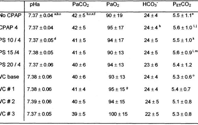
No CPAP CPAP4 PS 1 0 / 4 PS 15/4 PS 2 0 / 4 VC base V C # 1 V C # 2 V C # 3 pHa 7.37 ± 0.04 a4,c 7.37+0.04 7.37 ± 0.05 d 7.38 ± 0.05 7.37 + 0.06 7.38 ±0.06 7.38 ± 0.06 7.39 + 0.06 7.37 ±0.05 PaC02 42 + 5 bc,e,f 42 ± 5 41 ±5 41 ±5 40 ± 6 40 ± 6 41 ± 4 40±5 39 ± 5 Pa02 90 ±19 95 ±17 94 + 17 90 ±13 94 + 13 93 + 13 95+159 94±15 100 ±15 HCCV 24 ± 4 2 4 ± 4h 24 + 5 24 ± 5 23 ± 6 2 4 ± 4 24 ± 4 24 ± 5 22 ± 5 P E T C 02 5.5±1.1a 5.6±1.0l j 5.5±1.0k 5.6±0.9l m 5.4 + 1.2 5.3 ±0.6° 5.4 + 0.7 5.1 ±0.8 5.3 ± 0.8
Values are expressed as mean ± SD. All superscript letters are p < 0.05 : a CPAP 0 vs.
VC # 1 ; b CPAP 0 vs VC # 2 ; c CPAP 0 vs VC # 3 ; d PS 10/4 vs VC # 2 ; e CPAP 0 vs
VC base ;f CPAP 0 vs PS 2014 ; 9 VC # 1 vs VC # 2 ; h CPAP 4 vs PS 2014 ; ' CPAP 4
vs VC # 1 ;j CPAP 4 vs VC # 2 ;k PS 10 / 4 vs VC # 1 ; ' PS 1514 vs VC # 1 ; m PS 15 /
Tabl e 3 : Respirator y parameter s durin g n o CPAP , CPA P 4 c m H 2 0 an d nasa l intermitten t positiv e pressur e ventilatio n i n quie t slee p N o CPA P CPA P 4 P S 10/ 4 P S 15/ 4 P S 2 0 / 4 V C bas e VC# 1 VC# 2 VC# 3 C T insp i EM G 0.8 5 (0.18;0.66-1.07) bod e 0.4 1 (0.27;0.16-0.92) od e 0.2 6 (0.25 ; 0.10-0.85 ) 0.22(0.08;0.11-0.36 ) 0.2 1 (0.13:0.04-0.35 ) 0.5 2 (0.50;0.11-1.35) h J 0.39(0.57;0.09-1.67 ) 0.2 1 (0.08:0.09-0.28 ) a J 0.1 8 (0.06;0.09-0.21) a T A insp i EM G Q e gd. e 0.0 7 (0.06:0.02-0.22 ) e 0.13(0.08;0.05-0.29 ) 0.20(0.19;0.02-0.48 ) 0.0 8 (0.06,0.03-0.20) ' 0.13(0.10:0.04-0.31) ' 0.10(0.02,0.09-0.13) ' 0.2 9 (0.17;0.17-0.48) a Di a insp i EM G 1. 1 (0.2:0.8-1.5 ) bod e 0. 8 (0.2:0.6-1.2 ) °' d e 0. 6 (0.2;0.3-.09 ) 0.5(0.3:0.1-1.1 ) 0. 4 (0.3:0.1-0.7 ) 0. 9 (0.5;0.3-1.7 ) ah J 0. 4 (0.1:0.3-0.7 ) a '' 0. 3 (0.2;0.1-0.5 ) a 0. 3 (0.1:0.1-0.4 ) a Insp i TUA P (c m H2 0 ) -0.7(1.2;-2.5-+0.6) bod e 1. 1 (0.6:0.4-2.3) ° 0. 7 (0.7:0.1-2.4 ) 0. 8 (0.7:0.0-2.5 ) e 1.8(1.3:0.7-4.2 ) 3. 3 (1.1;1.9-5.1 ) afth ' 6. 7 (3.9;2.7-14.4 ) a W 9. 8 (1.9;7.7-11.9 ) * ' 17. 6 (4.4:13.4-23.6 ) a R R (breath s / min 4 2 (9;29-53 ) bcd e 3 5 (10;24-51 ) d e 3 3 (9;24-47) d e 2 5 (8 ; 13-37 ) e 16(4;10-21 ) 4 0 (9;31-54 ) 40(1,38-40 ) 4 0 (0;40 ) 3 8 (5;30-40 ) R R : respirator y rate ; Dia , CT , T A insp i EM G : diaphragm , cricothyroid , thyroarytenoi d phasi c inspirator y etectrica l activity TUA P : tran s uppe r airwa y pressure ; CPA P : continuou s positiv e airwa y pressure ; P S : pressur e support ; V C : volum e contro l ventilation . Not e tha t th e statistica l powe r i s < 80 % fo r Dia , C T an d T A insp i EM G in V C baselin e an d V C #3 . Al l superscrip t letter s ar e P < 0.0 5 : a vs. n o CPA P ; b vs. CPAP 4 ; c vs. P S 1 0 / 4 ; d vs. P S 15 IA ; e vs. P S 2 0 / 4 ; f vs. bas e ; g vs. V C # 1 ; h vs. V C # 2 ; ' vs. V C # 3 .
FIGURE LEGENDS :
Figure 1: Electrical activities of thyroarytenoid, cricothyroid and diaphragm muscles
during nasal intermittent positive pressure ventilation (left: no CPAP; right: pressure support 1 0 / 4 ) during wakefulness. Nasal ventilation inhibits diaphragm and CT EMG and triggers inspiratory TA EMG, which limits sub-glottal (tracheal) pressure until late inspiration. Abbreviations: TA: thyroarythenoid muscle EMG; JTA: moving time averaged TA; CT: cricothyroid muscle EMG; JCT: moving time averaged CT; Dia: diaphragm muscle EMG; jDia: moving time averaged Dia; Pulmonary volumes: sum signal of the respiratory inductance plethysmograph (inspiratory upwards). Inspiration (i) and expiration (e) are delimited according to lung inflation duration.
Figure 2: Variations in inspiratory cricothyroid (CT, left), thyroarytenoid (TA, center) and
diaphragm (Dia, right) EMG from baseline breathing to nasal intermittent positive pressure ventilation in the pressure support (top diagrams, A) and volume control (bottom diagrams, B) modes. The x axis represents varying levels of ventilation while the y axis represents the % of variation from baseline activity (CT and Dia) or % of maximal activity during swallowing (TA) as defined in data analysis.
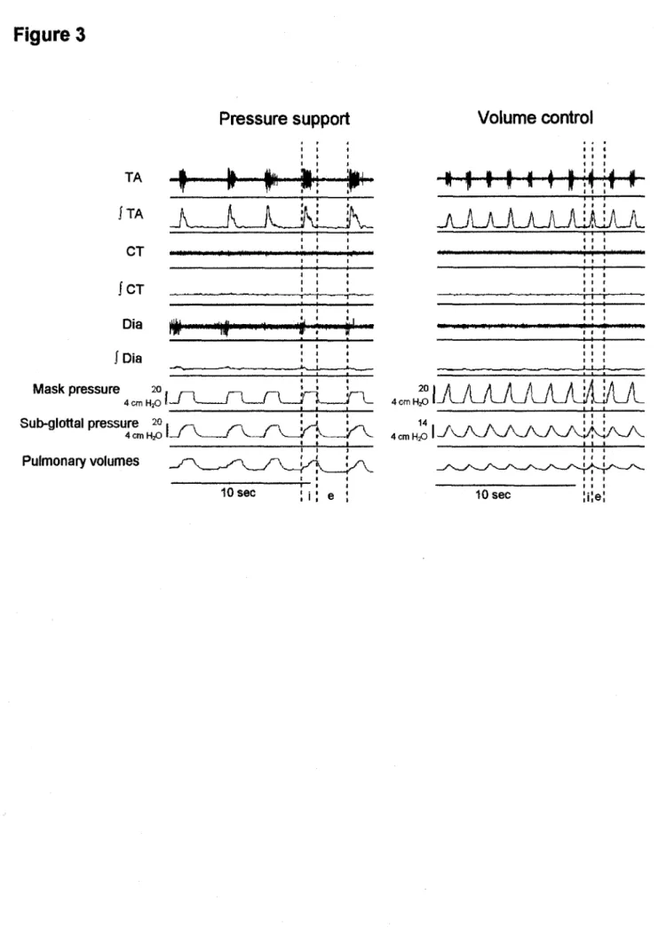
Figure 3: Differences in respiratory parameters between pressure support (left) and
volume control modes (right) during wakefulness. Note: 1) the sudden increase in TA EMG in pressure support compared to a progressive increase in volume control mode; 2) the disappearance of TA EMG in late inspiration in pressure support compared to
continuous TA EMG throughout inspiration in volume control mode; 3) the sub-glottal pressure plateau in late inspiration in pressure support, which is not observed in volume control mode. See figure 1 for abbreviations.
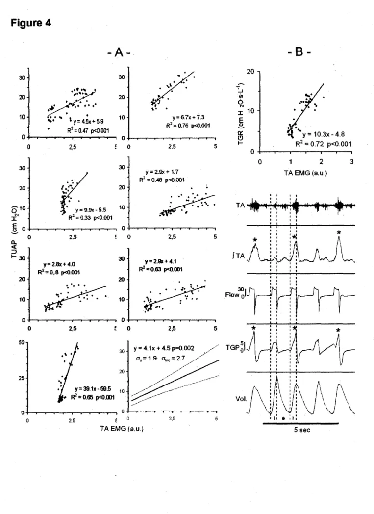
Figure 4: Increase in upper airway resistance during nIPPV in the volume control mode. Figure 4A depicts the relationship between trans-upper airway pressure (TUAP, cm H20) and thyroarytenoid muscle electrical activity (TA EMG, arbitrary units) in seven
lambs during nIPPV in the volume control mode (VC#3) during wakefulness. Note that the bottom left graph has a different y axis scale. The middle lower graph represents the 95% confidence interval equation (ax and oint: SD of the slope and intercept with x axis),
for the six upper lambs (the last lamb being excluded because of significant differences from the other lambs). The increase in TUAP with TA EMG at constant airflow (VC#3) indicates that upper airway resistance increases when TA EMG increases, suggesting that an active glottal closure occurs in response to pulmonary inflations. See text for further explanation.
Figure 4B : The above hypothesis is further supported by the significant relationship between trans-glottal resistance (TGR), and TA EMG in one lamb during nIPPV in the volume control mode during wakefulness (upper graph). Lower right graph shows increase in trans-glottal pressure (TGP, cm H20) during peak TA EMG activity (asterisks)
with constant airflow (Is"1). Dashed lines delimit the inspiratory (i) and expiratory (e)
phases of the respiratory cycle. See figure 1 for other abbreviations.
Figure 5: Complete laryngeal closure during bursts of TA EMG (asterisks) with no transmission of the ventilator strokes (mask pressure, black arrowheads) to the lungs
(sub-glottal pressure, white arrows) in the volume control mode in active sleep (right column). Indeed, with TA inspiratory EMG activity, maximal mask pressure increased from 17 to 32 cmH20 and sub-glottal pressure dropped from 12 to 7 cmH20. High
amplitude, tonic cricothyroid EMG activity was usually present in active sleep in the 5 lambs studied in this particular state. The left traces were obtained during wakefulness in the volume control mode in the same lamb.
Figure 1
No CPAP Pressure support 20 / 4
JA it4'*W^»HWl<H""f"l|f *0* ^fcfiw
1TA
« ^ l ~vA / w W W v J \si ^ ^ ^ W V v N « - /l- * » ) "n^J V - ^ ^ J V-«^_!/ V - ^ - A - ^ "
C T I | ^ I )|M I ^ I I I I > I < | N > I ^ I ^ ; # | I * I I I ^ ) > ' * ' ' ' I I 4 | | I| |I|I ipn mm milium tompip v in % • Nil »mn
1CT Dia f Dia Mask pressure Sub-glottal pressure 4 0 cm H20 Pulmonary volumes 0 cmH2Q
k
yiH|lwll»ptip)mi»|w>»ii'it mm frww n))iiiET
4 cm 4 cm H,0 20 , , , . 5 sec 5 secFigur
e
2
Inspirator
y
C
T
Inspirator
y
T
A
Inspirator
y
diaphrag
m
1. 6 i 1, 2 A 0, 8 0, 4 0, 0 J r I' * I * — i " 7 f " I-U
T
I 1 T * • * 4 10/ 4 15/ 4 20/ 4 4 10/ 4 15/ 4 20/ 4 | -K , I * ' * _ r T (— — *~— | I |— "k —-\ 4 10/ 4 15/ 4 20/ 1, 6 1, 2 B 0, 8 0, 4 0, 0 i" ,L • * • I II I * 1 I — *— I , * — _ — *— I T r ' I T 0 bas e # 1 # 2 # 3 i i i— '• 0 bas e # 1 # 2 # 3 fdimmmi , ^ «••»«• » 0 bas e # 1 # 2 #Figure 3
TA CT ICT Pressure support,TA
_A__A_Jl_A
M N M « M > « > |Dia Mask pressure 4 cm 20in
HO I - / ( -Sub-glottal pressure 20. 4cmH2oLf v—~J v -Pulmonary volumes 10 sec I"*?K
JTL Volume control\ 1 I t M | i< I H
JLAJLAJLJLAliliAJ
4cmHSIAA7UblAAi/Li/Ul
10 secFigure 4
A -
B
- 30- 20- 10-0> •»* *y=4.5x + 5.9 R2 = 0.47p<0.001 T1 "> r—'T I" I i ' i "' I 30- 20- 100 -5* •0
y = 6.7x + 7.3 R2 = 0.76 p<0.001 20 n 2,5 30-20 \ 10- y = 9.9x - 5.5 R2 = 0.33 p<0.001 Q. < — I r~ 2,5 30- 20- 100 -y = 2.9x + 1.7 R2 * 0.48 pO.001 • • 304 20-10 \ y=2.8x + 4.0 R2=0,.8 p<0.001 30 20 10 4 y=2.9x + 4.1 1^ = 0.63 1X0.001 to 6 10 E (!) H y = 10.3x-4.8 R2 = 0.72 p<0.001 - i 1 1 1 r 1 0 1 2 3 TA EMG (a.u.)lfr
illiit>ir<|H
i Flow 0I-M 50 n 25 J 30 20 \ y=39.1x-58.5 10 %* R2 = 0.65 pO.001 - T — r — — f — — - r — i I1" ' I ' 'I 'I I I ' I1 I I ' I 2,5 5 y = 4.1x + 4.5p=0.002 . / TGP0I ox=1.9 0lnt = 2.7 / 2,5 2.5Figure 5
Wakefulness
TA •*f'"''*""*f"^"#""<|tiwf"
|TA
J U J U U U J L
Q"J" «»•««'iiwi»|i|11|ni ii| ii mn n| |ii |ntmnniii|iii |ln m»1\v
I C T , • Q ja I K I W I ^ * I I * I * lltpmM UMiiimiiMMmnMIHin J Dia Mask pressure 17, 1 „ 1 „ 4 cm H20 l - Z L y u A J U ^ REM * * * * * * * * * -A . . . A
hi^iiiiit'liW^iiiM
1!
4 cm H^ I JUJJL/L/L/LJJUL/UL/JL
Sub-glottal pressure 121 4 cm H20 I Pulmonary volumes 121
4 cm H2o <II I i I I I I I
10 sec 10 secDiscussion
Les resultats obtenus dans cette etude effectuee chez des agneaux nouveau-nes demontrent que la VPPIn entratne a I'inspiration une diminution de I'activite des muscles dilatateurs glottiques (CT) et une augmentation des muscles constricteurs glottique (TA). De plus, nos resultats suggerent que ces modifications de I'activite des muscles larynges sont responsables d'une fermeture glottique active qui limite la ventilation pulmonaire en VPPIn. Ce sont done les premieres preuves directes que le retrecissement glottique deja observe chez les adultes par endoscopie lors de la VPPIn est cause par une modification de I'activite des muscles intrinseques du larynx. De plus, cette etude nous offre pour la premiere fois des donnees sur 1'impact de la VPPIn au niveau de la dynamique laryngee des nouveau-nes. Nous crayons que de tels resultats sont hautement pertinents pour I'etude de la physiologie respiratoire et pourraient etre a I'origine de modifications significatives des traitements respiratoires en periode neonatale.
Activite Snspiratoire des muscles glottiques en nlPPV
Activite inspiratoire du muscle thyroarytenoTdien: Le muscle TA est bien
etabli comme un constricteur glottique expiratoire qui se contracte normalement au debut de I'expiration (HARDING et al., 1986, KUNA et al., 1988, LU et al., 2005, LUDLOW, 2005, SAMSON et al., 2006, ZHOU et al., 1989). La contraction expiratoire du muscle TA est particulierement importante en periode neonatale comme mecanisme de freinage expiratoire, favorisant le maintien d'une capacite
residuelle fonctionnelle (CRF) plus elevee. Les nouveau-nes ayant une cage thoracique moins rigide et un parenchyme pulmonaire moins compliant, ce maintien actif de la CRF previent Patelectasie pulmonaire et ameliore Poxygenation (BARTLETT, 1989, DIAZ et al., 1996, HARDING et al., 1986). Par ailleurs, la presence d'activite inspiratoire du TA est tres rarement rapportee lors de la respiration normale chez un sujet sain (PRAUD et al., 1995). Une telle activite inspiratoire du muscle TA est cependant decrite dans differentes experiences ou dans des conditions pathologiques incluant: le gasping anoxique
in vivo (HUTCHISON et al., 2002, THUOT et al., 2001) et in vitro (LIESKE et al.,
2000); I'hypoxie ou I'anoxie (DUTSCHMANN et PATON, 2002); lors de Pinjection intraveineuse de capsa'icine (LU et al., 2005); lors de I'occlusion des voies aeriennes superieures (KIANICKA et PRAUD, 1997); ou chez les patients souffrants de sclerose laterale amyotrophique (ISOZAKI et al., 1994). Les mecanismes physiologiques a I'origine de I'activite phasique inspiratoire du TA ainsi que les consequences associees sont mal connus.
A notre connaissance, I'activation du muscle TA lors de inflation pulmonaire en VPPIn est une observation unique dont I'origine vient probablement de la stimulation des recepteurs des voies aeriennes intra et/ou extra-thoraciques par ('augmentation de la pression transmural. En effet, I'apparition plus precoce d'EMG du TA inspiratoire en mode pression de support (PC) qu'en mode volume controle (VC) (voir figures 1 et 3 de Particle) pourrait etre due a Paugmentation plus rapide et soudaine de la pression inspiratoire positive en mode PS. Premierement, I'apparition inspiratoire dEMG du TA pourrait etre stimulee par les
afferences des recepteurs broncho-pulmonaires intra-thoraciques. Une revue de litterature recente concernant I'impact des afferences pulmonaires sur la modulation des muscles des voies aeriennes superieures (BAILEY et FREGOSI, 2006) montre qu'une stimulation des recepteurs pulmonaires a adaptation lente inhibe I'activite du TA (BARTLETT et al., 1973, HARDING et al., 1986, HUANG et al., 1989). Par ailleurs, quelques donnees suggerent que la stimulation des recepteurs pulmonaires a adaptation rapide et des fibres C pourraient augmenter I'activite phasique de I'EMG du TA (HOLMES et REMMERS, 1989, STRANSKY et al., 1973). Une augmentation des afferences vagales provenant de ces recepteurs pourrait done expliquer les changements de I'activite inspiratoire du TA observes dans notre etude. Cependant, les changements de I'activite du TA cites dans ces etudes se retrouvaient tous dans la phase expiratoire du cycle respiratoire. Deuxiemement, il est aussi possible que I'apparition de TA inspiratoire soit stimulee par des afferences venant des baror^cepteurs a pression positive situes dans les voies aeriennes superieures sur la muqueuse laryngee et pharyngee (MATHEW et al., 1984, SANT'AMBROGIO et al., 1983). En effet, une etude basee sur un modele de larynx isole chez le cochonnet a recemment demontre qu'une stimulation des barorecepteurs larynges par une pression positive expiratoire entrame une augmentation de I'activite EMG du muscle TA lors de I'expiration (STELLA et ENGLAND, 2001). Troisiemement, I'hypothese que les recepteurs larynges sensibles au debit (thermorecepteurs) (SANT'AMBROGIO et al., 1983, STELLA et ENGLAND, 2001) soient impliques est peu probable car I'air provenant du ventilateur etait rechauffe et humidifie dans notre protocole experimental, empichant de ce fait I'activation des
thermorecepteurs. Finalement, meme si I'hypocapnie entratnee par une hyperventilation passive en VPPIn chez des adultes a deja ete reliee a une augmentation du TA expiratoire (KUNA et al., 1993), il est peu probable que ce soit le cas ici. En effet, la grande majorite des agneaux de notre experience avaient des valeurs normocapniques durant tout le protocole. De plus, nous n'avons pas note de difference dans I'EMG du TA entre les agneaux qui ont obtenu quelques valeurs hypocapniques et ceux dont les valeurs etaient normales.
Activite inspiratoire du muscle cricothyroTdien: Comme demontre recemment chez les humains et les agneaux, le muscle CT agit comme un dilatateur de la glotte lorsque associe au muscle cricoaryteno'idien posterieur lors de I'inspiration (KUNA et al., 1994, LUDLOW, 2005, SAMSON et al., 2006). Nos donnees demontrant la disparition de I'activite EMG inspiratoire du muscle CT sont en accord avec des observations similaires chez des humains adultes lors de VPPIn en mode PS (KUNA et al., 1993). La disparition concomitante de I'activite phasique du diaphragme et du CT est probablement en partie causee par I'augmentation des afferences vagales venant des recepteurs bronchopulmonaires a adaptation lente (BAILEY et FREGOSI, 2006). Deuxiemement, il est possible que la perte des pressions inspiratoires negatives soit la cause de la diminution de I'activite inspiratoire du CT. En effet, les pressions negatives inspiratoires en eupnee augmentent normalement I'activite du muscle cricoaryteno'idien posterieur (STELLA et ENGLAND, 2001). Finalement, bien que I'hypocapnie entratne une diminution de I'activite
inspiratoire du CT (KUNA et al., 1993), ce mecanisme est peu probable dans notre etude comme nous I'avons precedemment explique pour le TA.
Fermeture qlottique active lore de I'inspiration en VPPIn
L'augmentation de I'activite EMG du TA et la diminution de celle du CT lors de reflation pulmonaire suggere fortement que la glotte se referme de fagon active en VPPIn, comme suppose par des observations endoscopiques en mode VC chez des adultes humains (RODENSTEIN, 2001). Cette equipe de chercheurs a emis I'hypothese que la fermeture glottique inspiratoire qu'ils observent est tres probablement secondaire a une activation des muscles larynges adducteurs sans pour autant pouvoir le prouver experimentalement. De notre cote, nous demontrons dans cette etude que l'augmentation de I'EMG du TA est fortement reliee a une augmentation de la pression a travers les voies aeriennes (PTVAS) inspiratoire chez tous nos agneaux. Puisque ces mesures ont ete effectuees dans un meme mode ventilatoire pour chaque agneau (VC-3), nous pouvons affirmer que, a debit constant, l'augmentation des PTVAS indique une augmentation proportionnelle des resistances des voies aeriennes superieures. En somme, l'augmentation de I'EMG du TA est reliee avec une augmentation des resistances des voies aeriennes superieures. Cette augmentation des resistances pourrait theoriquement etre reliee a une contraction des muscles constricteurs pharynges ou encore a des mecanismes passifs comme un retrecissement de I'ouverture laryngee cause par I'effet Venturi lors des hauts debits inspiratoires. Cependant, plusieurs resultats suggerent fortement qu'une fermeture glottique active est au moins partiellement responsable de
I'augmentation des resistances des voies aeriennes superieures observee dans notre etude. Premierement, nous avons montre dans I'article que I'augmentation du TA inspiratoire est fortement reliee a une augmentation des resistances au niveau des cordes vocales chez un agneau (figure 4B). Deuxiemement, les resultats obtenus chez des humains adultes demontrent que la glotte se retrecit lors de I'inspiration en VPPIN en mode VC (JOUNIEAUX et al., 1995(1),
JOUNIEAUX et al., 1995(2)). Finalement, I'arret complet de transmission des
pressions du ventilateur au niveau tracheal demontre en sommeil agite suggere fortement un mecanisme actif (figure 5).
La consequence principale d'une augmentation des resistances laryngees lors des inflations pulmonaires en VPPIn est de limiter la ventilation pulmonaire. Cela est evident sur riconographie en eveil de la figure 4B en eveil qui montre une diminution des volumes courants lorsque I'EMG du TA et les pressions trans-glottiques sont au plus haut. De plus, les bouffees d'activite du muscle TA observees en sommeil agite etaient assez importantes pour empecher totalement la transmission des insufflations du ventilateur a la trachee (figure 5). Bien qu'une telle activite du muscle TA ait deja ete decrite chez des humains adultes en sommeil agite en eupnee (KUNA et al., 1988) et chez I'agneau nouveau-ne en VPPIn (SAMSON et al., 2005), la relation avec la ventilation pulmonaire n'a pas ete discutee. Finalement, une autre consequence d'une augmentation des resistances laryngees est d'augmenter le risque de distension gastro-intestinale et de perforation gastrique si les pressions des voies aeriennes superieures