HAL Id: hal-00751760
https://hal.inria.fr/hal-00751760
Submitted on 14 Nov 2012
HAL is a multi-disciplinary open access
archive for the deposit and dissemination of
sci-entific research documents, whether they are
pub-lished or not. The documents may come from
teaching and research institutions in France or
abroad, or from public or private research centers.
L’archive ouverte pluridisciplinaire HAL, est
destinée au dépôt et à la diffusion de documents
scientifiques de niveau recherche, publiés ou non,
émanant des établissements d’enseignement et de
recherche français ou étrangers, des laboratoires
publics ou privés.
Active stabilization of ultrasound image for
robotically-assisted medical procedures
Caroline Nadeau, Alexandre Krupa, Pedro Moreira, Nabil Zemiti, Philippe
Poignet, Jacques Gangloff
To cite this version:
Caroline Nadeau, Alexandre Krupa, Pedro Moreira, Nabil Zemiti, Philippe Poignet, et al.. Active
stabilization of ultrasound image for robotically-assisted medical procedures. Hamlyn Symposium on
Medical robotics, Jul 2012, London, United Kingdom. �hal-00751760�
Active stabilization of ultrasound image
for robotically-assisted medical procedures
C. Nadeau
1, A. Krupa
1, P. Moreira
2, N. Zemiti
2, P. Poignet
2, J. Gangloff
31
IRISA, Inria Rennes-Bretagne Atlantique, France,
2
LIRMM, UMR 5506, CNRS Université de Montpellier 2, 34095 Montpellier, France
3
LSIIT, UMR 7005 CNRS-Université de Strasbourg I, 67400 Illkirch, France
[email protected]
INTRODUCTION
In the context of robotic assistance to medical gestures, we propose solutions to stabilize the ultrasound (US) image by actively compensating for the physiological motions of the patient. The considered applications are for instance the assistance for diagnoses or hepatic tumor biopsies where the liver and the tumors mainly undergo the respiratory motion. Other clinic applications, such as prostate cancer brachytherapy have been identified in [1] that could benefit from such robotic image stabilization.
The per-operative image provided by the US probe and the contact force applied to the probe are used to control the six degrees of freedom (dof) of the robot. To deal with the low quality of the US images, we propose to use the intensity of the image as visual features in a visual servoing control loop [2][3]. This vision control is associated with a force control to ensure a constant force applied to the probe. Finally, a predictive controller is implemented in the control law to take advantage of the repetitiveness of the physiological motions. We present first ex-vivo robotic results on animal tissues where we compensate for the 3D motions using successively 2D and 3D probes.
MATERIALS AND METHODS
2D and 3D intensity-based visual servoing
In order to avoid time-consuming and difficult US image processing step, we implement an intensity-based visual servoing to control the motions of the probe. This approach is applied first with a classical 2-5 MHz 2D probe (Sonosite, C60) with a frame rate of 25Hz and then with a 3D motorized one (Ultrasonix, 4DC7) that provides 5vol/s.
With a 2D probe, the visual features vector is built with the intensity values of the pixels of a region of interest (ROI) of the US image. The interaction matrix Ls that
links the variation of these image features to the motion of the probe is modeled in [2] and used in an image-based visual servoing control law. This interaction matrix is computed from the 3D image gradient that is estimated once at the initial pose of the probe, without being updated during the tracking task.
In the 3D case [3], a 3D deformable grid, containing a set of control points, is attached to the US volume and
the intensity-based approach is applied to estimate the control points displacement between the successive volume acquired by the 3D probe. From the displacement of these points, a thin-plate spline model is considered to compute the deformation of the grid that corresponds to the non-rigid motion of the target. A rigid transformation is finally extracted from this motion model and used to control the robot by position-based visual servoing.
Vision/force control
For guaranteeing that a constant force is applied by the probe to the skin during the tracking task, we implement an hybrid vision/force control based on an external control loop approach. In the meantime, it also improves the safety of the task. The force control is used to servo the normal translation motion of the probe while the five remaining dof are controlled by the vision.
Predictive controller
In the classic visual servoing control law, the controller does not take advantage of some knowledge on the disturbance model and more particularly of its periodicity. In this work, we implement in the control loop a repetitive predictive controller (R-GPC) in order to anticipate the effect of the disturbance [4] .
RESULTS
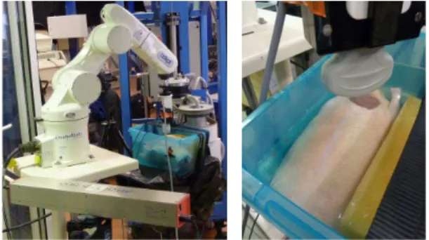
The first ex-vivo robotic results on real tissues with the US intensity-based visual servoing are presented here. For this validation, a chicken stuffed with pig liver and kidneys and immersed in a water tank to avoid air gaps inside its body is carried by a 6 dof robot. A 3D periodic motion composed of combined rotations and translations is applied to this phantom. The tracking task is performed successively with a 2D and a 3D US probe, mounted on a six dof anthropomorphic robotic arm, which is also equipped with a force sensor. The experimental setup is presented in Fig. 1. Two optical markers are fixed on the probe and on the phantom and provide the relative pose of both elements thanks to an EasyTrack system. This relative pose is only used as a ground truth to validate the tracking task.
The robotic arm is manually positioned above the phantom and the force control is applied with a desired force of 3N to put the probe in contact with the chicken surface.
Fig. 1 Robotic setup (left) with the animal phantom (right). A desired ROI is then delineated in the US image and a small back and forth motion is automatically realized to acquire parallel images around the desired image and compute the interaction matrix involved in the control law. A 3D periodic motion along all translations and one rotation (around the probe axis) is applied to the phantom. The disturbance has a period of 8s and generates amplitudes of motion of 18mm and 8mm along horizontal axes and 10mm along the vertical axis with a rotation of the phantom of 4deg.
The results of one tracking task with the 2D probe are displayed in Fig. 2. The ROI is initialized in the first US image (a) and the visual control is launched. The disturbance motion is applied at t=20s, then at t=160s the compensation is stopped. To visually validate the task, a visual error is defined as the Euclidean norm of this visual vector and is displayed on the curve (b). After one period of disturbance (at t=28s), this error is significantly reduced thanks to the predictive effect of the R-GPC. The curve (c) shows the respect of the force reference throughout the tracking task. Finally the curve (d), obtained thanks to the EasyTrack system, validate the robotic task in terms of pose since the relative pose of the probe with respect to the phantom is maintained constant during the tracking (t<160s).
(a) (b)
(c) (d) Fig. 2 Ex-vivo results : compensation with a 2D probe. Experiments were also performed with the 3D probe. In this case, no initialization step is required to compute the image gradient and the visual servoing is launched (at t=60s) while a sinusoidal disturbance, of 12.5s of period, occurs. The results of one tracking task are presented in Fig. 3. A 3D grid of 3!3!3 control points is defined in the first US volume provided by the 3D probe (a) to track the motion in successive volumes (see [3]). The central plane of the US volume is shown in
(b). The force control ensures a contact force of 3N during the whole robotic task (c) and the EasyTrack system gives a validation of the accuracy of the tracking in terms of pose (d).
(a) (b)
(c) (d) Fig. 3 Ex-vivo results : compensation with a 3D probe.
DISCUSSION
We have presented the first ex-vivo results of actively compensated periodic motions with a robot controlled along five directions with an intensity-based visual servoing and along one direction with force. These results show the asset of the approach that does not require features extraction or segmentation of the image and is therefore well adapted for dealing with any kind of anatomic images. With a 2D probe, a frame rate of 25Hz is reached but an initialization step is required before applying the tracking. The 3D probe, on the other hand, can be used to avoid this step, but the volume rate of 5Hz limits the dynamics of the tracking. In both cases, the vision control has been coupled with a force control running at 1kHz to ensure the contact of the probe.
Acknowledgement
This work was supported by the ANR project US-Comp of the French National Research Agency.
REFERENCES
[1] Krupa A., Fichtinger G., Hager G.D., Real time motion stabilization with B-mode US using image speckle information and visual servoing. Int. Journal of Robotic Research, 2009; 28:1334-1354.
[2] Nadeau C., Krupa A., Intensity-based direct visual servoing of an ultrasound probe. IEEE Int. Conf. on Robotics and Automation, 2011 May; 5677-5682. [3] Lee D., Krupa A., Intensity-based visual servoing for
non-rigid motion compensation of soft tissue structures due to physiological motion using 4D ultrasound. IEEE/RSJ Int. Conf. on Intelligent Robots and Systems, 2011 Sep.; 2831-2836.
[4] Gangloff J., Ginhoux R., De Mathelin M., Soler L., Marescaux J., Model predictive control for compensation of cyclic organ motions in teleoperated laparoscopic surgery. IEEE Trans. on Control System Technology, 2006; 14:235-246.