Faculty de g6nie
D^partement de g£nie chimique et de g6nie biotechnologique
CARACTERISATION ET OPTIMISATION D'UN
REACTEUR POUR L'INGENIERIE TISSULAIRE 3D
M6moire de maitrise Sp6cialit£: g£nie chimique
Justin DUBOIS
Jury : Patrick VERMETTE (directeur) Pierre PROULX
Nicolas ABATZOGLOU
Sherbrooke (Quebec) Canada mars 2011
Library and Archives Canada Published Heritage Branch Biblioth&que et Archives Canada Direction du Patrimoine de l'6dition 395 Wellington Street Ottawa ON K1A 0N4 Canada 395, rue Wellington Ottawa ON K1A 0N4 Canada
Your file Votre r6f6rence ISBN: 978-0-494-83747-4 Our file Notre reference ISBN: 978-0-494-83747-4
NOTICE:
The author has granted a non
exclusive license allowing Library and Archives Canada to reproduce, publish, archive, preserve, conserve, communicate to the public by
telecommunication or on the Internet, loan, distrbute and sell theses
worldwide, for commercial or non commercial purposes, in microform, paper, electronic and/or any other formats.
AVIS:
L'auteur a accords une licence non exclusive permettant £ la Biblioth&que et Archives Canada de reproduire, pubiier, archiver, sauvegarder, conserver, transmettre au public par telecommunication ou par I'lnternet, preter, distribuer et vendre des theses partout dans le monde, d des fins commerciales ou autres, sur support microforme, papier, 6lectronique et/ou autres formats.
The author retains copyright ownership and moral rights in this thesis. Neither the thesis nor substantial extracts from it may be printed or otherwise reproduced without the author's permission.
L'auteur conserve la propriety du droit d'auteur et des droits moraux qui protege cette these. Ni la th6se ni des extraits substantiels de celle-ci ne doivent dtre imprimis ou autrement reproduits sans son autorisation.
In compliance with the Canadian Privacy Act some supporting forms may have been removed from this thesis.
While these forms may be included in the document page count, their removal does not represent any loss of content from the thesis.
Conform6ment d la loi canadienne sur la protection de la vie priv6e, quelques formulaires secondares ont 6t6 enlev£s de cette thdse.
Bien que ces formulaires aient inclus dans la pagination, il n'y aura aucun contenu manquant.
RESUME
Vers l'objectif de crger ou remplacer des tissus endommag€s, il est essentiel d'6tablir un proc£d6 pouvant r6pondre aux multiples demandes biologiques. La culture sur p£tri ou en deux dimensions n'a qu'un potentiel tr£s limits dfi aux limitations, en autres, de 1'approvisionnement en oxygfcne.
Dans ce projet, un bioproc6d£ autonome et complet a 6t6 mis en place afin de r£pondre & ces besoins cellulaires. Le rgacteur est versatile puisque plusieurs designs de chambres de culture peuvent y etre connects. Le pH et la concentration en oxyg£ne y sont modules par deux contrdleurs PI. La pression, la temperature et le d£bit volum£trique sont constamment enregistrds. Un pulsateur permet de mimer la pulsation cr£ee par le cceur.
Dans ce memoire, afin de caracteriser le r£acteur, les r£sultats d'une analyse RTD seront presents. Des mesures de perm6abilit£ si I'aide d'une sonde fluorescente et de diffusion par imagerie par resonance magn&ique (IRM) permettront de caracteriser plus sp£cifiquement la chambre de culture cellulaire. II sera brifcvement d£montr£ comment le r£acteur a 6x6 optimist afin de rdduire les risques de contaminations, ligalement, la procedure pour d6velopper les outils de controle PI sera d£taill£e. Finalement, deux designs de chambre de culture seront pr6sent£s afin de cr6cr des micro-vaisseaux sanguins.
MOTS CLES : bior6acteur & perfusion, analyse RTD, permlabilitl, coefficient de diffusion apparent, controleur PI, chambre de culture cellulaire, orientation de micro-vaisseaux sanguins, cellules endothelials.
SUMMARY
Toward the objective to create or to replace impaired tissues, it is essential to establish a culture process allowing tissue growth in vitro. The Petri dish culture or the traditional 2D culture has only a limited potential, mainly caused by a poor oxygen mass transfer to feed larger tissue constructs.
In this project, an autonomous and complete bioprocess has been built to fullfill these needs. The developed reactor is versatile because many cell culture chamber designs can be connected to it. Two proportional-integral algorithms (PI) can control the dissolved oxygen concentration and the pH. The pressure, the temperature and mass flow rate are recorded in real time. An actuator allows mimicking the cardiac output flow.
In this memoire, it will be demonstrated how the reactor has been optimized to reduce the risk of bioburden. Also, the procedures to develop the control tools of the PI algorithm will be detailed. To characterize the reactor, a RTD study will be presented. To characterize the cell culture chamber, a permeability analysis using fluorescent microscopy and magnetic resonance imaging (MRI) will be exposed. Finally, two cell culture chamber designs to orient microvessel formation have been tested and these results will be presented.
KEYWORDS: RTD analysis, permeability, apparent diffusion coefficient, PI controller, cell culture chamber.
REMERCIEMENTS
J'aimerais remercier toutes les personnes qui ont apporte un support scientifique important k ce projet. Merci a l'6quipe de techniciens du Departement de genie et de g&iie biotechnologique de l'Universite de Sherbrooke, particuli&rement £ M. Serge Gagnon, M. Alain Levesque et M. Marc G. Couture.
Merci a l'equipe de recherche du Laboratoire de bio-ingenierie et de biophysique de rUniversite de Sherbrooke. Merci ^ M. Andriy Shkilnyy, M. Georges Sabra, M. Evan Dubiel, Mme Julie Chouinard, Mme Valerie Centis, Mme Carina Kuehn et M. Jamie Sharp.
Merci aux professeurs du departement pour leur aide, en particulier M. Pierre Proulx. Merci a M. Patrick Vermette qui a 6te un excellent superviseur dans ce projet.
Pour leur incroyable soutien moral, merci aux deux femmes de ma vie; ma conjointe Genevieve Leblanc et ma m&re Colette Aub£. Merci egalement a mon fr£re David Dubois, source d'inspiration par son ethique de travail. Merci a Andre Nault et a ma clique d'amis pour leur amitie.
TABLE DES MATIERES
ACRONYMES 8
Introduction g^n^rale... 10
Section 1.1: Mise en contexte 10
1.1.1 Les maladies cardiovasculaires et le genie tissulaire 10
1.1.2 L'ing£nierie vasculaire du systeme circulatoire 11
Section 1.2: Definition du projet de recherche et objectifs— .. 13
Section 1.3: Contributions originates ......a... 14
Section 1.4: Division des chapitres...„...15
Section 1.5: References ~ ... 16
1.1 Bag bioreactors / wave and undertow bioreactors... ... .. 19 1.2 Perfusion 1^ior63ctors ... ... 21 1*3 Biaxial liioreactors ... 2 3 1.4 Strain bioreactors ...2 3 1.5 Examples of commercially available bioreactors for tissue engineering 24 1.6 Conclusions...
1.7 References ..«...M...~...n...n...M.28 CHAPTER 2: FLOW DYNAMICS WITHIN A BIOREACTOR FOR TISSUE ENIGNEERING BY RESIDENCE TIME DISTRIBUTION ANALYSIS COMBINED WITH FLUORESCENCE AND MAGNETIC RESONANCE IMAGING TO INVESTIGATE FORCED PERMEABILITY AND APPARENT DIFFUSION COEFFICIENT IN A PERFUSION CELL CULTURE
CHAMBER 32
2.2 Introduction... 34
2.3 Materials and Methods.. —... ... ... 35
2.4 Results an^l ^ 4.^ 2.5 Conclusions ...~...m...~...^...~..~...49 2.6 Acknowledgements... 2.7 References...51 2.8 Tables 54 2.9 Figure captions ...5 5
CHAPTER 3: OPTIMIZATION OF A PERFUSION BIOREACTOR OPERATED WITH A MICRO-CHANNELLED CULTURE CHAMBER TO SUPPORT ENDOTHELIAL CELL
ATTACHEMENT 65
3.1 Abstract — 66
3.2 Introduction.. ... „ . .67
3.3 Materials and Methods 71
3.4 Results and Discussion 77
3.5 Conclusions 82 3.6 Acknowledgments... 83 3.7 References 84 3.8 Figure captions 87 3.9 Annexes 6
APPENDICE AU CHAPITRE 3 : AMELIORATION DU DESIGN POUR R&DUIRE LES RISQUES DE CONTAMINATION ET POUR AUGMENTER LA CROISSANCE CELLULAIRE
99
i ••• •••••• •••• •••• ... .... mm .... ...
A.2 Modifications du bior£acteur m...99
A.3 Designs des chambres de culture cellulaire , 101
A«4r Contamination •••••••••••••••••••••••••••••••••••••••a 10 3
L6gende des ii gures ....i 10 s
ACRONYMES
2D/3D: two dimensions/three dimensions ADC: apparent diffusion coefficient ADH: antidiuretic hormone
ANP: atrial natriuretic peptide BSA: bovine serum albumine CFD: computational fluid dynamics
CFSE: carboxyfluorescein succinimidyl ester CSTR: continuous-stirred tank reactor CVD: cardiovascular disease(s) DMSO: dimethyl sulfoxide DNA: deoxyribonucleic acid
DAPI: 4',6-diamidino-2-phenylindole DO: dissolved oxygen
ECGS: endothelial cell growth supplement ECM: extracellular matrix
eNOS: endothelial nitric oxide synthase FBS: fetal bovine serum
FOPDT: first-order plus dead time HBSS: Hank's balanced salt solution
HUVEC: human umbilical vein endothelial cell ID: internal diameter
LSQ: least square fit
PBS: phosphate buffer solution PDGF: platelet-derived growth factor PET: positron emission tomography P: proportional
PD: proportional-derivative
PFA: paraformaldehyde PI: proportional-integral
PID: proportional-integral-derivative MMP: matrix metalloproteinase MRI: magnetic resonance imaging mTG: m-transglutaminase
NO: nitric oxide
R&D: research and development ROI: region of interest
RGD: Arginine-Glycine-Aspartic acid RTD: residence time distribution SISO: single input single output
SOP: standardized operational procedures VEGF: vascular endothelial growth factor vWF: von Willebrand factor
Introduction generate
Section 1.1: Mise en contexte
1.1.1 Les maladies cardiovasculaires et le genie tissulaire
L'ing£nierie tissulaire ou la m&iecine r£g£n£ratrice est une science progressiste combinant la biologie fondamentale, la chimie (materiaux) et l'ingenierie. Le but de l'ingenierie tissulaire est d'ameiioier, de cr^er ou de Sparer des organes ou des tissus humains (ex.: os, tendon, valve cardiaque, etc.).
Malgrd un d&lin pendant les 40 derni&res ann^es, les maladies cardiovasculaires (CVD) etaient toujours en 2006 la cause la plus importante de mortality (30% de tous les d6c£s) [Heart and stroke foundation (http://www.heartandstroke.com/site/c.iklOLcMWJtE/b-.348399l/k.34A8/Statistics.htm#heartdisease)]. remplac^es depuis peu par le cancer de 2006
k aujourd'hui [1]. Les coflts engendrgs par ces maladies au Canada sont lvalues k 22.2
milliards de dollars par ann£e [2].
Les CVD mgritent amplement tout l'effort de recherche et d^veloppement (R&D) g6n£r6 dans le secteur du genie tissulaire. II existe une application indirecte tout aussi importante dans l'ingenierie vasculaire du systfcme circulatoire (artfcres, veines, capillaires). Puisque chaque cellule du corps humain doit 6tre & moins de 100pm d'une source sanguine, n'importe quel substitut de tissus humains devrait etre systematiquement accompagne d'un reseau vasculaire fonctionnel ou d'une alternative pour l'alimenter (bioprocedes). Les fonctions d'un reseau vasculaire sont [3]:
• Alimenter les cellules (sucies, O2, vitamines, etc.) ; • Enlever les dechets (metaboliques);
• Fournir une stimulation mecanique necessaire (contrainte de cisaillement, pulsation, etc.).
Dans les deux prochaines sections, l'ingenierie vasculaire du syst&me circulatoire et les biorgacteurs seront introduits comme les 2 voies essentielles pour cr6er des substituts de tissus
in vitro.
1.1.2 L'ingenierie vasculaire du syst&me circulatoire
L'ingenierie vasculaire du systeme circulatoire est seulement une sous-section de ringenierie tissulaire qui se concentre sur les veines, les art&res et les capillaires. Cette sous-section peut etre divisee comme suit:
• angiogenic (formation de vaisseaux sanguins a partir de structures preexistantes); • vasculogen^se (formation de vaisseaux sanguins utilisant des cellules progenitrices ou
cellules souches);
• substituts synthetiques pour petits et larges vaisseaux sanguins.
Depuis longtemps, la vasculogen£se in vivo etait un phenom^ne qu'on croyait arriver uniquement au stade embryonnaire. Cependant, des etudes ont demontre que la vasculogendse
in vivo pouvait survenir au stade adulte durant les cycles menstruels [4] et dans le
developpement de tumeurs [5]. La vasculogen&se in vitro, d'un autre c6te, est basee sur la capacite des cellules souches ou progenitrices k creer un equivalent de structure vasculaire en dehors du corps.
Les capillaires ont un contact direct avec les tissus et fournissent la zone d'echanges essentielle pour la survie et la croissance des tissus. Les capillaires sont les types de vaisseaux sanguins les plus simples puisqu'une unique couche de cellules endotheliales avec un basal
lamina forme sa structure. Quelquefois, une seule cellule peut couvrir enticement le diam&tre
de la lumi£re [6]. Malheureusement, la culture de cellules endotheliales jusqu'i la confluence est encore bien loin d'un vaisseau sanguin fonctionnel. Les cellules endotheliales de vaisseaux sanguins sont liees ensemble par des jonctions serrees ou relachees et sont allongees dans le
sens de l'ecoulement comparativement k la culture en statique. Le diamdtre moyen de la lumiere d'un capillaire est de 8-9|im avec une epaisseur de parois de 0.5jun [6].
II existe trois types de capillaires : continus, fenetres et discontinus [7]. Chacun de ces types est base sur la taille des pores presentes et consgquemment, sur les molecules qui peuvent pgnetrer ou sortir. Les capillaires continus n'ont aucune taille sp^cifique et permettent seulement aux ions et k l'eau de diffuser. Les capillaires fenetres ont des pores de 60-80 nm qui laissent passer les petites prolines par diffusion et finalement, les capillaires discontinus ont des pores de 30-40 tun qui, combines £ un manque de basal lamina et de fonctions gap entre les cellules, permettent k des globules rouges et des prolines du serum de penetrer [7]. L'endothelium peut aussi echanger des nutriments et des molecules par pinocytose et par les v6sicules de transport, comme transport actif.
L'endothelium interagit aussi avec son environnement en s£crgtant ou en r^agissant & divers stimuli. Plusieurs de ces stimuli sont essentiels pour l'h£mostase (ex. : epinephrine, hormones antidiuretiques (ADH), ANP) et ne sont pas naturellement disponibles pour tester dans des conditions in vitro puisque ces derniers sont produits par d'autres organes pour varier la pression et le volume sanguins [6]. Cependant, il existe d'autres stimuli chimiques qui jouent un role direct sur la reponse de l'endothelium comme l'angiotensine II, l'endotheiine, le PDGF, l'oxyde nitrique (NO), l'histamine, la prostacycline et plusieurs autres. La production de ces molecules ou la reponse a ces molecules (vaso-activite) pourrait etre un bon indicateur de la creation d'un vaisseau sanguin fonctionnel [6].
Les caracteristiques d'un vaisseau sanguin fonctionnel ne sont toujours pas etablies dans la litterature. Logiquement, un substitut vasculaire devrait avoir les memes attributs que les vaisseaux natifs [8]. Puisque l'hemostase est aussi reguiee par son environnement immediate (ex. : diaped&se, tissus/organes, etc.), il est alors impossible de valider in vitro
toutes les fonctions d'un vaisseau sanguin natif. Ainsi, quelques tests ont ete proposes dans le but de valider les vaisseaux sanguins.
• pression d'gclatement [9, 10]; • mesures de conformite [9,10];
resistance a la intention de suture [9]; fatigue dynamique [9];
Validation physique fatigue statique [9];
vessel ringlet pull tests [10];
indice de rigidity [10]; ^
module elastique maximal [10]; activation du factor XII [11];
temps d'activation partielle de la thromboplastine (aPPT) [11]
• eNOS [12]; >
Encore une fois, I'absence d'un consensus pour valider les fonctions et les proprietes d'un vaisseau sanguin est causee par les interactions tr&s complexes entre l'endothelium et les composantes sanguines. Par consequent, il existe aucune fa^on de valider parfaitement la fonctionnalite des vaisseaux sanguins in vitro. La meilleure fa9on de valider un substitut in
vitro reste l'implantation in vivo.
Section 1.2: Definition du projet de recherche et objectifs
L'objectif de ce projet est de caracteriser, d'optimiser et de valider un bioreacteur pour la croissance de structures semblables aux capillaires sanguins. Cela consiste & ameiiorer le systeme decrit par Chouinard et al. [13] par le design d'operations unitaires rgpondant aux
besoins cellulaires. La plupart des articles sur les biorgacteurs n'est pas toujours relig & l'ingenierie vasculaire, prgsente des syst£mes monovalents et montre uniquement des observations reliees & la culture cellulaire sans aucune autre information sur le controle, l'automatisation et l'ltude du proc£d£. Essentiellement, les biorgacteurs devraient etre : polyvalents, capables de maintenir une experience de longue durge sans contaminations ou du moins en minimiser les risques, confus pour optimiser l'efficacite des operations unitaires et construits pour permettre un veritable contrdle des conditions de culture.
La caracterisation de la dynamique d'ecoulement k I'aide de nombres adimensionnels ciblera les comportements non ideaux du syst&me en plus d'identifier les parametres les plus influents incluant la chambre de culture cellulaire. L'optimisation du procede sera developpee par l'introduction d'un controleur et par un meilleur design des operations unitaires. Les donnees experimentales, tant dans la caracterisation que dans l'optimisation doivent corrgler avec un module theorique base sur certaines hypotheses. Finalement, le bioprocede avec la chambre de culture cellulaire seront valides en performant une culture de 24 heures.
Section 1.3: Contributions originates
Tel que mentionne ci-dessus, les bioreacteurs rapportes dans la litterature sont mal caractgrises. Souvent, la dynamique des fluides calculee (Computational Fluid dynamics, CFD) est utilisee pour modeiiser l'ecoulement, malgre que cette technique est basee uniquement sur la simulation et non sur les donnees experimentales d'operation de procedes qui sont quasi inexistantes dans le secteur du genie tissulaire. Ici, une analyse de la distribution des temps de residence (.Residence Time Distribution, RTD) est presentee avec la dispersion comme source du comportement non-ideal en un axe. Une nouvelle equation pour l'ajustement des minima de la fonction RTD est decrite. Malgre que l'analyse demontre que la technique ne permet de detecter la presence de cellules a une certaine densite k 1'interieur d'un
hydrogel, l'imagerie par resonance magnetique (Magnetic Resonance Imaging, MRI) est utilisee in vitro, dormant ainsi un certain rep&re sur les limites de detection de l'appareil k partir du coefficient de diffusion apparent {Apparent Diffusion Coefficient, ADC). L'application de cette chambre de culture cellulaire represente une nouveaute dans la litterature. Meme si le design de la chambre de culture cellulaire est similaire, les cellules sont cultiv^es sur les parois des canaux, en peripheric de l'hydrogel comparativement a l'utilisation rapportee par Chouinard et al. [13] ou la culture des cellules se faisait k l'int^rieur de l'hydrogel. Egalement, l'utilisation d'un melange de gelatine produite avec de la transglutaminase comme agent d'enchevetrement en tant qu'echafaudage est nouvelle. L'interieur des canaux est recouvert d'une couche de fibronectine qui peut se lier k la gelatine. Ainsi, P6chafaudage mime en quelque sorte la presence de prolines de la matrice extracellulaire (ExtraCellular Matrix, ECM). Finalement, I'implantation d'un algorithme pour le contrdleur proportionnel-int^gral (PI) pour moduler le pH et la concentration d'oxygdne dissout {Dissolved Oxygen, DO) est presentee. Jusqu'ici, la litterature ne semble pas utiliser les controleurs pour les bioreacteurs relics au genie tissulaire. Ainsi, la demarche scientifique et le choix des valeurs de contrdle sont expliqu^s.
Section 1.4: Division des chapitres
Ce memoire est pi€sent£ sous forme d'articles scientifiques.
Le premier chapitre prgsente une revue de la litterature existante sur les bioreacteurs en ingenierie tissulaire (coauteurs: Yves Martin, Julie Chouinard, Roger Lecomte et Patrick Vermette).
Le second chapitre presente une analyse de la distribution des temps de residence (RTD) et la caracterisation de la dynamique d'ecoulement du procede et de la chambre de culture cellulaire. Dans cette partie se trouvera : les calculs des nombres de Per et Re, l'effet
fonction RTD, l'&juation pour pr&lire les minima de la fonction RTD, l'effet de la density cellulaire sur l'ADC de 1'eau mesur6 par MRI et sur la permSabilM Cette section correspond au premier article scientifique present^ dans ce memoire (coauteurs: Luc Tremblay, Martin Lepage et Patrick Vermette).
Le troisieme chapitre presente I'implantation du controleur proportionnel integral pour moduler le pH et la concentration en oxyg&ne dissout. Ce chapitre, sous forme d'article, d6crit les Stapes pour obtenir les variables et les simuler avec une Equation de premier ordre avec d£lai (FOPDT) (coauteurs: Andriy Shkilnyy, Georges Sabra, Jamie Sharp, Serge Gagnon, Pierre Proulx et Patrick Vermette). Finalement, un appendice au troisi&me chapitre presente les etapes qui ont men6 au design fuial du proc&te et aux differents designs de chambres de culture cellulaire. Les resultats de cultures cellulaires avec ces designs de chambre et les moyens utilises pour minimiser les risques de contamination sont Egalement montres dans cet appendice.
Section 1.5: References
1. Mortality, summary, list of Causes. 2006. Statistics Canada, 84F0209X (http://www.statcan.gc.ca).
2. Tracking heart disease and stroke in Canada. 2009. Public Health Agency of Canada, HP32-3/2009E (http://www.phac-aspc.gc.ca/publicat/2009/cvd-avc/index-eng.php). 3. Martin Y, Vermette P. 2005. Bioreactors for tissue mass culture: design, characterization,
and recent advances. Biomaterials 26:7481-7503.
4. Demir R, Yaba A, Huppertz B. 2010. Vasculogenesis and angiogenesis in the endometrium during menstrual cycle and implantation. Acta histochem. 112:203-214. 5. Mironov V, Komarova NL. 2005. On the role of endothelial progenitor cells in tumor
neovascularization. J. Theor. Biol. 235(3):338-349.
6. Tortora GJ, Derrickson BH. 2006. Principles of Anatomy and Physiology, 11th edition,
NY. Wiley. 1264 p.
7. Pavelka M, Jiirgen R. 2005. Functional Infrastructure: An Atlas of Tissue Biology and
Pathology. Springer, p. 232.
8. Nerem RM. 2003. Role of mechanics in vascular tissue engineering. Biorheology. 40:281-287.
9. Kongi G, McAllister TN, Dusserre N, Garrido S, Lyican C, Marini A, Fiorillo A, Avila H, Wystrychowski W, Zagalski K, Maruszewski M, Linthurst Jones A, Cierpka L, De La Fuente LM, L'Heureux N. 2009. Mechanical properties of completely autologous human tissue engineered blood vessels compared to human saphenous vein and mammary artery. Biomaterials 30(8): 1542-1550.
10. Stankus JJ, Soletti L, Fujimoto K, Hong Y, Vorp DA, Wagner W. 2007. Fabrication of cell microintegrated blood vessel constructs through electrohydrodynamic atomization. Biomaterials 28: 2738-2746.
11. Bots JGF, Van Der Does L, Bantjes A. 1986. Small diameter blood vessel prostheses from blendof polyethylene oxide and polypropylene oxide. Biomaterials 7(5):393-399.
12. Lovren F, Pan Y, Shukla PC, Quan A, Teoh H, Szmitko PE, Perterson MD, Gupta M, Al-Omran M, Verma S. 2009. Visfatin activates eNOS via Akt and MAP kinases and improves endothelial cell function and angiogenesis in vitro and in vivo: Translational implications for atherosclerosis. AJP-endocrinology and metabolism. 296(6):El440-E1449.
13. Chouinard AJ, Gagnon S, Couture GM, Levesque A, Vermette P. 2009. Design and validation of a pulsative perfusion bioreactor for 3D high cell density culture. Biotechnol. Bioeng. 104:1215-1223.
CHAPTER 1: BIOREACTORS AND THEIR
OPERATION IN TISSUE ENGINEERING**
** Le Chapitre 1 est adapts de Bioreactors for Tissue Engineering: Design, Applications and
Monitoring, £crit par Justin Dubois, Yves Martin, Julie A. Chouinard, Roger Lecomte et
Patrick Vermette et public dans "Comprehensive Biotechnology, 2nd edition", 6dit£ par Murray Moo-Young, Michael butler, Colin Webb, Antonio Moreira, Bernard Grodzinski, Z F Cui, Spiros Agathos.
Our understanding of the engineering of tissue substitutes has been evolving since the last decade, but still a lot remains to be done. It is evident that the growth of tissue substitutes must rely on combined physical and (bio)chemical stimuli rather than solely on (bio)chemical ones. As addressed in this section, bioreactors can provide the required physical stimuli and also, a better nutrient distribution within cell aggregates. The use of well-designed bioreactors allows to apply engineering concepts in some "tissue engineering" applications, opening the possibility of dynamic cultures of larger 3D structures, as opposed to 2D cultures. The use of bioreactors introduces new pieces to an already complex puzzle.
Bioreactors are often used to mimic in vitro the in vivo environment of the targeted tissue or organ. Given the embryonic status of bioreactor designs and their use in tissue engineering, each research team has its own vision of what parameters should be studied and what scale to select. This aspect added to the fact that a limited number of bioreactors are commercially available for tissue engineering results in a situation where researchers often build their own bioreactor based on their needs and specifications. One of the problems arising from the development and use of such custom-built bioreactors is the difficulty to transpose results or observations from one system to another, due to a lack of standardization. One way to avoid such a problem is to use dimensionless numbers in bioreactor design [1].
As reviewed by Martin and Vermette [1], many reactors used in mammalian tissue engineering are designed to produce 2D constructs such as skin substitutes. These reactors, however, are of less interest to us because of the small thickness of the tissues produced, modifying the needs for mass transport in these reactor systems. The resulting 2D oriented reactor designs are therefore very different from the expected design of reactors built to grow larger tissue mass. Examples of reactors studied and/or used in tissue engineering are Petri dishes, continuous stirred tank reactors (CSTR), hollow fiber reactors, and rotating-wall perfused / Couette-Taylor reactors, only to name a few. The reader is referred to the previous review article [1] for more details on these particular bioreactors.
The goal of this section is to review new development in the design and/or use of bioreactors in the tissue-engineering field since 2005. When possible, the rationale behind each reactor design is discussed. Also, the highlighted advantages and drawbacks related to the different reactor systems are presented. The reader is also invited to consult these reviews [2,3] and the following books [4-6] for supplement information on that subject.
1.1 Bag bioreactors / wave and undertow bioreactors
Possibly the simplest bioreactor to expose cells to dynamic and cyclic mechanical stimuli, bag bioreactors require minimum manipulations and adjustments. The principle behind simply consists in producing wave and undertow in a semi-closed vessel. Terrier and colleagues introduced such bioreactors for plant cell cultures, but the technology may also be transferred to anchorage-dependent cells with the introduction of a scaffold to support cells [7]. The intermittent rising and descending movement of the bag induces the formation of a wave and an undertow. Some parameters can be tuned to control the mixing, such as the time for the platform to raise and/or to descend as well as the time to remain in the upper or lower positions, the culture volume, the raising angle and the air inlet flow rate. A sterilizable
oxygen probe can be used to monitor the dissolved oxygen (DO) concentration. Bag bioreactors are disposable and can be autoclaved to achieve sterility.
This technique has some advantages including facility to be sterilized, good heat transfer, aeration and oxygen monitoring. Moreover, wave-mixed bag bioreactors can be adapted in batch, fed-batch, continuous or continuous perfusion mode [7]. Some companies such as GE Healthcare and Sartorius Stedim Biotech sell pre-filled bag bioreactors already sterilized with the desired medium.
Cell proliferation rates achieved in bag bioreactors can be equal and in some cases even better than other culture techniques such as the spinner flask and other rigid-wall reactors [18]. Also, Kilani and colleagues have shown higher oxygen transfer rate coefficient (faa) (up to 80h"') in vibrated pouch versus wave-induced bag bioreactor in yeast cultures [8]. To characterize the flow inside bag bioreactors, a new Reynold number has been introduced [7]:
Re kVC (I)
v(2h + B)
where V represents the culture volume, C is a correction factor dependent on the bag type, the rocking rate, the rocking angle and the culture volume, k is the rocking rate, v is the kinematic viscosity, h is the liquid level and B the width of the culture bag. Other parameters such as the mixing time t„ (tm = time to reach 95% homogeneity) and the residence time
distribution (RTD) could also be used to characterize such bioreactors. Bag bioreactors are often disposable and their working volume can range from 2L to 500L.
However, the author believes that their applications are limited to thin tissue constructs or even just cells in suspension on microcarriers, as it is a challenge to perfuse culture medium within cell-seeded scaffolds using this reactor setup.
1.2 Perfusion bioreactors
Perfusion bioreactors allow the convective flow of a culture medium through a bed of cells [5]. The bioreactor can either consist of scaffolds (seeded with cells) enclosed in a cell culture chamber with hollow fibers or with packed bed, fluidized bed and membranes to attach the cells.
Our group has developed a perfusion bioreactor system [9]. This system has been used to culture endothelial cells seeded in fibrin which was perfused by hollow fibers inside the cell culture chamber [9]. Cell proliferation was very high using this perfusion system [9]. Also, perfusion of culture medium was necessary to keep cells alive, since cell death was mostly observed when cells were cultured in static inside a fibrin gel of similar dimensions incubated in a traditional CO2 incubator [9]. The system is made of many unit operations including a peristaltic pump, mass flow meters, gas and heat exchangers, a bubble trap, a pressure probe, O2 and pH probes and pulsation system. The process is able to control temperature, pH and DO concentration. These parameters are monitored by a computer program and recorded. The pulsation system can mimic physiological pulsative flow conditions to stimulate cells. Perfusion was characterized by injecting a food dye into the circulation and was found to be efficient [9].
The major advantages of this type of bioreactor are their versatility allowing the control over many process parameters. It also offers the possibility to carry out many cultures in parallel, as many cell culture chambers can be connected to the system at the same time. Drawbacks of such systems include the use of a larger volume of culture medium than that needed to fill the cell culture chamber, this extra volume is required to allow positioning the different probes and instruments for their use. Also, the sterilization procedure requires many steps and different methods. Furthermore, wash out in perfusion reactors has been reported
[10], although the system described by us [9] eliminates such problems, as the cells cannot exit the cell culture chamber given the small pore size of the hollow fibers.
Bjork and colleagues [11], in a similar way than us [9], investigated the effect of flow pattern (axial flow, translumenal flow and combination of both) on cell growth inside a fibrin gel. The use of both axial and translumenal flows improved O2 distribution. However, detrimental effects related to burst pressure observed with axial flow and shear stress with translumenal flow, respectively, may occur in these systems. In perfusion bioreactors, as in other reactor systems, computational fluid dynamic (CFD) can be used to predict and optimize fluid flow distribution [10,12].
Jaasma et al. [13] used a simple form of perfusion bioreactor to evaluate the effects of different flow types over osteoblast proliferation and prostaglandin production. A decrease in cell proliferation rate and a significant boost of prostaglandin production was observed compared to static cultures. However, no significant differences were observed between the samples exposed to different types of flow (steady, pulsative, or oscillatory). A decrease in cell proliferation caused by dynamic flow has also been reported elsewhere [14]. Apart from cell proliferation, dynamic flow conditions has numerous other impacts on endothelial cells and these include elongation and orientation of cells in the direction of flow, alteration in F-actin localization, disturbance in endocytosis, increase of LDL receptor expression and prostacyclin production and inhibition of cyclin-dependent kinase [14,15]. Mechanical stimuli have the potential to slow down DNA synthesis by inducing new shifts in the cell metabolism. However, it is not well understood how mechanical stimuli induce all these important changes in cells. Davies and colleagues [16] showed that for the same shear stress value, only turbulent flow resulted in an increase in cell proliferation suggesting that the flow type has considerable impact over the cell behavior for this cell type. The impact of turbulent flow has also been confirmed later [17].
1.3 Biaxial bioreactors
The principle behind biaxial bioreactors involves the rotation of an entire spherical reactor in two axes. Essentially, biaxial bioreactors are similar to the Slow Turning Lateral Vessel, as previously reviewed [1]. A scaffold is anchored inside the spherical vessel by pins. The reactor is connected with tubing to a reservoir that replenishes the medium by perfusion. Because the reservoir is placed in a CO2 incubator, pH, DO concentration and the temperature remain constant. However, the fact this system relies on an incubator limits its range of operating conditions and its control.
Zhang et al. [18] used a biaxial bioreactor for the making of bone tissues from human fetal mesenchymal stem cells. Increase in cell proliferation, calcium deposition, alkaline phosphatase production and bony nodule formation were observed, when compared to static cultures. Also, in a biaxial bioreactor culture, cells remained alive up to a distance of 2000pm away from the oxygen source, while systems relying only on diffusion reduced that distance by 10 folds [ref. 14 in 1].
Biaxial reactors offer the advantage to enhance cell viability and to work with 3D constructs. It also induces low shear stress due to the rotating wall.
1.4 Strain bioreactors
In the culture of many tissues, especially tendon, ligament, bone, skin and vocal fold, one of the fundamental properties of the tissue substitute is its ability to resist a mechanical strain. Thus, many bioreactors have an actuator to test the tissue resistance towards a mechanical strain, but also to induce phenotype changes during cell growth. Titze and colleagues built a system based on standard T-flasks with voice coil actuator and vibration bar to investigate the effects of tensile strain and vibration on primary cultures of human laryngeal fibroblasts [19]. Previous evidences in literature demonstrated significant effects of
hyaluronate [ref. 10 in 20] and proteoglycan synthesis [ref. 5 in 30], ECM reorganization [ref. 9 in 19]) and on fibroblasts (200-fold increase in matrix metalloproteinase MMP production) [ref. 12 in 19]. Their strain/vibrational reactor experiments revealed that strain had a significant positive effect on elastin (+), procollagen (+), fibronectin (+) and fibromodulin (+) [19]. Moreover, the positive or negative effects of strain combined with vibration were even more pronounced on procollagen (-), fibronectin (+), MMP-1 (+), hyaluronic acid synthase 2 (+), CD44 (+), fibromodulin (+) and decorin (+), compared to cultures carried out with strain alone.
Meyer et al. [20] used a piezoelectric actuator to induce homogeneous deformations of cell-containing substrates in predetermined regions of the 3D construct with precise control of the strain field. Deformation occurred in one axis, resulting in a biaxial elongation. A continuous flow induced by a pump fed osteoblasts or chondrocytes located in the central region of the collagen gel. Deformation caused by the actuator resulted in an increase in cell proliferation and ECM production in both cell types up to a value of 2000(xstrain. Higher loads produced a decrease in cell numbers, suggesting a transitory strain zone between positive and negative effects.
1.5 Examples of commercially available bioreactors for tissue
engineering
1.5.1. Rotating bed bioreactor from Z®RP Technology (Glen Mills, USA)
The Z®RP bioreactor is designed for the growth of adherent cells in 3D environment. The scaffold of ceramic-based (each disk has a diameter of 65mm and a thickness of 3mm) doped with zirconium oxide, is rotated to pass intermittently through a cell culture medium and air. In some aspects, this system is similar to rotating drums used in solid-state fermentations [1]. Initially, cells are suspended in the culture medium and over time, they are supposed to penetrate the pores of the scaffold. We have some reserve as whether or not
sufficient number of cells would penetrate the scaffolds. The system can be autoclavable and is offered in volumes of 0.5L or 5L. Also, cells are inoculated with a syringe through a septum, allowing multiple inoculations without affecting sterility.
Diederichs and colleagues successfully cultured human adipose tissues derived from mesenchymal stem cells using the Z®RP bioreactor [21]. Using this system, they concluded to an increase in cell proliferation, as deduced from glucose consumption and lactate production, when compared to controls cultured in static conditions. Furthermore, the Z®RP bioreactor has also been tested by RQker et al. to culture human mesenchymal stem cells [22]. These authors reported a 47-day culture using a differentiation medium with a continuous increase in glucose consumption, lactate production and calcification (after staining).
The use of such a bioreactor to culture cells at high density in a solid matrix remains to be further demonstrated.
1.5.2. Fixed-bed bioreactor from Medorex and Netv-Brunswick
Packed-bed (or fixed-bed) bioreactors are common reactors found in the culture of mammalian cells because they have been well characterized in the chemical industry in solid catalysis. Instead of immobilizing or coating a chemical on a solid support to create a catalyst, cells are anchored to or trapped into the bed. Inside the vessel, a constant mixing allows feeding the bed for cell requirements. There is also a controller for temperature, pH and DO concentration. New Brunswick (Edison, NJ, USA) offers FibraCel scaffold disks as bed for their CelliGen bioreactor. In the fermentation industry, fixed-bed fermentors provide low-foaming and low shear environment. The technology is easily adaptable to fed-batch, batch or even continuous systems. Fixed-bed reactors are less common in tissue engineering.
1.5.3. TheFlexcell Tissue Train Culture System
This system consists in a modified 6-well plate (9.62 cm2/well) to create uniaxial or biaxial cyclic strain to 3D constructs. The principle is based on the flexible bottomed culture
plate and the Flexcell Tissue Train® technology that improves strain. Each well can be cell-seeded in a gel to a matrix-bonded foam that improved cell attachment. The device is optically clear and adapted to phase contrast, fluorescence or confocal microscopy; to allow the use of such microscopy techniques, very thin constructs have to be considered. Culture plates are available with covalently bonded amines, collagen, elastin, RGD and laminin. Riboh and colleagues have used the Flexcell technology to test both continuous and intermittent cyclic uniaxial strain on epitenon tenocytes, sheath fibroblasts, bone marrow-derived mesenchymal steam cells and adipo-marrow-derived stem cells for the growth of tendons [23]. The authors observed a decrease in cell proliferation when using continuous cyclic strain for all cell types but they also observed an increase in adipo-derived stem cells proliferation with intermittent cyclic strain. The increase in collagen I production was significant only with continuous cyclic strain for adipo-derived stem cells, while total collagen production was significant only with a specific intermittent cyclic strain. Although perhaps interesting to investigate the effect of mechanical strain on cells, this system does not represent a bioreactor (based on our definition) and cannot be used to culture tissue mass for subsequent clinical use.
1.6 Conclusions
Mammalian tissue growth in reactors is limited by strict requirements such as oxygen, nutrient and waste transfer and shear stress. Many types of reactors have been proposed to meet these putative requirements, the two most successful arguably being the rotating vessel reactors and the hollow fiber reactors.
As stated before, the main objectives for mammalian tissue growth in reactor conditions must be to facilitate nutrient delivery and waste elimination to the center of the tissues. As previously discussed [1], the uterus should be a good basis of a model for the in vitro growth of mammalian tissues. In fact, if someone wants to construct clinically sized tissues, the presence of some sort of circulation within the tissues will be mandatory. If this circulation
can be established, the placental exchange model could become very valuable to provide adequate transfer to the growing tissues. One can ultimately imagine a reactor configuration using forced convection through a seeded polymer matrix making the cells align in the direction of the flow (thus forming "vessels"), and continuous medium regeneration via a placental-like exchange membrane, creating a friendly environment closer to the uterus.
Most commercially available bioreactors are not well suited to support the growth of large tissue mass. Also, many bioreactors reported in the scientific literature are characterized in term of unit operations and process conditions, perhaps due to the emergence of this field, opening the door to important misinformation and misconception. As such, it is difficult to establish the effects of dynamic culture conditions over tissue substitute development and function. The use of dimensionless numbers may provide one of the tools to alleviate this problem, allowing to rigorously designing, studying and comparing different reactors. Unfortunately, the use of sound engineering concepts in some existing reactor configurations, such as the bag bioreactor, is almost impossible, reducing the situation to a trial-and-error approach for very specific cellular systems. Moreover, one imprecise definition of a bioreactor refers to any devices or systems that support a biologically active environment. Thus, almost everything responds to this definition of bioreactor, even a yogurt. This has often led to confusion on whether or not a system is a bioreactor.
The implantation of culture protocols involving a bioreactor introduces many challenges, often hidden or not addressed in scientific papers. Among these challenges is the maintenance of sterility in long-term cultures, the optimization of each unit operation, leakage and breakage. Introducing the use of bioreactor system is a challenge not only about biology perspectives, but it is rather an interdisciplinary challenge that should involve the application of sound engineering approaches.
monitoring of the tissue growth (e.g., nutrient and waste concentration). A serious difficulty in the culture of engineered tissue constructs, probably as important as tissues and cells availability, is the absence of standardized, universally accepted methods that could be recommended or prescribed to monitor and control the growth of large tissue engineered constructs. This is clearly an area where further research is urgently warranted.
A major limitation in understanding bioreactor performance to grow tissue-like mass lies in our inability to obtain direct information on conditions within the reactor and their effect on the biochemistry of the cells. It is very difficult to find inexpensive commercial biosensors that can be used to monitor the growth of tissue-engineered constructs. Non invasive and non-destructive imaging methods such as PET and MRI are well suited for this purpose and should be further developed and validated.
Seeding cells into scaffolds at high densities is another important aspect that should dictate the development of bioreactors for tissue engineering applications. It is a significant challenge to distribute a high density of cells efficiently and uniformly throughout a scaffold. Cell seeding strategies need to be developed. The use of automated and controlled processes can reduce the safety risks associated with the handling and transfer of constructs between separate reactors. In addition to create a physiological environment, bioreactors for tissue engineering purposes should be used in a closed manufacturing system to allow the seeding of cells, as well as the growth, shipping, and storage of the engineered tissue products all within the same container, improving the chance to maintain sterility, reducing labor, and eliminating the need for repackaging [ref. 1 in 1].
1.7 References
1. Martin Y, Vermette P. 2005. Bioreactors for tissue mass culture: design, characterization, and recent advances. Biomaterials 26:7481-7503.
2. Bilodeau K and Mantovani D. 2006. Bioreactors for tissue engineering: focus on mechanical constraints. A comparative review. Tissue engineering 12(8):2367-2383. 3. Vunjak-Novakovic G, Radisic M and Obradovic B. 2006. Cardiac tissue engineering:
effects of reactor flow environment on tissue constructs. Journal of chemical technology and biotechnology 81:485-490.
4. Catapano G, Czermak P, Eibl R, Eibl D and P6rter R. 2009. Bioreactor design and scale-up. In Eibl R, Catapano G, Pdrtner R and Eibl D. Cell and tissue reactor engineering. 1st edn., chapter 5., pp 173-259. Heidelberg: Springer.
5. Chaudhuri J and Al-Rubeai M. 2005. Bioreactors for tissue engineering: principle, design and operation. Netherland: Springer.
6. Kasper C, Van Griensven M and Portner R. 2009. Bioreactor systems for tissue engineering. Germany: Springer.
7. Eibl R, Werner S and Eibl D (2009) Bag bioreactor based on wave-induced motion: characteristics and applications. Advances in biochemical engineering/biotechnology. 115:55-87.
8. Kilani J and Lebeault J-M (2007) Study of the oxygen transfer in a disposable flexible bioreactor with surface aeration in vibrated medium. Applied microbiology and biotechnology 74:324-330.
9. Chouinard JA, Gagnon S, Couture MG, L^vesque A and Vermette P. 2009. Design and validation of a pulsative perfusion bioreactor for 3D high cell density culture. Biotechnology and bioengineering. 104(6): 1215-1223.
10. Maes F, Van Ransbeeck P, Van Oosterwyck H and Verdonck P. 2009. Modeling fluid flow through irregular scaffolds for perfusion bioreactors. Biotechnology and
bioengineering. 103:621-630.
11.Bjork JW and Tranquillo RT. 2009. Transmural flow bioreactor for vascular tissue engineering. Biotechnology and bioengineering. 104(6): 1197-1206.
12. Hutmacher DW and Singh H. 2006. Computational fluid dynamics for improved bioreactor design and 3D culture. Trends in biotechnology 26:166-172.
13. Jaasma MJ, Plunkett NA, O'Brien FJ. 2008. Design and validation of a dynamic flow perfusion bioreactor for use with compliant tissue engineering scaffolds. Journal of biotechnology. 133:490-496.
14. Levesque MJ and Nerem RM. 1990. Vascular endothelial cell proliferation in culture and the influence of flow. Biomaterials 11:702-707.
15. Akimoto S, Mitsumata M, Sasaguri T and Yoshida Y. 2000. Laminar shear stress inhibits vascular endothelial cell proliferation by inducing cyclin-dependent kinase inhibitor p21 Sdil/Cipl/Wafl. Circulation research 86:185-190.
16. Davies PF, Remuzzi A, Gordon EJ, Dewey FC and Grimbrone MA. 1986. Turbulent fluid shear stress induces vascular endothelial cell turnover in vitro. Proceeding fo the national academy of sciences USA. 83: 2114-2117.
17. Chiu J-J, Wang DL, Chien S, Skalak R, Usami S. 1998. Effects of disturbed flow on endothelial cells. Transactions of the american society of mechanical engineers. 120(1): 2-8.
18. Zhang Z-Y, Teoh SH, Chong W-S., Foo TT, Chng Y-C, Choolani M, Chan J. 2009. A biaxial rotating bioreactor for the culture of fetal mesenchymal stem cells for bone tissue engineering. Biomaterials 20:2694-2704.
19. Titze IR, Hitchcock RW, Broadhead K et al. 2004. Design and validation of a bioreactor for engineering vocal fold tissues under combined tensile and vibrational stresses. Journal of biomechanics 37:1521-1529.
20. Meyer U, BOchter A, Nazer N and Wiesmann HP. 2006. Design and performance of a bioreactor system for mechanically promoted three-dimensional tissue engineering. British journal of oral and maxillofacial surgery 44:134-140.
21. Diederichs S, R5ker S, Marten D. Peterbauer A, Scheper T, Van Griensven M, Kasper C. 2009. Dynamic cultivation of human mesenchymal stem cells in a rotating bed bioreactor system based on the Z®RP platform. Biotechnology progress 25:1762-1771.
22. ROker S, Diederichs S, Stark Y Bdhm S, Ochoa I, Sanz JA, Garcia-Aznar M, Doblare M. Van Griensven M, Scheper T, Kasper C. 2009. Novel 3D biomaterials for tissue engineering based on collagen and macroporous ceramics. Materialwissenschaft und werkstoffetchnik 40:54-60.
23. Riboh J, Chong K.S. A, Pham H, Longaker M Jacobs C, Chang J. 2008. Optimization of flexor tendon tissue engineering with a cyclic strain bioreactor. The journal of hand surgery 33:1388-1396.
CHAPTER 2: FLOW DYNAMICS WITHIN A
BIOREACTOR FOR TISSUE ENIGNEERING BY
RESIDENCE TIME DISTRIBUTION ANALYSIS
COMBINED
WITH
FLUORESCENCE
AND
MAGNETIC
RESONANCE
IMAGING
TO
INVESTIGATE FORCED PERMEABILITY AND
APPARENT DIFFUSION COEFFICIENT IN A
PERFUSION CELL CULTURE CHAMBER
Justin Dubois1,2, Luc Tremblay3, Martin Lepage3, Patrick Vermette1,2*
1. Laboratoire de bio-ingenierie et de biophysique de l'Universit^ de Sherbrooke, Department of Chemical and Biotechnological Engineering, Universite de Sherbrooke, 2500, blvd de 1'Universite, Sherbrooke, Quebec, Canada, J1K 2R1.
2. Research Centre on Aging, Institut universitaire de g&iatrie de Sherbrooke, 1036, rue Belvedere Sud, Sherbrooke, Quebec, Canada, J1H 4C4.
3. Department of Nuclear Medicine and Radiobiology and Centre d'imagerie moteculaire de Sherbrooke, Universite de Sherbrooke, 3001, 12e Avenue Nord, Sherbrooke, QC, Canada, J1H 5N4.
•Corresponding author:
Department of Chemical and Biotechnological Engineering, Universite de Sherbrooke, 2500, boul. de 1'University, Sherbrooke, Quebec, Canada, J1K 2R1.
Phone: 819-821-8000, ext. 62826; Fax: 819-821-7955. E-mail: [email protected]
Running title: RTD and MRI study of bioreactor operation.
2.1 Abstract
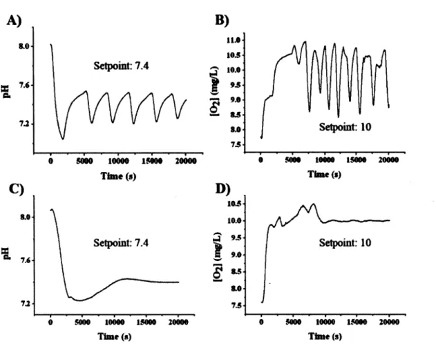
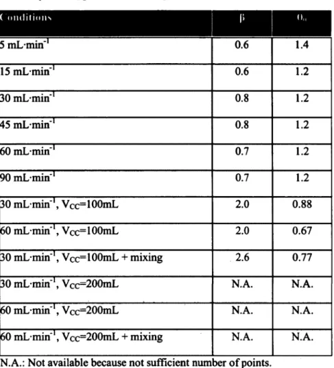
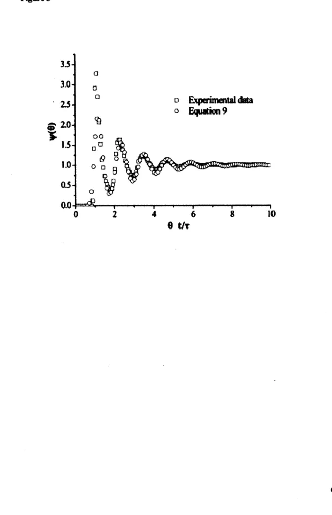
This study reveals that residence time distribution (RTD) analysis with pH monitoring after acid bolus injection can be used to globally study the flow dynamics of a perfusion bioreactor, while fluorescence microscopy and magnetic resonance imaging (MRI) were used to locally investigate mass transport within a hydrogel scaffold seeded or not with cells. The bioreactor used in this study is a closed-loop tubular reactor. A dispersion model in one dimension has been used to describe the non-ideal behavior of the reactor. From open-loop experiments (single-cycle analysis), the presence of stagnant zones and back mixing were observed. The impact of the flow rate, the compliance chamber volume and mixing were investigated. Intermediate flows (30, 45, 60 and 90 mL-min"1) had no effect over RTD function expressed in reduced time (6). Lower flow rates (5 and 15 mL-min"1) were associated to smaller extent of dispersion. The compliance chamber volume greatly affected the dynamic of the RTD function, while the effects of mixing and flow were small to non-significant. An empirical equation has been proposed to localize minima of the RTD function and to predict Per.
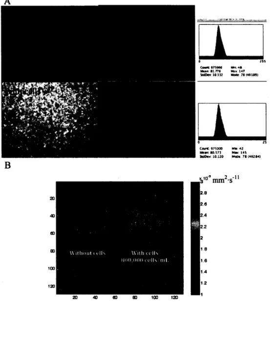
Finally, cells seeded in a gelatin gel at a density of 800,000 cells-mL'1 had no effect over the permeability and the apparent diffusion coefficient, as revealed by fluorescent microscopy and MRI experiments.
Keywords: Residence time distribution (RTD); Bioreactor; Magnetic resonance imaging (MRI); Tissue engineering; Endothelial cells; Cell density; Micro-channeled gelatin scaffolds.
2.2 Introduction
From the last decades, there has been an emergence in the development and use of bioreactors to culture human tissue substitutes. One of the principal challenges that tissue engineering is facing is the oxygen supply to large cell and tissue aggregates and waste removal from these living assemblies (Martin et al., 2004; Martin and Vermette, 2005). Oxygen solubility in cell culture media (i.e., atmospheric pressure, 37°C) is 45 times lower compared to in vivo conditions (Martin and Vermette, 2005). To overcome this limitation, oxygen carriers (Naruto et al., 2007; Sullivan et al., 2008) or convective forces must be used. Convective forces applied simultaneously with mechanical constraints can be used to increase oxygen transport within cell aggregates by creating interstitial flow (Bjork and Tranquillo, 2009). Thus, oxygen mass transfer in tissue is no more solely diffusion dependant and hypoxia inside cell pellet can be avoided. Convective mass transport that can be introduced and modulated through the use of bioreactors becomes essential to the successful growth of large tissue substitutes. In bioreactor culture conditions, phenotypes more related to those found in native physiological conditions can be achieved (e.g., cell elongation in flow direction) (Davies et al., 1986). However, as previously reviewed (Martin and Vermette, 2005), bioreactors are often poorly characterized and this may sometimes lead to misconceptions. For example, Davies and colleagues showed that for the same shear stress level, only disturbed (turbulent) flow induced cell proliferation (Davies et al., 1986). The observed cell responses, expected to be related to shear stress level, might not be correlated only to shear stress. Then, reproduction of similar experiments in other reactor set-ups could lead to completely different cell responses. The characterization of bioreactors and flow dynamic with dimensionless numbers is expected to improve our understanding of the different bioreactor designs and operation procedures found in literature.
Another example of the necessity to understand the flow dynamics within a bioreactor for mammalian cell culture is when considering that cells respond to growth factors often at low level (nM range) and small deviation (2-3 folds) of growth factor concentration or exposure time could switch on/off gene expression pattern (Akeson et al., 2010; Kawamoto et al., 1984; Nelson et al., 2008). In the scientific literature, the effects of growth factors are almost always investigated under static culture conditions with a growth factor concentration specified only at time zero. One of the objectives of dynamic cultures involving the use of a bioreactor is to prolong culture time to form larger substitutes and thus, one cannot assume a constant concentration over time. Hence, the periodic feeding of a fresh amount of growth factors could be required. Before studying the effect of growth factors on cells under bioreactor conditions, it becomes necessary to understand the flow dynamics within such bioreactors to further introduce and use sound chemical reaction engineering principles.
We have previously reported the design and validation of a perfusion bioreactor to sustain high cell density cultures of human endothelial cells dispersed in fibrin (Chouinard et al., 2009). This system can support cell viability and cell proliferation. Here, the same reactor system was used with some improvements. The aim was to use residence time distribution (RTD) to characterize the bioreactor, as RTD analysis can be applied to a wide range of bioreactor designs (Fogler, 2006). The effects over RTD of flow rate, compliance chamber volume and mixing inside the compliance chamber were investigated without a cell culture chamber. Furthermore, forced permeability and the apparent diffusion coefficient within a gelatin-filled culture chamber with and without cells were investigated by fluorescence microscopy and MRI, respectively.
2.3 Materials and Methods
MaterialsCulture medium 199 (Ml99, M5017), endothelial cell growth supplement (ECGS, E2759), heparin (HI027) and porcine type A gelatin (G1890) were purchased from Sigma-Aldrich (Oakville, ON, Canada). Antibiotics (penicillin G/streptomycin sulfate (15140-122)), L-glutamine (#25030) and FBS were from Invitrogen (Burlington, ON, Canada).
Bioreactor Set-Up and Operation
The block diagram of the bioreactor for RTD analysis is presented in Figure 1A. The reactor is a closed-loop system consisting of a peristaltic pump (Masterflex Model A-77924-10, Cole-Parmer, Montreal, Canada), a pump head (Easyload, Model 7518-60, Cole-Parmer), a custom-made glass spiral heat exchanger coupled to a heat bath (Haake DC10-P5, Thermo Fisher Scientific, Whaltam, MA, USA), a Coriolis mass flow meter (Promass 83A, Endress + Hauser Canada Ltee, St-Laurent, Quebec, Canada), two custom-made gas exchangers with Tygon® biopharm silicone tubing (cat. # 95702-05, Cole-Parmer), a pH probe (InPro 3100/120/ptl00, Mettler-Toledo, Columbus, OH, USA), a dissolved oxygen probe (InPro 6800, Mettler-Toledo), a pH transmitter (2100e, Mettler-Toledo), an O2 transmitter (4100e, Mettler-Toledo), a pressure probe (PX4200-005G, Omega, Laval, Quebec, Canada), a custom-made housing for pH and DO probes, a pneumatic pulsator (Vermette et al., 1998), and three gas flow controllers (M100B00812CS1BV, CCR, Kanata, Ontario, Canada). The tubing used to connect the units consists either of Masterflex pharmapure LSI5 (cat. # EW-06435-15) or fluorinated ethylene propylene tubing (cat. # EW-06450-04) and the pump tubing is Masterflex chemdurance-bio LS 14 (cat. # EW-06442-14), all purchased from Cole-Parmer. Four-way valves are from Smiths medical (Markham, Ontario, Canada, MX5341LN).
Cell Culture Chamber
The custom-made cell culture chamber is made from polysulfone and microscope slides (Fig. 1C). The chamber is filled with 3 mL of a 15% (w/v) gelatin solution at 37°C that
was cross-linked by adding 50jiL per mL of gelatin of a 25 U-mL'1 m-transglutaminase (mTG). Human umbilical vein endothelial cells (HUVEC) P8 were trypsinized, centrifuged (1200 rpm, 5 minutes), suspended in supplemented media (M199 with 2.2 g-L"1 sodium bicarbonate, 90 mg-L"1 porcine heparin, lOOU-mL"1 penicillin, 100 (ig mL'1 streptomycin, 10% (v/v) FBS, 2mM L-glutamine, and 20ng-mL"1 ECGS) and counted by hemacytometer. Then, the volume needed to get a final concentration of 8x10s cells-mL"1 was poured in a falcon tube prior to a second centrifugation step. Supernatant was removed and cells were suspended with gelatin + mTG solution. The liquid solution (gelatin + m-transglutaminase + medium (with or without HUVEC)) was then introduced into the cell culture chamber with a syringe. Four 20G medical-grade needles (20G x 1.5", cat. #305176, Becton Dickinson, ON, Canada) were used to close the four lateral holes of the chamber. The jellification of the solution was carried out overnight at room temperature. After jellification, the needles were removed from the cell culture chamber to create channels. Two tube adaptors were screwed to the chamber to orientate flow within these 4 channels for permeability experiments.
Residence Time Distribution (RTD) Analysis
One milliliter of 0.01M HC1 was injected into the system through an injection port positioned after the pH probe (Fig. 1A). For single-cycle analysis, the system was not a close-looped system but rather an open system in which fresh solution was fed in the entrance and the solution at the exit was discarded. For multiple-cycle analysis, the reactor was treated as a 100%-recycle system in which the same volume was circulated in a closed loop. From both techniques, pH values were translated into non-logarithmic scale i.e., molar H+ concentration. The H+ concentration before injection was set to zero to facilitate mathematical treatment.
When performing single-cycle experiments, the concentration function of the tracer
E ( 8 ) - r * E ( t ) - l *C ( t ) [11
f C { t ) d t
o
The residence time (r) of a given compound is calculated with the estimated reactor volume (148mL in this case) divided by the volumetric flow rate measured by the Coriolis mass flowmeter. The integral part of Equation 1, representing the area under the C(t) curve, is calculated by a numerical technique. 6 is the reduced time (t/x).
When performing multiple-cycle analysis, the steady state tracer concentration (Cr,) is known. Thus, concentrations recorded following injection are divided by this value (Equation 2). This leads to a final steady state value (adimensional) of one unit.
^r.«
Mathematically, Equation 1 is the same as Equation 2 (Annex). From single-cycle analysis, first and second moments (mean time, tm and variance, (f) of the RTD function are
calculated with Equation 3.
00
tm - J tE(t)dt 0
[3 a,b] 0
Dispersion Model and the Per to Explain Non-Ideal Behavior
From single-cycle or multiple-cycle analysis, we hypothesize that one of the main parameters affecting the non-ideal behavior, and possibly limiting the fit of our data, is the dispersion that broadens the initial impulse (Levenspiel, 1999; Fogler, 2006). Taking mass transport equation with no reaction and with dispersion and convection gives (Fogler, 2006):
~~ m DabV2Ct - V • (C/Cr) [4]
at
where DAB is the "isotropic" diffusion/dispersion coefficient, Cr the tracer concentration, and U is the mean molar velocity. The first term of Equation 4 corresponds to diffusion, while the second is the convective term. Considering that it is not possible in our case to evaluate the tracer concentration over the tubing radius, only the molar average velocity in the z-direction is considered. This latter simplification means that the reactor is approximated to an ideal plug flow reactor (with no laminar parabolic profile).
v11 v-'
—11 - D,„ f_lL _ u ^JL [5] dt B dz1 dz
Equation 5 can be further expressed in terms of dimensionless numbers with the following substitutions: y/ =Ct/Ct,, X = z/L, and 6 - t/x. CR» is the tracer concentration at
time /=oo, z is any distance from the impulse position within the reactor, and L corresponds to the characteristic length of the reactor.
<*i> ^Dab r61
dB " UL d% ~ dk
Given that Peclet reactor number Per - UL/DAB, only one unknown parameter
remains. Here, Per is a dimensionless number that characterizes the flow within the reactor. If
Per —* 0, there is an important dispersion (i.e., CSTR behavior), while if Per —• ao, there is negligible dispersion within the reactor (i.e., plug-flow behavior). By substituting Per into
Equation 6 and by introducing the perfect impulse function (Dirac delta function (<?)), Equation 6 becomes (Levenspiel and Bischoff, 1963):
«<A<1 [7]

![Figure 2 nyh A) Add1 COSConMw -0.01276 0037W A<M2 0.0199 Tmpot TitmtefoH Dtiayl HI— 0.Q127Q 14W»*1 Tmrnm Tw+mF<*Q 0tiay2 Transport TmfcrFert ]Ug $009*1 €HM®ri](https://thumb-eu.123doks.com/thumbv2/123doknet/2838948.69221/95.921.121.749.193.690/figure-cosconmw-tmpot-titmtefoh-dtiayl-tmrnm-transport-tmfcrfert.webp)