DEPARTMENT OF BIOCHEMISTRY
THESIS
Presented by
DEHIMI Khadidja
For the fulfillment of the requirements for the degree of
DOCTORATE OF SCIENCES
BIOLOGY
Special field: BIOCHEMISTRY
TOPIC
Study of biological activities of extracts from Thymelaea
microphylla Coss. and Dur.
Presented publically in 26/10/2017 JURY
President Baghiani Abderrahmane Pr. UFA Sétif 1
Supervisor Dahamna Saliha Pr. UFA Sétif 1
Examiners Lalaoui Korrichi Pr. Univ Constantine 1
Necib Youcef Pr. Univ Constantine 1
Khettal Bachra Dr. Univ Bejaia
Laboratory of Phytotherapy Applied to Chronic Diseases
University of Ferhat Abbas Setif 1
Faculty of Nature and Life Sciences
1
List of publications
Article:
Dehimi K., Speciale A., Saija A., Dahamna S., Raciti R., Cimino F., Cristani M. Antioxidant and anti-inflammatory properties of Algerian Thymelaea microphylla Coss. and Dur. extracts. Pharmacognosy magazine. 2016; 12: 203-10.
Communications
Dehimi K., Dahamna S., Merghem M., Saija A., Cristani M., Smeriglio A., Trombetta D. In vitro antioxidant activity of extracts from leaves and flowers from Thymelaea
microphylla Coss. and Dur. The first international seminar on medicinal plants, health
and environment.
Dehimi K., Dahamna S., Speciale A., Cimino F., Cristani M. Biological properties of Algerian Thymelaea microphylla Coss. & Dur. extracts. FARMACOGNOSIA nuove opportunita terapeutiche dal mondo vegetale. 20-21, june 2014. Napoli, Italy.
Dehimi K., Dahamna S., Speciale A., Cimino F., Cristani M., Saija A. Thymelaea
microphylla acetone extract protects endothelial cells against TNF- -induced
inflammation. The 15th international congress of the international society of ethnopharmacology. 5-8 May, 2015. Petra- Jordan.
Dehimi K., Dahamna S. Preliminary toxicity study on aqueous extract from Algerian
Thymelaea microphylla Coss. and Dur. Seminaire international sur la valorisation des
ressources naturelles dans les zones semi-arides: Potentialités et perspectives
-ACKNOWLEDGEMENTS
First praise is to Allah, the Almighty; only due to His blessings I could finish this thesis.
I would like to express my sincere gratitude to my supervisor Pr. Dahamna Saliha for the continuous moral support during my Ph.D. period of research.
I am deeply grateful to all the committee members: Pr. Baghiani, Pr. Lalaoui, Pr. Necib and Dr. Khettal to give the valuable feedback to evaluate and improve the contents of this thesis.
I acknowledge Pr. Saija Antonina, Dr. Cristani Mariateresa, Pr. Cimino Francesco and Dr. Speciale Antonio from the department of Drug and Health products in the University of Messina-Italy, for helping and hosting me in their laboratories where I realized the biggest part of this work.
Finally, a special thank goes to the ERASMUS-MUNDUS program of the European Union EMMAG for giving me the chance to benefit from their scholarships and their financial support.
Thymelaea microphylla Coss. and Dur. Thymelaea T. microphylla HPLC in vitro Folin DPPH FRAP ABTS ORAC BCB SOD IC50 AGEs in vitro IC50 33.24 IC50 ex vivo prostaglandin E2 thromboxane B2 cyclooxygenase TNF- E-selectin GSH Cyclooxygenase-2 Thymelaea microphylla
Abstract
Thymelaea microphylla Coss. and Dur. (Methnane) is a medicinal plant with Saharan affinity,
belonging to the Mediterranean genus Thymelaea. Leaves decoction is used in traditional medicine for anticancer, inflammatory, and diabetic properties. Herein, the antioxidant and anti-inflammatory activities of five extracts prepared using solvents with different polarities from leaves and flowers of Algerian T. microphylla, collected from the region of Draa El- were
evaluated. In addition, preliminary studies on phytochemical composition and toxicity estimation were realized. Extracts were first characterized for their content in some secondary metabolites, where acetone extract contained the highest amounts of flavonoids (137.56 µg Cat eq/mg) and flavonols (94.13 µg Que eq/mg), and ethyl acetate extract was the richest in tannins (29.23 µg Cat eq/mg). HPLC analysis revealed the presence of phenolic acids (gallic, caffeic, ferulic and p-coumaric acids) especially in aqueous extract, and flavonoids (luteolin and kaempferol) in organic extracts. Furthermore, antioxidant/free radical scavenging activity which was carried out by in vitro cell-free assays, showed that aqueous and acetone extracts exhibited the best potential in Folin, DPPH , FRAP, ABTS and ORAC assays, while in BCB, acetone and hexane extracts were very likely richer in antioxidants able to reach the lipophilic phase. In the same way, acetone extract was the most potent in SOD mimetic assay; with IC50 of 1.81mg/ml. Extracts were also tested for their inhibitory activity on
in vitro AGEs formation, where aqueous extract was the most potent (IC50 = 33.24 µg/ml). The
anti-inflammatory activity, which was first evaluated using albumin heat-induced denaturation assay, revealed that acetone extract was the most active with IC50 of 0.15 mg/ml, and it was further tested in
ex vivo experiments, to estimate its inhibitory potential on prostaglandin E2 and thromboxane B2
release in human whole blood, where it showed excellent anti-inflammatory and cyclooxygenase-inhibitory activity, together with lack of toxicity on normal human blood cells; furthermore, it was able to protect human endothelial cells against dysfunction induced by TNF- , as shown by decrease in cell death, E-selectin expression, leukocyte adhesion and prevention of depletion in GSH cell content. Moreover, ethanol and aqueous extracts had a significant cytotoxicity on peripheral blood mononuclear cells; however, aqueous extract did not cause mortality or any serious toxicity signs when tested in vivo. This experimental study support the use of this plant in phytotherapy and prevention of diseases related to oxidative stress and inflammation.
Keywords: Cyclooxygenase-2, flavonoids, inflammation, oxidative stress, radical scavenger activity,
Résumé
Thymelaea microphylla Coss. et Dur. (Methnane) est une plante médicinale à affinité saharienne,
appartenant au genre Méditerranéen Thymelaea. La décoction préparée des feuilles est utilisée en médecine traditionnelle pour ses propriétés anticancéreuse, anti-inflammatoire et antidiabétique. Dans ce travail, les activités antioxydante et anti-inflammatoire de cinq extraits préparés en utilisant des solvants de polarités différentes à partir des feuilles et fleurs de T. microphylla collectée de la région de Draa El- Algérie, ont été évaluées. De plus, des études préliminaires sur la composition
phyto-plus grande quantité de flavonoïdes (137.56 µg Cat eq/mg) et de flavonols (94.13 µg Que eq/mg), (29.23 µg Cat eq/mg)
HPLC a révélé la présence des acides phénoliques (acide gallique, caféique, férulique et
p--oxydante/anti-radicalaire, qui a été mesurée par des essais acellulaires in vitro, a montré que les extraits aqueux et acétonique avaient le meilleur potentiel dans
; avec IC50 de
1.81mg/ml. Les extraits étaient également testés pour leur pouvoir inhibiteur de la formation des AGEs in vitro ontré la meilleure activité (IC50 = 33.24 µg/ml)
avec IC50 de 0.15 mg/ml, cet extrait a été encore
testé dans des expériences ex vivo pour estimer son potentiel inhibiteur sur la libération du prostaglandine E2 et du thromboxane B2 dans le sang entier humain, où il a montré une excellente
activité anti-inflammatoire et inhibitrice de la
cyclo-les cellucyclo-les sanguines humaines; De plus, cet extrait était capable de protéger cyclo-les cellucyclo-les endothéliales humaines contre le dysfonctionnement induit par le
TNF-diminution de la mort cellulaire, l'expression de la sélectine E, l'adhésion aux leucocytes et la prévention de l'épuisement du GSH cellulaire. En outre, les extraits éthanolique et aqueux étaient significativement toxiques sur les cellules mononucléées du sang périphérique; Cependant, l'extrait aqueux n'a provoqué aucune mortalité ou autres signes graves de toxicité lorsque testé in vivo. Cette étude expérimentale supporte
Mots clés: Cyclooxygénase-2, flavonoïdes, inflammation, stress oxydatif, activité anti-radicalaire,
Summary
Introduction
... 1Chapter 1: Literature Review
... 41. Thymelaea microphylla ... 5
1.1. Thymelaeaceae family ... 5
1.1.1. Morphology ... 5
1.1.2. Phytochemical properties ... 5
1.1.3. Therapeutic interests ... 6
1.2. The genus Thymelaea ... 6
1.3. Description of the plant ... 6
1.3.1. Morphology ... 6
1.3.2. Classification ... 7
1.3.3. Traditional uses ... 7
1.3.4. Chemical composition of the plant ... 8
2. Polyphenols ... 8
2.1. Flavonoids ... 9
2.2. Phenolic acids ... 11
2.3. Tannins ... 12
3. Oxidative stress and antioxidants ... 13
3.1. Reactive oxygen species (ROS) ... 13
3.1.1. Superoxide anion (O2-) ... 13
3.1.2. Hydroxyl radical (OH ) ... 14
3.1.3. Hydrogen peroxide (H2O2) ... 14
3.1.4. Singlet Oxygen (1O2) ... 15
3.1.5. Nitric oxide (NO ) ... 15
3.2. Biological roles of reactive oxygen species ... 17
3.3. Cell damages caused by reactive oxygen species... 17
3.4. Antioxidants ... 18
3.4.1. Endogenous antioxidants ... 19
3.4.1.1. Glutathione ... 19
3.4.1.2. Superoxide dismutase (SOD) ... 19
3.4.1.3. Catalase (CAT) ... 20
3.4.2.1. Vitamins C and E... 20 3.4.2.2. Carotenoids ... 21 3.4.2.3. Minerals ... 21 3.4.3. Pro-oxidative effect ... 22 4. Inflammation ... 22 4.1. Definition ... 22 4.2. Mechanisms ... 22
4.3. Inflammation related pathologies ... 25
4.4. Anti-inflammatory drugs ... 26
Chapter 2: Materials and Methods
... 271. Materials... 28 1.1. Chemicals ... 28 1.2. Animals ... 28 1.3. Plant material ... 28 2. Methods ... 29 2.1. Extraction procedure ... 29 2.2. Phytochemical study ... 30
2.2.1. Screening for phytoconstituents ... 30
2.2.2. Determination of total flavonoids content in plant extracts ... 30
2.2.3. Determination of total flavonols content in plant extracts ... 31
2.2.4. Determination of Condensed tannins content in plant extracts ... 31
2.2.5. Phenolic profile study using high performance liquid chromatography - diode array detector (HPLC-DAD) ... 32
2.3. Antioxidant activity of plant extracts ... 36
2.3.1. Folin test ... 36
2.3.2. 2,2-diphenyl-1-picrylhydrazyl (DPPH) scavenging activity ... 36
2.3.3. 2,2'-azinobis-(3-ethyl-benzothiazoline-6-sulfonic acid) (ABTS) quenching capacity... 37
2.3.4. Ferric reducing/antioxidant power assay ... 37
-Carotene bleaching assay ... 38
2.3.6. Scavenging activity against the superoxide anion (Superoxide dismutase assay) ... 39
2.3.7. Oxygen radical absorbance capacity assay ... 39
2.4. Inhibition of advanced glycation end products formation ... 40
2.5. Anti-inflammatory potential of plant extracts ... 41
2.5.2. Release of prostaglandin E2 and thromboxane B2in whole blood ... 41
2.5.2.1. Blood samples collection ... 42
2.5.2.2. Release of PGE2 ... 42
2.5.2.3. Release of TXB2 ... 42
2.5.2.4. Enzyme immunoassay (EIA) ... 43
2.5.3. Protective effects on human umbilical vein endothelial cells exposed to tumor necrosis factor- ... 43
2.5.3.1. Cell cultures ... 43
2.5.3.2. Experiment procedure ... 44
2.5.4. RNA isolation and quantitative RT-PCR analysis ... 44
2.5.5. Leukocyte Adhesion assay ... 45
2.5.5.1. Mononuclear cells and cocultures preparation ... 45
2.5.6. Determination of intracellular reduced glutathione (GSH) content ... 46
2.6. Preliminary toxicity evaluation of Thymelaea microphylla extracts ... 46
2.6.1. Evaluation of extracts toxicity on peripheral blood mononuclear cells ... 46
2.6.2. Estimation of aqueous extract toxicity in mice ... 47
2.7. Statistical analysis ... 47
Chapter 3: Results and Discussion
... 481. Extraction yield... 49
2. Phytochemical profile of Thymelaea microphylla extracts ... 50
2.1. Preliminary phytochemical investigation ... 50
2.2. Total flavonoids, flavonols and condensed tannins contents in extracts ... 51
2.3. Extracts analysis using high performance liquid chromatography - diode array detector (HPLC-DAD) ... 53
... 56
3.1. Folin test ... 56
3.2. DPPH radical scavenging activity of extracts ... 57
3.3. ABTS radical scavenging activity of extracts ... 58
3.4. Ferric reducing/antioxidant power assay ... 59
3.5. Antioxidant activity of extracts using -Carotene bleaching assay ... 60
3.6. Superoxide anion scavenging activity of extracts ... 61
3.7. Oxygen radical absorbance capacity assay ... 61
4. AGEs formation inhibition by T. microphylla extracts ... 65
... 67
5.2. Inhibitory activity on PGE2 and TXB2 release ... 69
- -induced cytotoxicity ... 74 - ... 76 ... 78 ... 80 ... 80 ... 81
Conclusion and perspectives
... 82Annexes
References
... 88List of figures
Figure 1: Basic flavonoids structures ... 10
Figure 2: Basic structures of phenolic acids ... 11
Figure 3: Tannins chemical structure ... 12
Figure 4: Steps of acute inflammatory response Figure 5: Figure 6: Thymelaea microphylla Coss. and Dur. A: Plant in environment. B: Leaves and flowers of the plant ... 29
Figure 7: Catechin calibration curve (total flavonoids contents determination). ... 30
Figure 8: Quercetin calibration curve (total flavonols contents determination) ... 31
Figure 9: Catechin calibration curve (condensed tannins contents determination) ... 32
Figure 10: Gallic acid calibration curve ... 33
Figure 11: Caffeic acid calibration curve ... 33
Figure 12: Coumaric acid calibration curve ... 34
Figure 13: Ferulic acid calibration curve ... 34
Figure 14: Kaempferol calibration curve ... 35
Figure 15: Luteolin calibration curve ... 35
Figure 16: Gallic acid calibration curve (Folin-Ciocalteau assay) ... 36
Figure 17: Structures of flavonoids identified in Thymelaea microphylla extracts... 54
Figure 18: Structures of phenolic acids identified in Thymelaea microphylla extracts ... 55
Figure 19: Antioxidant potency of T. microphylla extracts expressed as gallic acid equivalents ... 56
Figure 20: DPPH scavenging activity of T. microphylla extracts ... 57
Figure 21: Mean scavenging concentration (SC50) of T. microphylla extracts against ABTS radicals 58 Figure 22: Mean inhibitory concentrations of T. microphylla extracts obtained in FRAP assay ... 59
Figure 23: Mean inhibition concentrations (IC50) of different extracts from leaves and flowers of T. microphylla -Carotene bleaching assay ... 60
Figure 24: Mean inhibitory concentrations of T. microphylla extracts against superoxide anion ... 61
Figure 25: Antioxidant capacity expressed as Trolox equivalents of extracts from T. microphylla as obtained in ORAC assay ... 62
Figure 26: Inhibition of Advanced Glycation End products formation by T. microphylla extracts ... 66
Figure 27: Antiglycation activity of quercetin used as positive control in AGEs formation inhibition test ... 66
Figure 28: Mean inhibition concentrations of extracts from T. microphylla obtained in heat- induced protein denaturation assay ... 68
Figure 29: Effect of T. microphylla acetone extract on release of ( A) TxB2 (in comparison with that of indomethacin) and of (B) PGE2 in human whole blood ... 71
Figure 30: Effect of T. microphylla ethyl acetate extract on release of A) TxB2 (in comparison with
that of indomethacin) and of B) PGE2 in human whole blood ... 72
Figure 31: Effect of T. microphylla aqueous extract on release of A) TxB2 (in comparison with that of
indomethacin) and of B) PGE2 in human whole blood ... 73
Figure 32: Cell viability in human umbilical vein endothelial cells pretreated with Thymelaea
microphylla acetone extract and then exposed to tumor necrosis factor- ... 75
Figure 33: Effect of T. microphylla acetone extract on TNF- -selectin adhesion molecule, in HUVECs... 77 Figure 34: Leukocyte adhesion in HUVECs pretreated with T. microphylla acetone extract and then exposed to TNF- ... 78 Figure 35: Cell viability, evaluated in the trypan blue assay, of human PBMCs exposed for 24 h to three doses of T. microphylla extracts ... 81 Figure 36: HPLC chromatogram of ethanol extract
Figure 37: HPLC chromatogram of aqueous extract Figure 38: HPLC chromatogram of ethyl acetate extract
List of tables
Table 1: Classification of T. microphylla Coss and Dur. ... 7 Table 2: Some biological systems capable of activating Molecular Oxygen ... 16 Table 3: Mediators in acute inflammatory response... 24 Table 4:Yeild of aqueous and organic extractions from Thymelaea microphylla leaves and flowers .49 Table 5: Phytochemicals investigation in T.microphylla aqueous and organic extracts. ... 51 Table 6: Content of total flavonoids, flavonols and condensed tannins determined in different T.
microphylla extracts. ... 52
Table 7: HPLC-DAD identification and quantification of phenolic compounds contained in extracts obtained from Thymelaea microphylla. ... 54 Table 8: Changes in intracellular GSH levels in HUVECs after 24 h of cell pretreatment with T.
microphylla acetone extract (20-40 µg/ml) and then 2 h of TNF- ... 79
Abbreviations
ABTS AGEs BCB BSA CAT COX DMSO DPPH FL FRAP GAPDH GPx GSH GSSH HPLC HUVECs IL LDL LOX LPS NADP NF- B NOS 1 O2 - ORAC PBMCs PBS PGE2 RNS ROS SOD TXB2 TNF- 2,2'-azino-bis(3-ethylbenzothiazoline-6-sulphonic acid) advanced glycation end products-carotene bleaching assay bovine serum albumin catalase
cyclooxygenase dimethyl sulfoxide
1,1-diphenyl-2-picrylhydrazyl radical fluorescein
ferric reducing/antioxidant power assay glyceraldehyde-3-phosphate dehydrogenase glutathione peroxidase
reduced glutathione oxidized glutathione
high performance liquid chromatography human umbilical vein endothelial cells interleukin
low density lipoprotein lipoxygenase
lipopolysaccharides
nicotinamide adenine dinucleotide phosphate nuclear factor-kappa B
nitric oxide
nitric oxide synthase singlet oxygen superoxide anion hydroxyl radical
oxygen radical absorbance capacity peripheral blood mononuclear cells phosphate buffered saline
prostaglandin E2
reactive nitrogen species reactive oxygen species superoxide dismutase thromboxane B2
1
people returning to natural therapies. Currently, and according to the World Health Organization (WHO), about 80% of the world population still uses herbs for their primary health care needs, where medicinal plants have a big importance in public health maintenance, especially in developing areas such as African countries (Muthamizhe Selvan et al., 2013). According to the WHO, medicinal plants are probably the best source of a variety of drugs; in fact, about 40% of all medicines on the market today have been derived directly or indirectly from natural sources, of which 25% being from plants (Varalakshmi et al, 2011; Asare et al., 2012).
Oxidative stress consists of disturbed equilibrium between pro-oxidant and antioxidant homeostasis, which means overproduction of free radicals causing damage to biomolecules. Oxidative stress has become a major topic in all areas of medical knowledge, since it is an important etiologic factor of the pathologic process of many chronic diseases such as atherosclerosis, cancer, diabetics, rheumatoid arthritis, cardiovascular diseases, aging and other degenerative diseases in humans. There is also evidence that oxidative stress plays a crucial role in the development and perpetuation of inflammation, and thus contributes to development of chronic inflammation-related pathologies (Lugrin et al, 2014). Inflammation ystem against injury, however, chronic inflammation is believed to be, alongside with oxidative stress, a main contributing factor and a root cause of chronic degenerative diseases. Medicinal plants and derivative phytochemicals, especially polyphenols, have proven their ability to minimize oxidative stress damages and possess powerful antioxidant and anti-inflammatory activities (Srivastava and Mishra, 2015).
2
The Algerian Sahara is a place where climatic conditions are very extreme and severe. Plant species growing in this area have developed a very efficient secondary metabolism and are generally rich in polyphenols to cope with severe stress conditions. T. microphylla is a medicinal plant with Saharan affinity, very common in arid and desert pastures. The leaves decoction is used in folk medicine to treat abscesse, skin diseases, abdominal pain, cancer, inflammation and diabetes (Boukef 1986; Benhammou et al., 2009). Another similar species of Thymelaea genus (Thymelaea hirsuta Endl.), possessing the same vernacular name (Methnane), is traditionally used for its several and well documented antioxidant, anti-inflammatory and anticancer properties (Miyamae et al., 2009; Akrout et al., 2011; Trigui et al., 2013; Amari et al., 2014). However, only few data dealing with antibacterial and antioxidant activities of extracts from T. microphylla are available (Benhammou et al., 2009; Djeridane et al., 2010; Ladjel et al., 2011), as well as on its chemical composition (Mekhelfi
et al. 2014, Kerbab et al., 2015).
The present work aims to investigate, by means of in vitro cell-free chemical assays, ex vivo experiments and cell culture based tests, the antioxidant/free radical scavenger and anti-inflammatory properties of several extracts (obtained by water, ethanol, acetone, ethyl acetate and hexane) from leaves and flowers of Algerian Thymelaea microphylla, and also to preliminary estimate phytochemical composition and toxic effect of these extracts. At this purpose prepared extracts were first screened for different phytochemicals which may be present in their structures, characterized for their total content in flavonoids, flavonols and tannins, and also analyzed using HPLC, to identify and quantify some of their flavonoids and phenolic acids. Extracts were then tested for their antioxidant potency by means of a battery of in vitro chemical assays differing in the mechanisms involved, the chemical environment used and the stressor applied (folin-Ciocalteau assay; bleaching of the stable DPPH radical; ABTS assay; oxygen radical absorbance capacity assay; beta-carotene bleaching test;
3
scavenging activity against superoxide anion and ferric reducing/antioxidant power assay). Extracts were also tested for their inhibitory effect on in vitro advanced glycation end products (AGEs) formation, which, alongside with ROS, have an important role in diabetic complications and cell damage. Anti-inflammatory effect of extracts was determined using prevention of heating-induced albumin denaturation assay, and on the basis of results in previous experiments and also taking into account the evidence of cytotoxicity shown by these extracts on in vitro cultured peripheral blood mononuclear cells (PBMCs), acetone extract was further investigated about its capability to inhibit cyclooxygenase (COX) activity, and consequently PGE2 and TXB2 release, in whole blood. The capacity of this extract to
protect vessel endothelial cells against the damage induced by the proinflammatory cytokine tumor necrosis factor- (TNF- ) was also evaluated in terms of cell viability and E-selectin expression which was estimated after RNA extraction and evaluation of E-selectin mRNA levels by means reverse transcription polymerase chain reaction (RT-PCR) technique. Leukocyte adhesion to endothelial cells and intracellular reduced glutathione (GSH) content were also evaluated. To further investigate the toxicity of T. microphylla, and view the use of its decoction in folk medicine, aqueous extract toxic effect was tested in vivo using male and female Albino mice.
Chapter 1
5 1. Thymelaea microphylla
1.1. Thymelaeaceae family 1.1.1. Morphology
Plants belonging to Thymelaeaceae family are shrubs or under-shrubs prevalent in tropical and moderate regions. This family contain up to 1200 species distributed in 67 genera. Species of this family are dicotyledons with persistent alternate leathery leaves. Flowers are hermaphrodites, dioecious or polygamous (4 to 5 merous), with tubular calyx and dry or drupaceous monospermous fruit. Perianth with welded parts at the base, often of coralline appearance more or less yellow or greenish. Stamina (8 to 10) are inserted in two rows (Quezel and Santa, 1962, 1963; Borris et al., 1988).
1.1.2. Phytochemical properties
Concerning phytochemical composition, many compounds were isolated from species belonging to this family. Among identified compounds groups, essential oils and monoterpenes confer to these plants aromatic properties, hence their use in perfumes fabrication. Diterpenes represent one of the most important chemical groups in this family; in general these compounds are derivative from daphnane and tigliane. Among characterizing compounds in Thymelaeaceae family, coumarins are present under different forms: simple coumarins such as daphnetin and umbelliferone, bis-coumarins like daphnoretin and tri-coumarins like edgeworoside. Many groups belonging to flavonoids are present in these plants, including flavones, flavonols, flavanones, biflavonoids and glycosylflavones; most common identified flavonoids are apigenin, luteolin, genkwanin, quercetin and kaempferol. Lignans also were identified in this family in free or linked form (Heywood, 1996; Julien, 2002).
6 1.1.3. Therapeutic interests
In folk medicine, plants belonging to Thymelaeaceae family have a large spectrum of use in treating different disorders. Prepared remedies from these species are used as vomitive, purgative, depurative, diuretic, against chronic rheumatism and skin diseases (Fournier, 1999a). Many species of this family were reported as toxic plants. In fact, as mentioned above, diterpenes which are known to have toxic properties are one of the most frequent compounds in Thymelaeaceae members, and responsible for their toxic effects (Borris et al., 1988; Fournier, 1999a).
1.2. The genus Thymelaea
The genus Thymelaea comprises thirty one species of annual xerophyllous shrubs and herbs (Kabbaj et al., 2013), with very small leaves and yellowish or greenish flowers (Fournier, 1999b). Among them, eight species are present in Algeria: T. microphylla, T. hirsuta, T.
passerina, T.velutina, T. virgata, T. nitida, T. virescens and T. tartonraira (Quezel and Santa,
1962). Plants from this genus were found to be rich in flavonoids, phenolic acids, coumarins and lignans (Trigui et al., 2013; Ghanem et al., 2014; Kerbab et al., 2015). Many of these species are well-documented for their antioxidant, anti-inflammatory, anticancer and hypoglycemic effects (Diogo et al., 2009; Elamrani et al., 2009; Benhammou et al., 2009).
1.3. Description of the plant
1.3.1. Morphology
Thymelaea microphylla is an annual under shrub with dioic clusters. The leaves are very small
(1 4 mm), ovoid, scattered and distant on the branches. Flowers are dioic and form glomeruli (2 5), with yellowish aspect. Branches are slender and canescent (Quezel and
7
Santa, 1963). T. microphylla has a Saharan affinity; it is very common in arid and desert pastures.
1.3.2. Classification
The vernacular name of T. microphylla is «Methnane». The classification of the plant is
represented in table 1.
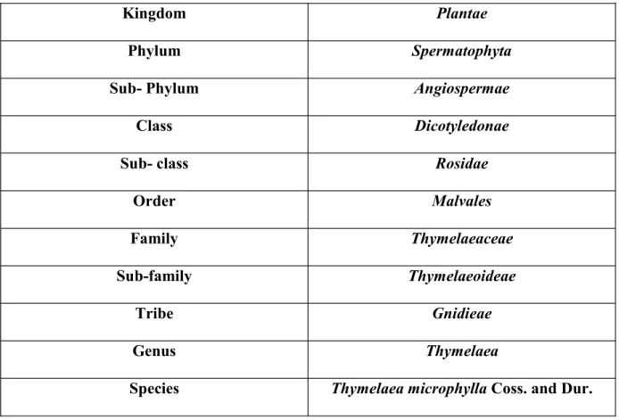
Table 1: Classification of T. microphylla Coss and Dur. (Quezel and Santa, 1962, 1963).
Kingdom Plantae
Phylum Spermatophyta
Sub- Phylum Angiospermae
Class Dicotyledonae
Sub- class Rosidae
Order Malvales
Family Thymelaeaceae
Sub-family Thymelaeoideae
Tribe Gnidieae
Genus Thymelaea
Species Thymelaea microphylla Coss. and Dur.
1.3.3. Traditional uses
T. microphylla is used in folk medicine for its anti-inflammatory, anticancer and antidiabetic
properties. Decoction and natural compress prepared from aerial parts are used to treat abscess, skin diseases, abdominal pain and rheumatism; this plant is also used as antihelmentic (Boukef, 1986; Benhammou et al., 2009).
8 1.3.4. Chemical composition of the plant
Only few studies were reported in literature concerning the phytochemical composition of
Thymelaea microphylla. In a study realized by Cheriti and Sekkoum in 1995, Oleanolic acid,
-sitosterol and 3-O- -D-glucopyranosyl- -sitosterol were detected in aerial parts of the plant (Dohou et al., 2003).
Other recent studies were reported about compounds isolated from this species. Among them, one study allowed isolation of two spiro- -lactone glycosides together with five biflavonoids (neochamaejasmin A, neochamaejasmin B, daphnodorin B, genkwanol A and stelleranol), one bis-coumarin (daphnoretin), two lignans (pinoresinol and matairesinol), one flavonoid glucoside (tiliroside) and a sinapyl alcohol glucoside (syringing), all isolated from ethyl acetate extracts of the aerial parts and roots of the plant (Ghanem et al., 2014).
Analysis of chloroform and ethyl acetate soluble parts of the aqueous-ethanol extract from aerial parts of T. microphylla led to the isolation of six compounds: vanillin, syringaresinol, daphnoretin, (Z)-8-hydroxylinalool, chrysoeriol and luteolin (Mekhelfi et al., 2014).
Another two studies (Kerbab et al., 2015; Noman et al., 2017) revealed the presence of monoterpene glucosides, phenolic acid derivatives (protocatechuic acid, chlorogenic acid butyl ester and ethyl gallate), phenylpropanoid glucosides, flavonoids (yuankanin, Kaempferol derivative and stenopalustroside A), lignans (such as matairesinol and prestegane B) and coumarins (such as umbelliferone and daphnoretin) in this species extracts.
2. Polyphenols
Phenolic compounds constitute one of the most extensive groups of chemicals in the plant kingdom. It is estimated that more than 8000 compounds have been isolated and described. These compounds are generally involved in plant defense against ultraviolet radiation or
9
aggression by pathogens. Their structure is characterized by the presence of, at least, an aromatic ring bearing one or more hydroxyl groups. Plant phenolic compounds have considerable significance as bioactive compounds with substantial health benefits, in fact, these polyhydroxylated phytochemicals are known to possess strong antioxidant, anti-inflammatory, anticancer, anti-diabetic and neuro-protective effects. Polyphenols may be classified into different groups as a function of the number of phenol rings that they contain and on the basis of structural elements that bind these rings to one another (Hennebelle et al., 2004; Ramos, 2007; Pandey and Rizvi, 2009; Ameer et al., 2017). Herein, we report three sub-classes: flavonoids, phenolic acids and tannins.
2.1. Flavonoids
Flavonoids are considered as the most studied group of polyphenols, where more than 4000 compounds belonging to this sub-class were identified. In plants, flavonoids are more abundant in aerial parts, in which are responsible for attractive colors of flowers, fruits and leaves (Pandey and Rizvi, 2009). Basic chemical structure of flavonoids consists of a fifteen-carbon skeleton containing two benzene rings linked via a heterocyclic pyrane ring. According to the level of oxidation and pattern of substitution of the heterocyclic ring, flavonoids are divided to six groups: flavonols, flavones, flavanones, flavanols, anthocyanins and isoflavones (Fig. 1) (Kumar and Pandey, 2013). Flavonoids are associated with strong therapeutic properties, including antioxidant, anti-inflammatory and anticancer effects; in fact, there is an epidemiological evidence that a higher intake of flavonoids was associated with a lower risk of coronary heart disease mortality and cancer (Beecher, 2003; Castro-Vazquez et al., 2016; Suen et al., 2016). As antioxidants, and due to their phenolic hydroxyl groups attached to ring structures, flavonoids can act as reducing agents, hydrogen donators, singlet oxygen quenchers, superoxide radical scavengers and metal chelators; furthermore, these compounds can inhibit enzymes involved in ROS production, like xanthine oxidase, and
10
stimulate antioxidant enzymes (Tsao and Yang, 2003; Montoro et al., 2005). As anti-inflammatory agents, flavonoids can modulate the function of the immune system and inflammatory cells by affecting enzyme systems critically involved in the generation of inflammatory processes, especially tyrosine and serine-threonine protein kinases. Furthermore, flavonoids are able to inhibit expression of isoforms of inducible nitric oxide synthase, cyclooxygenase, and lipooxygenase, which are responsible for the production of a great amount of nitric oxide, prostanoids, leukotrienes and other mediators of the
et
al., 2007; Kumar and Pandey, 2013).
11 2.2. Phenolic acids
Phenolic compounds are a group of secondary metabolites in plants. These compounds are usually divided in two main groups (Fig. 2), the hydroxycinnamic acids, which form the largest class, comprise a three-carbon side chain (C6 C3) structure; as examples from this class, caffeic, ferulic, p-coumaric and sinapic acids are the most common. The second group is formed by hydroxybenzoic acids, which comprise a C6 C1 structure; it includes gallic, p-hydroxybenzoic, protocatechuic, vanillic and syringic acids. These metabolites can be found in free or derivative forms. They exhibit multiple physiological functions, such as antioxidant, anti-inflammatory, anti-microbial, anti-diabetic and anticancer activities. As antioxidants, phenolic acids counteract both ROS and RNS-induced cell damage by their direct free radical scavenging activity as well as the up-regulation of superoxide dismutase (SOD), and catalase (CAT). In inflammation, Chlorogenic acid was proven to possess antiplatelet and antithrombotic effects; furthermore, this phenolic acid was able to cause suppression of proinflammatory cytokines such as IL-1 , TNF- , and IL-6, and suppress the production of NO and PGE2 via down-regulating of the expression of iNOS and COX-2 enzyme (Robbins,
2003; Saibabu et al., 2015; Taofiq et al., 2017).
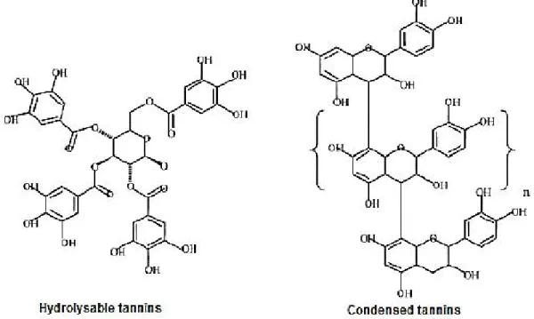
12 2.3. Tannins
Tannins are complex phenolic compounds, which are widely distributed in the bark of trees, insect galls, leaves, stems and fruits. They are the chief plant constituents responsible for astringency. These compounds have a high molecular weight (500-5000), and are classified as hydrolysable tannins or condensed tannins (Fig. 3). Hydrolysable tannins are derived from simple phenolic acids, which are linked to sugar by esterification. Condensed tannins or proanthocyanidins are polymers of flavan-3-ols (catechins) and flavan-3,4-diols (leucoanthocyanins), they are only partially soluble in water and alcohol (Berthod et al., 1999; Pengelly et al., 2004). Most biological properties of tannins are linked to their ability to form complexes with macromolecules, particularly with proteins, in fact they have an enzymatic inhibitory effect on lipoxygenase and protein-kinase C; also these compounds can inhibit lipid peroxidation and are good free radical scavengers (Bruneton, 2002).
13 3. Oxidative stress and antioxidants
Oxidative stress results from an imbalance between oxidants and antioxidants in favor of the oxidants, which are normal products of aerobic metabolism, however, their excessive presence can lead to cell damage. The damaging effect of free radicals is counteracted by the action of antioxidants (Sies, 1997; Quiñonez-Flores et al., 2016).
3.1. Reactive oxygen species (ROS)
Free radicals are molecules with electrons that are unpaired, which make them unstable and reactive. These reactive species steal electrons from other stable molecules in order to become stable themselves, and so a destructive effect begins (Cooper, 1997). In fact, the term reactive species may refer, not only, to designate radicals characterized by an unpaired electron, but either to Non-radical oxygen derivatives, such as hydrogen peroxide H2O2, singlet oxygen 1O2
and nitroperoxyde (ONOOH), which are also reactive and may be precursors of free radicals (Favier, 2003). Herein, we report some of the most important ROS and their sources.
3.1.1. Superoxide anion (O2-)
The uptake of one electron by molecular oxygen results in the formation of the superoxide anion radical (Marquardt et al., 1999).
O2 + e- O2
.-This reaction is mainly catalyzed by membrane NADPH oxidase. The superoxide anion may be formed in some cell organelles such as mitochondria where 2-5% of consumed oxygen is converted to superoxide radicals (Favier, 2003). O2- is a primary ROS (the precursor of most
14 3.1.2. Hydroxyl radical (OH )
It is the neutral form of the hydroxide ion. This radical is generated following interaction of Hydrogen peroxide (H2O2) with transition metal ions (Fenton-type reactions) (Marquardt et
al., 1999; Eboh, 2014).
Fe3+ + OH + OH- H2O2 + Fe2+
Or through reaction of Hydrogen peroxide with superoxide anion (reaction of Haber and Weiss) (Sorg, 2004).
H2O2 + O2.- Fe(III)/Cu(II) O2 + OH- + OH
Other reactions can lead also to the formation of hydroxyl radical, like the decomposition of peroxonitric acid and the reaction of hypochloric acid with superoxide anion (Bartosz, 2003).
The hydroxyl radical is a highly reactive oxidizing agent that can react with a wide variety of organic molecules (proteins, DNA, lipids), it can abstract Hydrogen atoms from essentially any Hydrogen-Carbon bond (Eboh, 2014).
3.1.3. Hydrogen peroxide (H2O2)
Hydrogen peroxide is a stable molecule generated as an end product of a variety of oxidative reactions in living cells. It may, in fact, act as both oxidizing and reducing agent. It is the least reactive molecule among reactive oxygen species and is stable under physiological pH and temperature in the absence of metal ions. H2O2 is produced by spontaneous or enzymatic
dismutation of superoxide radical. Enzymatic dismutation is catalyzed by the superoxide dismutase (SOD) (Ahsan et al., 2003; Eboh, 2014; Nita and Grzybowski, 2016):
2 O2.- + 2H+
SOD
15
Additionally to SOD, there are other enzymes producing H2O2, such as oxidases present in
peroxisomes. However, some of these enzymes like glycoxylate oxidase and D-aminoacide oxidase can directly catalyze the bivalent reduction of molecular Oxygen to give Hydrogen peroxide without formation of superoxide radical. Unlike the superoxide anion, the Hydrogen peroxide is capable to cross cells and organelles membranes causing damages away from his production site (Pham-Huy et al., 2008; Lobo et al., 2010).
3.1.4. Singlet Oxygen (1O2)
Although singlet Oxygen is not a free radical, it is a reactive oxygen species with strong oxidative properties; this molecule can quickly oxidize lipids and convert them into lipid peroxides. It is generated during some physiological and pathophysiological reactions (phagocytosis and prostaglandin biosynthesis), through oxidation reactions including those mediated by peroxidase and lipooxigenase between different ROS, or in the presence of light, oxygen and a photosensitizer (Sorg, 2004; Chen et al., 2012).
3 O2 light + photosensitizer 1 O2 O2.- + M(n+1)+ 1O2 + Mn+ H2O2 + ONOO- 1O2 + NO2- + H2O H2O2 + ClO- 1O2 + Cl- + H2O
3.1.5. Nitric oxide (NO )
Nitric oxide is produced by oxidation of one of N-terminal atoms in L-arginine. This reaction is catalyzed by the Nitric oxide synthase enzyme (NOS) (Sorg, 2004):
O2 + Arginine + NADPH
NOS
NO + Citrulline + H2O + NADP+
This production is important physiologically and plays a major role in neurotransmission, regulation of blood pressure, defense mechanism, smooth muscle relaxation, immune regulation (Valko et al., 2007). But at high concentrations, NO is deleterious for cells in
16
particular when reacting with O2.- to form a powerful oxidizing agent which is the
peroxynitrite (ONOO ) that can secondarily decompose and give other oxidants like NO2 and
OH (Densiov and Afanas'ev, 2005).
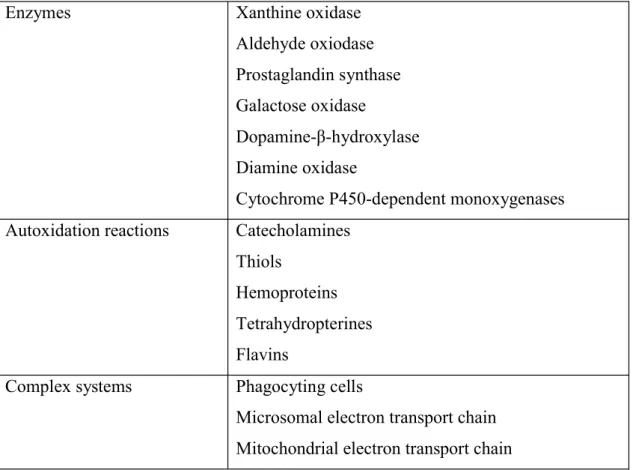
Furthermore, various soluble enzymes generate free radicals by reducing molecular oxygen in their catalytic cycles. Membrane-bound enzymes and electron transport system can also release oxygen radicals and related ROS. Xenobiotic-metabolizing enzymes located in the endoplasmic reticulum and the nuclear membrane are also capable of activating molecular oxygen. This was shown to be the case for the cytochrome P450-dependent monooxygenases, the NADPH-dependent cytochrome reductases, and the flavin-dependent monooxygenases. Phagocytic cells are another major biological source of ROS (Table 2) (Marquardt et al., 1999; Phaniendra et al., 2015).
Table 2: Some biological systems capable of activating Molecular Oxygen (Marquardt et al., 1999).
Enzymes Xanthine oxidase
Aldehyde oxiodase Prostaglandin synthase Galactose oxidase
Dopamine- -hydroxylase Diamine oxidase
Cytochrome P450-dependent monoxygenases Autoxidation reactions Catecholamines
Thiols
Hemoproteins Tetrahydropterines Flavins
Complex systems Phagocyting cells
Microsomal electron transport chain Mitochondrial electron transport chain
17 3.2. Biological roles of reactive oxygen species
It is well known since decades that living organisms use free radicals they produce in some fundamental physiological processes. Free radicals contribute in immune defense; phagocytosis of bacteria and parasites by macrophages is accompanied with a brutal and intense production of ROS. Inside phagosome, NADPH oxidase activation, superoxide dismutase (SOD) and NO synthase (NOS) actions, lead to a very corrosive mixture of O2-,
H2O2, OH and ONOOH, besides the presence of HOCl and 1O2 in the polynuclear; this
reactive mixture destroys by oxidation the bacterial constituents. ROS can act also as signal molecules in apoptosis, metabolism, aging, and hypoxic signaling pathways, by interacting with some membrane receptors and activating them. The presence of ROS in extra-cellular medium causes the activation of some transcription factors, inducing then expression of corresponding genes. These biological functions of ROS confer to them an important role in cell cycle progression and cell proliferation (Favier, 2003; Delattre et al., 2005; Zhang et al., 2016).
3.3. Cell damages caused by reactive oxygen species
Reactive oxygen species may attack biological macromolecules, giving rise to protein, lipid,
attacking the nitrogenous bases and the sugar phosphate backbone and can evoke single- and double-stranded DNA breaks. The mitochondrial DNA is more vulnerable to the ROS attack than the nuclear DNA, because it is located in close proximity to the ROS generated place. In fact, RNA is subjected to more oxidative damage than DNA in humans, due to its single stranded nature, lack of an active repair mechanism for oxidized RNA, less protection by proteins than DNA and moreover cytoplasmic RNAs are located in close proximity to the mitochondria where loads of ROS are produced.
18
ROS attack also structural and enzymatic proteins and oxidize amino-acids present in their structure, causing formation of protein protein cross linkages, results in the denaturing and loss of functioning of proteins, such as enzyme activity, function of receptors and transport proteins. Membrane lipids are also a target of reactive species; in fact, polyunsaturated fatty acids present in phospholipids are the most attacked by free radicals view the double bounds in their structures. This oxidation lead to loss of membrane functioning (inactivation of membrane bound enzymes and receptors) and formation of toxic end products from lipid peroxidation like malondialdehyde and 4-hydroxyl nonenal that cause damage to DNA and proteins. Furthermore, isoprostanes which are prostaglandin-like compounds, result from peroxidation of arachidonic acid, and are considered as markers of lipid peroxidation in oxidative stress.
Oxidative stress can then induce apoptosis, alters the growth signals and gene expression causing continuous proliferation of cells, alters the central nervous system due to the presence of high lipid content and causes atherosclerosis. All these conditions lead to several diseases cardiovascular diseases, rheumatoid arthritis, liver damage and diabetes complications (Phaniendra et al., 2015; Nita and Grzybowski, 2016).
3.4. Antioxidants
It has long been known that antioxidants fight the effects of free radicals and do much to slow down the aging process and prevent various types of diseases. Antioxidants are molecules which can safely interact with free radicals and terminate the chain reaction before vital molecules are damaged. These molecules exhibit great structural diversity and ability to function in cooperation; when one antioxidant reacts with a free radical, another antioxidant is present to regenerate the first. There are several endogenous enzyme systems and substances
19
within the cells that scavenge free radicals. The importance of antioxidant systems is emphasized by the fact that compounds with other primary biological roles like albumin, glucose, uric acid, polyamines and fibrinogen, also can function as antioxidants. Cells can also attain antioxidants through the circulation after consumption of antioxidant rich beverages and food (Cooper, 1997; Olinescu and L.Smith, 2002; Panglossi, 2006).
3.4.1. Endogenous antioxidants
3.4.1.1. Glutathione
ne (GSH), a tripeptide (g-glutamylcysteinylglycine) with a free thiol group, is a major antioxidant in human tissues. It has been stated that low levels of glutathione may cause illness and premature death. It is instrumental in the detoxification of drugs and pollutants and for healthy liver function. Glutathione is important for strong immune system. It is also stated that boosting glutathione can possibly reverse age-related retardation of immune system. GSH provides reducing equivalents for the glutathione peroxidase (GPx) catalyzed reduction of hydrogen peroxide and lipid hydroperoxides to water and the respective alcohol. During this process GSH becomes oxidized glutathione (GSSG). GSSG is then recycled to GSH through interaction with the reduced form of nicotinamide adenine dinucleotide phosphate (NADPH), catalyzed by glutathione reductase (GR) (Panglossi, 2006; Bains and Bains, 2015):
H2O2 + 2GSH
GPx
GSSG + H2O
GSSG + NADPH + H+ GR 2GSH + NADPH+
3.4.1.2. Superoxide dismutase (SOD)
Since superoxide is the primary ROS produced in a variety of sources, its dismutation by SOD is of primary importance for each cell. SOD is an enzyme with a generalized presence in
20
the body, it has three variants, copper-zinc SOD found in the cytoplasm, manganese SOD located in the mitochondria and extra-cellular SOD. SODs catalyze transformation of superoxide anions to oxygen and hydrogen peroxide (view reaction above), protecting cells against the toxic effects of oxygen metabolism (Al-Dalaen and Al-Qtaitat, 2014; Jeeva et al., 2015).
3.4.1.3. Catalase (CAT)
Catalase is an antioxidant enzyme that acts as a catalyst for the conversion of hydrogen peroxide produced by the action of SODs or oxidases, such as xanthine oxidase, to molecular oxygen and water.
2H2O2 2H2O + O2
Catalase has one of the highest turnover rates of all enzymes; one molecule of catalase can convert approximately 6 million molecules of hydrogen peroxide to water and oxygen each minute (Panglossi, 2006; Al-Dalaen and Al-Qtaitat, 2014; Jeeva et al., 2015).
3.4.2. Dietary antioxidants
This group comprises a variety of structurally very different compounds that have been known to exhibit antioxidant properties. Diet, particularly fruits, vegetables, nuts and seeds form a rich source of antioxidant vitamins, phytochemicals and minerals (Panglossi, 2006).
3.4.2.1. Vitamins C and E
Vitamin C is a water-soluble antioxidant, which prevents oxidative damage to cells by scavenging free radicals, recycling vitamin E (alpha-tocopherol) and raising intracellular glutathione levels, and then plays an important role in protein thiol group protection against oxidation. However, vitamin E, which is a fat-soluble compound, is considered as major
21
powerful membrane-bound antioxidant, employed by the cell as a protection against lipid
peroxidation; in fact, the anti- -tocopherol have been related to its
chain-breaking properties, preventing lipid peroxidation and formation of atherogenic oxidized LDL (Panglossi, 2006; Vance et al., 2013; Al-Dalaen and Al-Qtaitat, 2014).
3.4.2.2. Carotenoids
Carotenoids represent a very diverse group of natural pigments of the polyene type. They occur ubiquitously in all organisms capable of conducting photosynthesis. More than 700 carotenoids have been described, of which about 50 become constituents of the human diet. Carotenoids function as potent antioxidants and have also been shown to influence cell growth and induce apoptosis; some carotenoids serve as vitamin A precursors. Antioxidant activity of this group arises due to their ability to delocalize unpaired electrons, and thus -carotene has been found to react with peroxyl
2-.)
radicals (Vance et al., 2013; Al-Dalaen and Al-Qtaitat, 2014; Fiedor and Burda, 2014).
3.4.2.3. Minerals
Food derived minerals are crucial for antioxidant enzymes functions. Selenium is part of at least 25 different selenoproteins, such as glutathione peroxidase. This essential trace element is known for its antioxidant and anticarcinogenic activities. Furthermore, Iron, Copper, Zinc and Manganese are the co-factors of SODs, catalase, ceruloplasmin and metallothionein, which are implicated in free radicals removal. Deficiency of copper or zinc increases the cytochrome P450 activity in microsomes of liver and lungs, and thus enhances the ROS
22 3.4.3. Pro-oxidative effect
Some antioxidants may be actually becoming pro-oxidants under certain conditions; in fact, antioxidants function by being converted in the reaction to a live, but less reactive, free radical, and later are regenerated, such as vitamin E which is regenerated by vitamin C and glutathione. This is the case for most redox compounds that react as pro- or antioxidants depending on their environment and concentration. Some of the popular and well known antioxidants, flavonoids have been reported to act as pro-oxidants also when a transition metal is available, these have been found to be mutagenic in vitro (Olinescu and L.Smith, 2002; Rahal et al., 2014).
4. Inflammation
4.1. Definition
Inflammation is a vital part of the human immune system; it is a normal biological process in response to harmful stimuli, such as tissue injury, microbial pathogen infection, and chemical troy or isolate the underlying source of the disturbance, remove damaged tissue, and then restore tissue homeostasis. Normal inflammation is rapid and self-limiting, but aberrant resolution and prolonged inflammation cause various chronic disorders (Kumar and Pandey, 2013; Saibabu
et al., 2015).
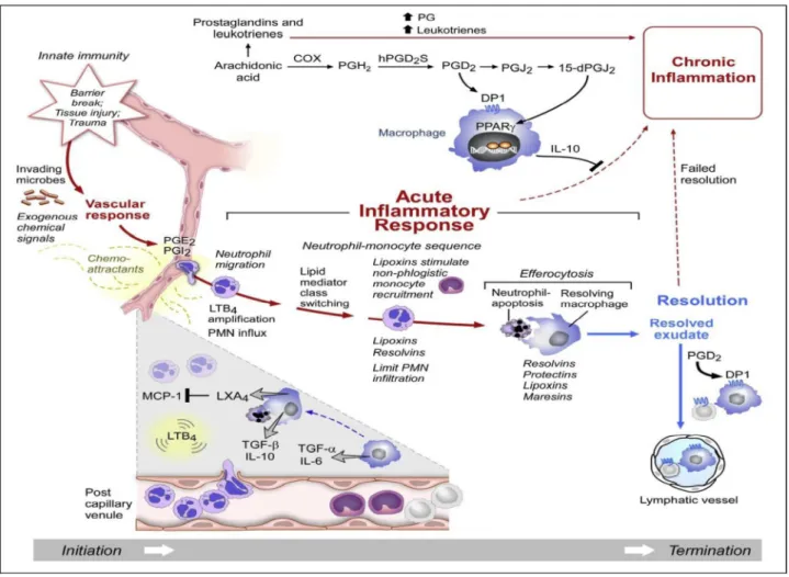
4.2. Mechanisms
After detection of infection or damage via a set of receptors that recognize pathogen-associated molecular patterns (PAMPs) or via alarmins, inflammation is initiated by migration of immune cells from blood vessels and release of
23
factor-alpha (TNF- such as PGE2 (table
3), facilitate the recruitment of effector cells, such as monocytes and neutrophils, to the site of disturbance (Fig. 4). During this recruitment, an up-regulation of adhesion molecules is induced, to facilitate adhesion of inflammatory cells to endothelium. These cells release ROS, RNS, and various proteinases which are destructive to both foreign pathogens and hosts. The last step is resolution, which is set into motion by tissue-resident and recruited macrophages.
cells produce
pro-leukotrienes, but rapidly switch to lipoxins, which block further neutrophil recruitment and instead favor enhanced filtration of monocytes important for wound healing (Zhang and An, 2007; Ashley et al., 2012).
24
Table 3: Mediators in acute inflammatory response (Lawrence et al., 2002; Janeway et al., 2003).
Mediator classes Mediators Functions
Amines histamine, bradykinin Released by mast cells, cause vasodilation, increase capillary
permeability Lipid mediators PGE2, PGI2, LTB4, LTC4 Vasodilators, attraction and
activation of leukocytes Adhesion molecules E-selectin, ICAM1,
VCAM1
Capture and adhesion of leukocytes to endothelial cells
Cytokines TNF, IL- -6 Released by macrophages, T cells
and monocytes, their functions are cytokine production, cell proliferation and apoptosis
Chemokines IL- Released by macrophages, epithelial
cells and endothelial cells, they have role in chemotaxis and angiogenesis
Among lipid mediators, prostaglandin E2 (PGE2) and thromboxane A2 (TXA2) are eicosanoids
resulting from arachidonic acid metabolism, which is liberated from membranes by phospholipase A2 (Fig. 5). TXA2 has potentially pro-inflammatory actions; it is implicated in
activation and aggregation of platelets and also in production of TNF- -monocytes. The PGE2 has actions which can be considered both pro- and anti-inflammatory,
depending on cells and target receptors; it is implicated in vasodilatation and cause exudation but also can inhibit chemotaxis and production of O2 by neutrophils (James et al., 2001).
25
Fig. 5: Arachidonic acid metabolism (Harizi et al., 2008).
4.3. Inflammation related pathologies
Inflammation can become potentially deleterious if excessive or deregulated. In fact, the persistent chronic inflammation increases the development of the degenerative diseases such as cancer, metabolic disorder, type II diabetes, arthritis, autoimmune diseases, neurological diseases, multiple sclerosis, pulmonary diseases, and cardiovascular complications. These various disorders have been linked to increased expression of pro-inflammatory mediators which activates inflammatory cells by increasing the expression of pro-inflammatory
cytokines, up-regulating genes that produce NF- ipase A2,
COX-1 and COX-2, 5-LOX, myeloperoxidase, iNOS, increasing oxygen consumption and producing many deleterious oxygen-free radicals (Iwalewa et al., 2007; Khansari et al., 2009; Prasad and Aggarwal, 2014).
26 4.4. Anti-inflammatory drugs
There are two main groups of anti-inflammatory drugs; non-steroidal and steroidal drugs. Non-steroidal anti-inflammatory drugs (NSAIDs) are a diverse group of compounds, able to inhibit prostaglandins production, by non-selective activity against COX-1 and COX-2 enzymes; NSAIDs can then eliminate the erythema, swelling, elevated temperature and pain (Vonkeman and Van de Laar, 2010). Steroidal anti-inflammatory drugs (SAIDs) are substances derived from cortisol, able to increase lipocortin production, and hence, inhibit phospholipase A2 enzyme implicated in arachidonic acid release. Furthermore, these drugs
decrease immune cells migration and release of cytokines and other inflammatory mediators. However, SAIDs are known for their numerous side effects, like hypertension, gastrointestinal disorders and osteoporosis (Payne and Adcock, 2001; Henzen, 2003). Anti-inflammatory effects of secondary metabolites from plants are also well documented, especially for flavonoids which exercise their activity via inhibition of tyrosine and serine-threonine protein kinases and COX/LOX enzymes. Besides flavonoids, other secondary metabolites exhibit also anti-inflammatory properties, such as phenolic acids, anthocyanins and tannins (Derbel and Ghedira, 2005; Kumar and Pandey, 2013).
Chapter 2
28 1. Materials
1.1. Chemicals
Pure HPLC reference standards were purchased from Extrasynthèse (Genay-France). Folin-Ciocalteau phenol reagent, analysis grade methanol, HPLC grade methanol, fluorescein sodium nitrite, and hydrochloride acid were obtained from Carlo Erba (Milan, Italy). HPLC grade water and acetonitrile, aluminium chloride anhydrous and potassium peroxodisulfate were from VWR. All other reagents, if not specified, were purchased from Sigma-Aldrich (Milan, Italy).
1.2. Animals
Sixty adult male and female Albino mice (average weight 25-30 g) were purchased from the institute of Pasteur, Algiers. Standard diet and water were available ad libitum. Animals were acclimated for two weeks before the experiment under the same laboratory conditions of photoperiod and room temperature (25 ± 2°C).
1.3. Plant material
Aerial parts of Thymelaea microphylla (Fig. 6) were collected in April 2011 from the region of Draa Elhadjj Algeria. The identification of the plant was based on the work of Quezel and Santa (1963), and validated by Pr. Laouar Hocine (laboratory of valorization of biological resources, university Ferhat Abbas Setif 1, Algeria). Samples were dried under shade at room temperature. Leaves and flowers were then separated from stems and used for extraction.
29
-A- -B-
Fig. 6: Thymelaea microphylla Coss. and Dur. (April 23rd, 2017). A: Plant in environment. B: Leaves and flowers of the plant. (Photographed by author).
2. Methods
2.1. Extraction procedure
The extraction was realized according to the methods described by Gnanaprakash et al. (2010) and by Anushia et al. (2009) with slight modification. Five extracts (aqueous, ethanol, ethyl acetate, acetone and hexane) were prepared from dried leaves and flowers of Thymelaea
microphylla. To obtain the aqueous extract, 10 g of the sample were put in contact with 100
ml of distilled water during 30 min at 70°C and then left for 2 days under occasional stirring. To obtain ethanol, ethyl acetate, acetone and hexane extracts, 10 g of sample were put in contact with 100 ml of each solvent during 48 hours with agitation in darkness. Then, the mixtures were filtered by Whatman N. 1 filter paper and the filtrates evaporated to dryness under vacuum by using a rotavapor. The residues were kept into brown vial in freezer until used.
30 y = 1,4577x + 0,0057 R² = 0,9992 0 0,2 0,4 0,6 0,8 1 1,2 1,4 1,6 0 0,25 0,5 0,75 1 1,25 Catechin concentration (mg/ml) 2.2. Phytochemical study
2.2.1. Screening for phytoconstituents
Extracts were screened for the presence of phytoconstituents (tannins, saponins, flavonoids, steroids, alkaloids, terpenoids, cardiac glycosides and quinones) using the standard procedures described by Sunil H. Ganatra et al. (2012) and by Doughari et al. (2012).
2.2.2. Determination of total flavonoids content in plant extracts
The total content of flavonoids in Thymelaea microphylla extracts was determined by the method described by Tomaino et al. (2010). Fifty microliters of the solution containing the extracts to be tested (or catechin used as standard) were diluted with distilled water to a final
2 were added. After 5 min the mixture was added
10% AlCl3
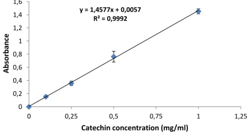
distilled water. Absorbance was recorded at 510 nm using a Shimadzu UV-1601 spectrophotometer. Total flavonoids content was expressed as µg of catechin equivalents (CatE) per mg of dried extract (Fig. 7). All determinations were carried out in duplicate and repeated three times.
31
2.2.3. Determination of total flavonols content in plant extracts
The flavonols content in extracts was measured according to the method described by Tomaino et al. (2010). 125 µl of each extract were added to an equal volume of AlCl3 (2
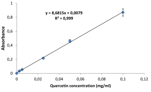
mg/ml), and 750 µl of sodium acetate (50 mg/ml). After incubation during 2.5h, absorbance was recorded at 440 nm. Quercetin was used as standard with concentrations ranging from 2.5 to 100 µg/ml (Fig. 8). Total flavonols content was expressed as µg of quercetin equivalents (QE) per mg of dried extract. Each treatment was performed in duplicate and the whole experiment was repeated three times.
Fig. 8: Quercetin calibration curve. Each point represents the mean ± SD (n=6). 2.2.4. Determination of Condensed tannins content in plant extracts
The determination of condensed tannins content in extracts was carried out by the method described by Sun et al. (1998) and by Tounsi et al. (2009). 200 µl of extracts were added to 500 µl of vanillin methanolic solution (11 mg/ml) and 500 µl of H2SO4 (10%). After
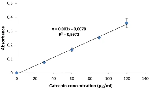
incubation for 15 min, absorbance was measured at 500 nm. Catechin was used as standard with concentrations ranging from 30 to 120 µg/ml (Fig. 9). Condensed tannins contents were expressed as µg of catechin equivalents (CatE) per mg of dried extract. Each determination was carried out in duplicate and repeated three times.
y = 8,6815x + 0,0079 R² = 0,999 0 0,2 0,4 0,6 0,8 1 0 0,02 0,04 0,06 0,08 0,1 0,12 Quercetin concentration (mg/ml)
32
Fig. 9: Catechin calibration curve. Each point represents the mean ± SD (n=6). 2.2.5. Phenolic profile study using high performance liquid chromatography - diode array detector (HPLC-DAD)
Extracts from Thymelaea microphylla were analyzed using high performance liquid chromatography according to the method described by Siracusa et al. (2011). Qualitative and quantitative determination of phenolic profile of Thymelaea microphylla extracts was carried out using HPLC Varian Prostar system (Varian Prostar 220/230/240 pumps, Prostar 410 autosampler, Varian Prostar 325LC detector, Phenomenex Luna C18 column: 250 x 4.6 mm, 5 µm particle size). Extracts (5 mg/ml) and standards (50 µg/ml) were all dissolved in dimethylformamide (DMF) and analyzed using the following elution gradient of B (Acetonitrile) in A (2.5% formic acid in water): 0 min: 5% B, 10 min: 15% B, 30 min: 25% B, 35 min: 30% B, 50 min: 55% B, 55 min: 90% B, 57 min: 100% B, 65 min: 100% B, 70 min: 5% B. The flow rate was set to 1 ml/min, the temperature was kept at 25°C and the injector volume was 10 µl. UV-detection was performed at 280 and 370 nm for all samples.
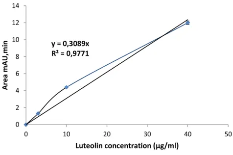
Quantification of detected compounds was carried out at 280 nm and realized by means of the external standard method using corresponding compounds (Fig. 10 to 15). The results were
y = 0,003x - 0,0078 R² = 0,9972 0 0,1 0,2 0,3 0,4 0,5 0 20 40 60 80 100 120 140 Catechin concentration (µg/ml)
33 y = 0,4802x + 0,2582 R² = 0,9977 0 2 4 6 8 10 12 14 0 5 10 15 20 25 30
Gallic acid concentration (µg/ml)
obtained from the average of three determinations and are expressed as mg/g dried extract ± percent relative standard deviation (% RSD).
Fig. 10: Gallic acid calibration curve. Each point represents the mean ± SD (n=3).
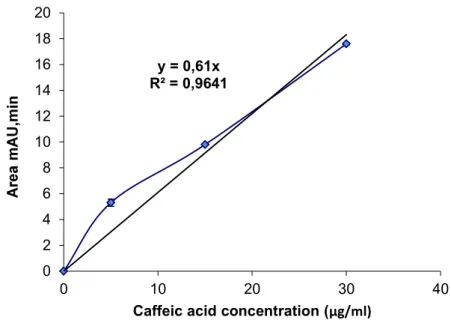
Fig. 11: Caffeic acid calibration curve. Each point represents the mean ± SD (n=3).
y = 0,61x R² = 0,9641 0 2 4 6 8 10 12 14 16 18 20 0 10 20 30 40
34
Fig. 12: Coumaric acid calibration curve. Each point represents the mean ± SD (n=3).
Fig. 13: Ferulic acid calibration curve. Each point represents the mean ± SD (n=3).
y = 0,834x R² = 0,9952 0 5 10 15 20 25 30 35 40 45 0 10 20 30 40 50 60
Coumaric acid concentration (µg/ml)
y = 0,5273x R² = 0,9879 0 2 4 6 8 10 12 14 0 5 10 15 20 25 30
35
Fig. 14: Kaempferol calibration curve. Each point represents the mean ± SD (n=3).
Fig. 15: Luteolin calibration curve. Each point represents the mean ± SD (n=3).
y = 0,2735x R² = 0,9987 0 1 2 3 4 5 6 7 8 9 10 0 10 20 30 40 Kaempferol concentration (µg/ml) y = 0,3089x R² = 0,9771 0 2 4 6 8 10 12 14 0 10 20 30 40 50 Luteolin concentration (µg/ml)