UNIVERSITÉ DU QUÉBEC
À
MONTRÉAL
MÉMOIRE PRÉSENTÉ
À
UNIVERSITÉ DU QUÉBEC
À
MONTRÉAL
COMME EXIGENCE PARTIELLE
DE LA MAITRISE EN BIOLOGIE
UNCOVERING NOVEL PROTEIN PARTNERS OF NUCLEOLAR
PROTEIN 6 (NOP6) BY YEAST TWO-HYBRID ANAL YSIS AND
THEIR ROLE IN RIBOSOME BIOGENESIS
PAR
HOSSAIN
,
AHMED
UNIVERSITÉ DU QUÉBEC À MONTRÉAL Service des bibliothèques
Avertissement
La diffusion de ce mémoire se fait dans le respect des droits de son auteur, qui a signé le formulaire Autorisation de reproduire et de diffuser un travail de recherche de cycles supérieurs (SDU-522 - Rév.01-2006). Cette autorisation stipule que «conformément à l'article 11 du Règlement no 8 des études de cycles supérieurs, [l'auteur] concède à l'Université du Québec à Montréal une licence non exclusive d'utilisation et de publication de la totalité ou d'une partie importante de [son] travail de recherche pour des fins pédagogiques et non commerciales. Plus précisément, [l'auteur] autorise l'Université du Québec à Montréal à reproduire, diffuser, prêter, distribuer ou vendre des copies de [son] travail de recherche à des fins non commerciales sur quelque support que ce soit, y compris l'Internet. Cette licence et cette autorisation n'entraînent pas une renonciation de [la] part [de l'auteur] à [ses] droits moraux ni à [ses] droits de propriété intellectuelle. Sauf entente contraire, [l'auteur] conserve la liberté de diffuser et de commercialiser ou non ce travail dont [il] possède un exemplaire.»
AVERTISSEMENT
La diffusion de ce mémoire se fait dans le respect des droits de son auteur, qui a signé le formulaire Autorisation de reproduire et de diffuser un travail de recherche de cycle supérieurs (SDU-522 -Ré v .0 1-2006). Cette autorisation stipule que «conformément à l'article 11 du Règlement no 8 des études de cycles supérieurs, Ahmed Hossain concède à l'Université du Québec à Montréal une licence non exclusive d'utilisation et de publication de la totalité ou d'une partie importante de son travail de recherche pour des fins pédagogiques et non commerciales. Plus précisément, Ahmed Hossain autorise l'Université du Québec à Montréal à reproduire, diffuser, prêter, distribuer ou vendre des copies de son travail de recherche à des fins non commerciales sur quelque support que ce soit, y compris l'Internet. Cette licence et cette autorisation n'entraînent pas une renonciation de la part de Ahmed Hossain à ses droits moraux ni à ses droits de propriété intellectuelle. Sauf entente contraire, Ahmed Hossain conserve la liberté de diffuser et de commercialiser ou non ce travail dont Il possède un exemplaire.»
ACKNOWLEDGEMENTS
1 would like to thank Dr. François Dragon for giving me the opportunity to work with him and to advance my career in research. More importantly, 1 would like to thank him for ali of his help.
1 would also like to thank Dr. Benoit Barbeau and Dr. Jean Danyluk for being members of my evaluation committee.
I would also like to thank the secretary of Graduate Studies, Ginette Lozeau, for her help during my degree.
1 greatly appreciated the advice and technical details offered by my colleagues.
TABLE OF CONTENTS
AVERTISSEMENT ... i
ACKNOWLEDGEMENTS ... ii
LIST OF FIGURES ... v
LIST OF TABLES ... vi
LIST OF ABBREVIA TI ONS ... vii Résumé ... x
CHAPTER 1: INTRODUCTION ... 1
1.1. The nucleolus ... 1
1.2. Maturation of ribosomal RNA ... 3
1.3. Small nucleolar ribonucleoprotein complexes ... 6
1.4. Box H/ACA snoRNA associated proteins ... 9
1.5. Nucleolar Protein 6 (Nop6) ... 11
1.6. The coiled-coil do mains ... 13
CHAPTER 2: HYPOTHESIS and OBJECTIVES ... 16
2.1 Hypothesis ... 16
2.2. Objectives ... 16
CHAPTER 3: RIBOSOME BIOGENESIS FACTOR NOP6 INTERACTS WITH RRT5 ... 17
3 .1. Abstract. ... 20
3.2. Introduction ... 22
3.3. Materials and Methods ... 24
3.3.1 Y east strains and plasmids ... 24
3.3.2. Sporulation of diploid cells ... 26
3.3.3. Immunoprecipitation experiments (IPs) ... 27
3.3.4. GST-Pulldown ... 27
3.3.5 Immunofluorescence microscopy ... 28
3.4. Results and Discussion: ... 30
3.4.1 Nop6 interacts with Rrt5 in the two-hybrid system ... 30
3.4.2. Nop6 and Rrt5 may be invo1ved in the regulation of sporulation ... 31
3.4.3. The interaction between Nop6, and Rrt5 is not RNA-mediated ... 33
3.4.4. Nop6 and RrtS co-localize to the cell wall in spores ... 34
3.5. Tables ... 36
3.6. Figure and Table Legends ... 44
CHAPTER 4: GENERAL DISCUSSION AND PERSPECTIVES ... 51
CHAPTER 5: REFERENCES ... 55
Appendice ... 61
6.1 Prediction ofNop6 coiled-coil domain ... 61
6.2. Cloning possible prey partners ofNop6 into prey vector pGADT7 ... 63
6.3. Screen of genomic DNA libraries Cl, C2, and C3 with pGBKT7-Nop6 bait... ... 64
6.4 Analysis ofplasmid DNA from positive clones oftwo-hybrid screen ... 64
6.4 BLAST analysis of plasmids from positive clones and identification of prey proteins 67 6.5 Analysis of fusion protein products from baits and prey proteins ... 76
6.6 Nop6 interacts with Firl in the yeast two-hybrid system ... 80
LIST OF FIGURES
Figure 1. 1 Structure of snoRNAs ... 8
Figure 1. 2 Schematic representation ofNop6 domains ... 13
Figure 1. 3 Helical wheel diagram of coiled coi! motifs ... 15
Figure 3 .1. Two-hybrid interaction ofN op6 and Rrt5 do es not require the CC domains ... 45
Figure 3. 2 Rrt5 is highly expressed during sporulation ... .45
Figure 3. 3 The interaction ofRrt5 and Nop6 is not RNA-mediated ... 46
Figure 3.4 Nop6 and Rrt5 co-localize to the cel! wall spores ... 46
Figure 6. 1 Prediction of coiled-coil domains ofNop6 ... 62
Figure 6. 2 Example of PCR amplification and cloning of possible Nop6 partners into prey vector pGADT7 ... 63
Figure 6. 3 Analysis of positive clones by digestion with Pstl and Bglii ... 66
Figure 6. 4 An example of bioinformatics analysis using BLAST of positive clones from Two-Hybrid Screen from S. cerevisiae . ...... 73
Figure 6. 5 Example of analysis on polyacrylamide gel of protein extractions of expression of bait and prey proteins to confirm two-hybrid interactions ... 79
Figure 6. 6 Two-hybrid interaction of Nop6 and Fir1 fragments retrieved from a genomic DNA screen includes two regions ... 81
Figure 6. 7 The interaction ofFirl and Nop6 is not RNA-mediated ... 84
LIST OF TABLES
Table 3. 1. Nop6 deletion alone lowers sporulation frequency ... 36
Table 3. 2. Mid-sporulation elements and their regulation ... 37
Table 3. 3 Y east Strains used in study ... 38
Table 3. 4 Oligonucleotides used in study ... .40
Table 3. 5 Plasmids used in study ... 43
Table 6. 1 Sequenced plasmids from potential positive clones ... 68
LIST OF
ABBREVIATIONS
A a Amino Acids
Amp Ampicillin
DNA Deoxyribonucleic acid
eDNA Complementary deoxyribonucleic acid
RNA Ribonucleic acid
mRNA Messenger ribonucleic acid
cc
Coiled-coildNTP Deoxyribonucleic acid triphosphate DTT Dithiothréitol
EDTA Ethylenediamine tetraacetate
ETS External transcribed spacer
GST Glutathione sulfo transferase
HRP Horseradish peroxidase ITS Internai transcribed spacer Kan Kanamycin
Me OH Methanol
LacZ Gene of ~-Galactocidase in yeast LB Luria Bertani
Liüac Lithium Acetate
Me OH Methanol
MBP Maltose binding protein
MgCh Magnesium Chloride
MSE Middle sporulation element Na Cl Sodium Chloride
NOR Nucleolar organizer region Oligo Oligonucleotide
PCR Polymerase chain reaction PEG Polyethylene Glycol Pol 1 Polymerase 1 PVDF Polyvinyldifluoride RNase Ribonuclease SD Synthetic Dropout SDS Sodium Dodecyl Sulfate
SDS-PAGE SOS polyacrylamide gel electrophoresis snoRNA Small nucleolar ribonucleic acid
snoRNP Small nucleolar ribonucleoprotein TBS Tris-Buffered Saline
TBS-T TBS-Tween
TE Tris-EDTA
TEMED N,N,N, N'-Tetramethylethylenediamine Tris Tris (hydroxymethyl) aminomethane
uv
X-Gal YPDUltraviolet
X-a-Gal 5-Bromo-4-chloro-3-indolyl alpha-D-galactopyranoside Y east Ex tract Peptone Dextrose
RÉSUMÉ
La biogenèse des ribosomes, localisée dans le nucléole, est un processus
essentiel à la croissance des cellules et à leur prolifération. Les cellules cancéreuses présentent souvent de multiples nucléoles de grandes tailles, résultat d'une demande
accrue en synthèse protéique nécessaire à une croissance cellulaire rapide. Afin de mieux comprendre les mécanismes qui régulent la biogenèse des ribosomes, nous
avons utilisé la levure Saccharomyces cerevisiae comme modèle. snR30 (U17 chez
l'humain) est une petite ribonucléoprotéine nucléolaire (snoRNP) essentielle à la maturation des ARN ribosomiques (ARNr). Nous avons purifié la snoRNP snR30 par
chromatographie d'affinité et les protéines du complexe ont été identifiées par
spectrométrie de masse. Une de ces protéines est Nop6, une protéine nucléolaire qui
contient un domaine de liaison à I'ARN (RRM) et deux domaines
« coiled-coil
»(CC). La présence des CC suggère que Nop6 pourrait interagir avec d'autres
protéines. Un partenaire possible de Nop6, Fir1, a été trouvée, au cours d'un criblage
double hybride. De plus, des tests doubles hybrides indiquent que Nop6 interagit
fortement avec Rrt5, une protéine associée à la transcription de l' ARNr. La délétion des deux domaines CC, n'a pas d'effet sur l'interaction entre Nop6 et Rrt5. Nos expériences in vivo et in vitro ont confirmé 1' association entre Nop6 et Rrt5. Nos expériences d'immuno-microscopie ont montré la co-localisation de Nop6 et Rrt5 durant la sporulation. Nop6 semble avoir un rôle important dans la biogénèse des
ribosomes. Cependant, l'interaction entre Rrt5 et Nop6 durant la sporulation suggère
une autre fonction inconnue de Nop6 dans la méiose.
CHAPTER 1: INTRODUCTION
1.1. The nucleolusThe nucleolus is the most distinctive sub-nuclear compartment and it is the site of both rRNA transcription and ribosome biogenesis. Ribosomal genes (r-genes) are transcribed in the form of Christmas trees on active chromatin. Electron microscopy images ofthese Christmas trees have depicted areas of the nucleolus with active r-genes with RNA Polymerase 1 (Pol 1) transcription, or inactive r-genes onto nucleosomes, areas with condensed chromatin and no transcription (Brown & Dawid 1968, Gall 1968, Miller & Beatty 1969).
Different macromolecule localizations suggest the nucleolus has various roles in the cell. For instance, in addition to being involved in ribosome biogenesis, the nucleolus has been implicated in viral infection control, maturation of non-nucleolar RN As, senescence and regulation of telomerase function (Raska et al 2006b ). The best characterised of these other activities is the assembly of the signal recognition particle, an RNA-protein complex that targets translation of proteins to the endoplasmic reticulum.
Electron microscopy analysis of eukaryotic nucleoli identified severa! features of the nucleolus that allow it to be subdivided into its three sub-compartments, the fibrillar center (FC), dense fibrillar component (DFC) and granular component (GC). These compartments may differ in their arrangement and prevalence between species
the RNA and protein molecules to diffuse freely (Politz et al 1998). Ribosomal genes are primarily repeated and can be grouped into chromosomal regions called nucleolar organizer regions (NOR). Clusters of NORs organize to help form the nucleolus (Dammann et al 1993). During mitosis, NOR chromatin is present in two configurations: one favouring transcription and another, the inhibition of transcription. In higher organisms, the nucleus disassembles and the nucleolus disappears during the start of mitosis and is reformed at the end of mitosis. This pattern is not observed in Saccharomyces cerevisiae, where the nucleolus remains intact throughout mitosis (Dammann et al 1993, Dammann et al 1995).
Interestingly, the nucleolus is not a static structure. It changes its form prior to the start of the cell cycle from puffed and as it enters the cell cycle to threaded (Fuchs & Loi dl 2004 ). This compartment is also involved in the sequestration of proteins involved in cell cycle regulation (Shou et al 1999). Pol 1 and its factors remain at the sites of rDNA repeats at the NORs of the chromosomes, while other pre-rRNA factors form a sheath around the condensed chromosomes, the perichromosomal space (Dundr et al 2000). These perichromosomal components are partitioned to the daughter nuclei together with the chromosomes and used for re-forming the nucleoli at telophase (Fuchs & Loidl 2004). In meiosis of plants and animais, nucleoli are resolved and only reform during telophase II (Loidl et al 1998). ln Saccharomyces
cerevisiae, the dynamics of the nucleolus differ from other organisms, and like other
fungi, this is not weil understood. Nucleoli maintain their integrity during both mitosis and meioisis which is linked to their endonuclear type of division (Loidl
2003). In meiosis in S. cerevisiae, nuclei do not completely separate at the end of the first division but form a dumbbell-shaped structure. The two meiosis II spindles are formed in the half-nuclei within the confines of the single parental-nuclear mass, and the haploid set chromatids migrate into four protrusions of the nucleus (Moens & Rapport 1971 ). A recent study viewed the dynamics of nucleolar division during meJos1s. In meiosis II, the rDNA was present in one or two dots at the center of the cruciform tetrad until very late in anaphase when the majority of DNA had already entered the spores (Fuchs & Loidl 2004). Immunostaining for the nucleolar proteins Nopl, Nop5 and Nhp2, revealed the nucleoli to be attached to the NORs during the first meiotic division until anaphase II. At the end of the second meiotic division, nucleolar proteins remained within the cytoplasm of the ascus and no nucleolar material was detected in the prospores. It was only in late telophase or at the beginning of spore wall formation that low amounts of nucleolar proteins could be detected, probably due to the reformation of nucleolus (Moens & Rapport 1971 ).
1.2. Maturation of ribosomal RNA
The synthesis of ribosomes in eukaryotes involves the processing of precursor ribosomal RNA (pre-rRNA) and the sequential assembly of a large number of ribosomal proteins on the rRNAs. This is essential for both cell growth and proliferation, and is the most transcribed process at any given time (Martin et al 2004). Due to the energy expenditure and requirement for protein synthesis, there is
coordination between transcription, ribosome biogenesis and the formation of RNA-RNA and RNA-RNA-protein complexes (de la Cruz et al 1999).
Several diseases are caused by defects in ribosomal maturation factors including nucleolar proteins. For example, in the autosomal recessive disorder, Werner syndrome, there is a mutation in the gene encoding the nucleolar protein Wrn. Wrn appears to be involved in rRNA transcription by Pol I and its Joss is hypothesized to result in premature aging, a phenotype of this disorder (Marciniak et al 1998, Shiratori et al 2002). Treacher Collins syndrome is another autosomal recessive disease caused by a mutation in the nucleolar protein Treacle which may affect mRNA and rRNA formation (Dixon et al 2007). In addition, Diamond-Blackfan anemia (DBA) is a disease characterized by abnormal pre-rRNA maturation. lt includes mutations in ribosomal proteins essential for the correct assembly of ribosomal subunits. RPS19 is one ofthe most frequently mutated genes (Ellis & Gleizes 2011). Thus, gaining a better understanding of the roles of these factors and characterizing these proteins may potentially contribute to the development of a treatment for diseases caused by defects
in ribosome biogenesis (Louvet 2004).
Previously, there was little information regarding the dynamics of rRNA processing, the development of affinity purification procedures and mass-spectrometry analyses has allowed the characterization of complexes, and hence the proteins likely involved in the various stages of ribosome biogenesis (Granneman & Baserga 2004).
In 1969, the Miller and Beaty analysis of electron micrographs identified
actively transcribed rDNA genes, where the 5' ends of nascent pre-rRNA transcripts in Xenopus laevis oocytes were decorated with condensed terminal knobs. These
terminal knobs represented large rRNA processing complexes that include the U3
snoRNP (Dragon et al 2002, Osheim et al 2004).
Ribosomal rRNA maturation and its assembly into ribosomal subunits involve
severa! hundred proteins and small nucleolar ribonucleoprotein complexes (snoRNPs) (Kressler et al 1999, Venema & Tollervey 1999). First, Pol l generates a large
pre-rRNA. This RNA contains the sequences for the mature ribosomal RNAs 18S, 5.8S
and 25S, two external transcribed spacers (ETS) and two internai transcribed spacers
(ITS). A major component of PolI is its upstream activating factor (UAF). UAF acts as an activator of Pol 1 and an inhibitor of RNA polymerase Il, a protein involved in the
transcription of severa! ribosomal proteins (Siddiqi et al 2001). This primary transcript
is chemically modified at numerous sites, and subsequently the subject of endo- and exonucleolytic cleavages to produce its mature forms. The fourth rRNA, 5S rRNA, is
independently transcribed as a precursor by RNA polymerase III.
In yeast, the earliest detectable 35S pre-rRNA is cleaved at sites AO, Al (5' ETS) and A2 (ITS 1 ), which rem oves the 5' ETS and separates the 18S precursor (20S) from the 25S and 5.8S precursor (27SA2). The 20S pre-rRNA is then
transported to the cytoplasm where it is cleaved at site D, yielding the mature 18S
is cleaved at site A3 in ITS 1 by the endonuclease MRP, followed by trimming to site B 1 S by Rat1 exonuclease; 15% is cleaved directly at site B 1 L by an unknown enzyme. Cleavage at the 3' end of the 25S (site B2) occurs alongside the cleavage at BI. The 27SB1 and 27SB2 are processed similarly at sites C1-C2 where they undergo 3' and 5' exonucleolytic digestion site E by the exosome complex (Fromont-Racine et al 2003, Granneman & Baserga 2004). The 35S RNA, non-ribosomal and ribosomal proteins form a large RNP complex that is rapidly converted into precursors of the 40S and 60S ribosomal subunits. The pre-40S parti cl es are further processed in the cytoplasm, whereas the pre-60S mature in the nucleus before being transported to the cytoplasm. Thus, ribosome biogenesis is a process requiring severa] factors and the export of pre-ribosomal subunits to the cytoplasm for final maturation (Granneman & Baserga 2004).
1.3. Small nucleolar ribonucleoprotein complexes
Modification of residues on ribosomal precursors is essential for processing and maturation. The isomerization of uridines to pseudouridines and 2' -0-methylation of riboses are the most predominantly modified nucleotides in rRNAs (Fromont-Racine et al 2003). These modified residues include two key features: 1)
they are not vital alone but globally play a role in RNA conformation, and 2) they are localized in functional regions of rRNAs (Green & Noller 1996); (Kowalak et al 1995); (Nissen et al 2000)).
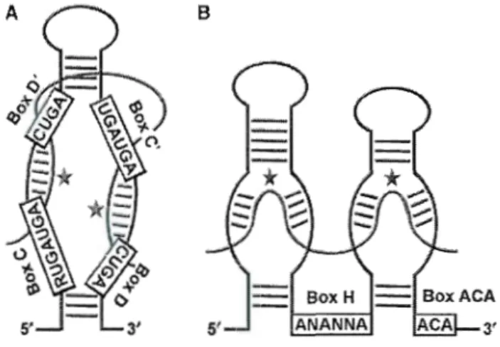
SnoRNAs are short molecules of 60-600 nucleotides, but are mostly in the range of 70-200 nucleotides each (Dragon et al 2006). They can be divided into two major classes that contain evolutionarily conserved sequence elements (Kiss 2001). One major class of snoRNAs is the box CID snoRNAs. Members of this class contain the motifs box C (PuUGAUGA) and D (CUGA), while the HIACA snoRNAs contain box H (ANANNA) and the triplet ACA, a motif located exactly 3 nucleotides from the 3' end. Both classes of guide snoRNAs specify sites of modification by forming direct base pairing interactions with substrate RNA. The 2' -0-methylation guide snoRNAs establish long (10-21 bp) duplexes with the target sequence, white pseudourydilation guide snoRNAs form small duplexes (3-1 0 bp in length). In both classes, short stems bring the conserved boxes close to one another to form core structural motifs. In the box CID snoRNAs, base pairing of 5' and 3' ends is common and favours the exposure of boxes C and D. Boxes C and D are required for snoRNA stability and accumulation. Base pairing between motifs box C and D leads to the formation of a K -turn motif th at binds Snu 13 (Dragon et al, 2006).
The CID snoRNAs sometimess conta in C and D-like motifs, C 'and D',
which may reproduce the helix formed by boxes C and D. In addition, a conserved sequence specifie for rRNA upstream of D or D' regions is present. Box HIACA snoRNAs fold into a hairpin-hinge-haripin-tail structure; the hinge and tai! are single-stranded regions that con tain the H and ACA motifs (Figure 1. 1)
B
Box H ANAN NA
Figure 1. 1 Structure of sn oRNAs.
Members of CID box family contain short sequence elements RUGAUGA (box C) and CUGA (box D) positioned near the 5' and 3' termini. C' and D' boxes are also present. Gui ding 2' -0-methylation involves base pairing of the l 0-to-21-nucleotide-long sequence positioned upstream of box D (or D') to RNA. The H/ACA class consists of a hairpin-hinge-hairpin-tail secondary structure. One or both of the hairpins are interrupted by an internai loop, the pseudourydilation pocket, which con tains two short (3-1 0 nts) nucleotide sequences complementary to sequences flanking the site of isomerisation. (Reichow et al 2007).
The 7-2/MRP RNA (component of RNase MRP) is another snoRNA although it is not classified as part of the two major groups of snoRNAs. This molecule has no known sequence motifs, however, its structure and certain sequences resemble the RNA component of RNase P, the endonuclease that cleaves the 5' extension of transfer RNA (tRNA) precursors. As a mutation in RNase P in yeast affects
rRNA processing, a role in ribosome biogenesis IS suggested (Chamberlain et al 1996).
Biogenesis of functional rRNAs, tRNAs and snRNAs includes the post-transcriptional covalent modification of many carefully selected ribonucleotides. These modifications are important for correct maturation, although these processes are not weil understood (Kiss 2002). Although most snoRNAs participate in rRNA modification reactions, sorne members also take part in processing events. The box
Hl ACA sn oRNA snR30 participates in 18S rRNA synthesis; however, its specifie role remains to be characterized. Processing complexes form immediately after the initiation of pre-rRNA transcription. These complexes are viewed as terminal knobs at the edge of 'Christmas trees' in chromatin spreads of rRNA transcription units. Terminal knob formation requires a large complex that includes the U3 snoRNA (known as the small subunit (SSU) processome) (Dragon et al2002).
1.4. Box H/ACA snoRNA associated proteins.
HIACA RNAs are found in complexes with the following proteins: Cbf5 ( dyskerin in hum ans), Gar 1, Nhp2 and Nop 10 (Kiss 2001 ). Ail four Hl ACA proteins are required for cell viability and rRNA maturation. Cbf5 is a 55-kDa protein and a member of the TruB protein family of pseudouridine synthases (Koonin 1996). Pseudouridylation is catalyzed by Cbf5 (Hoang & Ferre-D'Amare 2001); (Zebarjadian et al 1999). Cbf5 was recently shown to interact directly with the guide
sn oRNA and the remaining proteins (Youssef et al 2007) and con tains a nuclear localization signal (NLS) and the acidic/lysine-rich domain (KKE/D) found in many
nucleolar proteins. Mutations in dyskerin have been shawn to cause a genetic disease called dyskeratosis congenita. This includes progressive bone marrow failure,
abnormal skin pigmentation and mucosal leucoplakias.
Garl, a 25 kDa nucleolar protein, is composed of a core domain containing ali components needed for its localization and function. The core region is flanked by glycine and arginine-rich (GAR) domains notable for their RNA binding capacity (Girard et al 1992). Garl is associated with every H/ACA snoRNA (Girard et al
1992). In vitro binding assays showed the core domain to be inefficient in RNA binding, which may suggest a role for an accessory domain in mediating binding,
including the GAR domain (Bagni & Lapeyre 1998).
Nop10 is a small nucleolar protein of 58 amino acids with no known motif. lt is essential for 18S rRNA synthesis. lt is thought that Nop10, along with Nhp2, are essential for uridine selection, association with pre-rRNA particles, and the stabilization of snoRNP complexes through snoRNA contacts (Henras et al 1998). Nop 10 is an elongated prote in th at associates tightly with Cbf5 near the site of its catalytic domain and stabilizes its active site structure (Hamma et al 2005); (Manival et al2006).
Nhp2 is a protein of 22 kDa and is similar to Snu13 (box CID protein),
suggesting they evolved from a common ancestor. In contrast to Snu 13, Nhp2 does not bind specifically to RNA in vitro. However, it may do so when bound to another
Hl ACA linked prote in. Recently Nhp2 was shown to interact with RN As containing
irregular stem-loop structures, but only has weak affinity for double-stranded RNA.
The central region ofNhp2p is believed to function as an RNA-binding domain, since
it is related to motifs found in various RNA-binding proteins. Removal oftwo amino acids within the putative B-strand element of the central domain impairs its ability to
interact with H/ACA snoRNAs (Henras et a12001).
1.5. Nucleolar Protein 6 (Nop6)
The snoRNA snR30 is essentia1 for the maturation of pre-rRNA. 1t was
earlier demonstrated that snR30 was required for early processing events leading to
18S rRNA production (Morrissey & Tollervey 1993). We recently purified snR30 by
affinity chromatography and identified proteins by mass spectrometry (Lemay et al
2011). One of the proteins was nucleolar protein 6, Nop6, a protein of 26 kDa
implicated in ribosome biogenesis (Garcia-Gomez et al2011). Interestingly, depletion
of snR30 disrupts the association of Nop6 with U3, U14, snR4 and snR35 where
Nop6 associates most strongly with snR35 (Lemay et al 2011 ). Nop6 was originally identified as a member of the hydrophillins, a group of proteins known for their high glycine content and hydrophilicity (Garay-Arroyo et al 2000a). A network-based algorithm of large scale data used to assign gene function predicted Nop6 to be a
fungal-specific nucleolar protein involved in rRNA processing (Samanta & Liang
2003). Subsequent studies using GFP-tagged Nop6 confirmed its localization to the
NEP 1 as the deletion of NOP6, SNR57 or TMA23 genes suppressed a mutation of Nepl that affects ribosome biogenesis. This further highlights the role ofNop6 in this process (Buchhaupt et al 2007). Moreover, Nep 1 is a factor that was shown to methylate pseudouridine residue 1191 of 18S rRNA, and is involved in the assembly of ribosomal protein Sl9 (Rpsl9) into pre-40S ribosome subunits. Nepl is a highly conserved factor that has pseudo-NI-specifie methyltransferase activity such that it can catalyze methylation at the Nl of pseudouridines (Piekna-Przybylska et al 2007, Thomas et al 2011). Loss ofNep1 results in a Joss of cleavage at site A2, leading to an accumulation of21S rRNA species. In fact, Nepl is mutated in Bowen-Condradi syndrome, a lethal genetic disease characterized by low birth weight and a small head (Armistead et al 2009). A recent study on the role of Nop6 in ribosome biogenesis reported that deletion of Nop6 leads to a 20 % drop in 18S rRNA production and a drop in 40S subunit formation, which was suggested to be due to mild inhibition of pre-rRNA processing at cleavage site A2, and deletion did not affect snoRNA formation (Garcia-Gomez et al 2011). Tandem affinity purification followed by mass spectrometry and Northern blot analysis displayed that Nop6 is a component of the 90S pre-ribosomal complex. Localization of Nop6 was shown to be slightly dependant on the transcription of Pol I whereas a Joss of the Pol 1 Rpa49 subunit led to a partial localization in the nucleus. Thus localization of Nop6 to the nucleolus is dependent on pre-rRNA transcription. The intracellular localization of Nop6-eGFP after in vivo shutdown of rRNA transcription suggested that Nop6 binds to pre-rRNA early during transcription.
-- --· - -·
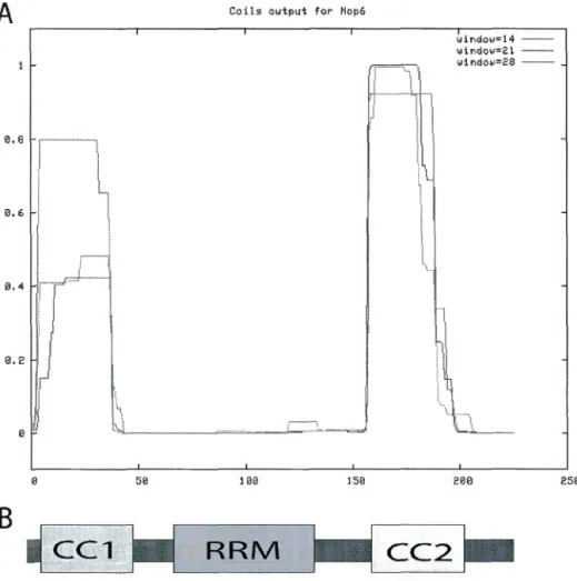
---1Our bioinformatics results suggest that Nop6 contains two Coiled-coil (CC) motifs, which flank an RNA Recognition Motif (RRM) (Figure 1. 2). Previous analysis also concluded Nop6 to be a basic protein as it is rich in both lysine and arginine, and contains monopartite and bipartite nuclear localization signal (NLS) (Garcia-Gomez et al2011).
3 35 79 151 159 187 225
Figure 1. 2 Schematic representation ofNop6 domains.
Coiled-coil 1 (CCl), RNA-binding domain (RRM) and coiled-coil 2 (CC2) are indicated in the schematic representation. The amino acid position of each domain within Nop6 is also shown, as predicted from bioinformatics tools COILS and SMART.
1.6. The coiled-coil domains
Coiled-coil (CC) domains mediate protein-protein interactions and include a repetitive heptad sequence of (abcdefg) n. The first and fourth residues are usually hydrophobie and non-polar, together forming the hydrophobie core. In contrast, the charged and polar fifth and seventh residues form complementary side chains which can interact to form stabilizing salt-bridges. These domains contain two or five alpha helices that are wrapped in a superhelical fashion (Ulijn & Woolfson 201 0). In
addition, there are severa! parallel and anti-parallel CC domains such as the parallel trimeric CC which is illustrated in a helical wheel diagram highlighting their different inter-chain interactions (Figure 1. 3). Dimeric coiled coils such as the anti-parallel MADS box transcription factor is frequently involved in gene regulation, either as activators or as other proteins that are involved in the DNA transcriptio(Changela et al 2003, Lavigne et al 1998, Santelli & Richmond 2000, Walters et al 1997). Using the two-hybrid system, one can verify whether these domains mediate specifie protein-protein interactions. The probability of proteins containing CC domains can also be found through bioinformatics tools such as SMART or COILS (Lupas et al 1991, Newman et al 2000).
14
-· o A
- o
··
··
·· .
. --"
·--
b o B b ----o
·
·
-
-
---
b ~ . ~ ' f f , / \ ,'P a . . ' f . . ' ' .(f . \ ' d. ·•. / c c ' ; ·· :d -.. / c--
-c(~\
((i. / ' ' " -' bO
x o
.
·
.ct
..., ' -; 1 a layero.-a
~ ~-o
tJ'
ç.o
Ota
dlayer0~
Cff
~
C
D-o~
' 1..
' J 1 ' : c a ' •, dc
,
"•<.._dXol
T ' a .' i
~à
' :--- ' , ' Mixed a and d layersO.
*a
qê?
Çi0
éfo
Figure 1. 3 Helical wheel diagram of coiled coil motifs.
(A) parallel dimeric coiled coi!, (B) anti-parallel dimeric coiled coi!, (C) parallel trimeric coiled coi!, and a (D) parallel tetrameric coiled coi!. The curved arrows indicate salt bridges while the crossed-arrows indicate hydrophobie interactions. The knobs-in-holes configuration are found in (E) parallel dimeric, trimeric and tetrameric coiled coils and (F) antiparallel dimeric, trimeric and tetrameric coiled coils. Taken from (Apostolovic et al2010)
- - - ---·---
---CHAPTER 2: HYPOTHESIS and OBJECTIVES
2.1 Hypothesis
Recent studies indicated that Nop6 is a nucleolar protein that is potentially involved in ribosome biogenesis. Moreover, bioinformatics searches have indicated that Nop6 contains two predicted CC motifs and an RNA recognition motif (RRM). The presence of the RRM would suggest that Nop6 may interact with snoRNAs, rRNA, or another RNA molecule. Given the presence of CC motifs in Nop6, which are sites of protein-protein interactions, Nop6 could regulate ribosome biogenesis through contacts with other proteins.
Our main hypothesis is that Nop6 associates with protein partners through its CC domains in complexes, and this helps to regulate ribosome assembly. In addition, due to the presence of an RRM, Nop6 may also affect ribosome maturation through contacts with RNA.
2.2. Objectives
The first objective was to identify partners of Nop6 using a yeast-two-hybrid screen with Nop6 as bait. The second objective was to confirm results of the two-hybrid screen by immunoprecipitation experiments (IPs). ln order to conduct the IPs, strains expressing epitope-tagged protein partners of Nop6 were prepared by PCR amplification of modules containing the tag of interest and introduced through homologous recombination. The third objective was to determine if the interactions were direct using GST-pulldown experiments.
CHAPTER 3:
RIBOSOME BIOGENESIS FACTOR NOP6
INTERACTS WITH RRTS
Ahmed Hossain et Francois Dragon
Département des sciences biologiques, Centre de recherche BioMed, Université du Québec à Montréal, Montréal, Québec, Canada, H3C 3P8
Résumé
Le nucléole est le site de la biogenèse des ribosomes, un processus essentiel pour la survie et la prolifération cellulaire. L'assemblage et la maturation des ribosomes est complexe et comprend de nombreux facteurs impliqués dans le clivage des précurseurs d'ARN ribosomiques. Nous avons récemment découvert que la protéine Nop6 est associée à snR30, une petite RNP nucléolaire impliquée dans la maturation du pré-ARNr, ce qui suggère que Nop6 pourrait être impliquée dans la biogenèse des ribosomes. Des études bioinformatiques ont suggéré que Nop6, une protéine nucléolaire de 26 kDa, contient deux motifs coiled-coil (CC) qui flanquent un motif de liaison à I'ARN (RRM). Nous faisons l'hypothèse que Nop6 se lie à d'autres protéines à travers ses motifs CC, et que ces interactions participent à la maturation des pré-ARNr. Un partenaire potentiel de Nop6, Rrt5, a été retrouvé par une recherche de base de données. Nos résultats doubles hybrides chez la levure montrent que Nop6 interagit avec Rrt5 et que les CC de Nop6 ne sont pas nécessaires pour son association avec Rrt5. Ces résultats ont été confirmés par des tests de GST pulldown,
- - - -- - -- - - -- -- - - ,
qui ont révélé que l'interaction Nop6/Rrt5 n'est pas dépendante d'ARN. En outre, nous avons confirmé par une analyse de type « western
»
que le gène RRT5 est peu exprimée au cours de la croissance végétative et donc d'autres expériences ont été menées dans des conditions de sporulation. Des analyses par microscopie à immunofluorescence ont montré que Nop6 et Rrt5 sont localisées à la paroi cellulaire exclusivement pendant la sporulation, suggérant une fonction spécifique pour la sporulation.Ribosome biogenesis factor Nop6 interacts
with
Rrt5
Ahmed Hossain and Francois Dragon
Département des sciences biologiques and Centre de recherche BioMed, Université du Québec à Montréal, Montréal, Québec, Canada, H3C 3P8
3.1. Abstract
The nucleolus is the site of ribosome biogenesis, an essential process for cel! survival and proliferation. The assembly and maturation of ribosomes is complex and includes many factors involved in the cleavage of ribosomal RNA precursors. We have recently found protein Nop6 in a complex with snR30, a small nucleolar RNP
involved in pre-rRNA processing, which could implicate Nop6 in ribosome
biogenesis. Bioinformatics studies have suggested that Nop6, a nucleolar protein of 26 kDa, contains two coiled-coil (CC) motifs which flank an RNA recognition motif (RRM). We hypothesize that Nop6 binds to other proteins through its CC motifs, and these interactions effectively participate in ribosomal subunit maturation. A potential partner of Nop6, Rrt5, was found by data base mining. Our yeast two-hybrid results show that Nop6 interacts with Rrt5 and that the CC regions ofNop6 are not necessary for its association with Rrt5. These results were confirmed by GST pulldown assays, which revealed that the Nop6/Rrt5 interaction is not RNA dependent. In addition we confirmed using western blot analysis that Rrt5 is poorly expressed during vegetative
growth and thus further experiments of Rrt5 were conducted under sporulating
conditions. Immunofluorescence imaging localized Nop6 and Rrt5 to the cel! wall exclusively during sporulation, suggestive of a sporulation-specific function for Nop6.
Key Words: Meiosis, nucleolus, Nop6, ribosome biogenesis, Saccharomyces cerevisiae
3.2.
IntroductionThe nucleolus is the most distinctive sub-nuclear compartment. It is the site of rRNA transcription and ribosome biogenesis (Gerbi et al 2001). Ribosomal genes are
repeated genes that are grouped into chromosomal regions, nucleolar organizer regions
(NOR), where many NORs organize to help form the nucleolus (Dammann et al 1993).
This compartment is also involved in the sequestration of proteins involved in cel! cycle regulation (Shou et al 1999). RNA polymerase 1 (Pol 1) and its factors remain at the sites of rDNA repeats at the NORs of the chromosomes, while other pre-RNA
factors form a sheath around the condensed chromosomes, the perichromosomal space
(Dundr et al 2000). Maturation of ribosomal rRNA and its assembly into ribosomal subunits involves severa! hundred proteins and small nucleolar ribonucleoprotein
complexes (snoRNPs) (Kressler et al 1999, Venema & Tollervey 1999).
Ribosome biogenesis begins with Pol I generating a large 35S pre-rRNA
precursor that contains the sequences for mature ribosomal RNAs (18S, 5.8S, 25-28S),
two external transcribed spacers (ETS) and two internai transcribed spacers (ITS).
This primary transcript is subsequently chemically modified at numerous sites and subjected to endo- and exonucleolytic cleavages to produce its mature forms. The 35S pre-rRNA and non-ribosomal and ribosomal proteins form a large RNP complex that is rapidly converted into precursors of the 40S and 60S ribosomal subunits. The pre-40S particles are further processed in the cytoplasm, whereas the pre-60S particles mature in the nucleus prior to transport to the cytoplasm for final assembly.
The snoRNA snR30 is essential for the maturation of pre-rRNA. lt was previously shown that snR30 is required for early processing events leading to 18S
rRNA production (Morrissey & Tollervey 1993). We have recently purified snR30 by
affinity chromatography and proteins were identified by mass spectrometry. One of the
proteins was Nop6, a protein of 26 kDa. lnterestingly, a depletion of snR30 disrupted
the association of Nop6 with U3, Ul4, snR4 and snR35 snoRNAs; Nop6 associated
most strongly with snR35 (Lemay et al2011).
Nop6 was originally identified as a member of the group of proteins called hydrophillins due to its high glycine content and hydrophilicity (Garay-Arroyo et al 2000a). A network-based algorithm of large scale data used to assign gene function predicted Nop6 to be a fungal-specific nucleolar protein involved in rRNA processing
(Samanta & Liang 2003). Studies using GFP-tagged Nop6 confirmed its Jocalization to
the nucleolus (Huh et al 2003) . A recent study on the role of Nop6 in ribosome
biogenesis reported that the deletion of Nop6 leads to a 20 % decrease in 18S rRNA
and a decrease in 40S subunit formation; it is suggested that this results from a mild inhibition of pre-rRNA processing at cleavage site A2 (Garcia-Gomez et al 2011 ). The
intracellular localization of Nop6-eGFP after in vivo shutdown of pre-rRNA
transcription suggested that Nop6 binds to pre-rRNA early during transcription (Garcia-Gomez et al 2011). These authors showed Nop6 to be a basic protein as it is rich in lysine and arginine, and contains both monopartite and bipartite nuclear Jocalization signais (NLS). Our bioinformatics analyses suggest that Nop6 contains two coiled-coil (CC) motifs, which flank an RNA recognition motif (RRM) (Figure 3.
lA). This suggests Nop6 could mediate RNA maturation through its interactions with
other proteins using its CC motifs as this structural motif is known for mediating such
interactions (Ulijn & Woolfson 2010). Since Nop6 has already been implicated in ribosome biogenesis, identifying protein effectors for its function during this process is
of interest. One interesting protein partner already identified in a genome-wide two-hybrid screen is Rrt5, a protein that is highly expressed during sporulation and possibly involved in regulating rDNA transcription (Hontz et al 2009, Naitou et al 1997).
We have determined that deletion of both Nop6 CC domains does not impede its association with Rrt5. ln addition, Nop6 and Rrt5 co-localize only during
sporulation, which suggests Nop6 could have a separate function from ribosome biogenesis.
3.3. Materials and Methods
3.3.1 Y east strains and plasmids
Y east strains that were used in this study are listed in Table 3 .l. Rrt5-Myc
was prepared through the integration of a 9xmyc epitope tag at the C-terminus using
standard PCR amplification (Longtine et al 1998). The knockouts of Rrt5 and Nop6 were obtained by integrating a cassette of auxotrophic markers at the NOP6 and
RRT5 loci. Strains expressing mye- or HA-tagged constructs under the control of
their natural promoter were generated as described (Longtine et al 1998). Strains
YPH499, YPH500 were modified to express Ndt80 and Suml under the control of the GALl promoter (Longtine et al 1998).
The sequence corresponding to the open reading frame (ORF) of NOP6 was amplified from genomic DNA extracted by standard procedures (Ausubel 1999) using oligonucleotides NOP6-For- (5'- CCC CCC GGG TAI GGG GTC CGA GGA AGA TAA AAA G-3') and NOP6-Rev- (5'- CGC CTC GAG ICA TTT AAG TAG TTT GGC TC-3'). The deletion mutant ofNop6 lacking the coiled-coil 1 domain was amplified using oligonucleotides NOP6 ~CCl-For- (5'- CGC CCC GGG TAI GCC AGA AGG AAA AAG ACC C-3') and NOP6-Rev- (5'- CGC CTC GAG ICA TTT AAG TAG TTT GGC TC-3'). The deletion mutant lacking the CC2 domain was amplified using oligonucleotides NOP6-For- (5'- CCC CCC GGG TAI GGG GTC CGA GGA AGA TAA AAA G-3') and NOP6 ~CC2-Rev- (5'- GCG CTC GAG CTA TCC GCC AAC AGI TAG CTC-3'). The double deletion mutant ofNop6 was amplified using oligonucleotides NOP6 ~CCl-For- (5'- CGC CCC GGG TAI GCC AGA AGG AAA AAG ACC C-3') and NOP6 ~CC2-Rev (5'- GCG CTC GAG CTA TCC GCC AAC AGI TAO CTC-3'). The PCR products were cloned between restriction sites Xmal and Xhol in pGBKT7 (Clontech) and pOEX4Tl (Amersham Pharmacia) (Table 3.3 and 3.5). A clone ofNOP6 containing a mutation in the RNPl motif was generated a two-step PCR mutagenesis strategy using a first amplification with oligonucleotides NOP6-For and NOP6-MutRNPl-R- (5'- GOC TGC CGC AGC TOC AGC AGC TGC OTC AGC TCT GAG GCG GAT CT-3') and separate
- -
--
---
----
---GCT GCA ---GCT GCG GCA GCC GAT ---GCT GAC AAA GAT CGC AC-3 ') and
NOP6-R-. The two PCR products from the first amplification were further amplified
together using oligonucleotides NOP6-F-and NOP6-R-. This RNPI mutant was
cloned between sites Xmal and Xhol in both pGBKT7 and pGEX4Tl. Similarly, the
ORF of RRT5 was amplified using oligonucleotides RRT5-For- (5'- GCG GAA TTC
ATG ACA GAA CAA GTT AAC AAT GAC-3') and RRT5-Rev- (5'- CGC GAA
TTC CTA AGT AGC AGC CAC GGT-3') and cloned in the site EcoRl in pGADT7.
The ORF of RRT5 was subcloned from pGADT7 into pMal-c5x (New England
Biolabs). Two-hybrid plasmids, pGBKT7 containing Nop6 or mutant derivatives,
and pGADT7 containing fulllength Rrt5 were transformed in AH109 The integrity of
ali constructs was verified by automated sequencing at the McGill University and
Génome Québec Innovation Centre.
3.3.2. Sporulation of diploid cells
In arder to verify whether deletion of NOP6 and RRT caused changes in
sporulation, strains YPH50!, N op6MN op6L1, Rrt5 LVRrt5 L1,
Nop6L1Rrt5MNop6L1Rrt5L1, (Table 3. 1) were sporulated as previously described
(Nickas & Neiman 2002a). Cells were sporulated after incubation in YPD (1% yeast
extract, 2% peptone, 2% dextrose), grown to mid-log phase in YP acetate 0.1% and
transferred to 2% potassium acetate at an optical density of 2 A600 units with rapid shaking at 30°C in liquid.
3.3.3. Immunoprecipitation experiments (IPs)
Cells expressing myc-tagged Rrt5 were grown m synthetic media to exponential phase (A600 ~ 0.5), sporulated as described (Nickas & Neiman 2002b),
and harvested by centrifugation. Cells were stored at -80°c. The cell pellet was
washed twice in ice-cold sterile water and resuspended in TMN100 buffer (25 mM
Tris-HCl (pH 7.6), 10 mM MgCh, 100 mM NaCI, 1 mM 1,4-dithiothreitol (DTT),
0.1% NP-40) complemented with Complete protease inhibitor (Roche). The buffer to
cell ratio was 100 ~-tl! A600 unit. Who le cell extract was prepared with glass beads (Sigma). The lysate was cleared by centrifugation (5 min, 10000 x g). A volume of 0.5 ml of cell extract was incubated with 25 ).tl of ProteinA (Roche) beads coupled to
mye mAb at 4°C for 2 hon a nutator. Beads were washed with 1 mL of TMN100
lacking NP40 and DTT several times. Elution from Protein A beads was carried out with SDS sample buffer. Samples were separated on polyacrylamide gels and
analyzed by Western blotting.
3.3.4. GST-Pulldown
GST-Nop6 fusion constructs were expressed in Rozetta 2pLys (Novagen)
grown to exponential phase in LB (1% tryptone, 0.5% NaCJ, 0.5 % yeast extract)
containing ampicillin (100 11g/ml). Fusion proteins were expressed by induction with
1mM IPTG for 2 hours. Rozetta 2pLys cells expressing MBP-Rrt5 were grown to
exponential phase in LB broth and 0.2 %dextrose, and then induced with IPTG (0.3
pellets were resuspended in PBS buffer (13.7 mM NaCI, 0.27 mM KCI, 10 mM Na2HP04, 0.2 mM KH2P04 pH 7.4). Total protein extracts were prepared by
sonication and debris removed by centrifuging for 10 min. MBP-Rrt5 was bound to amylose column washed with NETN500 (0. 5% NP40, 0.1 mM EDTA, 20 mM Tris-HCI pH 7.4, 500 mM NaC1) and eluted with NETN200 (20 mM Tris-HCI pH7.4, 200 mM NaCI, 0.1 mM EDTA) containing 10 mM maltose. GST-pulldown assays were carried out as described (Frangioni & Nee) 1993). Essentially, GST or GST-Nop6 fusions were bound to 25 f.LL of GST coupled beads (GE healthcare), washed with NETN150 mM [0. 5% NP40, 0.1 mM EDTA, 20 mM Tris pH 7.4, 150 mM NaCI, Complete protease inhibitor cocktail (Roche)] and Rrt5-myc yeast extracts or MBP-Rrt5 were incubated with GST columns followed by elution with SDS 2X sample buffer. Samples were migrated in a polyacrylamide gel and analyzed by Western blotting. Rrt5 was detected through Western blotting with myc (9E1 0) and anti-MBP (New England Biolabs) antibodies.
3.3.5 Immunofluorescence microscopy
Yeast cells were processed as described to detect changes in protein expression during sporulation (Nickas & Neiman 2002a). Purified anti-Nop6 rabbit polyclonal antibodies, and 9El 0 anti-myc mouse monoclonal antibody were used to recognize Nop6 and Rrt5-myc, while mou se monoclonal 17C 12 anti-fibrillarin antibody was used to identify the nucleolar marker Nop1 (Yang et al 2001). Antibodies were diluted 1/500 in blocking buffer (0.5% BSA, 0.5% Tween-20 in
PBS) and incubated overnight at 4°C. Samples were washed in blocking buffer several times and incubated for 30 minutes at room temperature with secondary antibodies Alexa Fluor 488 goat anti-rabbit lgG and Alexa Fluor 555 goat anti-mouse
lgG (lnvitrogen) diluted 1/1000 in blocking buffer. Nuclear DNA was stained for 15
min with 1 j.lg/ml of 4' -6-diamidino-2-phenylindole (DAPI). Coversl ips were washed
with blocking buffer and slides were mounted with ProLong Gold antifade reagent (Invitrogen). FITC-concanavalin A staining was used to determine the cell wall
association ofNop6 and Rrt5, and Alexafluor 555 was used to detect Nop6 and Rrt5
(Nickas & Neiman 2002a). Specimens were observed with an Eclipse Ti inverted
microscope (Nikon) using an Apochromat objective (numerical aperture 1.4). Images were acquired with a Monochrome CCD camera (CFW1312 from Scion Corporation)
and processed with ImageJ version 1.34s (Wayne Rasband, National Institutes of
3.4. Results and Discussion:
3.4.1 Nop6 interacts with RrtS in the two-hybrid system
After conducting a literature search for potential partners of Nop6 we have found that a previous array screen showed that Nop6 interacts with Yfr032c, a protein of unknown function. However, further studies have not confirmed this interaction (Uetz et al 2000). Northern analyses studying the expression levels of severa! genes of chromosome VI indicated that YFR032C is highly expressed during sporulation
and mRNA was undetectable in vegetative growth (Naitou et al 1997). Yfr032c was also identified as a modulator of transcription of ribosomal DNA (rDNA). It has been re-named Rrt5 for "Regulator of [Ïbosomal DNA !ranscription" (Hontz, Niederer et al. 2009).
To examine the interaction between Nop6 and Rrt5 in the yeast two-hybrid system, Nop6 was cloned in the bait vector pGBKT7 and Rrt5 was cloned into the prey vector pGADT7. These proteins showed a strong interaction with the ability to bind as cells can grow on media containing a high concentration of 3-aminotriosol (15 mM), an inhibitor of the product of the HIS3 reporter gene. Nop6 contains two
coil-coiled domains and an RNA- recognition motif (RRM) (Figure 3. 1). The RNA-recognition motif (RRM), also known as RBD (RNA-binding domain) or RNP (ribonucleoprotein domain) is the most frequent RNA binding domain in higher vertebrates and most studied (Clery et al2008). We created deletion mutants ofNop6 lac king one or both of the CC motifs (Figure 3. 1 ). The Joss of both motifs, leaving
just the RRM and a portion of flanking sequences intact, stiJl produced a strong association with Rrt5 (Figure 3. ID). This suggests that the CC motifs ofNop6 do not mediate its interaction with Rrt5. Rrt5 also contains a RBD domain; however we were unable to express Rrt5 deletion mutants lacking the RRM. In addition NOP6-RNPI mutant cloned into pGBKT7 was unstable and could not be expressed in AH109 cells.
3.4.2. Nop6 and Rrt5 may be involved in the regulation of sporulation
RRT5 mRNA expression was previously shown to be strongly expressed during sporulation (Na itou et al, 1997). Rrt5 prote in bearing a 9-myc tag was barely detectable under vegetative growth and highly expressed during sporulation (Figure 3. 2, lanes 3 and 5). Rrt5 was slightly immunoprecipitated under vegetative conditions, and since it is highly expressed during sporulation a large amount was immunoprecipitated under these conditions (Figure 3. 2, lanes 4 and 6). Naitou et al, (1997) indicated that RRT5 does not contain an URS 1 regulatory site found in most early sporulation genes, and thus, is most likely a middle or late-meiotic sporulation gene. The loss of RRT5 does not affect sporulation, however Joss of NOP6 alone Jowers sporulation (Table 3. 1). Interestingly the double knockout of RRT5 and NOP6
shows normal levels of sporulation compared to single NOP6 KO. There appears to be a global Joss in sporulation in NOP6 KO cells, however this is just due to Jess cells being counted. This indicates that the effects of NOP6 Joss could be alleviated by the
Joss of RRT5 and Rrt5 mediates the effects of Nop6 during sporulation. Ectopie overexpression of two regulators of middle sporulation genes, NDT80 and SUMJ
under the inducible GALl promoter did not affect the levels of RRT5 transcript (data not shown).
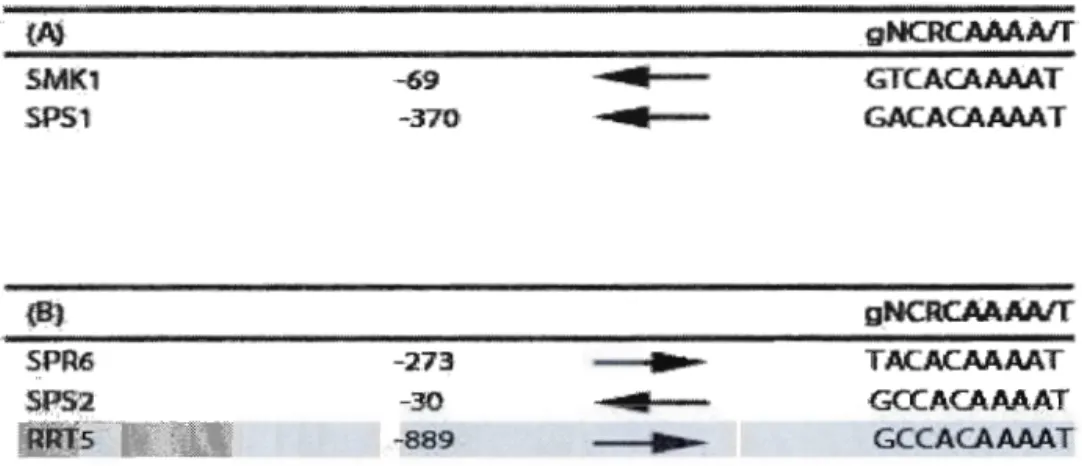
RRT5 gene appears to also contain a MSE element in its promoter region,
which contains the consensus sequence of an MSE (Table 3. 2) MSEs of genes regulated by global middle sporulation gene activator Ndt80are indicated in Table 3.
2A and genes with MSEs and not regulated by Ndt80 are indicated in Table 3.2B. Many middle sporulation genes are activated by the global activator Ndt80, which also positively regulates its own expression. Ndt80 is a transcriptional activator that binds to the conserved middle sporulation element that is upstream of middle sporulation genes. These MSE elements could have dual function as during sporulation they function as activator sites and during vegetative growth as repressors sites. The fact that overexpression of Ndt80 does not increase the levels of RRT5
would indicate that RRT5 may be a mid-late sporulation gene (data not shown). In contrast to RRT5, the Joss of NOP6 alone lowers sporulation, which suggests that this normally, nucleolar protein could have a function during sporulation (Table 3. 1).
In Saccharomyces cerevisiae, the dynamics of the nucleolus are different from other organisms and like other fungi, this is not weil understood. Nucleoli maintain their integrity during both mitosis and meiosis which is linked to their endonuclear type of division (Loidl 2003). ln meiosis, nuclei do not completely separate at the end
of the first division but form a dumb-bell shaped structure. Immunostaining against nucleolar proteins Nopl, Nop5 and Nhp2 revealed that the nucleoli are attached to the NORs during the first meiotic division until anaphase II. At the end of the second meiotic division, nucleolar proteins remained with the cytoplasm of the ascus and no nucleolar material was detected in the prospores (Fuchs & Loidl 2004). This may indicate that Nop6 as a nucleolar protein may have a separate function during meiosis due to possible changes in localization.
3.4.3. The interaction between Nop6, and Rrt5 is not RNA-mediated
As the yeast two-hybrid system can sometimes produce artefacts, we used standard in vitro methods to confirm the association ofNop6 with Rrt5. Mye tagged
Rrt5 was used in pull-down assays with GST-Nop6 or its mutant derivatives immobilized on glutathione Sepharose.
In order to verify the association of Nop6 and Rrt5, Nop6 was immunoprecipitated from whole cell extracts expressing Rrt5-myc, by coupling it to Agarose protein-A beads, which were bound to anti-Nop6 rabbit polyclonal antibodies. Nop6 was able to co-immunoprecipitate Rrt5 (Figure 3. 3A). In addition, the association of GST-Nop6 expressed from E. coli with Rrt5-myc expressed from
yeast whole cell extracts was tested to confirm that the association ofNop6 with Rrt5 was direct. Glutathione Sepharose beads were coupled to bacterial extracts that expressed GST alone, GST-Nop6 or GST-Nop6 mutant derivatives followed by
incubation with yeast whole cell extracts expressing Rrt5-myc. Rt15 was able to
associate with GST-Nop6 and its mutant derivatives, but not GST alone (Figure 3.
3B). Finally, to confirm the direct interaction ofNop6 and Rrt5, GST and GST-Nop6
constructs expressed from bacteria were coupled to glutathione beads followed by the addition of Rrt5 expressed in fusion with MBP. MBP-Rrt5 could be pulled down with GST-Nop6 constructs, but not GST alone (Figure 3.3C). We confirmed that the association ofNop6 with Rrt5 was not mediated through its coiled-coil (CC) domains as observed in the two-hybrid tests as Joss of both CCs did not disrupt this association (Figure 3.3 Band C). To examine if the association between Nop6 and Rrt5 is RNA-mediated, Nop6 was immunoprecipitated from whole cell extracts expressing
Rrt5-myc in the presence or absence of RNase A. The association between Nop6 and Rrt5
was not disrupted in the presence of RNase A, which indicates a protein-protein
interaction with Nop6 (Figure 3.3A). The RNPl motif in the RRM is known to be
vital for RNA recognition and its disruption was hypothesized to affect the interaction with Nop6 via an RNA intermediate. The mutation of RNPl did not alter the stability of the association ofNop6 and Rrt5 (Figure 3.3B and C), which further indicates this interaction is not RNA-mediated.
3.4.4. Nop6 and Rrt5 co-localize to the cell wall in spores
Nop6 was previously demonstrated to be a potential ribosome biogenesis factor. lt was localized to the nucleolus, the site of ribosome biogenesis ((Huh et al
2003);Garcia-Gomez et al, 2011; Lemay et al, 2011). Rrt5-myc cells were grown under vegetative conditions or sporulated in 2 % acetate solution in order to conduct
immunolocalization. During vegetative growth, Nop6 co-localized with the nucleolar
marker Nop1, while Rrt5 localized to the cell periphery (Figure 3. 4A and 3B). After
induction of sporulation, Rrt5 and Nop6 both co-localized to the cel! membrane of
asci (Figure 3. 4A). Unlike Nop6, the nucleolar marker Nop1 remained in discrete
points after sporulation (Figure 3. 4A). In order to confirm this localization to the cel!
wall, cells were stained with the cell wall marker concanavalin A (ConA). lndeed,
Nop6 and Rrt5 co-localized with ConA to the cel! wall (Figure 3. 4C). This result could indicate an unexpected function ofNop6 during sporulation.
Ribosome biogenesis is a complex process involving severa! hundred factors. Nop6 is a factor that is involved in ribosomal maturation whose function is still not weil understood. We have shown that Nop6 interacts physically with Rrt5 and they co-localizes to the cell wall of spores. Thus Nop6 seems to be an interesting factor that may be altered after sporulation.
3.5. Tables
Table 3. 1. Nop6 deletion alone lowers sporulation frequency.
Strain Sporulated Unsporulated Percent Sporulated
YPHS01 494 1309 27 o/o
rrt51J/rrt51J 568 2130 20.9%
nop61J/nop6!J 66 468 11.7%
nop61Jrrt51J Y/nop61Jrrt51J 345 1193 22.2%
Table 3. 2. Mid-sporulation elements and their regulation. :SMK1 SPS1 SPR6 SPS2 RRJ5 -69 -370' -273 -30 -889 . . m
..
..
gNCRCAAAA!T GTCACAAAAT GACACAMAT gNCIKAAMIT TACACAAAAT GCCACAAAAT GCCACAAAATTable 3. 3 Y east strains used in study
Strain Alias Genotype Source
MA Ta ura3-52
lys2-(Sikorski &
801 amber
ade2-YPH499
-101_ochre trp1-1163 his3- Hieter 1989)
11200 leu2-111 MATa ura3-52 lys2-801 amber ade2-(Sikorski & 101_ochre trp1-1163 his3-YPH500
-11200 leu2-111 Hieter 1989) (Sikorski &YPH501
-
YPH499/YPH500 Hieter 1989)(Lemay et al,
AHY1 NOP611 YPH499 YPH499, NOP611:.HISJ 2011)
(Lemay et al,
AHY2 NOP6/1 YPH500 YPH500, NOP611::TRP 1 2011)
YPH499,
RRTS-9-RRTS-9- Mye ::TRP1/YPH500,
AHY3 Myc/RRTS-3-Myc RRTS-3-Myc ::HIS1 This work
NOP611 YPH499/ YPH499, NOP611::HISJ/
AHY4 NOP611 YPH500 YPH500, NOP611::TRP 1 This work
RRTS/1 YPH499/ YPH499, RRT511::TRP 11
AHY5 RRTS/1 YPH500 YPH500, RRT511::HIS1 This work
YPH499,
PGALl-NDT80::TRPJI YPH499,
PGAu-NDT80::TRP JI
YPH500,
PGALl-AHY6 GAL::NDT80 NDT80::HIS1 This work
RRTS/1 YPH499/ YPH499, RRT511::TRP JI
AHY7 NOP611 YPH500 YPH500, NOP611::HISJ This work
MATa, trp1-901, leu2-3,
112, ura3-52, his3-200,
gal4/1, gal80/1,
L YS2::GAL1
UAS-GALl TATA -HIS3,
GAL2uAs-GAL2TATA-ADE2, URA3::MEL1 uAs
-AH109
-
MELl TATA-IacZ, MEL1 ClontechSame as AH1 09 except has pGBKT7+pGADT7- pGBKT7 and pGADT
7-AHY8 Rrt5 (AH109) Rrt5 This work
pGBKT7- Sames as AH1 09 except Nop6+pGADT7- has pGBKT7-Nop6 and
AHY9 Rrt5 (AH 1 09) pGADT7-Rrt5 This work
pGBKT7- Same as AHl 09 except has
N op6+pGADT7 (AH pGBKT7-Nop6 and
AHY10 109) pGADT7 This work
Same as AH1 09 except has pGBKT7-Nop6 pGBKT7-Nop6 ~CC1and
AHY11 ~CC 1 +pGADT7 pGADT7 This work
pGBKT7-Nop6 Same as AH1 09 except has
~CC 1 +pGADT7- pGBKT7-Nop6 ~CC1and
AHY12 Rrt5 pGADT7-Rrt5 This work
Same as AHl 09 except has pGBKT7-Nop6 pGBKT7-Nop6 ~CC2 and
AHY13 ~CC2+pGADT7 pGADT7 This work
pGBKT7-Nop6 Same as AH1 09 except has ~CC2+pGADT7- pGBKT7-Nop6 ~CC2 and AHY14 Rrt5 pGADT7-Rrt5 This work pGBKT7-Nop6 Same as AH 1 09 except has ~CC 11 ~CC2+pGAD pGBKT7-Nop6 AHY15 T7 ~CC1/~CC2 and pGADT7 This work Same as AHl 09 except has pGBKT7-Nop6 pGBKT7-Nop6 ~CC 11 ~CC2+pGAD ~CC 11 ~CC2+pGADT 7-AHY16 T7-Rrt5 Rrt5 This work
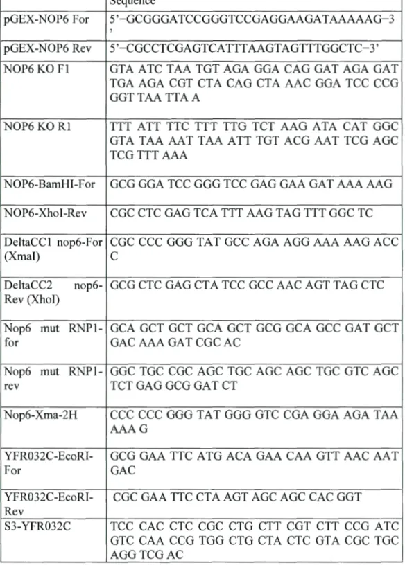
Table 3. 4 Oligonucleotides used in study
Sequence
pGEX-NOP6 For 5 '-GCGGOA TCCOGGTCCGAGGAAGA T AAAAAG-3
'
pGEX-NOP6 Rev 5'-CGCCTCGAGTCATTTAAGTAGTTTGGCTC-3'
NOP6 KO Fl OTA ATC TAA TOT AGA GOA CAO GAT AGA GAT
TGA AGA CGT CTA CAO CTA AAC GOA TCC CCG GGTTAA TTAA
NOP6 KO R1 TTT ATT TTC TTT TTG TCT AAG ATA CAT GGC
OTA TAA AAT TAA ATT TOT ACG AAT TCG AOC TCGTTT AAA
NOP6-BamHI-For GCG GOA TCC GOG TCC GAG GAA GAT AAA AAO
NOP6-Xhol-Rev CGC CTC GAG TCA TTT AAG TAO TTT GGC TC
DeltaCC1 nop6-For CGC CCC GOG TAT GCC AGA AGG AAA AAG ACC
(X mal)
c
DeltaCC2 nop6- GCG CTC GAG CTA TCC GCC AAC AGT TAO CTC
Rev (Xhol)
Nop6 mut RNPl- GCA OCT OCT GCA OCT GCG GCA GCC GAT OCT
for GAC AAA GAT CGC AC
Nop6 mut RNPl- GGC TOC CGC AOC TOC AOC AOC TOC GTC AOC
re v TCT GAG GCG GAT CT
Nop6-Xma-2H CCC CCC GOG TA T GOG GTC CGA GOA AGA T AA
AAAG
YFR032C-EcoRI- GCG GAA TTC A TG ACA GAA CAA OTT AAC AA T
For GAC
YFR032C-EcoRI- CGC GAA TTC CTA AGT AOC AOC CAC GOT
Re v
S3-YFR032C TCC CAC CTC CGC CTG CTT CGT CTT CCG A TC
GTC CAA CCO TGG CTO CTA CTC GT A CGC TOC AGGTCGAC
S2-YFR032C TTT ATG TTA AGI TCT CAC TGT TTA TAC CCC CTT TTC AAT TTT ATA AAG ATA TCG ATG AAT TCGAGCTCG
YFR032CcheckCtF CGCGACCCACCTCCAGCAGC OR
YFR032CcheckCtR CTT AAA AGG GGT TCC CCC AGC CGC
EV
F4-YFR032C TCT TTT TCC TCC TT A TGT GCC ACA AAA TGG
AAG CGT CAC AAA TTA ATA ACG AAT TCG AGC TCGTTT AAAC
R2-YFR032C A TG GIA GTG GIA GTG ICA CTG GT A GTG ICA
TTG TTA ACT TGT TCT GTC A TT TTG AGA TCC GGGTTTT
YFR032CcheckNtF TTT ATC TCAATA TCC AGI ACC GTTTTC OR
YFR032CcheckNt CGC CAT AGI TGT TTA GGA AGG C
REV
Yfr032c Mut delta GAC CA TA TG ACA CGT TCC TT A CGA GGC
RRM Ndei-For
Yfr032c Mut delta AAC CCC GGG GT A GAG TCT GCC TCG T AA GGA
RRM c-Xmal- ACG TGT AAC TTT AGA AA T AGI TTC TGA TTT
RRT5-Check-KO- CCA CAA GCA TGC CCI ACA CGG CAC For
Rl-RRT5-KO TTT ATG TTA AGI TCT CAC TGT TTA TAC CCC CTT TTC AAT TTT ATA AAG ATG AAT TCG AGC TCGTTT AAAC
Fl-RRTS~KO AAC ACA TAG AGA AGI ATA AGG CCI CAC ATA AGC ATA CAA ACA AGC CCG CAC GGA TCC CCG GGTTAA TTAA
F4-NDT80 GAC AAA GCT CCA GAA CGG TTG TCT TTT GTT
TCG AAA AGC CAA GGT CCC TTG AAT TCG AGC TCGTTT AAAC
R3-NDT80 TTG GAT ACG AGA ICA TCC TGT AA T ACT GGA TCT GTG TTT TCC A TT ICA TT A GCA CCA CCG CAC TGA GCA GCG TAA TCT G
GTG TTT TCC A TT TCA TTC A TC A TT TTG AGA
TCC GGGTTTT
NDT80-CheckNt- TAC TTC CGC GGC TA T TTG ACG TTT TCT
For
NDT80-CheckNt- TCT TTT ATA ACC TAC CCA CTC TTC ATC AAT
Re v ATG
F4-SUM1 ACA GGC ATA TTT TAT CAA AAG TOT CAO CAA
ACA GAG CAC AAG GOA CTT GTG AA T TCG AOC TCGTTT AAAC
R3-SUM1 AGT CTC TOT TCA TTG OTT ATG TTA TCA GAA GOG OCT GTG GTG TTC TCA GAA GCA CCA CCG CAC TGA GCA GCG T AA TCT G
R2-SUM1 CTC TOT TCA TTG OTT A TG TT A TCA GAA GOG
OCT GTG GTG TTC TCA GAC ATC ATT TTG AGA TCCGGGTTTT
anti-ySRP CCC ACC AGA AAG CCA TTA CAO CC
anti-18S CAT GGC TTA ATC TTT GAG AC


