HAL Id: inserm-00369695
https://www.hal.inserm.fr/inserm-00369695
Submitted on 15 Mar 2010HAL is a multi-disciplinary open access
archive for the deposit and dissemination of sci-entific research documents, whether they are pub-lished or not. The documents may come from teaching and research institutions in France or abroad, or from public or private research centers.
L’archive ouverte pluridisciplinaire HAL, est destinée au dépôt et à la diffusion de documents scientifiques de niveau recherche, publiés ou non, émanant des établissements d’enseignement et de recherche français ou étrangers, des laboratoires publics ou privés.
arrhythmias.
Sarah Fernandes, Harold van Rijen, Virginie Forest, Stéphane Evain,
Anne-Laure Leblond, Jean Mérot, Flavien Charpentier, Jacques de Bakker,
Patricia Lemarchand
To cite this version:
Sarah Fernandes, Harold van Rijen, Virginie Forest, Stéphane Evain, Anne-Laure Leblond, et al.. Cardiac cell therapy: overexpression of connexin43 in skeletal myoblasts and prevention of ventricular arrhythmias.: Cardiac cell and Cx43-gene therapy for arrhythmias. Journal of Cellular and Molecular Medicine, Wiley Open Access, 2009, 13 (9B), pp.3703-12. �10.1111/j.1582-4934.2009.00740.x�. �inserm-00369695�
For Peer Review
Cardiac Cell Therapy: Overexpression of Connexin43 in Skeletal Myoblasts and Prevention of Ventricular Arrhythmias
Sarah Fernandes1,2, Harold V.M. van Rijen3, Virginie Forest1,2, Stéphane Evain1,2,4, Anne-Laure Leblond1,2, Jean Mérot1,2,5, Flavien Charpentier1,2,5, Jacques M.T. de Bakker3,6, Patricia Lemarchand1,2,4,*.
1
INSERM, UMR915, l’institut du thorax, IFR26, Nantes, France 2
Université de Nantes, UFR Médecine, Nantes, France 3
Department of Medical Physiology, University Medical Center Utrecht, Utrecht, The Netherlands
4
CHU Nantes, l'institut du thorax, Nantes, France 5
CNRS, ERL3147, Nantes, France 6
Department of Experimental Cardiology, Academic Medical Center, Amsterdam, The Netherlands
Short title: Cardiac cell and Cx43-gene therapy for arrhythmias
*Correspondence should be addressed to P.L. (patricia.lemarchand@univ-nantes.fr)
INSERM UMR915, l’institut du thorax, Faculté de Médecine, 1 rue Gaston Veil, F-44035 Nantes cedex 1, France
Tel: (33) 2 40 41 29 91 Fax: (33) 2 40 41 29 50 3 4 5 6 7 8 9 10 11 12 13 14 15 16 17 18 19 20 21 22 23 24 25 26 27 28 29 30 31 32 33 34 35 36 37 38 39 40 41 42 43 44 45 46 47 48 49 50 51 52 53 54 55 56 57 58 59 60
For Peer Review
ABSTRACT
Cell-based therapies have great potential for the treatment of cardiovascular diseases.
Recently, using a transgenic mouse model Roell et al. reported that cardiac
engraftment of connexin43-overexpressing myoblasts in vivo prevents post-infarct
arrhythmia, a common cause of death in patients following heart attack [Nature 2007; 450: 819-24]. We carried out a similar study but in a clinically relevant context via transplantation of autologous connexin43-overexpressing myoblasts in infarcted rats.
Seven days after coronary ligation, rats were randomized into 3 groups: a Control
group injected with myoblasts, a Null group injected with myoblasts transduced with
an empty lentivirus vector (Null), and a Cx43 group injected with myoblasts
transduced with a lentivirus vector encoding connexin43. In contrast to Roell’s report,
arrhythmia occurrence was not statistically different between groups (58%, 64% and
48% for the Control (n=12), Null (n= 14) and Cx43 (n=23) -groups, respectively,
p=0.92). Using ex vivo intramural monophasic action potential recordings synchronous
electrical activity was observed between connexin43-overexpressing myoblasts and
host cardiomyocytes, whereas such synchrony did not occur in the Null-transduced
group. This suggests that ex vivo connexin43 gene transfer and expression in
myoblasts improved intercellular electrical coupling between myoblasts and
cardiomyocytes. However, in our model such electrical coupling was not sufficient to
decrease arrhythmia induction. Therefore, we would suggest a note of caution on the
use of combined Cx43 gene and cell therapy to prevent post-infarct arrhythmias in
heart failure patients.
Keywords: cell therapy, gene therapy, arrhythmia, connexin43, myoblast
3 4 5 6 7 8 9 10 11 12 13 14 15 16 17 18 19 20 21 22 23 24 25 26 27 28 29 30 31 32 33 34 35 36 37 38 39 40 41 42 43 44 45 46 47 48 49 50 51 52 53 54 55 56 57 58 59 60
For Peer Review
INTRODUCTION
The hypothesis behind cell-based therapy for cardiac injury is that adding healthy cells
to injured myocardium increases the rate of recovery and, in so doing, improves
cardiac function and prevents life-threatening arrhythmias, the major cause of sudden
death in heart failure patients. Yet, to date, success with cell therapies has been
limited, and under some conditions, such therapy results in arrhythmias, a documented
risk of skeletal muscle myoblast delivery into the heart[1]. The exact mechanism of
these arrhythmias is unknown, but it has been suggested that they result from a lack of
electrical coupling between the skeletal myoblasts and the host cardiomyocytes[2].
Electrical coupling between ventricular cardiomyocytes is very efficient in healthy
myocardium, and depends mainly on Connexin43 expression (Cx43, the primary
ventricular gap junction protein). Interestingly, proliferating myoblasts express Cx43
but down-regulate Cx43 expression progressively upon fusion, mature skeletal
myofiber (myotube) formation and further differentiation. Several preclinical and
clinical studies have shown that once injected into the heart, myoblasts differentiate
into myotubes, and thus, are not coupled to neighboring cardiomyocytes[3,4].
Interestingly, transplanted myotubes are able to contract spontaneously occasionally,
but these contractions do not spread to neighboring cardiomyocytes[2]. In vitro and
ex vivo studies have shown that a mixture of myotubes and cardiomyocytes without
sufficient functional gap junctions results in slower conduction velocities and greater
tissue heterogeneity[5,6]. Such heterogeneity predisposes to wave breaks and reentry,
both key elements for inducing ventricular arrhythmias[7].
Recently, in a well-designed study using an in vivo infarcted mouse model, Roell et al.
showed that cardiac transplantation of myoblasts from transgenic mice overexpressing
3 4 5 6 7 8 9 10 11 12 13 14 15 16 17 18 19 20 21 22 23 24 25 26 27 28 29 30 31 32 33 34 35 36 37 38 39 40 41 42 43 44 45 46 47 48 49 50 51 52 53 54 55 56 57 58 59 60
For Peer Review
connexin43 (Cx43, the main cardiac gap junction protein) not only eliminates
myoblast pro-arrhythmogenic effect but also provides potent protection against
ventricular arrhythmias[8]. They concluded that an increase in intercellular coupling
by cell-based therapy may be an effective therapy to prevent post-infarction
ventricular arrhythmias[8].
In a previous study[9], we transplanted autologous myoblasts or autologous bone
marrow cells into infarcted heart of Wistar rats. Like Roell et al, using in vivo
programmed electrical stimulation (PES), we showed that transplantation of myoblasts
but not of bone marrow mononuclear cells increases arrhythmia induction. As a follow
up, the purpose of this new study was to evaluate arrhythmogenicity after autologous
cell therapy and Cx43 ex vivo gene transfer. This combination of cells and genes
represents a clinically relevant and pragmatic approach to Roell’s hypothesis. Despite
electrical coupling between transplanted cells and host cardiomyocytes (as
demonstrated by Roell and confirmed in our study), we did not observe any reduction
in post-infarct arrhythmias. 3 4 5 6 7 8 9 10 11 12 13 14 15 16 17 18 19 20 21 22 23 24 25 26 27 28 29 30 31 32 33 34 35 36 37 38 39 40 41 42 43 44 45 46 47 48 49 50 51 52 53 54 55 56 57 58 59 60
For Peer Review
MATERIALS AND METHODS
Experimental model
All animal experiments were performed in accordance with the Guide for the Care and
Use of Laboratory Animals published by the US National Institute of Health (NIH
Publication No. 85-23, revised 1996).
Autologous myoblasts were injected into the infarcted area of the myocardium of
Wistar rats 7 days after coronary ligation. As previously described, intramyocardial
injections of a total of 10.106 autologous myoblasts were performed under direct
observation via left thoracotomy[9]. Myoblast primary cultures were sourced from
tibialis anterior muscles of male Wistar rats as previously described[9,10].
Lentivirus vector construction and production
A self-inactivating HIV-derived gene-transfer plasmid (pHR’-CMV-Cx43-W-sin18;
Figure Ia) containing the cDNA for rat Cx43 downstream of the cytomegalovirus
(CMV) promoter elements was kindly provided by Pr P. Meda (University of Geneva,
Switzerland). As controls we used a lentivirus vector containing the same expression
cassette but without the Cx43 cDNA (Null) or a lentivirus vector containing the same
expression cassette and the Green Fluorescent Protein (GFP) cDNA. Lentivirus vector
production was performed by the LentiVirus Production Unit (LVPU, Geneva,
Switzerland).
Lentivirus vector transduction
Transduction was carried out by adding lentivirus vector to myoblast primary culture
24 hr after cell isolation (40 transducing units (TUs) /cell). Transduced cells were
3 4 5 6 7 8 9 10 11 12 13 14 15 16 17 18 19 20 21 22 23 24 25 26 27 28 29 30 31 32 33 34 35 36 37 38 39 40 41 42 43 44 45 46 47 48 49 50 51 52 53 54 55 56 57 58 59 60
For Peer Review
cultured in vitro for 6 days before intramyocardial transplantation. Non-transduced
myoblasts and Null-transduced myoblasts served as controls.
FACS analyses
Quantification of myoblasts and of lentivirus vector transduction efficacy in primary
culture was performed using desmin (a specific marker for muscle cells) and GFP
expression, respectively, in flow cytometry analyses. A mouse anti-human desmin
antibody (D33, Dako-Cytomation, Denmark), and a second fluorescent antibody (alexa
red anti mouse IgG; Molecular Probes) were used to detect desmin. For all GFP
analyses thresholds were chosen using a cell sample from the same primary culture
that has not been transduced with GFP-lentivirus and that did not undergo desmin
immunolabeling. Analyzes were performed using a FACSCalibur instrument (BD
Biosciences, San Jose CA, CellQuestPro software).
RNA isolation
Total RNA was isolated from myoblasts and from myocardial tissue injected with
myoblasts, using a RNeasy Mini kit (QIAGEN) and a RNeasy fibrous tissue Mini kit
(QIAGEN), respectively. DNase treatment was performed after each RNA extraction
to eliminate genomic DNA (RNase free DNase set; QIAGEN). Absence of RNA
degradation was verified by capillary electrophoresis on a 2100 Bioanalyser (Agilent).
Real time RT-PCR
First-strand cDNA was synthesized from 2 µ g of total RNA using the High-Capacity
cDNA Archive Kit (Applied Biosystems) and was preamplified using
TaqMan®PreAmp Master Mix Kit (Applied Biosystems). On-line PCR was performed
3 4 5 6 7 8 9 10 11 12 13 14 15 16 17 18 19 20 21 22 23 24 25 26 27 28 29 30 31 32 33 34 35 36 37 38 39 40 41 42 43 44 45 46 47 48 49 50 51 52 53 54 55 56 57 58 59 60
For Peer Review
with the following primers: desmin (Rn00574732_m1), and Cx43 (Rn01433957_m1).
Fluorogenic TaqMan probes were labeled on the 5’-end with the fluorescent reporter
dye 6-carboxyfluorescein (FAM®, Applera), and on the 3’-end with non-fluorescent
quencher (Applied Biosystems). Data were collected with instrument spectral
compensations by the Applied Biosystems SDS 2.3 software and analyzed using the
threshold cycle (CT) relative quantification method. Fluorescence levels were
normalized to the hypoxanthine guanine phosphoribosyl transferase (HPRT,
Rn01527838_g1), used as reference gene. Specific mRNA quantifications were
performed in duplicate. Absence of DNA contamination in RNA samples was verified
by performing real time PCR on RNA samples that were not reverse transcribed. All
data were averaged and then used for the 2-∆CT calculation. 2-∆CT corresponds to the
ratio of each gene expression versus HPRT.
Immunolabeling
Serial cryosections (10µm) were performed 2 weeks after myoblast transplantation. A
mouse monoclonal antibody against the fast skeletal myosin heavy chain (clone My32,
NCL-MHCf, Novocastra) and a rabbit polyclonal antibody against Cx43 (Zymed
Laboratories, USA) were used for identification of differentiated myotubes and Cx43,
respectively.
Ex vivo intramural electrophysiologic recordings
Animals were sacrificed 2 weeks after autologous myoblast transplantation by
pentobarbital injection (100mg/kg ip; Pentobarbital sodique®, Cerva Santé Animale).
After heparin injection (3750 UI/kg ip; Héparine Choay), hearts were harvested for
Langendorff perfusion at 37°C with a Krebs modified solution (NaCl, 118.3 mM;
3 4 5 6 7 8 9 10 11 12 13 14 15 16 17 18 19 20 21 22 23 24 25 26 27 28 29 30 31 32 33 34 35 36 37 38 39 40 41 42 43 44 45 46 47 48 49 50 51 52 53 54 55 56 57 58 59 60
For Peer Review
KCl, 3.8 mM; MgSO4, 1.2 mM; NaHCO3, 25 mM; KH2PO4, 1.2 mM;
glucose, 11.1 mM; CaCl2, 1.25 mM), saturated with carbogen (O2 95% and CO2 5%).
Monophasic action potentials (MAPs) were recorded during sinus rhythm (250ms) at
different sites of the myocardium (in the healthy myocardium, in the infarct border
zone and in the transplanted area of the infarct). These different sites were probed
serially with a single MAP sharp, tungsten needle-electrode that was isolated except at
the tip, as previously described [11]. Recordings in the tibialis muscle were performed
in situ from a nerve/tibialis muscle preparation. The nerve was stimulated and the
same MAP-electode was inserted in the tibialis. Trains of 1 ms stimuli (S1-S1 250ms)
were applied to the nerve and the MAPs were recorded. Because the nerve was
stimulated, no pacing artifacts were present. Because the tip was in the extracellular
space, it also recorded extracellular potentials [12].
In vivo programmed electrical stimulation
Ventricular electrical instability related to cell transplantation was evaluated in all
groups using the PES procedure, as described previously[9]. Briefly, an epicardial
electrode was tied to the viable left ventricular myocardium during surgery for
coronary ligation. For PES stimulation, animals were sedated with etomidate
(8 mg/kg ip; Hypnomidate®, Janssen-Cilag) and pentobarbital (40 mg/kg ip). The distal
tip of the epicardial electrode was externalized to be used as the negative lead. Another
electrode was placed on the thorax to be used as the positive lead, allowing unipolar
stimulation (UHS 20, Biotronik). Surface six-lead ECGs were recorded for monitoring
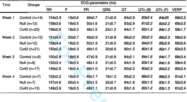
and later analyses. Standard criteria were used for interval measurements (RR, PR,
QRS and QT). For further comparison between groups, QT interval were corrected
using bot Fredericia and Bazett fomulas (QTc(F) = QT/ (RR/150)1/3and QTc(F) = QT/
3 4 5 6 7 8 9 10 11 12 13 14 15 16 17 18 19 20 21 22 23 24 25 26 27 28 29 30 31 32 33 34 35 36 37 38 39 40 41 42 43 44 45 46 47 48 49 50 51 52 53 54 55 56 57 58 59 60
For Peer Review
(RR/150)1/2 respectively; Table I). Standard clinical PES protocols were used,
including single, double and triple extrastimuli applied under spontaneous rhythm or
following a train of 9 stimuli at 100-ms drive cycle length. The coupling interval of the
last extrastimulus was decreased to the ventricular effective refractory period (VERP).
Protocols were interrupted if sustained ventricular tachycardia (VT) was induced.
Sustained VT was defined as fast ventricular rhythm of 15 or more beats, according to
the Lambeth Conventions[13].
Data Analyses
Data were expressed as mean±SEM and frequencies (expressed as percentages).
Statistical analyses were performed using MedCalc 9.1 software. Real time RT-PCR
data and cell count data were assessed using the Student t-test. Occurrences of
sustained VT were compared with Cox’s model and were analyzed as failure time data
(rats without event were considered as censored). The assumption of proportional
hazards between groups was confirmed, and the group was the unique covariate
selected in the Cox’s model. Overall mortality between groups was compared using
Fisher’s exact test. ECG parameters (P, RR, PR, QRS, QT and QTc and VERP values)
were assessed by a linear mixed model with random slope and intercept in the control,
Null, and Cx43 groups. The fixed effects were the group and the time. Interaction
between group and time was tested but not included in the model (not significant). The
power of the study was 0.40 for all statistical analysis. A p-value <0.05 was considered
significant. 3 4 5 6 7 8 9 10 11 12 13 14 15 16 17 18 19 20 21 22 23 24 25 26 27 28 29 30 31 32 33 34 35 36 37 38 39 40 41 42 43 44 45 46 47 48 49 50 51 52 53 54 55 56 57 58 59 60
For Peer Review
RESULTS
In vitro Cx43 overexpression
A lentivirus vector was used to overexpress Cx43 in rat myoblast primary cultures
ex vivo, prior to autologous intramyocardial injection (Figure 1a). As controls we used
lentivirus vectors containing an empty expression cassette (Null) or the Green
Fluorescent Protein (GFP) cDNA. Efficacy of lentivirus transduction was evaluated
in vitro using flow cytometry analyses for both GFP and desmin after GFP lentivirus
vector transduction. GFP was expressed by 50% of the desmin positive cells,
suggesting that 50% myoblasts expressed the transgene before transplantation (Figure
1b). The transduction rate of non-myoblast contaminating cells (i.e. GFP+ desmin –
cells) was 16.3±4.1% (Figure 1b). Seven days after Cx43 or Null lentivirus
transduction, cell counts and desmin expression levels were similar in both Null- and
Cx43-transduced myoblasts, whereas Cx43 expression level was 2.5 fold higher in
Cx43- than in Null-transduced myoblasts (p<0.05), showing that Cx43 overexpression
did not alter myoblast expansion (Figure 2a).
To evaluate exogenous Cx43 expression due to Cx43 lentivirus vector transduction, we
used gene expression quantification of the post-transcriptional regulatory element
Woodchuck hepatitis virus (Wpre), that is located within the expression cassette of the
lentivirus vector in 3’ of the Cx43 cDNA and proximal to the polyadenylation signal
(Figure 1a). Wpre gene expression was detected only in Cx43-transduced myoblasts
6 days after Cx43- and Null-transduction (Figure 2b). In Cx43-transduced myoblasts,
total Cx43 gene expression level correlated with Wpre expression level (R2=0.8710;
Figure 2b). Finally, in vitro time-course studies demonstrated that Cx43 expression
remained at least 2.5 fold higher in Cx43-transduced myoblasts than in
Null-transduced myoblasts (p<0.05, Figure 2c) for at least 35 days after Cx43 transduction.
3 4 5 6 7 8 9 10 11 12 13 14 15 16 17 18 19 20 21 22 23 24 25 26 27 28 29 30 31 32 33 34 35 36 37 38 39 40 41 42 43 44 45 46 47 48 49 50 51 52 53 54 55 56 57 58 59 60
For Peer Review
Moreover, in Cx43-transduced myoblasts, Wpre gene expression remained stable. In
view of these results, Wpre was used as a marker to detect exogenous Cx43 expression
in vivo after intramyocardial myoblast transplantation.
In vivo Cx43 overexpression
Seven days after coronary ligation, rats were randomized into 3 groups: a Control
group injected with autologous myoblasts, a Null group injected with autologous
myoblasts transduced with the Null lentivirus vector, and a Cx43 group injected with
autologous myoblasts transduced with the lentivirus vector encoding Cx43.
Using real time RT-PCR, Wpre gene expression was detected in 9/9 hearts injected
with Cx43-tranduced myoblasts up to 35 days after their in vivo injection (Figure 3a),
suggesting that Cx43-transduced muscle cells overexpressed Cx43 in vivo. Two weeks
after Cx43-transduced myoblast transplantation, Cx43 protein was detected in
cryosections of infarcted myocardium in cells expressing fast skeletal myosin heavy
chain (Figures 3b-h), suggesting that ex vivo Cx43 lentivirus vector transduction lead
to in vivo Cx43 protein expression in differentiated myotubes.
Electrophysiological analyses
PES was performed at 1, 2, 3 and 4 weeks after intramyocardial myoblast
transplantation. No differences between groups were observed in ECG parameters
prior to the first PES procedure (week 1). Neither standard ECG measurements nor
VERP at 100-ms pacing cycle length were significantly altered by the repeated PES
procedures (Table I), suggesting that lentivirus vector transduction (in Null group) or
Cx43 overexpression (in Cx43 group) in transplanted myoblasts did not modify ECG
parameters. Overall the percentage of rats that underwent at least one arrhythmia event
3 4 5 6 7 8 9 10 11 12 13 14 15 16 17 18 19 20 21 22 23 24 25 26 27 28 29 30 31 32 33 34 35 36 37 38 39 40 41 42 43 44 45 46 47 48 49 50 51 52 53 54 55 56 57 58 59 60
For Peer Review
during one of the PES were similar between groups (58%, 64% and 48% of animals in
the Control (n=12), Null (n=14) or Cx43 (n=23) group, respectively, Cox’s model,
p=0.92, Figure 4a). Additionally, the percentage of newly inducible rats did not differ
among groups (Figure 4b). In each group, ECG parameters of rats that underwent
sustained VT during PES did not differ from those that did not show sustained VT (not
illustrated). Mortality was similar in Control, Null or Cx43 groups (42%, 50%, 43%,
respectively, p=0.87).
Ex vivo measurements of intramural monophasic action potentials (MAPs)
To evaluate electrical coupling between Cx43-overexpressing myoblasts and host
cardiomyocytes, ex vivo intramural monophasic action potential (MAP) recordings
were performed 14 days after myoblast injection in Langendorff-perfused hearts, using
a tungsten electrode that recorded both local MAP and remote electrical activity. For
each rat, MAPs were recorded during sinus rhythm at 7 different sites (1 located in the
right ventricle and 6 located in the left ventricle). Control included recordings from rat
tibialis muscle (Figure 5a) and from healthy non-infarcted myocardium (Figure 5b). A
total of 16 rats were evaluated, 6 in the Cx43 and in the Null groups, and 4 in the
Control group. Recordings in the infarct solely showed the remote signal of healthy
myocardium in both Null and Cx43 groups in 9/16 animals (Figures 5c,d). In 5/16 rats
monophasic action potentials (MAPs) were recorded with average duration of
52.9±4.6ms, while in 2/16 rats significantly shorter MAPs were recorded of 2.8±1.9ms
(p<0.001, Figures 5e,f, and Figures 5h,i (asterisks)). These short MAPs were similar to
those recorded in rat tibialis muscle (MAP duration 3.5ms, Figure 5a,g), while the
longer MAPs recorded in the infarct compared well to MAPs recorded in healthy
cardiac muscle (MAP duration 55.1±2.4ms, p=0.96, n=16). No such short MAPs were
3 4 5 6 7 8 9 10 11 12 13 14 15 16 17 18 19 20 21 22 23 24 25 26 27 28 29 30 31 32 33 34 35 36 37 38 39 40 41 42 43 44 45 46 47 48 49 50 51 52 53 54 55 56 57 58 59 60
For Peer Review
recorded in the non-transplanted area of the heart, strongly suggesting that the short
MAPs reflected electrical activity of transplanted skeletal muscle cells, while the
longer MAPs reflected electrical activity of host cardiomyocytes.
In myocardium transplanted with Null-transduced myoblasts, skeletal muscle cell
MAPs and myocardial electrograms were not synchronized (Figures 5e,h), suggesting
that skeletal muscle cells were not activated in synchrony with host cardiomyocytes. In
contrast, in left ventricle from the Cx43 group, skeletal muscle cells exhibited
electrical activity in synchrony with surrounding healthy myocardium (Figures 5f,i).
Multiple registrations of 2 seconds of myoblast spikes were performed from the same
location in these rats. In the Null group, the mean interval for myoblast spikes was 545
ms (range 541 – 549 ms) and for myocyte spikes was 224 ms (range 214 – 233). In the
Cx43 group, the mean interval for myoblast spikes was 225 ms. Myoblast spikes were
synchronous with myocyte activity. These results suggest that ex vivo Cx43 gene
transfer and expression in myoblasts enhanced electrical coupling and frequency
entrainment of skeletal muscle cells and host cardiomyocytes.
3 4 5 6 7 8 9 10 11 12 13 14 15 16 17 18 19 20 21 22 23 24 25 26 27 28 29 30 31 32 33 34 35 36 37 38 39 40 41 42 43 44 45 46 47 48 49 50 51 52 53 54 55 56 57 58 59 60
For Peer Review
DISCUSSION
In this study, using an in vivo model of cardiac cell and gene therapy with autologous
myoblasts (in which arrhythmic risk related to myoblast transplantation has been
previously evaluated[9]), ex vivo Cx43 gene transfer prior to intramyocardial
transplantation enhances Cx43 expression and in vivo electrical coupling between
transplanted myoblasts and host cardiomyocytes. However, in our model, improved
electrical coupling was not sufficient to significantly decrease arrhythmogenicity
related to myoblast transplantation.
Several in vitro studies showed that in myoblast/cardiomyocyte coculture models,
lentiviral-mediated overexpression of Cx43 in myoblasts was sufficient to induce gap
junction formation between both cell types[5,14-16]. Although we did not demonstrate
gap junction formation in the present study, the functionality of these gap junctions
was demonstrated by others, using western blot analyses and in vitro dye transfer
techniques[5,15,16]. In our study, gap junction formation and functionality were
suggested in vivo by electrical coupling between skeletal muscle cells and
cardiomyocytes that occurred only in myocardium injected with Cx43-transduced
myoblasts. Cx43 overexpression in myoblasts and gap junction formation have also
been successfully obtained using retrovirus[15] or adenovirus[17] vectors. In contrast
with our present study, increased cell death was observed in vivo after Cx43
adenovirus vector transduction and cell transplantation[17]. This was clearly linked to
very high vector transduction rate, as usually obtained with adenovirus vectors[18] in
contrast to retrovirus or lentivirus vectors. In our study, 50% myoblasts were
transduced by the lentivirus vectors, and Cx43 expression increased only 2.5-fold as
compared to baseline levels, a level compatible with studies using similar gene transfer
conditions[15]. In one study, no Cx43 overexpression was observed after cell
3 4 5 6 7 8 9 10 11 12 13 14 15 16 17 18 19 20 21 22 23 24 25 26 27 28 29 30 31 32 33 34 35 36 37 38 39 40 41 42 43 44 45 46 47 48 49 50 51 52 53 54 55 56 57 58 59 60
For Peer Review
transplantation of Cx43 retrovirus vector-transduced myoblasts, an observation that
was linked to promoter silencing[15]. In our study, this was clearly not the case since
Wpre, a regulatory element within the expression cassette, was expressed in vivo more
than 8 weeks after cell transplantation. In summary, although each vector type lead
in vitro to significant Cx43 overexpression and gap junction formation, lentivirus
vector transduction offered long in vivo expression without deleterious effects.
An in vitro study showed an increase in electrical coupling between cardiomyocytes
and myoblasts transduced with a lentivirus vector encoding Cx43 associated to a
reduction (but not elimination) of myoblast arrhythmogenicity[5], an hypothesis that
needed to be evaluated in vivo. Both Roell’s study and ours provided evidence of
in vivo host cardiomyocyte and transplanted myoblast electrical coupling when
myoblasts overexpressed Cx43. In this regard, our results confirm the feasibility of
ex vivo gene transfer to modify in vivo electrophysiological properties of injected
cells[19]. The rat number in which myoblast MAPs were observed in the heart was
very low, most probably because MAP recordings were performed each time using one
single electrode and that this electrode recorded only local electrical activity.
Therefore the chance that the electrode was placed in close proximity with myoblasts
was low, as myoblast number was also probably low. Furthermore, myoblast spikes
might have been hidden in the upstroke of myocyte MAPs. Some myocyte MAPs in the
Cx43 group showed fractionated upstrokes (not shown), suggesting that myoblast
spikes caused fractionation of the upstroke of these myocyte MAPs. Nevertheless,
fractionated upstrokes were not counted, since there was no final proof that these
deflections were indeed myoblast spikes. MAP recordings were performed at a pacing
cycle length of 250 ms. This cycle length was chosen because this is the rat normal
spontaneous sinus cycle length. Evaluating the level of electrical coupling between
3 4 5 6 7 8 9 10 11 12 13 14 15 16 17 18 19 20 21 22 23 24 25 26 27 28 29 30 31 32 33 34 35 36 37 38 39 40 41 42 43 44 45 46 47 48 49 50 51 52 53 54 55 56 57 58 59 60
For Peer Review
transduced myoblasts and cardiomyocytes in vivo was not possible by changing pacing
rate. Conduction is determined by 3 parameters (excitability, cell-to-cell coupling and
myocardial architecture), and it is not possible to determine the contribution of a
single parameter by changing stimulation frequency. In addition, in remodeled
infarcted myocardium, all three parameters are changed. If conduction delay increased
between myoblasts and cardiomyocytes at a higher stimulation frequency it is unclear
whether this would be due to an inadequate coupling between host and donor cells, a
reduced coupling between cardiomyocytes, impaired sodium current of the
cardiomyocytes or the changed myocardial architecture.
In contrast to Roell’s study, electrical coupling was not sufficient to significantly
decrease arrhythmogenicity related to myoblast transplantation in a clinically relevant
model combining gene and autologous cell therapy. This result highlights the
differences between the in vivo study by Roell et al and ours, including the animal
model and the level of Cx43 overexpression in transplanted cells. Although the type of
injury may have an impact on arrhythmia triggering, arrhythmias associated to cell
therapy were evaluated in different models of myocardial injury (cryolesion[8],
ischemia/reperfusion[20] or even pharmacological models[21]. This suggests that myoblast-induced arrhythmias are not dependent from the myocardial infarction
model. As in Roell’s study, we did not evaluate spontaneous ventricular tachyarrhythmias, because their frequency is low in rodents[9]. Although in Roell’s
study VT frequency was 100% in the myocardial infarction model, VT frequency was
60% in ours (a result similar to that of our previous study, confirming the
reproducibility of our model), probably because we counted only VT>15 beats.
Although we did not measure the extent of myocardial infarction in the present study,
histological analysis of the same model in our previous study showed a reproducible
3 4 5 6 7 8 9 10 11 12 13 14 15 16 17 18 19 20 21 22 23 24 25 26 27 28 29 30 31 32 33 34 35 36 37 38 39 40 41 42 43 44 45 46 47 48 49 50 51 52 53 54 55 56 57 58 59 60
For Peer Review
scar of 25+3% of the left ventricle 7 weeks after coronary ligation[9], an infarct size
comparable to that in other studies using the same animal model[22]. Finally, as
mentioned above in our study only 50% transduced myoblasts expressed Cx43,
inducing an increase of the overall Cx43 expression only 2.5 fold as compared to
baseline levels. Because Roell et al. used myoblasts from a genetically modified
mouse overexpressing Cx43, both percentage of Cx43-overexpressing myoblasts and
Cx43 expression level may have been significantly higher, favoring extensive
electrical coupling between myoblasts and cardiomyocytes. Importantly, only
frequency but not waveform entrainment (typical for low coupling between cells with
different intrinsic action potential waveforms) was observed in our study, suggesting
that electrical coupling was too low to significantly affect the electrical stability of the
heart. It has also been suggested that the occurrence of arrhythmias depends on cell
distribution within the infarcted area, an hypothesis that was not confirmed in a recent
study on rabbits[23]. Notably, in the transgenic mouse study, electrical stability
occurred even in animals whose stem cell grafts were physically isolated from the
native myocardium. Finally, because myoblasts do not transdifferentiate into
cardiomyocytes and because their action potential duration remains significantly
shorter than host cardiomyocytes[24], an electrical coupling between both cell types
might induce locally heterogeneous distribution of action potential duration, another
risk factor for arrhythmia[1]. Although this potential adverse effect has not been
detected in vitro or in rodent models, because early preclinical studies did not reveal
the tendency of myoblasts to induce life-threatening arrhythmias such hypothesis
needs to be evaluated in larger animal models.
Our study has some limitations. First, we did not correlate the injected-cell number
with arrhythmia inducibility, and Cx43 overexpression might have increased in vivo
3 4 5 6 7 8 9 10 11 12 13 14 15 16 17 18 19 20 21 22 23 24 25 26 27 28 29 30 31 32 33 34 35 36 37 38 39 40 41 42 43 44 45 46 47 48 49 50 51 52 53 54 55 56 57 58 59 60
For Peer Review
cell engraftment and/or proliferation as compared to engraftment of Null-transduced
cells. Although a dose-dependence between cell engraftment rate and arrhythmia might
be expected, results of a recent randomized double-blind clinical trial did not support a
dose-response increase in arrhythmic episodes[25]. In our study, we did not evaluate
the level of myoblast contamination with smooth muscle cells nor their level of viral
infection with Cx43 transgene. In humans and in rats, myoblast primary culture from
muscle samples does not lead to pure myoblast preparations[10, 3, 26], the culture
being contaminated mostly with fibroblasts. In Roell’s study, myofibroblasts induced
arrhythmias, suggesting that if myofibroblasts contaminated myoblast culture, they
may induce arrhythmia when injected in vivo. Therefore we cannot rule out the
contribution to arrhythmia triggering of transduced or untransduced contaminating
cells with Cx43 lentivirus vector. In Roell’s study, Cx43 was expressed under the
control of a promoter specific for myotubes. Therefore contaminating smooth muscle
cells did not overexpress Cx43 in Roell’s study. Nevertheless, there was a significant
decrease in arrhythmias following injection of Cx43-overexpressing myoblasts,
suggesting that contaminating cells within myoblast preparations did not play a major
role in arrhythmias.
Finally, improvement of the heart function after myoblast transplantation was not
studied. Myoblast transplantation in the failing heart has been initially motivated by
the hope that transplanted cells would actively improve systolic contraction. It has
been shown that myoblasts could organize in fibers with contractile capacity, with the
right orientation such that their synchronous contraction would increase the heart
contraction strength. Without electrical coupling, skeletal muscle cells are incapable of
contributing to contraction. Myoblasts have been shown to improve heart function
(possibly by paracrine effect), and one important question would be to evaluate if an
3 4 5 6 7 8 9 10 11 12 13 14 15 16 17 18 19 20 21 22 23 24 25 26 27 28 29 30 31 32 33 34 35 36 37 38 39 40 41 42 43 44 45 46 47 48 49 50 51 52 53 54 55 56 57 58 59 60
For Peer Review
enhanced electrical coupling (leading to synchronous contraction) improves efficacy of
myoblast therapy[24].
Although Cx43 expression level in Cx43 lentivirus vector-transduced myoblasts was
compatible with in vitro studies using similar gene transfer conditions[5], it may be
ineffective to restore full electromechanical coupling in an injured heart, as expression
of other junction proteins such as N-cadherin may also be necessary[27]. Therefore,
based on our present electrophysiological study we would suggest a note of caution on
the use of combined gene and cell therapy to prevent post-infarct arrhythmias. Current
technologic limitations to gene therapy, including low gene transduction and low
foreign gene expression may explain these results. In this regard, further studies to
improve gene therapy vectors will be rewarding.
3 4 5 6 7 8 9 10 11 12 13 14 15 16 17 18 19 20 21 22 23 24 25 26 27 28 29 30 31 32 33 34 35 36 37 38 39 40 41 42 43 44 45 46 47 48 49 50 51 52 53 54 55 56 57 58 59 60
For Peer Review
Acknowledgements
Lentivirus vector production was performed by the Lentivirus vector Production Unit.
This work was supported in part by INSERM avenir grant, by the Association
Française contre les Myopathies and by the GIS-maladies rares. Sarah Fernandes was,
in part, supported by the Association Française contre les Myopathies. The authors
declare no conflict of interest.
3 4 5 6 7 8 9 10 11 12 13 14 15 16 17 18 19 20 21 22 23 24 25 26 27 28 29 30 31 32 33 34 35 36 37 38 39 40 41 42 43 44 45 46 47 48 49 50 51 52 53 54 55 56 57 58 59 60
For Peer Review
Tables
Table I: ECG parameters and VERP values
ECG measurements were performed under sinus rhythm. Ventricular Effective Refractory Period was measured at a basic pacing cycle length (BCL) of 100ms at week 1, 2, 3 and 4 after myoblast transplantation. Abbreviations: P, P wave duration; RR, PR, QRS, QT, QTc(B) and QTc(F): RR, PR, QRS, QT intervals, QT interval corrected with Bazett formula (B) or Fredericia formula (F) respectively; VERP, Ventricular effective refractory period. All measurements were performed on lead I under general anesthesia. Results are expressed as mean±SEM.
Groups ECG parameters (ms)
RR P PR QRS QT Week 1 154±3.9 19±0.6 48±0.7 21±0.9 84±3.9 158±3.6 19±0.5 50±1.6 21±0.7 83±2.8 156±2.9 18±0.3 48±1.5 22±1.2 84±1.7 Week 2 151±4.0 20±0.7 49±0.9 21±0.8 86±3.0 158±4.4 19±0.5 50±1.9 21±0.5 86±2.9 150±2.3 18±0.3 48±1.0 20±0.6 80±1.5 150±2.6 18±0.6 47±0.9 21±0.8 84±2.1 152±3.4 18±0.5 48±1.3 21±0.5 84±1.9 149±2.8 18±0.4 48±1.0 21±0.7 80±2.2 152±2.2 19±0.5 49±1.7 19±1.3 85±2.3 157±4.6 20±0.4 50±1.5 22±0.7 84±1.8 149±3.9 18±0.5 48±1.1 21±0.8 83±1.6 VERP 69±3.2 65±3.5 59±1.7 59±2.9 65±5.6 62±3.5 68±3.4 59±4.6 60±2.4 61±2.1 52±3.6 61±2.4 83±2.4 81±2.2 83±1.2 85±2.3 84±2.5 80±1.8 84±1.6 83±1.6 80±2.3 86±2.2 83±1.5 83±1.2 84±25 82±2.2 84±1.3 86±2.3 85±2.6 80±1.7 84±1.7 84±1.6 80±2.2 86±2.3 83±1.5 83±1.3 QTc (B) QTc (F)
Time Groups ECG parameters (ms)
RR P PR QRS QT 154±3.9 19±0.6 48±0.7 21±0.9 84±3.9 158±3.6 19±0.5 50±1.6 21±0.7 83±2.8 156±2.9 18±0.3 48±1.5 22±1.2 84±1.7 151±4.0 20±0.7 49±0.9 21±0.8 86±3.0 158±4.4 19±0.5 50±1.9 21±0.5 86±2.9 150±2.3 18±0.3 48±1.0 20±0.6 80±1.5 150±2.6 18±0.6 47±0.9 21±0.8 84±2.1 152±3.4 18±0.5 48±1.3 21±0.5 84±1.9 149±2.8 18±0.4 48±1.0 21±0.7 80±2.2 Week 3 Week 4 152±2.2 19±0.5 49±1.7 19±1.3 85±2.3 157±4.6 20±0.4 50±1.5 22±0.7 84±1.8 Control (n=14) Null (n=12) Cx43 (n=23) Control (n=12) Null (n=12) Cx43 (n=21) Control (n=9) Null (n=9) Cx43 (n=17) Control (n=7) Null (n=7) Cx43 (n=13) 149±3.9 18±0.5 48±1.1 21±0.8 83±1.6 VERP 69±3.2 65±3.5 59±1.7 59±2.9 65±5.6 62±3.5 68±3.4 59±4.6 60±2.4 61±2.1 52±3.6 61±2.4 69±3.2 65±3.5 59±1.7 59±2.9 65±5.6 62±3.5 68±3.4 59±4.6 60±2.4 61±2.1 52±3.6 61±2.4 83±2.4 81±2.2 83±1.2 85±2.3 84±2.5 80±1.8 84±1.6 83±1.6 80±2.3 86±2.2 83±1.5 83±1.2 83±2.4 81±2.2 83±1.2 85±2.3 84±2.5 80±1.8 84±1.6 83±1.6 80±2.3 86±2.2 83±1.5 83±1.2 84±25 82±2.2 84±1.3 86±2.3 85±2.6 80±1.7 84±1.7 84±1.6 80±2.2 86±2.3 83±1.5 83±1.3 84±25 82±2.2 84±1.3 86±2.3 85±2.6 80±1.7 84±1.7 84±1.6 80±2.2 86±2.3 83±1.5 83±1.3 QTc (B) QTc (F)
Groups ECG parameters (ms)
RR P PR QRS QT Week 1 Week 1 154±3.9 19±0.6 48±0.7 21±0.9 84±3.9 158±3.6 19±0.5 50±1.6 21±0.7 83±2.8 156±2.9 18±0.3 48±1.5 22±1.2 84±1.7 Week 2 Week 2 151±4.0 20±0.7 49±0.9 21±0.8 86±3.0 158±4.4 19±0.5 50±1.9 21±0.5 86±2.9 150±2.3 18±0.3 48±1.0 20±0.6 80±1.5 150±2.6 18±0.6 47±0.9 21±0.8 84±2.1 152±3.4 18±0.5 48±1.3 21±0.5 84±1.9 149±2.8 18±0.4 48±1.0 21±0.7 80±2.2 152±2.2 19±0.5 49±1.7 19±1.3 85±2.3 157±4.6 20±0.4 50±1.5 22±0.7 84±1.8 149±3.9 18±0.5 48±1.1 21±0.8 83±1.6 VERP 69±3.2 65±3.5 59±1.7 59±2.9 65±5.6 62±3.5 68±3.4 59±4.6 60±2.4 61±2.1 52±3.6 61±2.4 83±2.4 81±2.2 83±1.2 85±2.3 84±2.5 80±1.8 84±1.6 83±1.6 80±2.3 86±2.2 83±1.5 83±1.2 84±25 82±2.2 84±1.3 86±2.3 85±2.6 80±1.7 84±1.7 84±1.6 80±2.2 86±2.3 83±1.5 83±1.3 QTc (B) QTc (F)
Time Groups ECG parameters (ms)
RR P PR QRS QT 154±3.9 19±0.6 48±0.7 21±0.9 84±3.9 158±3.6 19±0.5 50±1.6 21±0.7 83±2.8 156±2.9 18±0.3 48±1.5 22±1.2 84±1.7 151±4.0 20±0.7 49±0.9 21±0.8 86±3.0 158±4.4 19±0.5 50±1.9 21±0.5 86±2.9 150±2.3 18±0.3 48±1.0 20±0.6 80±1.5 150±2.6 18±0.6 47±0.9 21±0.8 84±2.1 152±3.4 18±0.5 48±1.3 21±0.5 84±1.9 149±2.8 18±0.4 48±1.0 21±0.7 80±2.2 Week 3 Week 3 Week 4 152±2.2 19±0.5 49±1.7 19±1.3 85±2.3 157±4.6 20±0.4 50±1.5 22±0.7 84±1.8 Control (n=14) Null (n=12) Cx43 (n=23) Control (n=12) Null (n=12) Cx43 (n=21) Control (n=9) Null (n=9) Cx43 (n=17) Control (n=7) Null (n=7) Cx43 (n=13) Control (n=14) Null (n=12) Cx43 (n=23) Control (n=12) Null (n=12) Cx43 (n=21) Control (n=9) Null (n=9) Cx43 (n=17) Control (n=7) Null (n=7) Cx43 (n=13) 149±3.9 18±0.5 48±1.1 21±0.8 83±1.6 VERP 69±3.2 65±3.5 59±1.7 59±2.9 65±5.6 62±3.5 68±3.4 59±4.6 60±2.4 61±2.1 52±3.6 61±2.4 69±3.2 65±3.5 59±1.7 59±2.9 65±5.6 62±3.5 68±3.4 59±4.6 60±2.4 61±2.1 52±3.6 61±2.4 83±2.4 81±2.2 83±1.2 85±2.3 84±2.5 80±1.8 84±1.6 83±1.6 80±2.3 86±2.2 83±1.5 83±1.2 83±2.4 81±2.2 83±1.2 85±2.3 84±2.5 80±1.8 84±1.6 83±1.6 80±2.3 86±2.2 83±1.5 83±1.2 84±25 82±2.2 84±1.3 86±2.3 85±2.6 80±1.7 84±1.7 84±1.6 80±2.2 86±2.3 83±1.5 83±1.3 84±25 82±2.2 84±1.3 86±2.3 85±2.6 80±1.7 84±1.7 84±1.6 80±2.2 86±2.3 83±1.5 83±1.3 QTc (B) QTc (F) 3 4 5 6 7 8 9 10 11 12 13 14 15 16 17 18 19 20 21 22 23 24 25 26 27 28 29 30 31 32 33 34 35 36 37 38 39 40 41 42 43 44 45 46 47 48 49 50 51 52 53 54 55 56 57 58 59 60
For Peer Review
References
1. Smith RR, Barile L, Messina E, Marban E. Stem cells in the heart: what's the
buzz all about? Part 2: Arrhythmic risks and clinical studies. Heart Rhythm. 2008; 5: 880-7.
2. Leobon B, Garcin I, Menasche P, Vilquin JT, Audinat E, Charpak S.
Myoblasts transplanted into rat infarcted myocardium are functionally isolated from their host. Proc Natl Acad Sci U S A. 2003; 100: 7808-11.
3. Hagege AA, Carrion C, Menasche P, Vilquin JT, Duboc D, Marolleau JP, Desnos M, Bruneval P. Viability and differentiation of autologous skeletal
myoblast grafts in ischaemic cardiomyopathy. Lancet. 2003; 361: 491-2.
4. Rubart M, Soonpaa MH, Nakajima H, Field LJ. Spontaneous and evoked
intracellular calcium transients in donor-derived myocytes following intracardiac myoblast transplantation. J Clin Invest. 2004; 114: 775-83.
5. Abraham MR, Henrikson CA, Tung L, Chang MG, Aon M, Xue T, Li RA, B OR, Marban E. Antiarrhythmic engineering of skeletal myoblasts for cardiac
transplantation. Circ Res. 2005; 97: 159-67.
6. Mills WR, Mal N, Kiedrowski MJ, Unger R, Forudi F, Popovic ZB, Penn MS, Laurita KR. Stem cell therapy enhances electrical viability in myocardial
infarction. J Mol Cell Cardiol. 2007; 42: 304-14.
7. Antzelevitch C. Basic mechanisms of reentrant arrhythmias. Curr Opin
Cardiol. 2001; 16: 1-7.
8. Roell W, Lewalter T, Sasse P, Tallini YN, Choi BR, Breitbach M, Doran R, Becher UM, Hwang SM, Bostani T, von Maltzahn J, Hofmann A, Reining S, Eiberger B, Gabris B, Pfeifer A, Welz A, Willecke K, Salama G, Schrickel JW, Kotlikoff MI, Fleischmann BK. Engraftment of connexin 43-expressing
cells prevents post-infarct arrhythmia. Nature. 2007; 450: 819-24.
9. Fernandes S, Amirault JC, Lande G, Nguyen JM, Forest V, Bignolais O, Lamirault G, Heudes D, Orsonneau JL, Heymann MF, Charpentier F, Lemarchand P. Autologous myoblast transplantation after myocardial
infarction increases the inducibility of ventricular arrhythmias. Cardiovasc Res. 2006; 69: 348-58.
10. Pouzet B, Vilquin JT, Hagege AA, Scorsin M, Messas E, Fiszman M, Schwartz K, Menasche P. Intramyocardial transplantation of autologous
myoblasts: can tissue processing be optimized? Circulation. 2000; 102: III210-5.
11. Coronel R, de Bakker JM, Wilms-Schopman FJ, Opthof T, Linnenbank AC, Belterman CN, Janse MJ. Monophasic action potentials and activation
recovery intervals as measures of ventricular action potential duration: experimental evidence to resolve some controversies. Heart Rhythm. 2006; 3: 1043-50.
12. Takei M, Sasaki Y, Yonezawa T, Lakhe M, Aruga M, Kiyosawa K. The
autonomic control of the transmural dispersion of ventricular repolarization in anesthetized dogs. J Cardiovasc Electrophysiol. 1999; 10: 981-9.
3 4 5 6 7 8 9 10 11 12 13 14 15 16 17 18 19 20 21 22 23 24 25 26 27 28 29 30 31 32 33 34 35 36 37 38 39 40 41 42 43 44 45 46 47 48 49 50 51 52 53 54 55 56 57 58 59 60
For Peer Review
13. Walker MJ, Curtis MJ, Hearse DJ, Campbell RW, Janse MJ, Yellon DM, Cobbe SM, Coker SJ, Harness JB, Harron DW, Higgins AJ, Julian DG, Lab MJ, Manning AS, Northover BJ, Parrat JR, Riemersma RA, Riva E, Russell DC. The Lambeth Conventions: guidelines for the study of arrhythmias
in ischaemia infarction, and reperfusion. Cardiovasc Res. 1988; 22: 447-55. 14. Suzuki K, Brand NJ, Allen S, Khan MA, Farrell AO, Murtuza B, Oakley
RE, Yacoub MH. Overexpression of connexin 43 in skeletal myoblasts:
Relevance to cell transplantation to the heart. J Thorac Cardiovasc Surg. 2001; 122: 759-66.
15. Tolmachov O, Ma YL, Themis M, Patel P, Spohr H, Macleod KT, Ullrich ND, Kienast Y, Coutelle C, Peters NS. Overexpression of connexin 43 using a
retroviral vector improves electrical coupling of skeletal myoblasts with cardiac myocytes in vitro. BMC Cardiovasc Disord. 2006; 6: 25.
16. Stagg MA, Coppen SR, Suzuki K, Varela-Carver A, Lee J, Brand NJ, Fukushima S, Yacoub MH, Terracciano CM. Evaluation of frequency, type,
and function of gap junctions between skeletal myoblasts overexpressing connexin43 and cardiomyocytes: relevance to cell transplantation. FASEB J.
2006; 20: 744-6.
17. Reinecke H, Minami E, Virag JI, Murry CE. Gene transfer of connexin43
into skeletal muscle. Hum Gene Ther. 2004; 15: 627-36.
18. Lemarchand P, Jaffe HA, Danel C, Cid MC, Kleinman HK, Stratford-Perricaudet LD, Stratford-Perricaudet M, Pavirani A, Lecocq JP, Crystal RG.
Adenovirus-mediated transfer of a recombinant human alpha 1-antitrypsin cDNA to human endothelial cells. Proc Natl Acad Sci U S A. 1992; 89: 6482-6. 19. Rissanen TT, Yla-Herttuala S. Current status of cardiovascular gene therapy.
Mol Ther. 2007; 15: 1233-47.
20. Fotuhi P, Song YH, Alt E. Electrophysiological consequence of
adipose-derived stem cell transplantation in infarcted porcine myocardium. Europace. 2007; 9: 1218-21.
21. Chen M, Fan ZC, Liu XJ, Deng JL, Zhang L, Rao L, Yang Q, Huang DJ.
Effects of autologous stem cell transplantation on ventricular electrophysiology in doxorubicin-induced heart failure. Cell Biol Int. 2006; 30: 576-82.
22. Fletcher PJ, Pfeffer JM, Pfeffer MA, Braunwald E. Left ventricular diastolic
pressure-volume relations in rats with healed myocardial infarction. Effects on systolic function. Circ Res. 1981; 49: 618-26.
23. McCue JD, Swingen C, Feldberg T, Caron G, Kolb A, Denucci C, Prabhu S, Motilall R, Breviu B, Taylor DA. The real estate of myoblast cardiac
transplantation: negative remodeling is associated with location. J Heart Lung
Transplant. 2008; 27: 116-23.
24. Cohen IS, Rosen AB, Gaudette GR. A Caveat Emptor for myocardial
regeneration: mechanical without electrical recovery will not suffice. J Mol Cell
Cardiol. 2007; 42: 285-8.
25. Menasche P, Alfieri O, Janssens S, McKenna W, Reichenspurner H, Trinquart L, Vilquin JT, Marolleau JP, Seymour B, Larghero J, Lake S, Chatellier G, Solomon S, Desnos M, Hagege AA. The Myoblast Autologous
3 4 5 6 7 8 9 10 11 12 13 14 15 16 17 18 19 20 21 22 23 24 25 26 27 28 29 30 31 32 33 34 35 36 37 38 39 40 41 42 43 44 45 46 47 48 49 50 51 52 53 54 55 56 57 58 59 60
For Peer Review
Grafting in Ischemic Cardiomyopathy (MAGIC) trial: first randomized placebo-controlled study of myoblast transplantation. Circulation. 2008; 117: 1189-200. 26. Al Attar N, Carrion C, Ghostine S, Garcin I, Vilquin JT, Hagege AA,
Menasche P. Long-term (1 year) functional and histological results of
autologous skeletal muscle cells transplantation in rat. Cardiovasc Res. 2003; 58: 142-8.
27. Pedrotty DM, Klinger RY, Badie N, Hinds S, Kardashian A, Bursac N.
Structural coupling of cardiomyocytes and noncardiomyocytes: quantitative comparisons using a novel micropatterned cell pair assay. Am J Physiol Heart
Circ Physiol. 2008; 295: H390-400. 3 4 5 6 7 8 9 10 11 12 13 14 15 16 17 18 19 20 21 22 23 24 25 26 27 28 29 30 31 32 33 34 35 36 37 38 39 40 41 42 43 44 45 46 47 48 49 50 51 52 53 54 55 56 57 58 59 60
For Peer Review
Figure legends
Figure 1: Cx43 transgene expression in myoblasts.
(a) The Cx43 lentivirus vector contained an expression cassette including the rat Cx43
cDNA under the control of the cytomegalovirus (CMV) promoter, followed by the
post-transcriptional regulatory element of the woodchuck hepatitis virus (Wpre). (b)
Evaluation of lentivirus vector transduction in myoblast primary culture 6 days after
GFP-lentivirus vector transduction, by flow cytometry analyses: cells from primary
culture that have not been transduced with GFP lentivirus and that have not been
labeled for desmin were used as controls to design thresholds (non-transduced
cells/Des-; top left panel). The same thresholds were further used for FACS analyses
of non-transduced cells/Des+ cells (non-transduced cells with desmin immunostaining;
top right panel), of GFP-transduced/Des- cells (GFP-transduced cells with no
immunostaining; bottom left panel) and of GFP-transduced/Des+ cells
(GFP-transduced cells with desmin immunostaining; bottom right panel).
Figure 2: Characterization of myoblast primary cultures after lentivirus vector transduction.
(a) Cell numbers (left panel) and desmin or Cx43 mRNA levels (right panel) in Null-
and Cx43-transduced myoblasts (n=10 and n=9, respectively), 6 days after lentivirus
vector transduction. Gene expression levels were measured using TaqMan real time
RT-PCR. Desmin and Cx43 gene expression levels were corrected by HPRT gene
expression levels. (b) Correlation between Cx43 and Wpre mRNA expression levels in
Cx43 transduced myoblasts, 6 days after lentivirus vector transduction (n=9). (c) Cx43
3 4 5 6 7 8 9 10 11 12 13 14 15 16 17 18 19 20 21 22 23 24 25 26 27 28 29 30 31 32 33 34 35 36 37 38 39 40 41 42 43 44 45 46 47 48 49 50 51 52 53 54 55 56 57 58 59 60
For Peer Review
gene expression from day 14 to day 35 post-transduction in Null- (dotted line) and
Cx43- transduced myoblasts (solid line, n=3 for both groups). * indicate p<0.05.
Figure 3: In vivo Cx43 and Wpre gene expression, following ex vivo lentivirus vector transduction and intramyocardial transplantation.
(a) Wpre expression in myocardium transplanted with Cx43 transduced myoblasts.
(b-h) Section of left ventricle transplanted with Cx43-transduced myoblasts, in
fluorescent microscopy (b to e) and in confocal microscopy (f to h), using
immunolabeling against the fast skeletal myosin heavy chain (b and f) and Cx43 (c and
g). Cell nuclei were labeled with DAPI (d). (e) Superposition of the b-d panels. (h)
Superposition of the f and g panels. Cryosections were performed within the infarcted
area, 14 days after myoblast transplantation.
Figure 4: Ventricular hyperexcitability of the myocardium after myoblast transplantation. Rats with myocardial infarction underwent in vivo Programmed
Electrical Stimulation (PES) procedures at 1, 2, 3 and 4 weeks after myoblast
transplantation. (a) Percentage of Control, Null and Cx43 rats with at least one episode
of sustained ventricular tachycardia (VT) during one of the PES procedures (p=0.92,
Cox’s model). (b) Percentage of rats with first episode of sustained VT between week
1 and 4 after myoblast transplantation.
Figure 5: Ex vivo intramural electrophysiological recordings. Recordings were
performed using a sharp, tungsten needle electrode that recorded both local
Monophasic Action Potentials (MAPs) and remote electrograms. (a) MAPs from rat
tibialis anterior muscle (paced at 250-ms intervals); stars indicate fast spikes of 5 to
3 4 5 6 7 8 9 10 11 12 13 14 15 16 17 18 19 20 21 22 23 24 25 26 27 28 29 30 31 32 33 34 35 36 37 38 39 40 41 42 43 44 45 46 47 48 49 50 51 52 53 54 55 56 57 58 59 60
For Peer Review
10ms length, typical for skeletal muscle. (b) MAPs from healthy myocardium in the
left ventricular free wall. Diamonds indicate typical rat cardiac MAPs of 80ms
duration. (c,d) Electrograms within the infarcted myocardium area (triangles), 14 days
after intramyocardial transplantation of Null-transduced myoblasts (c) or of
Cx43-transduced myoblasts (d). The MAP-needle only recorded the electrograms of remote
ventricular activity, as indicated by the triangles. (e,f) MAPs and electrograms from
the same infarcted regions as in c and d, but in the transplanted area. Asterisks and
triangles indicate MAPs from skeletal muscle cells and electrograms from remote
non-infarcted myocardium, respectively. Note the synchrony between MAPs from skeletal
muscle cells (asterisks) and ventricular electrograms (triangles) in the Cx43 group.
The extracellular complex (triangle) in tracing f was remote. The small deflection prior
to the MAP signal (arrows) suggests that myocardial activation preceded myoblast
activation, which suggests, but does not prove, that myocytes drove the myoblasts. (g,
h, i) enlargement of the recordings a, e and f, respectively. (j) higher enlargement of
the recording f/i. 3 4 5 6 7 8 9 10 11 12 13 14 15 16 17 18 19 20 21 22 23 24 25 26 27 28 29 30 31 32 33 34 35 36 37 38 39 40 41 42 43 44 45 46 47 48 49 50 51 52 53 54 55 56 57 58 59 60
For Peer Review
Cx43 transgene expression in myoblasts. 190x254mm (300 x 300 DPI) 3 4 5 6 7 8 9 10 11 12 13 14 15 16 17 18 19 20 21 22 23 24 25 26 27 28 29 30 31 32 33 34 35 36 37 38 39 40 41 42 43 44 45 46 47 48 49 50 51 52 53 54 55 56 57
For Peer Review
Characterization of myoblast primary cultures after lentivirus vector transduction. 190x254mm (300 x 300 DPI) 3 4 5 6 7 8 9 10 11 12 13 14 15 16 17 18 19 20 21 22 23 24 25 26 27 28 29 30 31 32 33 34 35 36 37 38 39 40 41 42 43 44 45 46 47 48 49 50 51 52 53 54 55 56 57
For Peer Review
Figure 3: In vivo Cx43 and Wpre gene expression, following ex vivo lentivirus vector transduction and intramyocardial transplantation.
108x170mm (300 x 300 DPI) 3 4 5 6 7 8 9 10 11 12 13 14 15 16 17 18 19 20 21 22 23 24 25 26 27 28 29 30 31 32 33 34 35 36 37 38 39 40 41 42 43 44 45 46 47 48 49 50 51 52 53 54 55 56 57
For Peer Review
Ventricular hyperexcitability of the myocardium after myoblast transplantation. 190x254mm (300 x 300 DPI) 3 4 5 6 7 8 9 10 11 12 13 14 15 16 17 18 19 20 21 22 23 24 25 26 27 28 29 30 31 32 33 34 35 36 37 38 39 40 41 42 43 44 45 46 47 48 49 50 51 52 53 54 55 56 57