Catherine Duclos, BA
(Hons)
Marie Dumont, PhD
Caroline Arbour, PhD
Jean Paquet, PhD
Hélène Blais, BA
David K. Menon, MD,
PhD
Louis De Beaumont, PhD
Francis Bernard, MD
Nadia Gosselin, PhD
Correspondence to Dr. Gosselin: [email protected] Editorial, page 226Parallel recovery of consciousness and sleep
in acute traumatic brain injury
ABSTRACT
Objective:
To investigate whether the progressive recuperation of consciousness was associated
with the reconsolidation of sleep and wake states in hospitalized patients with acute traumatic
brain injury (TBI).
Methods:
This study comprised 30 hospitalized patients (age 29.1
6 13.5 years) in the acute
phase of moderate or severe TBI. Testing started 21.0
6 13.7 days postinjury.
Conscious-ness level and cognitive functioning were assessed daily with the Rancho Los Amigos scale of
cognitive functioning (RLA). Sleep and wake cycle characteristics were estimated with
con-tinuous wrist actigraphy. Mixed model analyses were performed on 233 days with the RLA
(fixed effect) and sleep-wake variables (random effects). Linear contrast analyses were
per-formed in order to verify if consolidation of the sleep and wake states improved linearly with
increasing RLA score.
Results:
Associations were found between scores on the consciousness/cognitive functioning
scale and measures of sleep-wake cycle consolidation (p , 0.001), nighttime sleep duration
(p 5 0.018), and nighttime fragmentation index (p , 0.001). These associations showed strong
linear relationships (p , 0.01 for all), revealing that consciousness and cognition improved in
parallel with sleep-wake quality. Consolidated 24-hour sleep-wake cycle occurred when patients
were able to give context-appropriate, goal-directed responses.
Conclusions:
Our results showed that when the brain has not sufficiently recovered a certain level
of consciousness, it is also unable to generate a 24-hour sleep-wake cycle and consolidated
nighttime sleep. This study contributes to elucidating the pathophysiology of severe
sleep-wake cycle alterations in the acute phase of moderate to severe TBI.
Neurology®2017;88:268–275GLOSSARY
AR15 autoregressive; CS 5 compound symmetry; DAR 5 daytime activity ratio; GCS 5 Glasgow Coma Scale; ICU 5 intensive care unit; MCS5 minimally conscious state; RLA 5 Rancho Los Amigos scale of cognitive functioning; TBI 5 traumatic brain injury.
Nonsedated patients in the acute stage of a moderate to severe traumatic brain injury (TBI) have
serious alterations of their sleep-wake cycle,
1,2characterized by short sleep and wake bouts,
a few minutes in length, dispersed over 24 hours.
1Pain, medication, and the hospital
environ-ment are possible causes of these sleep-wake disturbances.
3However, recent experimental
models of TBI have shown that the injured brain itself has a direct effect on the sleep-wake
cycle by increasing fragmentation of sleep and wake periods.
4–6In patients with acute TBI, the reconsolidation of the 24-hour sleep-wake cycle predicts
emergence from posttraumatic amnesia at hospital discharge
1as well as cognitive impairment
in rehabilitation settings.
7,8Studies on chronic disorders of consciousness also suggest that the
circadian variation of the sleep-wake cycle reemerges with improving consciousness.
9Overall,
these observations point to an intrinsic association between recovery of the sleep-wake cycle,
consciousness, and cognition following a brain injury. However, we have yet to characterize how
From the Center for Advanced Research in Sleep Medicine (C.D., M.D., C.A., J.P., H.B., L.D.B., N.G.) and the Traumatology Program (F.B.), Hôpital du Sacré-Coeur de Montréal; Departments of Psychiatry (C.D., M.D.), Psychology (C.A., N.G.), and Medicine (F.B.), Université de Montréal, Canada; Division of Anaesthesia (D.K.M.), University of Cambridge, UK; and Department of Psychology (L.D.B.), Université du Québec à Trois-Rivières, Canada.
the sleep-wake cycle recovers on a day-to-day
basis in relation to improving consciousness
and higher cognitive functions in acute TBI.
The objective of this study was to verify
whether an association exists between the
evo-lution of the sleep-wake cycle and the recovery
of consciousness and cognition in acute
mod-erate to severe TBI. A second objective was to
determine which improved first, or whether
they evolved synchronously. We predicted
that the consolidation of sleep-wake states
would increase synchronously with improving
consciousness and cognition, because they
depend on overall brain integrity.
METHODS Patients.We recruited patients from Hôpital du Sacré-Coeur de Montréal, a level 1 trauma center affiliated with the Université de Montréal, between January 2010 and May 2015. We defined TBI as an alteration in brain function or other evidence of brain pathology caused by an external force,10and assessed TBI severity
upon emergency room admission, prior to intubation, using the Glas-gow Coma Scale (GCS).11We included patients if they were
hospi-talized in the intensive care unit (ICU) for their TBI. In order to characterize our study sample, we documented the following for all patients: mechanism of injury, GCS score at emergency room admis-sion, ICU and hospital lengths of stay, number of days with elevated intracranial pressure (.20 mm Hg), Marshall and Rotterdam scores,12,13which are qualitative CT classification systems, Disability
Rating Scale score within 72 hours of hospital discharge,14and patient
orientation at hospital discharge. We obtained written informed con-sent for study participation from patients’ families and the hospital ethical standards committee on human experimentation approved the study. We excluded patients if they were younger than 16 or older than 65 years; were quadriplegic; had a history of substance abuse,
psychiatric, or neurologic disorders; had a diagnosed sleep disorder prior to injury; had any damage to the eyes or the optic nerve (mod-ifying light perception); or had a history of prior TBI or concussion.
Experimental design.During the ICU and post-ICU hospital stay, patients wore an activity monitor to assess their sleep-wake patterns continuously for several days, during which a daily assessment of consciousness and cognition was also carried out.
Assessment of consciousness and cognitive level. We used the Rancho Los Amigos scale of cognitive functioning (RLA),15
a comprehensive behavioral rating scale developed specifically to monitor the stages of recovery in the adult TBI population,16
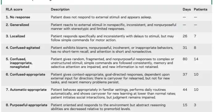
which can be easily administered at bedside. The RLA evaluates key features of consciousness and cognitive functioning, such as level of awareness of the environment, response to stimuli, ability to follow command, confusion, attention, and the appropriate-ness of verbalization and motor actions. The RLA scale consists of 8 hierarchical levels, with level 1 representing no response and level 8 representing purposeful and appropriate cognitive func-tion (table 1). Durafunc-tion of RLA assessment ranges from 5 to 40 minutes and is carried out when patients are fully awake and all aspects of the scale are assessable. Trained occupational therapists with experience with the acute TBI population assessed the RLA scale daily on weekdays.
Sleep-wake assessments. Patients wore a wrist actigraph (Actiwatch-L or Actiwatch-Spectrum; Philips Healthcare, Andover, MA) on a nonparalyzed arm starting in the ICU and continuing throughout hospitalization in regular wards. As described in a previous study,1actigraphy recording began when continuous sedation and
analgesia had ceased for at least 24 hours, and once patients reached a RLA score$3, indicative of a more apparent physical reactivity to internal and external stimuli. With its low invasiveness, actigraphy enables the long-term measurement of the rest-activity cycle, and is recognized as a proxy measure of the sleep-wake cycle.17
We measured activity counts per 1-minute epoch and derived 3 variables from actigraphic recordings to estimate sleep-wake quality: Daytime activity ratio (DAR). We estimated consolidation of the 24-hour sleep-wake cycle with the DAR.1The DAR
Table 1 Rancho Los Amigos scale of cognitive functioning (RLA), including the number of days and patients representing each RLA score
RLA score Description Days Patients
1. No response Patient does not respond to external stimuli and appears asleep. — — 2. Generalized Patient reacts to external stimuli in nonspecific, inconsistent, and nonpurposeful
manner with stereotypic and limited responses.
— —
3. Localized Patient responds specifically and inconsistently with delays to stimuli, but may follow simple commands for motor action.
26 7
4. Confused-agitated Patient exhibits bizarre, nonpurposeful, incoherent, or inappropriate behaviors, has no short-term recall, and attention is short and nonselective.
31 8
5. Confused, inappropriate, nonagitated
Patient gives random, fragmented, and nonpurposeful responses to complex or unstructured stimuli; simple commands are followed consistently, memory and selective attention are impaired, and new information is not retained.
80 14
6. Confused-appropriate Patient gives context-appropriate, goal-directed responses, dependent upon external input for direction; there is carryover for relearned, but not for new tasks, and recent memory problems persist.
37 10
7. Automatic-appropriate Patient behaves appropriately in familiar settings, performs daily routines automatically, and shows carryover for new learning at lower than normal rates; patient initiates social interactions, but judgment remains impaired.
44 10
8. Purposeful-appropriate Patient oriented and responds to the environment but abstract reasoning abilities are decreased relative to premorbid levels.
15 3
Adapted from Bagnato S, Boccagni C, Prestandrea C, Sant’Angelo A, Castiglione A, Galardi G. Prognostic value of standard EEG in traumatic and non-traumatic disorders of consciousness following coma. Clin Neurophysiol 2010;121:274–280,36
represents the percentage of total 24-hour activity occurring in the daytime ([daytime activity/24-hour activity]3 100). A high DAR reflects a more consolidated sleep-wake cycle, with a high concentration of activity (wake) during the day (7:00– 21:59 hours) and rest (sleep) during the night (22:00–6:59 hours). A DAR $80% represents a consolidated 24-hour sleep-wake cycle.1
Nighttime sleep duration. Given that sleep diaries could not be used, we defined nighttime as the period when light and noise were minimized in the hospital, which was from 22:00 to 06:59 hours. We estimated sleep duration based on periods of inactivity, using the designated actigraphy software (Actiware 5.0) with a medium wake threshold (40 activity counts per mi-nute). The total of 1-minute epochs scored as sleep between 22:00 and 06:59 hours defined nighttime sleep duration.
Nighttime fragmentation index. The dedicated software also computes a nighttime fragmentation index, which is an index of restlessness that reflects the frequency of changes between mobility and immobility, and is correlated to the arousal index, as measured by polysomnography.18,19This fragmentation index corresponds to
the summed percentage of mobile bouts and immobile bouts of 1 minute for the given interval, divided by the total number of immobile bouts of.1 minute ([% mobile bouts of 1 minute 1 % immobile bouts of 1 minute]/no. immobile bouts.1 minute). A mobile bout is a 1-minute epoch with$4 activity counts.
Statistical analyses.In order to assess the relationship between consciousness/cognition and consolidation of sleep and wake states on a day-to-day basis, we integrated the RLA score into linear mixed model analyses with DAR, nighttime sleep duration, and fragmentation index, using alternatively autoregressive (AR1) and compound symmetry (CS) covariance structures. The CS structure assumes that variance and covariance of observations of a single patient are homogenous, while the AR1 structure posits that covariance between observations on the same patient comes from the exponential decrease in covariance between observations as they get farther apart in time.20We entered the RLA as the fixed effect and the
DAR, nighttime sleep duration, and fragmentation index as random effects (each in a separate analysis).
In order to verify if consolidation of the sleep and wake states improve linearly with increasing RLA score, we performed linear
contrast analyses within the mixed model analyses for the 3 var-iables (DAR, nighttime sleep duration, fragmentation index).
Finally, we performed cross-correlation analyses, which enable the identification of the best-fit lag, in order to determine whether sleep parameters or consciousness and cognitive recovery improved first, or whether they evolved synchronously. We aver-aged the RLA score and actigraphy variables per day over 10 days, and performed cross-correlation analyses between RLA score and each actigraphy variable separately, with a maximum lag of 3 days (30%), to minimize bias.21
We set statistical significance at p, 0.01 and report only results from the best fitting mixed model, based on the smallest Akaike information criterion.
Control for potentially confounding variables.To ensure that our 4 variables of interest (i.e., RLA, DAR, nighttime sleep duration, and nighttime fragmentation index) were not indirect measures of time since ICU discharge, and were not influenced by the cumulative dose of sedatives and analgesics received in the ICU, we submitted these variables to Pearson correlations. We found no association (r, 0.45, NS for all). RLA and our 3 sleep-wake variables were therefore not indirect measures of the passage of time and the natural improvement of patients’ overall condition, nor were they influenced by the quantity of sedatives and analgesics received during the patients’ ICU stay.
To ensure that reactivity to internal/external stimuli (RLA score) was not simply an indirect measure of daytime sleep dura-tion, we evaluated the association between RLA and duration of daytime sleep using a Pearson correlation and found no associa-tion (r5 0.06, p 5 0.35).
Finally, we verified if time of morning increase in lighting ($10 lux) measured through the Actiwatch differed according to RLA score, and no association was found (r5 0.105, p5 0.122).
RESULTS Patient characteristics.
We recruited the 30
consecutive patients who fitted our inclusion criteria,
were hospitalized sufficiently long to participate in
the study, and provided consent for participation.
Pa-tients were 29.1
6 13.5 years old (range 17–58 years;
22 men) and the average GCS score at admission was
7.7
6 3.6 (range 3–14). Two patients had a GCS
score of 14 and 1 had a GCS of 13 at admission, but
received a diagnosis of moderate or severe TBI by the
neurosurgeon given that they had decompressive
cra-niectomy. Mechanisms of injury were motor vehicle
accident (n
5 20), fall (n 5 7), recreational/sports
injury (n
5 2), and blow to the head (n 5 1). Patients
had an average ICU stay of 22.9
6 14.2 days and
a hospital length of stay of 44.6
6 21.2 days. Fifteen
patients (50%) had elevated intracranial pressure
dur-ing their ICU stay of an average duration of 10.4
6
4.6 days. Twenty-eight (93.3%) patients had
evidence of traumatic injuries on their initial brain
CT scans, and average Marshall and Rotterdam
scores were 2.9
6 1.4 (range 1–5) and 3.3 6 1.3
(range 2
–6), respectively. Average score on the
Disability Rating Scale was 10.2
6 4.4 prior to
hospital discharge, corresponding to moderate to
severe deficits. Overall, 23 patients (76.7%) were
transferred to an inpatient rehabilitation center.
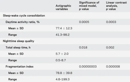
Table 2 Association between sleep-wake patterns and level of consciousness/cognition Actigraphic variables Significance of mixed model, p value Linear contrast analysis, p value Sleep-wake cycle consolidation
Daytime activity ratio, % 0.0005 0.0003
Mean6 SD 77.46 12.3
Range 41.3–98.2
Nighttime sleep quality
Total sleep time, h 0.018 0.002
Mean6 SD 5.76 2.0
Range 0.5–8.7
Fragmentation index 0.00000003 0.000008
Mean6 SD 78.86 39.8
Association between level of consciousness/cognition and sleep-wake patterns.
Patients wore the actigraph for
11.3
6 4.1 days, starting 21.0 6 13.7 days postinjury
(in the ICU for 60% of the patients). Overall, there
were 233 days of both actigraphy recording and RLA
assessment.
Daytime activity ratio.
We observed a strong
associ-ation between RLA and DAR (table 2). Our results
showed that an increase in RLA score was associated
with a linear improvement in the consolidation of the
24-hour sleep-wake cycle, as measured by the DAR
(figure 1A). When we used a DAR criterion of
$80%
to determine the occurrence of a consolidated
sleep-wake cycle,
1we observed that patients attained a
con-solidated 24-hour sleep-wake cycle when they evolved
from an RLA score of 5 (confused, nonpurposeful
response, but able to answer simple commands) to
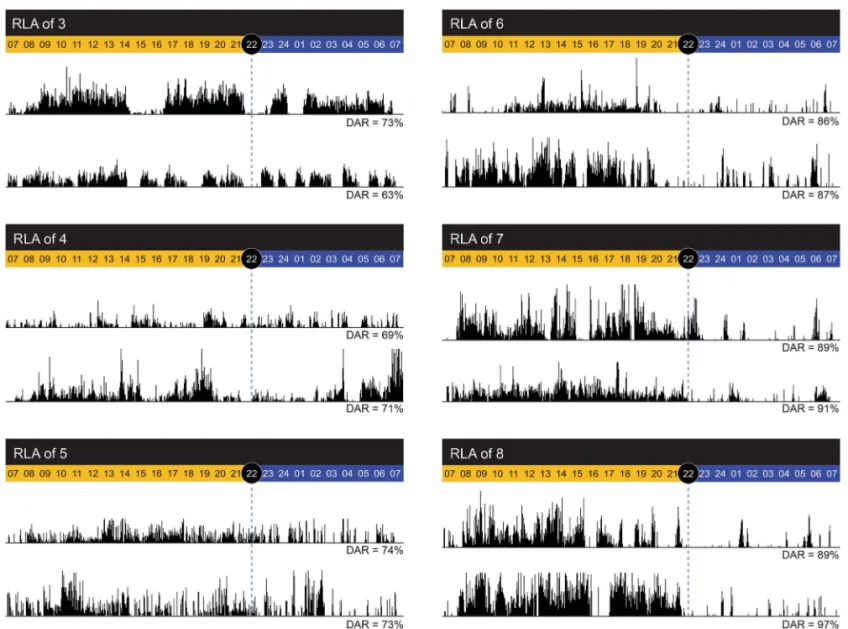
6 (goal-directed behavior). Figure 2 shows examples
of actigraphic findings in relation to RLA scores.
Nighttime sleep duration.
We observed a moderate
association between RLA score and nighttime sleep
duration (trend for significance when the Bonferroni
correction was applied), and an increase in RLA score
was associated with a linear improvement in
night-time sleep duration (table 2 and figure 1B).
Nighttime fragmentation index.
We also found a strong
association between RLA score and fragmentation
index, such that an increase in RLA score was
associ-ated with a linear decrease in nighttime fragmentation
index (table 2 and figure 1C).
Cross-correlations revealed that the best-fit lag
between RLA and DAR was 0 (R
25 0.816, p ,
0.001), suggesting that improvements in DAR were
simultaneous to that of RLA scores. Cross-correlations
with nighttime sleep duration and nighttime
fragmen-tation index were not significant, although a trend for
a correlation at lag 0 was observed between RLA and
fragmentation index (r
5 20.60, p 5 0.069).
DISCUSSIONIn this study of 30 hospitalized
pa-tients in the acute phase of moderate and severe
TBI, we demonstrate that the recovery of
conscious-ness and higher cognitive functions occurs in parallel
with improvements in consolidation of the
sleep-wake cycle, as measured with actigraphy. Increasing
consciousness and cognitive functioning was also
tightly timed with the increase in the estimated
nighttime sleep duration and the decrease in the
estimated nighttime fragmentation index. This study
establishes a clear link between acute sleep-wake
disturbances and recovery of brain functions after
TBI. No previous study investigated this temporal
association in acute TBI following emergence from
coma. Some research groups showed that the
presence of sleep elements measured by EEG (i.e.,
sleep spindles, K-complexes, and REM sleep) are
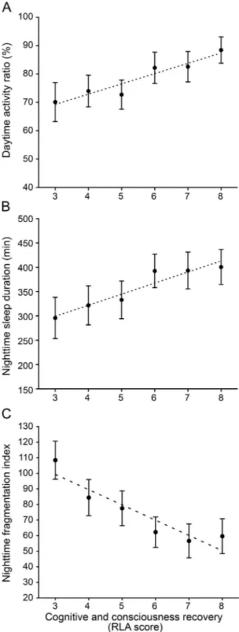
Figure 1 Association between cognitive and consciousness recovery and thesleep-wake cycle
(A) Parallel evolution of the Rancho Los Amigos scale of cognitive functioning (RLA) score and daytime activity ratio in the 30 patients assessed over 233 days. Black dots indicate the mean daytime activity ratio per score on the RLA scale, generated within the mixed model equation, and black bars represent SEM. The linear contrast analysis was statisti-cally significant (p , 0.001). (B) Parallel evolution of the RLA score and nighttime sleep duration. Black dots indicate the mean nighttime sleep duration (minutes) per score on the RLA scale, generated within the mixed model equation, and black bars represent SEM. The linear contrast analysis was statistically significant (p 5 0.002). (C) Parallel evolution of RLA score and nighttime fragmentation index. Black dots indicate the mean nighttime fragmentation index per score on the RLA scale, generated within the mixed model equa-tion, and black bars represent SEM. The linear contrast analysis was statistically signifi-cant (p , 0.001).
associated with level of consciousness, cognition or
prognosis in posttraumatic coma, in the subacute
phase of brain injuries, and in chronic disorders of
consciousness.
22–24Other studies focused on the
presence or absence of a 24-hour sleep-wake cycle in
chronic disorders of consciousness. For example,
a study
25compared the strength of the circadian
rest-activity cycle of patients in chronic unresponsive
wake syndrome (RLA score
;2) to that of patients in
chronic minimally conscious states (MCS; RLA score
;3–5), using 4-day actigraphy, and showed a more
robust circadian rhythm of rest-activity in patients in
MCS. Our study shows that this parallel improvement
continues with further improvement of the cognitive
state, and demonstrates the linearity of the
relationship. Our results also suggest that in acute
TBI, consolidation of a circadian sleep-wake cycle
attains an acceptable level (DAR
$80%) only when
patients emerge from MCS, marked by the capacity
for functional communication or functional use of
objects (RLA score
$6).
26Prior to this stage of
consciousness recovery, sleep and wake states are
present, but are fragmented and dispersed
throughout the day and night rather than
consolidated in a circadian rhythm. Although we
cannot confirm the causal relationship between the
injured brain and sleep-wake patterns, our results
suggest that when the brain has not sufficiently
recovered a level of consciousness to sustain both
arousal and awareness of one
’s surroundings, it is
also unable to generate consolidated sleep and
wake.
Though the linearity of the relationship
between RLA and the actigraphy variables is
strong, the 3 sleep-wake variables seem to plateau
at RLA scores of 6, 7, and 8. This plateauing
may reflect the optimal level of consolidation of
the sleep-wake cycle and nocturnal sleep quality
that patients can reach in this context, given the
limitations of the hospital environment, nursing
Figure 2 Examples of actigraphic findings in relation to Rancho Los Amigos scale of cognitive functioning (RLA) scoresExamples of typical actigraphic findings for RLA ranging from 3 to 5 (left panel), and RLA ranging from 6 to 8 (right panel). Total activity counts for each minute of recording are illustrated by vertical dark lines, on a scale of 0 to 1,000 activity counts. Daytime hours (07:00–22:00 hours) are shown in yellow and nighttime hours (22:00–07:00 hours) in blue. Daily daytime activity ratios (DAR) are indicated at the bottom of each actogram.
interventions, and residual pain. Future studies
should aim to assess what constitutes normal sleep
parameters among critically ill patients in the ICU
and regular wards without brain injury, to better
situate the sleep of TBI patients.
Given that in healthy individuals, sleep
restric-tion negatively affects cognirestric-tion, particularly
mem-ory formation,
27the inability to consolidate sleep
and wake may hinder the recovery of consciousness
and cognitive function after TBI. Impaired sleep is
hypothesized to impede memory by preventing
synaptic homeostasis.
28Without sleep, the brain
is less able to encode and consolidate new information
in memory. Synaptic plasticity and hippocampal
neurogenesis, 2 crucial processes for recovery following
TBI, are also highly sleep-dependent.
29In this
context, poor sleep consolidation may impede
cognitive recovery after a brain injury. However,
in the present study, cross-correlation analyses
suggest a synchronous recovery of sleep quality,
cognition, and consciousness, rather than a causal
relationship. This suggests that in the context of
acute TBI, it is most likely overall neuronal
recov-ery that drives the progressive return of
conscious-ness, cognition, and sleep.
Strengths and limitations.
Actigraphy is a measure of
physical motion and therefore indirectly measures
sleep and wake through assessment of the
rest-activity cycle. Actigraphy is closely correlated
to polysomnography in healthy individuals and
is well-validated for the estimation of sleep
parameters across age groups.
17Moreover, one
research team recently showed that actigraphy
correlated with polysomnography-measured total
sleep time and sleep efficiency among severe TBI
inpatients in a rehabilitation setting.
30Still, results
of the present study reflect an indirect measure of
sleep and wake, though actigraphy remains the
best-suited method for the long-term assessment of
sleep-wake cycles within this clinical population.
Results from cross-correlations are sometimes
criticized because they tend to overestimate the
strength of time-lagged relationships, mainly because
of data autocorrelations and intramultiplicity.
21However, given the strength of the
cross-correlation analysis between RLA and DAR (r
5
0.816) and a moderate autocorrelation (0.5) in our
data, we estimate our type I error rate bias to be
under 0.10,
21which is negligible. Moreover, given
the high variability of RLA scores and actigraphy
data on each day of actigraphy recording, averaging
our RLA and actigraphy data per day most likely
weakened interday differences. Such pooling of
data to create averages per day, as required to
perform
cross-correlation
analyses,
reduces
variability and the number of data points, and
may thus explain why no cross-correlation was
sig-nificant with nighttime sleep duration and
frag-mentation index (trend for significance only).
Clinical implications.
This study showed that the
con-solidation of sleep and wake states goes hand in hand
with the recovery of consciousness and cognition in
acute TBI, though the directionality (or
bidirection-ality) of this relationship remains unknown. Insight
into the association between neuronal recovery
and the sleep-wake cycle could help shed light on
the
pathophysiology
of
post-TBI
sleep-wake
disturbances, which frequently persist up to several
years postinjury.
31–35This association also suggests
that assessment of the sleep-wake cycle in acute TBI
may be a useful tool for monitoring patient
evolution and recovery. Moreover, the possibility
of a positive feedback action of improved
consolidation
of sleep and wake states on
consciousness and cognitive recovery may be
worthy of further investigation. The role of
hospital lighting and noise could be interesting to
assess in future studies in order to better appraise
their implications in sleep-wake disturbances.
However, given that patients in the present study
were hospitalized in the same environment but
had different sleep-wake cycle consolidation and
quality depending on their level of consciousness,
this may suggest that environmental factors only
partly account for the sleep-wake disturbances
observed in hospitalized TBI patients. Results from
the present study could have implications for the
development of interventions targeting sleep-wake
cycles and aimed at optimizing functional recovery
in
both
acute
and
chronic
disorders
of
consciousness.
AUTHOR CONTRIBUTIONS
Catherine Duclos: acquisition of data, analysis and interpretation of data, drafting and critically revising the manuscript. Marie Dumont: conception and design of the study, analysis and interpretation of data, drafting and revising the manuscript. Caroline Arbour: analysis and interpretation of data, drafting and revising the manuscript. Jean Paquet: analysis and interpretation of data, drafting and revising the manuscript. Hélène Blais: conception and design of the study, acqui-sition of data, analysis and interpretation of data, drafting and revising the manuscript. David K. Menon: conception and design of the study, interpretation of data, drafting and revising the manuscript. Louis De Beaumont: interpretation of data, drafting and revising the manuscript. Francis Bernard: conception and design of the study, interpretation of data, drafting and revising the manuscript. Nadia Gosselin: led con-ception and design of the study, analysis and interpretation of data, drafting and revising the manuscript.
ACKNOWLEDGMENT
The authors thank Elyse Laflamme and Jeanne Woo, occupational therapists at Hôpital du Sacré-Coeur de Montréal, for providing the RLA evaluations and scores of the patients included in this study; Dr. Harrison Westwick for scoring CT scans according to Marshall and Rotterdam criteria; the patients and their families for their
collaboration; as well as the nursing staff of the Intensive Care Unit and Neurological Ward for their help in monitoring patients during actigraphy recordings.
STUDY FUNDING
This study was supported by the Canadian Institutes of Health Research (grant no. 115172) and by the Fonds pour la Recherche du Québec– Santé (grant no. 24742).
DISCLOSURE
C. Duclos received studentship funding from the Université of Montréal. M. Dumont, C. Arbour, J. Paquet, H. Blais, D. Menon, L. De Beaumont, F. Bernard, and N. Gosselin report no disclosures relevant to the manuscript. Go to Neurology.org for full disclosures.
Received March 18, 2016. Accepted in final form August 30, 2016.
REFERENCES
1. Duclos C, Dumont M, Blais H, et al. Rest-activity cycle disturbances in the acute phase of moderate to severe traumatic brain injury. Neurorehabil Neural Repair 2013;28:472–482. 2. Chiu HY, Chen PY, Chen NH, Chuang LP, Tsai PS. Trajectories of sleep changes during the acute phase of traumatic brain injury: a 7-day actigraphy study. J Formos Med Assoc 2013;112:545–553.
3. Gabor JY, Cooper AB, Crombach SA, et al. Contribu-tion of the intensive care unit environment to sleep disruption in mechanically ventilated patients and healthy subjects. Am J Respir Crit Care Med 2003; 167:708–715.
4. Olson E, Badder C, Sullivan S, Smith C, Propert KJ, Margulies SS. Alterations in daytime and nighttime activ-ity in piglets after focal and diffuse brain injury. J Neurotrauma 2015;33:734–740.
5. Sabir M, Gaudreault PO, Freyburger M, et al. Impact of traumatic brain injury on sleep structure, electrocortico-graphic activity and transcriptome in mice. Brain Behav Immun 2015;47:118–130.
6. Skopin MD, Kabadi SV, Viechweg SS, Mong JA, Faden AI. Chronic decrease in wakefulness and disruption of sleep-wake behavior after experimental traumatic brain injury. J Neurotrauma 2015;32:289–296.
7. Makley MJ, Johnson-Greene L, Tarwater PM, et al. Re-turn of memory and sleep efficiency following moderate to severe closed head injury. Neurorehabil Neural Repair 2009;23:320–326.
8. Holcomb EM, Towns S, Kamper JE, et al. The relation-ship between sleep-wake cycle disturbance and trajectory of cognitive recovery during acute traumatic brain injury. J Head Trauma Rehabil 2016;31:108–116.
9. Blume C, Del Giudice R, Wislowska M, Lechinger J, Schabus M. Across the consciousness continuum: from unresponsive wakefulness to sleep. Front Hum Neurosci 2015;9:105.
10. Menon DK, Schwab K, Wright DW, Maas AI. Position statement: definition of traumatic brain injury. Arch Phys Med Rehabil 2010;91:1637–1640.
11. Teasdale G, Jennett B. Assessment of coma and impaired consciousness: a practical scale. Lancet 1974;2:81–84. 12. Marshall LF, Marshall SB, Klauber MR, et al. The
diag-nosis of head injury requires a classification based on com-puted axial tomography. J Neurotrauma 1992;9(suppl 1): S287–S292.
13. Maas AI, Hukkelhoven CW, Marshall LF, Steyerberg EW. Prediction of outcome in traumatic brain injury with
computed tomographic characteristics: a comparison between the computed tomographic classification and combinations of computed tomographic predictors. Neurosurgery 2005;57:1173–1182.
14. Rappaport M, Hall KM, Hopkins K, Belleza T, Cope DN. Disability rating scale for severe head trauma: coma to community. Arch Phys Med Rehabil 1982; 63:118–123.
15. Hagen C, Malkmus D, Durham P. Rancho Los Amigos Levels of Cognitive Functioning Scale. Downey, CA: Pro-fessional Staff Association of Rancho Los Amigos National Rehabilitation Center; 1972.
16. Dowling GA. Levels of cognitive functioning: evalua-tion of interrater reliability. J Neurosurg Nurs 1985;17: 129–134.
17. Martin JL, Hakim AD. Wrist actigraphy. Chest 2011;139: 1514–1527.
18. Han HJ. Comparison of results with actigraphy and poly-somnography in two sleep disorders: obstructive sleep apnea syndrome and primary insomnia. J Korean Neurol Assoc 2003;21:156–162.
19. Wang D, Wong KK, Dungan GC II, Buchanan PR, Yee BJ, Grunstein RR. The validity of wrist actimetry assess-ment of sleep with and without sleep apnea. J Clin Sleep Med 2008;4:450–455.
20. Littell RC, Pendergast J, Natarajan R. Modelling covari-ance structure in the analysis of repeated measures data. Stat Med 2000;19:1793–1819.
21. Olden DJ, Neff DB. Cross-correlation bias in lag analysis of aquatic time series. Mar Biol 2001;138:1063–1070. 22. Bergamasco B, Bergamini L, Doriguzzi T, Fabiani D. EEG
sleep patterns as a prognostic criterion in post-traumatic coma. Electroencephalogr Clin Neurophysiol 1968;24: 374–377.
23. de Biase S, Gigli GL, Lorenzut S, et al. The importance of polysomnography in the evaluation of prolonged disorders of consciousness: sleep recordings more adequately corre-late than stimulus-recorre-lated evoked potentials with patients’ clinical status. Sleep Med 2014;15:393–400.
24. Ron S, Algom D, Hary D, Cohen M. Time-related changes in the distribution of sleep stages in brain injured patients. Electroencephalogr Clin Neurophysiol 1980;48: 432–441.
25. Cruse D, Thibaut A, Demertzi A, et al. Actigraphy assess-ments of circadian sleep-wake cycles in the vegetative and minimally conscious states. BMC Med 2013;11:18. 26. Giacino JT, Ashwal S, Childs N, et al. The minimally
conscious state: definition and diagnostic criteria. Neurology 2002;58:349–353.
27. Walker MP, Stickgold R. Sleep, memory, and plasticity. Annu Rev Psychol 2006;57:139–166.
28. Tononi G, Cirelli C. Sleep and the price of plasticity: from synaptic and cellular homeostasis to memory consolidation and integration. Neuron 2014;81:12–34.
29. Kreutzmann JC, Havekes R, Abel T, Meerlo P. Sleep deprivation and hippocampal vulnerability: changes in neuronal plasticity, neurogenesis and cognitive function. Neuroscience 2015;309:173–190.
30. Kamper JE, Garofano J, Schwartz DJ, et al. Concordance of actigraphy with polysomnography in traumatic brain injury neurorehabilitation admissions. J Head Trauma Rehabil 2016;31:117–125.
31. Baumann CR, Werth E, Stocker R, Ludwig S, Bassetti CL. Sleep-wake disturbances 6 months after traumatic
brain injury: a prospective study. Brain 2007;130: 1873–1883.
32. Sommerauer M, Valko PO, Werth E, Baumann CR. Excessive sleep need following traumatic brain injury: a case-control study of 36 patients. J Sleep Res 2013;22: 634–639.
33. Imbach LL, Valko PO, Li T, et al. Increased sleep need and daytime sleepiness 6 months after traumatic brain injury: a prospective controlled clinical trial. Brain 2015; 138:726–735.
34. Duclos C, Dumont M, Wiseman-Hakes C, et al. Sleep and wake disturbances following traumatic brain injury. Pathol Biol 2014;62:252–261.
35. Ouellet MC, Beaulieu-Bonneau S, Morin CM. Sleep-wake disturbances after traumatic brain injury. Lancet Neurol 2015;14:746–757.
36. Bagnato S, Boccagni C, Prestandrea C, Sant’Angelo A, Castiglione A, Galardi G. Prognostic value of standard EEG in traumatic and non-traumatic disorders of conscious-ness following coma. Clin Neurophysiol 2010;121:274–280.