Université de Montréal
Polymeric micelles as versatile carriers for
drugs and nucleic acids
par
Mahmoud El Sabahy
Faculté de Pharmacie
Thèse présentée à la Faculté des études supérieures
en vue de l’obtention du grade de doctorat
en sciences pharmaceutiques
option technologie pharmaceutique
Université de Montréal
Faculté des études supérieures
Cette thèse intitulée :
Polymeric micelles as versatile carriers for
drugs and nucleic acids
Présentée par :
Mahmoud El Sabahy
A été évaluée par un jury composé des personnes suivantes :
Prof. Patrice Hildgen, président-rapporteur
Prof. Jean-Christophe Leroux, directeur de recherche
Prof. Yue Zhao, membre du jury
Résumé
Le cancer est la principale cause de mortalité au Canada. Les taxanes (e.g. le paclitaxel et le docétaxel (DCTX)) constituent des remèdes efficaces contre une série de tumeurs solides telles que les cancers du sein, du poumon et de l’ovaire. Par ailleurs, des acides nucléiques (e.g. les oligonucléotides antisens (AON) ou les petits ARN interférents (siRNAs)), capables de supprimer sélectivement certains oncogènes impliqués dans la carcinogénèse, sont actuellement étudiés pour traiter une large gamme de cancers. Bien que l’activité des taxanes et des acides nucléiques soit bien établie sur des modèles humains et/ou animaux, plusieurs aspects physico-chimiques et cliniques restent encore à améliorer. Leur solubilité limitée (pour les taxanes), leur dégradation rapide dans le sang (pour les acides nucléiques), leur élimination précoce, leur absence de sélectivité et leur toxicité envers les tissus sains sont les principaux facteurs limitant leur efficacité. C’est pourquoi de nombreux efforts ont porté sur l’élaboration de systèmes de vectorisation ciblés à base de polymères, dans le but de surmonter les problèmes associés aux thérapies actuelles. Dans cette thèse, deux types de micelles polymères ont été développés pour la vectorisation de DCTX et d’acides nucléiques. D’une part, des micelles de poly(oxyde d’éthylène)-bloc-poly(oxyde de butylène/styrène) ont été étudiées pour la première fois pour solubiliser le DCTX et le protéger de l’hydrolyse. Ces polymères se sont révélés moins toxiques que le surfactant utilisé commercialement pour solubiliser le DCTX (i.e. polysorbate 80) et ont permis une libération prolongée du principe actif. D’autre part, deux systèmes différents de micelles polyioniques (PICM) ont été mis au point pour la vectorisation d’acides nucléiques. De nouveaux conjugués de poly(éthylène
glycol) (PEG)-oligonucléotide ont été proposés pour la protection et la libération contrôlée d’AON. Lorsque ces conjugués ont été formulés avec des dendrimères de poly(amidoamine) (PAMAM), des complexes de taille homogène ont été obtenus. Ces PICM ont permis de prolonger la libération de l’AON et de le protéger efficacement contre la dégradation enzymatique. De plus, des polymères de poly(oxyde d’éthylène)-bloc-poly(méthacrylate de propyle-co-acide méthacrylique) ont été incorporés afin de conférer des propriétés acido-sensibles aux PICM. Dans ces micelles, formées de ce dernier polymère formulé avec le dendrimère PAMAM, des oligonucléotides (AON et siRNA) ciblant l’oncogène Bcl-2 ont été encapsulés. L’internalisation cellulaire fut assurée par un fragment d’anticorps monoclonal (Fab’) situé à l’extrémité de la couronne de PEG. Après l’internalisation cellulaire et la protonation des unités d’acide méthacrylique sous l’effet de l’acidification des endosomes, les micelles se sont affranchies de leur couronne. Elles ont ainsi exposé leur cœur composé d’acide nucléique et de dendrimère PAMAM, qui possède une charge positive et des propriétés endosomolytiques. En effet, ces PICM acido-sensibles ciblées ont permis d’augmenter la biodisponibilité des acides nucléiques vectorisés et se sont avérées plus efficaces pour silencer l’oncoprotéine Bcl-2 que les micelles non ciblées ou que le dendrimère de PAMAM commercial seul. Finalement, les nanovecteurs polymères présentés dans cette thèse se révèlent être des systèmes prometteurs pour la vectorisation des anticancéreux et des acides nucléiques.
Mots-clés : micelles polymères, micelles polyioniques, docétaxel, oligonucléotide
antisens, siRNA, dendrimères de poly(amidoamine), vectorisation de médicament, vectorisation d’acides nucléiques, sensibilité au pH, ciblage.
Abstract
Cancer is considered as the leading cause of premature death in Canada. Taxanes (e.g. paclitaxel and docetaxel (DCTX)) are effective against a range of solid tumors including breast, lung, and ovarian malignancies. In addition, nucleic acids (e.g. antisense oligonucleotides (AON) and short interfering RNA (siRNA)) which are capable of selectively suppressing oncogenes involved in carcinogenesis are currently being investigated for the treatment of a wide variety of cancers. Although the activity of taxanes and nucleic acid drugs is well-established in human and/or animal models, several physicochemical and clinical issues still need to be addressed. Low aqueous solubility (i.e. taxanes), rapid degradation in the blood (i.e. nucleic acids), fast clearance, non-selectivity and toxicity to normal tissues are limiting factors to their effectiveness. Hence, many efforts have been focused on developing targeted polymeric delivery systems to overcome the problems associated with the current therapies. In this thesis, two types of polymeric micelles have been developed for the delivery of DCTX and nucleic acids. On the one hand, poly(ethylene
oxide)-block-poly(butylene oxide/styrene oxide) micelles were tested for the first time to
solubilize and protect DCTX from hydrolytic degradation. The polymers showed less toxicity than the surfactant used commercially to dissolve DCTX (i.e. polysorbate 80) and released the drug in a sustained fashion. On the other hand, two different systems of polyion complex micelles (PICM) were developed for the sustained release and intracellular delivery of nucleic acids. Novel poly(ethylene glycol) (PEG)-oligonucleotide conjugates were assessed to protect AON against degradation and release them in a sustained manner. When these conjugates were mixed with
poly(amidoamine) (PAMAM) dendrimers, monodisperse PICM were formed. These PICM further slowed down AON release and significantly protected it against enzymatic degradation. In addition, the incorporation of poly(ethylene oxide)-block-poly(propyl methacrylate-co-methacrylic acid) was exploited to impart pH-sensitivity to PAMAM-based PICM. This system was composed of the previous copolymer mixed with PAMAM dendrimer. Such PICM were loaded with AON or siRNA targeting the Bcl-2 oncogene. Micelles uptake by the cancer cells was mediated by a monoclonal antibody fragment (i.e. Fab') positioned at the extremity of the PEG corona. Upon cellular uptake and protonation of the methacrylic acid units in the acidic endosomal environment, the micelles lost their corona, thereby exposing their positively-charged endosomolytic PAMAM/nucleic acid core. The targeted, pH-sensitive PICM were found to increase the intracellular bioavailability of the entrapped nucleic acids and knock down the Bcl-2 oncoprotein more than either non-targeted micelles or commercial PAMAM dendrimers. The polymeric nanocarriers reported in this thesis appear to be promising vehicles for the delivery of anticancer drugs and nucleic acids.
Keywords: Polymeric micelles, polyion complex micelles, docetaxel, antisense
oligonucleotide, siRNA, poly(amidoamine) dendrimers, drug delivery, nucleic acid delivery, pH-sensitivity, targeted delivery.
Table of contents
Résumé ... 3 Abstract ... 5 List of tables ... 13 List of figures ... 15 List of schemes... 24 List of abbreviations... 25 Acknowledgments ... 30CHAPTER 1 - General overview... 32
1. Formulations of taxanes ... 36
1.1 Polymeric macromolecular carriers... 38
1.2 Emulsions ... 39
1.3 Liposomes ... 40
1.4 Polymeric micelles ... 41
1.5 Nanoparticles... 42
2. Formulations of nucleic acids... 43
2.1 Chemical modifications... 45
2.2 Viral vectors ... 48
2.3 Non-viral vectors... 49
2.3.1 Non-complexing polymers ... 49
2.3.2 Lipoplexes and polyplexes ... 50
2.3.3 PEGylated lipoplexes and PICM... 51
2.3.4 Targeted complexes... 52
2.3.5 pH-sensitive PICM ... 53
CHAPTER 2 - Polymeric micelles as versatile carriers for drugs and nucleic acids
delivery... 66
Abstract ... 67
1. Introduction ... 68
2. Micellization... 72
3. Composition of the micellar carriers ... 74
4. Micelle stability... 77
4.1 Significance ... 77
4.2 Thermodynamic stability... 78
4.3 Kinetic stability ... 82
4.4 Stability of PICM towards dissociation... 84
4.5 Protection of the drugs towards enzymatic degradation ... 85
4.6 Towards greater micelle stability ... 87
5. Micelle dimensions and morphology ... 90
5.1 Theoretical prediction of the micelle morphology ... 90
5.2 Analysis of the micelle morphology ... 91
5.3 Transformation between different morphologies ... 94
5.4 Morphologies attainable with PICM ... 96
5.5 Significance in drug delivery ... 96
6. Drug incorporation ... 97
6.1 Drug loading procedures ... 97
6.2 Achieving high drug loading... 100
7. Drug release... 104
7.1 Release from PM ... 104
7.2 Release from other micellar systems... 108
7.3 Triggered drug release... 109
9. In vitro and in vivo applications ... 116 9.1 Non-targeted micelles... 118 9.2 Targeted micelles ... 121 9.2.1 pH-responsive micelles ... 121 9.2.2 Temperature-sensitive micelles... 122 9.2.3 Functionalized micelles... 123
9.3 PM for oral drug delivery... 128
9.4 Reverse PM ... 129
10. Conclusion... 130
11. Acknowledgements ... 132
12. References ... 133
Objectives and hypotheses of the thesis ... 150
CHAPTER 3 - Solubilization of docetaxel in poly(ethylene oxide)-block-poly(butylene/styrene oxide) micelles... 152
Abstract ... 153
1. Introduction ... 154
2. Materials... 156
3. Methods ... 156
3.1 Characterization of unloaded polymeric micelles ... 156
3.1.1 Synthesis and characterization of polymers ... 156
3.1.2 Determination of the critical association concentration (CAC) ... 158
3.1.3 Measurement of core viscosity... 158
3.1.4 Dynamic light scattering (DLS) measurements ... 159
3.1.5 Multiangle static light scattering (MASLS) measurements ... 160
3.2 TEM... 161
3.3 Characterization of DCTX-loaded polymeric micelles... 161
3.3.1 Preparation and characterization of DCTX-loaded micelles... 161
3.4 Chemical stability of DCTX-loaded micelles ... 163
3.5 Calculation of solubility parameters by the group contribution method... 163
3.6 In vitro release kinetics... 164
3.7 In vitro cytotoxicity assay ... 165
4. Results and discussion... 166
4.1 Characterization of the polymers and unloaded micelles... 166
4.2 Characterization of DCTX-loaded micelles ... 172
4.3 In vitro evaluation of micelle formulations... 175
5. Conclusion... 181
6. Acknowledgements ... 182
7. References ... 183
Supporting information ... 187
CHAPTER 4 - Synthesis and enzymatic stability of PEGylated oligonucleotide duplexes and their self-assemblies with polyamidoamine dendrimers ... 188
Abstract ... 189
1. Introduction ... 190
2. Materials... 193
3. Methods ... 194
3.1 Preparation of the PEGylated duplexes... 194
3.1.1 PEGylation of the SON ... 194
3.1.2 Hybridization of the PEG-SON with the AON ... 194
3.2 Characterization of PEG-ON duplex... 195
3.2.1 Determination of the quenching efficiency ... 195
3.2.2 Determination of melting temperature (Tm) ... 196
3.2.3 Agarose gel electrophoresis... 196
3.2.4 Capillary electrophoresis... 197
3.3.2 Characterization of the PICM... 198
3.4 Enzymatic degradation ... 199
4. Results and discussion... 200
4.1 Preparation and characterization of PEG-SON ... 200
4.2 Preparation and characterization of PEG-SON/AON ... 203
4.3 Preparation and characterization of the PICM ... 204
4.4 Enzymatic degradation ... 209
5. Conclusion... 213
6. Acknowledgement... 213
7. References ... 214
CHAPTER 5 - Delivery of nucleic acids through the controlled disassembly of multifunctional nanocomplexes ... 218
Abstract ... 219
1. Introduction ... 220
2. Results and discussion... 224
3. Experimental Section ... 234
4. Acknowledgement... 237
5. References ... 238
Supporting information ... 241
CHAPTER 6 – Discussion ... 253
1. Hurdles facing current delivery systems ... 253
2. PM for drug delivery ... 254
2.1 Micellar characterization... 254
2.2 Factors affecting drug-loading ... 255
2.3 In vitro evaluation of the micelle formulations ... 256
3. Polyion complex micelles for nucleic acid delivery ... 258
3.1 Preparation of polymeric nanocarriers for nucleic acid delivery ... 258
3.2 Enzymatic stability of entrapped nucleic acids ... 260
3.3 Active targeting ... 265
4. Designing polymeric nanocarriers for drug and nucleic acid delivery ... 268
Conclusion and perspectives ... 270
References ... 273
List of tables
CHAPTER 1 - General overview
Table 1. Examples of colloidal taxane formulations currently being tested in clinical
trials or approved for cancer treatment………... .37
Table 2. Some AON and siRNA currently in clinical trials for the treatment of
cancer………..………... .45
Table 3. Characteristics of viral vectors……… .49
CHAPTER 2 - Polymeric micelles as versatile carriers for drugs and
nucleic acids delivery
Table 1. Selection of polymers most often used for the preparation of micelles in
drug delivery………75
Table 2. Different parameters that can be used to predict the micelle morphology...92 Table 3. Examples of PM loaded with various antitumor drugs………..…117 Table 4. Examples of PM loaded with heparin, pDNA, AON or siRNA………….118
CHAPTER 3 - Solubilization of docetaxel in poly(ethylene
oxide)-block-poly(butylene/styrene oxide) micelles
Table 1. Molecular characteristics of PEO-b-PBO and PEO-b-PSO………...166 Table 2. Size, CAC and core viscosity of unloaded micelles. Mean of 3 independent
experiments……… ……...167
Table 4. Characteristics of DCTX-loaded micelles………..174 Table 5. Total (
δ
t) and partial (δ
d,δ
pandδ
h) solubility parameters of DCTX andindividual polymer blocks (MPa½)………175
CHAPTER 4 - Synthesis and enzymatic stability of PEGylated
oligonucleotide duplexes and their self-assemblies with
polyamidoamine dendrimers
Table 1. Characteristics of the ternary PICM prepared at an N/P 2:1 ratio; mean ± SD
(n=3)………..……205
Table 2. Michaelis-Menten kinetic parameters for SON degradation by DNase 1..210
CHAPTER 5 - Delivery of nucleic acids through the controlled
disassembly of multifunctional nanocomplexes
Table S1. Molecular characteristics of PEG-b-P(PrMA-co-MAA)……….…249 Table S2. Characteristics of the ternary PICM prepared at N/(P + COO-) molar ratio of 1.5 before and after lyophilization; mean ± SD (n=3)………..249
CHAPTER 6 - Discussion
Table 1. Comparison of the clinical pharmacokinetics of Taxol® (commercial formulation) and Genexol (micellar nanocarrier)………....……… 258
Table 2. Characteristics of ternary PICM-AON; mean ± SD (n=3)……….. ……...260 Table 3. Chemically modified siRNA………..…272
List of figures
CHAPTER 1 - General overview
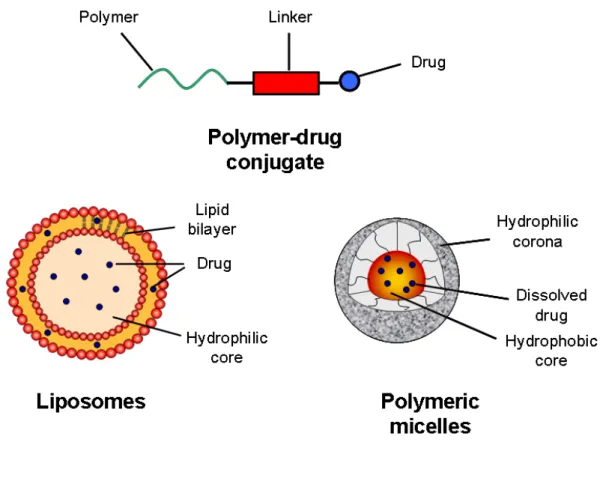
Figure 1. Some delivery systems (liposomes, PM and polymeric-drug conjugates)
currently being exploited for the delivery of anticancer drugs……….. .38
Figure 2. Mechanism of action of AON and siRNA. AON pair with their
complementary target RNA and block mRNA translation or induce its degradation. siRNA duplexes are assembled into the RISC where the sense strand is cleaved and unwound, leaving single-stranded RNA associated with the RISC. These complexes hybridize and cleave complementary target mRNA. Both actions result in target protein downregulation……….………...44
Figure 3. Chemical structure of PS DNA, 2′-O-methyl RNA, PNA, LNA and
2′F-ANA...47
CHAPTER 2 - Polymeric micelles as versatile carriers for drugs and
nucleic acids delivery
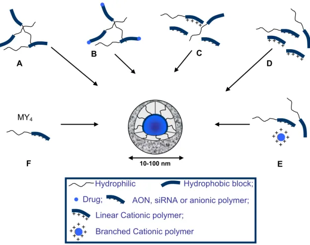
Figure 1. Conventional PM (A); drug-conjugated PM (B); and PICM with the
polyionic block consisting of cationic polymer (C) or polynucleic acid (antisense oligonucleotide (AON) or short interfering RNA (siRNA)) (D and E). In D and E, the core forming agent is either linear or branched cationic polymer, respectively. Polymer-metal complex micelles formed via the ligand substitution reaction where M and Y are the metal and the ligand, respectively (F)……….…………..69
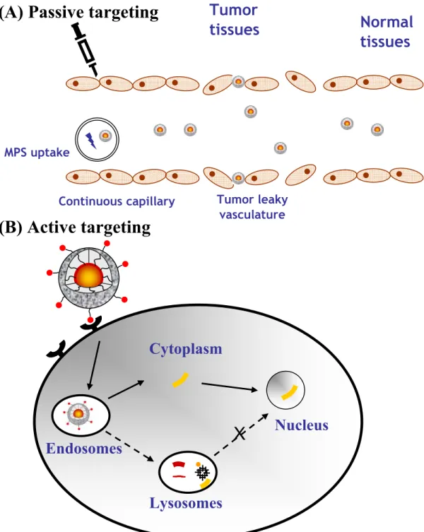
Figure 2. Passive (A) and active (B) targeting. In passive targeting, the
non-functionalized micelles extravasate in tissues presenting leaky vasculatures (e.g. tumors) and accumulate through the EPR effect. In active targeting, the micelles bind
to specific receptors expressed on the surface of the target cells, after which they are internalized. The entrapped drug should be able to escape from the endosomes in order to reach to the cytoplasm and/or nucleus (the solid lines)……….…………....71
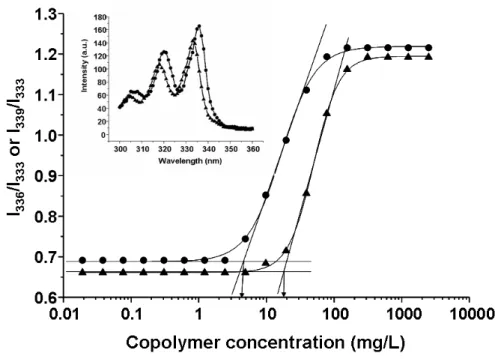
Figure 3. Plot of the intensity ratio I336/I333 or I339/I333 (from pyrene excitation spectra) as a function of PEG45-b-PBO15 (▲) and PEG45-b-PSO15K (●) concentration. Each value is the mean of three independent measurements. Values of the CMC are indicated by arrows. Inset: Excitation spectra of pyrene (2 × 10⁻⁷ M aqueous solution) monitored at λem 390 nm below (▲) and above (●) the PEG45-b-PSO15 copolymer’s CMC……….…………..80
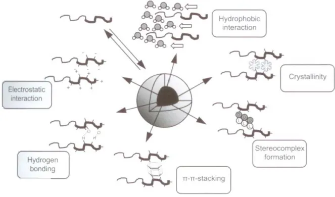
Figure 4. Interactions in the micellar core that enhance the kinetic stability of PM.
Reproduced from Carstens, M. et al. [56] Copyright 2008, with permission from Springer Science……….…….84
Figure 5. Dequenching of fluorescein-labeled sense oligonucleotide (SON)
fluorescence following the incubation of PEG10K-SON/AON-PAMAM generation 3 (Mw = 6,909) PICM at N/P ratios of 1:1 (■), 2:1 (▲), 3:1 (●), 4:1 (∆) and 5:1 (□) with DNase 1 (60 U/µg oligonucleotide, 37°C, pH 7.4). Mean ± SD (n=3). The λex and λem were measured at 490 and 520 nm, respectively. The SON release was indirectly assessed by measuring the dequenching of the SON fluorescence. The fluorescence intensity corresponding to 100% release was determined at the end of the experiment by adding an excess of heparin to destabilize the micelles followed by the addition of DNase 1 and letting the reaction run for another 24 h………86
Figure 6. Crosslinking of the micellar corona or core as means to interplay with the
Figure 7. The control of micelles morphology by changing the solvent conditions.
Representative TEM pictures showing the reversibility of various morphological transitions for a solution of 1% (w/w) poly(styrene)310-b-poly(acrylic acid)52. Reprinted from Shen, H. et al. [93] Copyright 1999, with permission from American Chemical Society……….………....95
Figure 8. Most often used drug-loading procedures; simple equilibrium (A), dialysis
(B), oil-in-water emulsion (C), solution casting (D) and freeze-drying (E). Reproduced from Dufresne et al. [46]………...100
Figure 9. (A) In vitro release (left panel) and plasma concentration-time curve (right
panel) after i.v. administration of hydroxycamptothecin loaded in PEG-b-PCL micelles to rats. (B) Destabilization of the PM possibly occurs due to the adsorption of plasma proteins. Reprinted from Shi, B. et al. [113] and Chen, H. et al. [116] Copyright 2005 and 2008, with permission from Springer Science and American Chemical Society, respectively……….. ……...108
Figure 10. Hydrolysis of acetals on the dendrimer periphery of the micelle-forming
copolymer 1 leads to a solubility change which disrupts micelle formation and triggers the drug release. Reprinted from Gillies, E. et al. [121] Copyright 2005, with permission from American Chemical Society………...113
Figure 11. Three-layered micelles (A) and micelles with diamine structure (B) as
strategies to enhance endosomal escape and transfection efficiency. Reprinted from Oishi, M. et al. and Itaka, K. et al. [10, 135] Copyright 2006 and 2004, respectively, with permission from American Chemical Society………... ……...115
Figure 12. Targeting can be achieved with the whole antibody (A) or its fragments
such as F(ab′)2 (B), (Fab') (C) or scFV (D)………125
Figure 13. Assembly of reverse PM in organic solvent (CH2Cl2 (DCM)) and crosslinking of the micelle core……….130
CHAPTER 3 - Solubilization of docetaxel in poly(ethylene
oxide)-block-poly(butylene/styrene oxide) micelles
Figure 1. Fluorescence emission spectra of 1,3-bis-(1-pyrenyl)propane in EO45-BO15 (○), EO45-BO24 (●), EO45-SO15 (□), EO45-SO26 (■), and SDS (▲) micellar aqueous solutions……….168
Figure 2. Change of diameter with the magnitude of K2 for EO45-BO15 (○), EO45 -BO24 (●), EO45-SO15 (□), and EO45-SO26 (■) micelles (detection angles 50-140°). Each value is the mean of 3 experiments ± SD. The mean diameter was found to be independent on the polymer concentration above the CAC………..170
Figure 3. TEM imaging of unloaded (i) and DCTX-loaded (ii) EO45-BO15 (A), EO45 -BO24 (B), EO45-SO15 (C) and EO45-SO26 (D) micelles………..172
Figure 4. Effect of micellar solubilization on DCTX stability in EO45-BO15 (○), EO45-BO24 (●), EO45-SO15 (□), and EO45-SO26 (■) micelles. The stability of free DCTX in water (▲) is also shown. The experiments were carried out at fixed temperature (50ºC) for 24 h. DCTX loading and concentration were set at 0.04 (%
w/w) and 2 µg/mL, respectively. Mean ± SD (n=3)………..177
Figure 5. In vitro release kinetics of DCTX from EO45-BO24 (0.7% DCTX) (●) and EO45-SO26 (0.7% DCTX) (□), and EO45-SO26 (3.5% DCTX) (■) micelles. DCTX diffusion in the absence of micelles is represented by (▲). The experiments were
carried out at 37ºC in PBS (pH 7.4). Mean ± SD (n=3). The dotted lines represent the simulated release kinetics………..179
Figure 6. Cell viability of PC-3 cells exposed to EO45-BO24 (0.7% DCTX) (●) and EO45-SO26 (□) (0.7% DCTX), and (3.5% DCTX) EO45-SO26 (■) DCTX-loaded micelles. A polysorbate 80/ethanol formulation of DCTX was used as control (▲). The data represent the mean of 3 experiments. For the purpose of clarity, the error bars of the standard deviation are omitted (CV < 12%)………181
Figure S1. Kinetic model for DCTX release from micelles in jacketed Franz
diffusion cells………. ……...187
CHAPTER 4 - Synthesis and enzymatic stability of PEGylated
oligonucleotide duplexes and their self-assemblies with
polyamidoamine dendrimers
Figure 1. Capillary electropherograms showing the migration of SON (a), PEG20K -SON (b), PEG20K-br-SON (c) and PEG10K-SON (d). The separation was carried out in borate buffer (55 mM, pH 8.22). The voltage was set at 25 kV and the capillary temperature was maintained at 25°C……….202
Figure 2. Migration profiles of (A) PEG10K-SON, PEG20K-SON and PEG20K-br-SON, and (B) PEG10K-SON/AON, PEG20K-SON/AON and PEG20K-br-SON/AON on 2%-agarose gel. Control SON and AON are shown in the first lane. The green and red spots correspond to the fluorescence emission of FAM (λex = 488 nm) and Cy3 (λex = 532 nm), respectively………. ……...202
Figure 3. Emission spectrum of FAM-labeled SON prior to (●) and after
Figure 4. AFM image of PEG20K-SON/AON/G5 PAMAM based PICM………....206
Figure 5. Effect of ionic strength on scattering intensity of PEG10K- (A), PEG20K- (B) and PEG20K-br-SON/AON/PAMAM (C) based PICM prepared with G3 (○) or G5 (●) PAMAM. Mean ± SD (n=3)………...208
Figure 6. Dequenching of FAM-labeled SON fluorescence following the incubation
with DNase 1 (60 U/µg ON, 37°C, pH 7.4) with SON/AON (■), PEG10K- (▲), PEG20K- (∆) and PEG20K-br-SON/AON (○). Mean ± SD (n=3)……….210
Figure 7. Migration profiles of PEG10K-, PEG20K- and PEG20K-br-SON/AON on 2%-agarose gel after degradation by DNase 1 (60 U/µg ON, 1 h, 37°C, pH 7.4) and of intact SON and AON. The gel was scanned for FAM-labeled SON (λex = 488 nm) (A) and Cy3-labeled AON (λex = 532 nm) (B) detection. The empty white box corresponds to the original bands of the non-degraded PEG-SON/AON duplexes..211
Figure 8. Dequenching of FAM-labeled SON fluorescence following the incubation
with DNase 1 (60 U/µg ON, 37°C, pH 7.4) of PEG10K- (triangles), PEG20K- (squares), PEG20K-br- (circles) SON/AON/PAMAM based PICM prepared with G3 (open symbols) or G5 (closed symbols) PAMAM. Mean ± SD (n=3)……… ……...211
CHAPTER 5 - Delivery of nucleic acids through the controlled
disassembly of multifunctional nanocomplexes
Figure 1. Gel electrophoresis of PICM prepared at N/(P + COO-) molar ratio of 1.5 using PEG115-b-P(PrMA28-co-MAA53), siRNA and G5 PAMAM before and after freeze-drying. First 2 lanes represent PEG115-b-P(PrMA28-co-MAA53) and siRNA controls. The white inset corresponds to the same gel but following iodine staining to reveal PEG……….225
Figure 2. a) Effect of pH on scattering intensity (●) and zeta-potential (▲) of
PEG115-b-(PrMA28-co-MAA53)/AON/G5 PAMAM based PICM. Mean ± SD (n=3). b) Proposed mechanism of shell dissociation………226
Figure 3. siRNA degradation following incubation with 50% FBS (10 µg/mL
siRNA, 37°C, pH 7.4) of free siRNA (◊), siRNA complexed with G3 (□) or G5 (■) PAMAM, PEG115-b-P(PrMA51-co-MAA33) (triangles) and PEG115-b-P(PrMA28 -co-MAA53) (circles) PICM prepared with G3 (open symbols) or G5 (closed symbols) PAMAM. Mean ± SD (n=3)………..228
Figure 4. Cell viability of PC-3 cells exposed to PEG115-b-P(PrMA28-co-MAA53) and G5 PAMAM (0.01-0.5 mg/mL) and PEG115-b-P(PrMA28-co-MAA53)/G5 PAMAM PICM (N/COO- molar ratio of 1.5). Mean ± SD (n=3)……….229
Figure 5. a) Flow cytometry experiment showing the fluorescence intensity of PC-3
cells incubated with Cy3-labeled AON (400 nM) free, complexed to G5 PAMAM or entrapped in plain or Fab'-PEG115-b-P(PrMA28-co-MAA53)/G5 PAMAM PICM (N/(P + COO-) molar ratio of 1.5). b) Confocal microscopy images of PC-3 cells after treatment with Cy3-AON (red, panel ii) complexed to (A) G5 PAMAM or (B) Fab'-PEG115-b-P(PrMA28-co-MAA53)/G5 PAMAM PICM. Endosomes/lysosomes were stained by LysoTracker (green, panel i), and the images were merged in panel iii. c) Bcl-2 gene silencing in PC-3 cells transfected for 5 h with AON (400 nM) or siRNA (25 nM) complexed to G5 PAMAM or entrapped in plain or Fab'-PEG115 -b-P(PrMA28-co-MAA53)/G5 or G3 PAMAM PICM. Control cells were treated with medium alone. The effect of preincubation with 10% FBS, free CD71 or bafilomycin is indicated on the graph. The inset is representative of immunoreactive Bcl-2 and
glyceraldehyde-3-phosphate dehydrogenase (GAPDH) as determined by immunoblotting. The molar ratio of the Fab'-PEG145-b-P(PrMA27-co-MAA58) vs. PEG115-b-P(PrMA28-co-MAA53) is 2% for all the experiments. The results are representative of several independent experiments………...233
Figure S1. Emission spectrum of fluorescein- and DY547-labeled siRNA prior to
(▲) and after (●) degradation in serum (λex = 488 nm). The inset is the gel electrophoresis of the siRNA before (1) and after (2) degradation in serum when scanned at λex/λem of 488/526 nm……….. ……...250
Figure S2. The stability of siRNA in PEG115-b-P(PrMA28-co-MAA53)/ G5 PAMAM PICM following incubation with 100% FBS could be enhanced by increasing the N/(P + COO-) molar ratio from 1.5 (▲) to 3 (●) (10 µg/mL siRNA, 37°C, pH 7.4). Mean ± SD (n=3)………...251
Figure S3. Confocal microscopy images of PC-3 cells after treatment with
uncomplexed Cy3-AON (red, panel b). Endosomes/lysosomes were stained by LysoTracker (green, panel a), and the images were merged in panel c……. ……...251
Figure S4. Red fluorescence intensity of PC-3 cells incubated with Cy3-labeled
AON entrapped in Fab'-modified PEG115-b-P(PrMA28-co-MAA53)/G5 PAMAM PICM either preincubated with free CD71 antibody or not. Fluorescence of unstained control cells is indicated………251
Figure S5. Bcl-2 gene silencing in PC-3 cells transfected for 5 h with AON (400 nM)
or siRNA (25 nM) complexed lipofectamine (1 µg/mL) or entrapped in Fab'-PEG115
-b-P(PrMA28-co-MAA53)/G5 PAMAM PICMs at N/(P + COO-) of 3 (final volume of the transfection medium is 2 mL). Control cells were treated with medium alone. The
effect of preincubation with 50% FBS is indicated on the graph. The molar ratio of the Fab'-PEG145-b-P(PrMA27-co-MAA58) vs. PEG115-b-P(PrMA28-co-MAA53) is 2% for all the experiments. The results are representative of several independent experiments………...252
CHAPTER 6 – Discussion
Figure 1. (A) siRNA: the fluorophore (FAM) and the quencher (Cy3) are placed at a
distance from each other so that FRET is the predominant mode of quenching. (B) ON duplex: the fluorophore and the quencher are placed close to each other so that contact quenching occurs. In both cases, upon the degradation of DNA or RNA duplexes, the fluorophore and quencher become apart from each other and fluorescence dequenching occurs……. ………...……...262
Figure 2. Effect of serum concentration on the stability of siRNA complexed in
PEG115-b-P(PrMA28-co-MAA53)/ G5 PAMAM PICM (10 µg/mL siRNA, 37°C, pH 7.4). The formulations were incubated in 20% (●), 50% (▲) or 100% (■) FBS. Mean ± SD (n=3)……….264
Figure 3. Effect of albumin, 40 mg/mL (▲), γ-globulins, 10 mg/mL (∆) and α- and
β-predominant globulins, 15 mg/mL (□) and heparin, 6 x 10⁻⁴ mg/mL (■) on the stability of siRNA formulated in PEG115-b-P(PrMA28-co-MAA53)/ G5 PAMAM PICM (10 µg/mL siRNA, 37°C, pH 7.4) when incubated with 20% FBS. The stability of the PICM in 20% (○) and 100% (●) FBS is also shown. Mean ± SD (n=3)………..264
List of schemes
CHAPTER 4 - Synthesis and enzymatic stability of PEGylated
oligonucleotide duplexes and their self-assemblies with
polyamidoamine dendrimers
Scheme 1. Approaches investigated to deliver nuclease-resistant AON: (A)
PEG-conjugation and (B) Self-assembly of PEG conjugate with PAMAMs to form PICM. The enzymatic degradation of the nuclease-sensitive SON releases the AON either free or complexed to the PAMAM………192
Scheme 2. Synthesis of PEG–SON conjugates and hybridization with AON. In this
example, the SON is a PD ON whereas the AON is a PS analog……….201
CHAPTER 5 - Delivery of nucleic acids through the controlled
disassembly of multifunctional nanocomplexes
Scheme 1. a) Approach investigated to deliver AON or siRNA: self-assembly of
PEG115-b-P(PrMA-co-MAA)/Fab'-PEG145-b-P(PrMA-co-MAA) and AON or siRNA with PAMAM to form PICM. b) Proposed mechanism by which the PICM can trigger the endosomal destabilization and facilitate endosomal escape of nucleic acids………...222
Scheme S1. Synthesis of the targeted block copolymer;
Fab'-S-S-PEG-b-(PrMA-co-MAA). n, x, and y represent the number PEG, PrMA and MAA repeat units, respectively………250
List of abbreviations
2′F-ANA 2′-deoxy-2′-fluoro-β-D-arabinonucleic acid
AON Antisense oligonucleotides
ASGPR Asialoglycoprotein receptor
ATRP Atom transfer radical polymerization
AUC Area under the concentration-time curve
BGE Background electrolyte
CAC Critical association concentration
CDN Candesartan cilexetil
CE-LIF Laser-induced fluorescence detection coupled to capillary electrophoresis
CMC Critical micelle concentration
DCTX Docetaxel
DEAE Sephadex® Diethylaminoethyl Sephadex®
dh Hydrodynamic diameter
DLS Dynamic light scattering
DOX Doxorubicin
EPR Enhanced permeation and retention
Fab' Fragment antigen binding
FAM 3′-Fluorescein
FOL Folic acid
FRET Fluorescence resonance energy transfer
GPC Gel permeation chromatography
HPLC High performance liquid chromatography
IP Inflection points
ITC Isothermal titration calorimetry
K2 Scattering vectors
Kv Partition coefficient
LCST Lower critical solution temperature
LNA Locked nucleic acid
MASLS Multiangle static light scattering
Mn Number-average molecular weight
MPS Mononuclear phagocyte system
MTD Maximum tolerated dose
MTT 3-[4,5-dimethylthiaolyl]-2,5-diphenyl-tetrazolium bromide
MW Weight-average molecular weight
N Nitrogen
Nagg Aggregation number
NHS N-hydroxysuccinimide
ON(s) Oligonucleotide(s)
P Phosphate
P Packing parameter
P(Al(M)A-co-MAA)s Poly(alkyl(meth)acrylate-co-methacrylic acid)
P(MAA-co-EA-co-BMA) Poly(methacrylic acid-co-ethyl acrylate-co-butyl methacrylate)
PAMAM Poly(amidoamine)
PBLG Poly(γ-benzyl-L-glutamate)
PBO Poly(butylene oxide)
PBS Phosphate buffered saline
PCL Poly(ε-caprolactone)
PCTX Paclitaxel
PD Phosphodiester
PDI Polydispersity index
PDLA Poly(D-lactide)
PDLLA Poly(D,L-lactide)
pDNA Plasmid DNA
PDPT Poly(3-[(3-aminopropyl)amino]propylaspartamide)
PEG Poly(ethylene glycol)
PEG-b-P(Asp) PEG-b-poly(aspartic acid) PEG-b-PBO PEG-b-poly(butylene oxide) PEG-b-PPS PEG-b-poly(propylene sulfide) PEG-b-PSO PEG-b-poly(styrene oxide)
PEG-PE PEG-phosphatidyl ethanolamine
PEI Polyethylenimine
PEO Poly(ethylene oxide)
PLGA Poly(D,L-lactide-co-glycolide)
PLL Poly(L-lysine)
PLLA Poly(L-lactide)
PM Polymeric micelles
PMPA Poly[(3-morpholinopropyl) aspartamide]
PNA Peptide nucleic acid
PNIPAM Poly(N-isopropylacrylamide)
PPO Poly(propylene oxide)
PS Phosphorothioate
PSO Poly(styrene oxide)
PVP Poly(N-vinylpyrrolidone)
Rg Radius of gyration
Rh Hydrodynamic radius
RISC RNA-induced silencing complex
RNAi RNA interference
S Specific surface area
scFV Single-chain variable SCP Solubilizing capacity
SDS Sodium dodecyl sulfate
siRNA Short interfering RNA
SNALP Stable nucleic-acid-lipid particles
SON Nuclease-sensitive sequence or Sense oligonucleotide SPARC Secreted protein acid and rich in cysteine
TBA tert-butanol
tBMA tert-butyl methacrylate
TEM Transmission electron microscopy
Tm Melting temperature
UPM Unimolecular polymeric micelles
VEGF Vascular endothelial growth factor
λem Emission wavelength
λex Excitation wavelength
Acknowledgments
First and foremost, Professor Jean-Christophe Leroux, Faculty of Pharmacy,
University of Montreal, deserves recognition for his tremendous contribution to this
work, in addition to recognition for the immeasurable impact he has had on my career and professional development. Professor Leroux provided guidance, knowledge, insight and direction whenever it was needed. He introduced me to many of the pioneers in multidisciplinary fields, giving me an opportunity to learn from them as well. He also supported me and my family financially during my Ph.D. studies. He is the one who gave me the chance to come to Canada and continue in my graduate studies. Honestly, most of my knowledge in the modern Pharmaceutical Sciences was acquired from Professor Leroux.
I am also thankful for everyone in my lab. All of them helped me in different ways. It is difficult to mention certain names because all of them contributed to my Ph.D. However, I acknowledge François Plourde, Marie-Ève Perron, Nicolas
Bertrand, Marie-Hélène Dufresne, Nada Wazen and Núria Bayó who were
directly involved in my research. I also acknowledge Jeanne Leblond for her critical reading of the thesis.
Other individuals deserve special recognition for their impact on this thesis.
Professor Masad Damha, Department of Chemistry, McGill University, and his
students introduced me to a new field (i.e. latest advances in the nucleic acid chemistry). Communication and attendance of meetings with this lab were very useful and contributed significantly to my Ph.D. Glen Deleavey has synthesized
Professor Marc Servant, Faculty of Pharmacy, University of Montreal, and
his students helped me a lot in evaluating the effect of my formulations on the expression of Bcl-2 (i.e. oncoprotein) which is a very important part in my thesis.
Jean-François Clément, Priscilla Doyon and Myriam Archambault are specially
acknowledged for helping me with the Bcl-2 assay.
I also would like to acknowledge Professor Patrice Hildgen, Faculty of
Pharmacy, University of Montreal and all the members of my jury for their
contribution to this thesis.
I also acknowledge the Faculté des études supérieures for financial assistance and the Egyptian cultural office for involving me in the exemption program.
I really appreciate the efforts that my WIFE did to support me throughout my Ph.D. She provided encouragement, help, care, time and understanding throughout my study.
Last but not least, I would like to thank my mother, father, grandmother,
wife, daughter (Jana), son (Mohamed), brother and sister for providing
encouragement and understanding throughout my educational career.
CHAPTER 1 - General overview
Cancer is the uncontrolled division of cells. Cancerous cells are abnormal and can invade and damage nearby tissues or separate from the tumor and spread to other parts of the body1. Cancer is considered to be the leading cause of premature death in Canada. In 2003, 2.5% of Canadian men and 2.8% of Canadian women were diagnosed with cancer. According to Canadian cancer statistics, 171,000 new cases of cancer and 75,300 deaths from cancer are anticipated in Canada in 2009.
Many factors, such as, tobacco smoking, prolonged exposure to radiation, free radicals and alcohol can cause cancer2-5. In addition, cancer can occur due to viral infections6. There are over 100 distinct types of cancer which are classified according to the type of initially-affected cell7.
Tumorigenesis, a multistep process, reflects the accumulation of genetic damages that drives the progressive transformation of normal cells into malignant cells8. These mutations usually produce oncogenes with dominant gain of function and tumor suppressor genes with recessive loss of function. Both activation of oncogenes and loss of tumor suppressor genes allow cancer cells to avoid apoptosis (programmed cell death). The normal function of proteins encoded by tumor suppressor genes is to provide growth restraint9. Oncogenes are mutated genes that allow unregulated cell growth by yielding a large number of proteins required for tumor progression10. These proteins are usually expressed in normal cells. However, they can be made oncogenic by mutation or overexpression. For example, epidermal growth factor receptors and platelet-derived growth factor receptors are either mutated or overexpressed in tumor cells10.
Cancer cells usually have distinct properties: evading apoptosis, limitless replicative potential, self-sufficiency of growth factors, insensitivity to anti-growth signals, angiogenesis (recruitment of new blood vessels from pre-existing ones), invasion and metastasis7. In addition, some morphological changes are characteristic of tumor cells, such as, variations in cell shape and nuclear size with loss of normal tissue organization11. These alterations can be characteristic of a given tumor type and stage.
Cancer is treated by different modalities, such as, surgery12,13, radiation13,14, immunotherapy15,16, hormonal therapy17,18, angiogenesis inhibition19-21 and chemotherapy22-26. The choice of treatment depends on tumor location and grade. Two or more of these therapies are often combined.
Tumor mass can be completely removed by surgery. However, the tendency of cancer to spread to other sites limits its effectiveness12,13. Therefore, dissection of cancer is usually followed by chemotherapy and/or radiotherapy13.
Radiotherapy employs ionizing radiation to kill cancer cells and shrink tumors. It usually acts by destroying genetic materials to prevent cell growth and division14. It is included after surgery, sometimes in combination with chemotherapy, to prevent the spread of cancer13. Different types of radiation can be administered, depending on the tumor site and grade. Some can reach deep areas while others can be controlled to treat only small sites14. Nevertheless, radiotherapy is toxic to normal tissues, and this restricts its clinical application27.
Immunotherapy can be specific or non-specific. Non-specific immunotherapy consists of cytokine-based regimens to modulate the immune response15,16. Specific
immunotherapies, designed to elicit or enhance the immune response to tumor antigens, involve the administration of cancer cells obtained from the patient (after certain manipulations and are usually combined with immunity activators) to strengthen immunity against specific tumor types28. In addition, specific immunotherapies deploy antibodies or immunotoxins against tumor antigens29. The main problem with immunotherapies is immunological tolerance. The tumor can violate regulatory mechanisms (e.g. loss of tumor antigens) which diminish the activities of these antibodies30.
Hormonal therapy generally targets tumors that are sensitive to hormones (e.g. breast and prostate cancers). In these tumors, certain hormones (e.g. estrogen and testosterone) can bind to cancer cell receptors and promote cellular proliferation. Blocking these receptors17, or inhibiting hormonal production18 can reduce tumor growth. Drugs that interfere with estrogen binding to breast cancer cell receptors (e.g. tamoxifen), or turn off its production from the ovaries (e.g. aromatase inhibitors), can serve to treat breast cancer17,18.
Angiogenesis inhibitors prevent the extensive growth of blood vessels required for tumor growth19-21. These inhibitors can prevent vascular endothelial cells from proliferating or migrating in response to a spectrum of pro-angiogenic proteins (e.g. vascular endothelial growth factor, VEGF)19-21. In addition, they can inhibit the expression of angiogenic proteins (e.g. via RNA interference, RNAi)31-34.
Cancer chemotherapy relies on the use of drugs that prevent the dissemination of neoplasias by interfering with specific molecules involved in tumor growth22, 23, 26,35. Although these drugs are effective in killing tumoral cells, they are usually
harmful to normal tissues and cause toxicity22. Alternatively, by focusing on the molecular and cellular changes that are specific to cancer (e.g. expression of specific extracellular receptors or differences in pH inside the cancer cells), targeted cancer therapies can be more effective and less harmful than current interventions23. Taxanes (e.g. paclitaxel and docetaxel) are used against a range of solid tumors including breast, lung, and ovarian malignancies36,37. In addition, nucleic acids (e.g. antisense oligonucleotides (AON) and short interfering RNA (siRNA)) which are capable of selectively suppressing oncogenes (e.g. Bcl-2) involved in carcinogenesis have been investigated for the treatment of a wide variety of cancers38,39. The mechanisms of action of taxanes, AON and siRNA will be discussed in details in the following sections. Although the effectiveness of both types of compounds is well-established in human and/or animal models, the clinical outcomes still need to be improved. Low aqueous solubility (i.e. taxanes), rapid degradation in the blood (i.e. nucleic acids), rapid clearance, non-selectivity and toxicity to normal tissues are limiting factors to an optimal activity. Hence, many efforts have been focused on developing targeted polymeric delivery systems to overcome the problems associated with current therapies40-43.
Targeted polymeric carriers, such as liposomes44, lipoplexes45, polyplexes46 and polymeric micelles47 (PM), have been tested for the delivery of taxanes, nucleic acids and other drugs. These delivery systems provide a reservoir for either solubilizing water-insoluble agents (e.g. antitumor drugs) or accommodating and protecting charged biomacromolecules (e.g. proteins and nucleic acids) from enzymatic degradation. These systems are usually coated with a non-ionic
hydrophilic shell to prevent the adsorption of opsonins, thereby limiting the rapid uptake by the mononuclear phagocyte system (MPS) and prolonging the circulation half-life of the encapsulated drug. Prolonged circulation time allows the passive targeting of the nanoparticles into tumors via the enhanced permeation and retention (EPR) effect (Chapter 2, Figure 2)48. The EPR effect is explained by the vascular leakage and impaired lymphatic drainage at tumor sites, resulting in the peripheral deposition of colloidal particles. In addition, colloids can be decorated with specific ligands that bind to extracellular receptors over-expressed by tumors. In this chapter, some delivery systems that have been examined for the delivery of hydrophobic drugs (e.g. taxanes) and nucleic acids will be discussed (Tables 1 and 2).
1. Formulations of taxanes
Taxanes are mitotic inhibitors that include paclitaxel and docetaxel. Their anticancer activity is achieved through binding to tubulins, thus inhibiting cell mitosis at the G2/M phase by stabilizing the microtubules, which triggers apoptosis49. Taxanes are effective against a range of solid tumors, such as breast, lung, and ovarian malignancies. However, conventional use of taxanes can be very toxic and only a small proportion of injected drug molecules reaches target cells, whereas the rest damages healthy cells and tissues. In addition, taxanes have poor aqueous solubility. For example, the aqueous solubility of paclitaxel and docetaxel is 1.6 and 2 µg/mL, respectively50,51. To improve their solubility, a vehicle composed of polyethoxylated castor oil (Cremophor® EL) and ethanol is used for paclitaxel (commercial formulation Taxol®) while docetaxel is formulated in polysorbate 80
(Tween® 80) and ethanol (commercial formulation Taxotere®). These solvents can cause histamine-mediated hypersensitivity reactions which usually requires premedication22. Even with prophylactic antihistaminic agents and corticosteroids, severe reactions can still occur. Furthermore, these surfactants modulate the disposition profiles of paclitaxel and docetaxel after intravenous administration, resulting in non-linear pharmacokinetics22,52. To overcome the disadvantages of current formulations and to increase the therapeutic index of taxanes, various colloidal drug carriers, such as emulsions, liposomes, nanoparticles, and PM, are currently being studied (Table 1 and Figure 1).
Table 1. Examples of colloidal taxane formulations currently being tested in clinical
trials or approved for cancer treatment.
Formulation Drug Carrier Reference
Taxol® Paclitaxel Cremophor® EL 53
Taxotere® Docetaxel Tween® 80 38
Xyotax® Polymeric conjugate Paclitaxel Poly(L-glutamic acid) 54
Tocosol® Emulsion Paclitaxel Tocopherol 55
Figure 1. Some delivery systems (liposomes, PM and polymeric-drug conjugates)
currently being exploited for the delivery of anticancer drugs.
1.1 Polymeric macromolecular carriers
Chemotherapeutic agents can be conjugated to hydrophilic polymeric backbones to increase their aqueous solubility (Figure 1). This conjugation can modulate the pharmacokinetics and decrease the side-effects of drugs. Moreover, it can be applied for active targeting by attaching a targeting moiety to the polymeric carrier so as to favor its recognition by a specific cell receptor. The idea was first proposed by Ringsdorf in the mid-1970s and then extended to many therapeutic applications57. Several polymers have been employed for these purposes, such as
poly(ethylene glycol) (PEG), poly(N-(2-hydroxypropyl) methacrylamide) and poly(glutamic acid)54,58. Among all these polymeric-drug conjugates, poly(L-glutamic acid)-paclitaxel (Xyotax®) (Table 1) has received special impetus. In this system, paclitaxel is conjugated to poly(L-glutamic acid) via ester linkage. The resulting conjugate is highly water-soluble. The active drug is then released by enzymatic cleavage of the poly(glutamic acid) backbone through the action of cellular proteases in tumors, particularly cathepsin B, which is upregulated by malignant tissues54. Xyotax® has demonstrated significantly enhanced antitumor efficacy and improved safety compared to paclitaxel in preclinical studies and advanced to phase III clinical trials59.
1.2 Emulsions
Emulsion is an heterogeneous mixture of two immiscible liquids with emulsifier that stabilizes the dispersed droplets. They can be utilized as carriers for hydrophilic or hydrophobic drugs for different therapeutic applications. For instance, oil-in-water emulsions have served as carriers for lipophilic drugs and many of them are available commercially60. These emulsions can alter the biodistribution of the incorporated drugs and enhance their accumulation in target tissues. For example, Constantinides
et al.61 have formulated a submicron emulsion of paclitaxel using vitamin E (tocopherol) as the internal phase (Tocosol®-paclitaxel) (Table 1). When the emulsion was injected into melanoma-bearing mice, it was found to be less toxic and had greater antitumor activity than Taxol®. However, Tocosol®-paclitaxel, unexpectedly, failed in a phase III study conducted in breast cancer patients. The response rate was 37% for Tocosol®-paclitaxel vs. 45% for Taxol®, and it was linked
to more side effects than Taxol®62. Although there is no clear explanation for this failure, the weak interactions between tocopherol and paclitaxel might explain the lower efficacy of the formulation.
1.3 Liposomes
Liposomes are spherical vesicles composed of lipid bilayers with an hydrophilic core (Figure 1). They are usually classified according to the number of lipid bilayers into unilamellar and multilamellar vesicles. Normally, both hydrophilic and lipophilic drugs can be loaded into the core and lipid bilayer of liposomes, respectively63. However, the loading of hydrophobic drugs can be limited by the space in the hydrophobic lipid layers. Steric stabilization is usually required to avoid the rapid clearance by the MPS and is usually achieved by grafting hydrophilic polymers (i.e. PEG) to the surface of the liposomes. Active targeting can also be carried out by attaching targeting moieties to the surface coating. Liposomes have been employed as delivery vehicles for various chemotherapeutic agents63. In particular, doxorubicin-loaded liposomes (DoxilTM) have been approved for the treatment of ovarian cancer44. These liposomes showed extended circulation time and enhanced accumulation in tumors, compared to the free drug. Liposomes have been tested with several other anticancer drugs, such as paclitaxel and docetaxel51,64. For instance, Immordino et al.64 reported that upon intravenous injection of docetaxel formulated in polysorbate 80, the β half-life was 52.3 min. Conversely, the half life rose to 665 min when docetaxel was formulated in PEGylated liposomes. Nevertheless, the low loading capacity of liposomes for taxanes and poor physical stability limited their progress to the clinic51.
1.4 Polymeric micelles
PM are formed via the self-assembly of amphiphilic copolymer chains in aqueous milieu. They present a core/shell architecture wherein the hydrophobic core serves as a microenvironment for the solubilization of poorly water-soluble drugs while the hydrophilic corona acts as a stabilizing interface between the core and the external medium (Figure 1). In water, hydrophobic interactions are generally the main driving force behind the micellization process. PM usually have fairly narrow size distributions with diameters ranging from 10 to 100 nm. Incorporation of the drug inside the micelles often decreases the toxicity of the entrapped drug, allowing for higher doses to be administered and greater efficacy. For example, D. Le Garrec
et al.65 demonstrated that paclitaxel incorporated in poly(N-vinylpyrrolidone)-b-poly(D,L-lactide) (PVP-b-PDLLA) did not reach the maximum tolerated dose (MTD) even at 100 mg/kg and showed greater antitumor activity than Cremophor® EL micelles whose MTD was established at 20 mg/kg. Because of the higher MTD, paclitaxel could be injected at higher doses (60 mg/kg) where it induced three- and twofold increases in the plasma and tumor area under the concentration-time curves, respectively, vs. Cremophor® EL (20 mg/kg). Indeed, paclitaxel formulated in
PEG-b-PDLLA micelles is now in phase II clinical trials, either alone or in combination
with cisplatin66,67. The applications of PM to deliver various therapeutic agents will be discussed in detail in Chapter 2.
1.5 Nanoparticles
Nanoparticles are colloidal particles with a rigid core. They are either made from i) a polymeric or lipidic matrix in which a drug is dissolved or dispersed or ii) from drug nanocrystals stabilized by a polymer56, 68,69. They are usually larger than micelles (100-200 nm) and are generally more polydisperse. Amphiphilic copolymers have been exploited as emulsifiers for the preparation of nanoparticles. They form stable films, where the hydrophobic block is oriented toward the core while the hydrophilic block creates a hydrated corona and provides steric stabilization for the nanoparticles. Many copolymers have been employed in the preparation of nanoparticles for the delivery of anticancer drugs. Examples of these polymers include PEG-b-PDLLA and PEG-b-poly(lactide-co-glycolide)68-71. Alternatively, albumin nanoparticles (nab-paclitaxel; Abraxane®) have been used for paclitaxel delivery (Table 1)56,72. These protein nanoparticles consist of albumin-bound paclitaxel with a mean particle size of 130 nm. They facilitate drug transport into tumors through albumin receptors and caveolae-mediated transport across endothelial cells, which increase the intratumoral accumulation of paclitaxel56. Preliminary evidence suggests that this process may be facilitated through binding of albumin to SPARC (secreted protein acid and rich in cysteine), an extracellular matrix glycoprotein that is overexpressed and associated with poor prognosis in a variety of cancers. In a recent in vivo study, intratumoral paclitaxel accumulation was found to be 33% higher for nab-paclitaxel compared to Cremophor® EL-paclitaxel when each formulation was administered using equal doses of paclitaxel37. These nanoparticles have been recently approved for patients with metastatic breast cancer.
2. Formulations of nucleic acids
The introduction of nucleic acids into eukaryotic cells to investigate gene function and regulate gene expression has drawn considerable attention. Nucleic acids, such as plasmid DNA (pDNA), AON and siRNA have been tested to treat several diseases including cancer33,34,38,73-76. For example, AON and siRNA have been implicated in the downregulation of oncogenes involved in the carcinogenesis, such as Bcl-2, an important antiapoptotic protein found in a wide variety of human cancer cells. Bcl-2 protein controls the mitochondrial outer membrane permeabilization. Overexpression of Bcl-2 causes stabilization of the mitochondrial membrane and prevents the release of cytochrome c (which plays a role in apoptosis) thus interrupting the intrinsic apoptotic pathway. The latter confers resistance to malignant cells against chemotherapeutic agents that work by inducing apoptosis39. Bcl-2 Inhibition would sensitize cancer cells to chemotherapy. Bcl-2 expression can be downregulated by both AON and siRNA (Figure 2)77. AON cause protein knockdown by selectively binding to mRNA, blocking mRNA translation or inducing its degradation by activating RNase H endonuclease activity that cleaves RNA in RNA–DNA heteroduplexes. This leads to the selective degradation of target mRNA while leaving AON intact. Alternatively, RNAi cleaves the mRNA and knocks down a target protein via a different mechanism. In the RNAi process, long double stranded RNA are cleaved into shorter double stranded RNA segments (siRNA) by the Dicer protein, ribonuclease III type protein. siRNA possesses a well-defined structure consisting of a short (19-22 nucleotides) double stranded RNA with 2-nucleotides 3′ overhangs on each strand. After cellular entry, the siRNA duplex is assembled into
large protein assemblies, called the RNA-induced silencing complexes (RISC), where the sense strand of siRNA is removed by a helicase associated with the RISC. The RISC with the antisense strand specifically cleaves target mRNA which has a complementary sequence to the antisense strand78. Both AON and siRNA lead at the end to the target gene downregulation (Figure 2). To date, one AON has been approved for local administration to treat cytomegalovirus (fomivirsen). However, other AON and siRNA are currently in various stages of clinical development (Table 2)73,79,80.
Figure 2. Mechanism of action of AON and siRNA. AON pair with their
complementary target RNA and block mRNA translation or induce its degradation. siRNA duplexes are assembled into the RISC where the sense strand is cleaved and unwound, leaving single-stranded RNA associated with the RISC. These complexes hybridize and cleave complementary target mRNA. Both actions result in target protein downregulation.
Table 2. Some AON and siRNA currently in clinical trials for the treatment of
cancer73,79,80.
AON or siRNA Molecular target Status Sponsor
Oblimersen AON (Genasense, G3139)
Bcl-2 Phase II/III Aventis/Genta LY900003 AON
(Affinitak, ISIS 3521)
Protein kinase C-α Phase II/III Lilly/Isis
ISIS 2503 AON Ha-ras Phase II ISIS Pharmaceuticals Oncomyc-NG AON c-myc Phase I/II AVI BioPharma
CALAA-01 siRNA RPM2 Phase I Calando Pharmaceuticals
ALN-VSP siRNA KSP and VEGF Phase I Alnylam Pharmaceuticals
However, the clinical use of nucleic acid-based drugs is still largely hampered by their inability to reach their site of action in sufficient amounts. Parameters, such as circulation time in the blood stream81,82, deposition at the target site and intracellular transport83, play a detrimental role in their in vivo activity. Unmodified nucleic acids have very limited cellular uptake due to their polyanionic nature and high molecular weight. In addition, naked AON or siRNA usually have a short half-life in serum (usually a few minutes) due to enzymatic degradation, and are rapidly cleared from the body. Numerous efforts have been made to develop efficient vectors for nucleic acid delivery and various methods have been proposed to accomplish their transfer into eukaryotic cells.
2.1 Chemical modifications
Unmodified oligonucleotides (ONs) are seldom tested in animal models as they rapidly degrade in biological fluids and cells84-87. The chemical instability issue has been partially solved through specific modifications of the ON backbone.
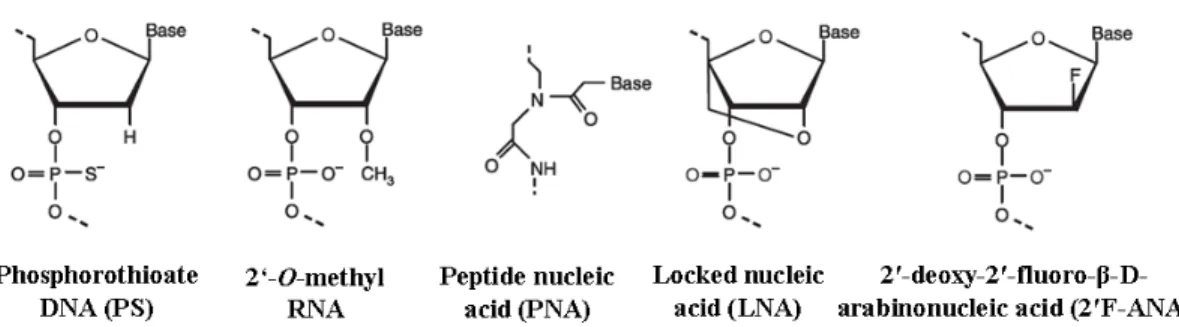
Among all ON derivatives, phosphorothioate (PS) analogs have been widely studied (Figure 3)79,84. In PS modifications, one of the non-bridging oxygen atoms of the phosphodiester linkage is substituted by a sulfur atom. PS modifications confer moderate resistance against nuclease degradation and increase the half-life in human serum compared to the native ON. The major disadvantage of the PS modification is that the sulfur atom promotes non-specific binding to certain proteins, which can cause severe adverse reactions (especially at high doses)82,88. Another shortcoming is the reduced affinity towards the mRNA compared to the phosphodiester ON82,88.
Regarding sugar modifications, 2'-O-methylation is one of the most widely- studied sugar modifications for both AON and siRNA (Figure 3) because it increases their nuclease stability79,84. However, many groups have found that large numbers of 2'-O-methyl modifications do not induce the RNase H endonuclease activity and decrease siRNA potency89,90. Other modifications of ribose sugar include peptide nucleic acid (PNA) and locked nucleic acid (LNA) (Figure 3)73,79. PNA is a synthetic DNA in which the deoxyribose phosphate backbone is replaced by polyamide linkages. LNA is a conformationally restricted nucleotide containing a 2'-O, 4'-C-methylene bridge in the β-D-ribofuranosyl configuration. Both PNA and LNA can bind to complementary DNA or RNA with high affinity and specificity. In addition, they have high stability against nuclease degradation. However, both PNA and LNA do not elicit target RNA cleavage by RNase H. Therefore, their applications might be limited as antisense agents and they can be exploited to modulate gene expression91.
Alternatively, 2′-deoxy-2′-fluoro-β-D-arabinonucleic acid (2′F-ANA) sugar modifications of the ON have been found to maintain high intracellular
concentrations for prolonged periods of time leading to long-term gene silencing (Figure 3)92. This was explained by increased serum stability and higher binding affinity to the target mRNA. The 2′F-ANA modifications have been successfully applied to both AON and siRNA78. They are well-tolerated throughout both sense and antisense strands of siRNA92-94. Although the use of modified nucleic acids has partially overcome the problem of rapid degradation, most chemical modifications are partially ineffective in reducing their renal clearance or improving their intracellular and site-specific delivery.
Figure 3. Chemical structure of PS DNA, 2′-O-methyl RNA, PNA, LNA and
2′F-ANA
Likewise, conjugates have been employed to enhance transfection efficiency and resistance to nucleases. Covalent conjugation to hydrophilic polymers (e.g. PEG) or lipophilic moieties (e.g. cholesterol) have been shown to improve the pharmacokinetics and enhance the efficacy of many nucleic acids95-97. For instance, conjugation to cholesterol has been reported to slow ON clearance95,98 and enhance cellular association and transport96. However, these conjugates are mainly taken up
by the liver by interacting with lipoprotein particles97, and their applications to target other tissues are limited.
2.2 Viral vectors
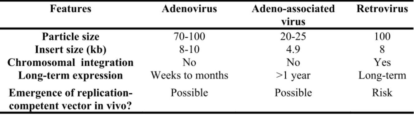
Viruses are composed of an envelope or capsid that contains genetic material (DNA or RNA) in a compact form. They possess sizes ranging from 20 to 100 nm99. A variety of these viruses have been converted to vectors to deliver genes to cells (e.g. adenoviruses, retroviruses and adeno-associated viruses). Some viruses and their characteristics are presented in Table 3. Although a detailed discussion of viral vectors in nucleic acid delivery is beyond the scope of this introduction, it is worth mentioning that they have been implicated in 70% of gene therapy clinical trials99. Indeed, viral vectors offer superior transfection efficiency than their synthetic counterparts, can infect most kinds of cells and are among the first carriers adopted for the delivery of different nucleic acid materials. Nevertheless, their use is limited because of inherent safety concerns. The use of viral vectors for gene therapy can be associated with severe inflammation and immunological problems100,101. The toxicity of viral vectors is usually due to random integration of the transported genes99,102. In addition, the size of the DNA and the type of the genetic material that can be encapsulated into viral vectors restrict their applicability. Hence, there is a need for alternative synthetic approaches for the delivery of nucleic acids.
Table 3. Characteristics of viral vectors. Adapted from reference99.
Features Adenovirus Adeno-associated virus Retrovirus
Particle size 70-100 20-25 100
Insert size (kb) 8-10 4.9 8
Chromosomal integration No No Yes
Long-term expression Weeks to months >1 year Long-term
Emergence of replication-competent vector in vivo?
Possible Possible Risk
2.3 Non-viral vectors
2.3.1 Non-complexing polymers
Amphiphilic copolymers such as, poloxamers, have been exploited as non-viral gene vectors to deliver different genetic materials103. Poloxamers consist of linear triblock copolymers with an A-B-A structure ((ethylene oxide)x-(propylene oxide)x-(ethylene oxide)x). For instance, poloxamer 407 has been used to deliver AON by intravenous injection into mdx mice for the treatment of Duchene muscular dystrophy (DMD). DMD is a degenerative muscle disease caused by the low expression of dystrophin protein in skeletal muscles evoked by a mutation in the dystrophin gene. Repeated injections of this formulation enhanced dystrophin induction in skeletal muscles by restoring the dystrophin gene104.
Polymeric nanocapsules have also been deployed for the delivery of nucleic acids105. This method was developed by Lambert et al.106 who prepared poly(isobutylcyanoacrylate) nanocapsules containing an aqueous core. The incorporation of AON into the aqueous core of nanocapsules improved their stability
against enzymatic degradation and increased their serum half-lives as compared to naked AON. Alternatively, the incorporation of AON and siRNA into the aqueous core of non-ionic polymersomes was achieved recently by Discher and coworkers107. The exact mechanism of interactions between these charged (i.e. non-complexing) polymers and nucleic acids is still unclear and needs further investigation. Indeed, the delivery of nucleic acids by complexing them with cationic lipids and polymers has stirred more interest and has produced promising results.
2.3.2 Lipoplexes and polyplexes
A common approach to enhance the stability of nucleic acids and improve their transfection efficiency consists in complexing them with positively-charged lipids and polymers to form lipoplexes and polyplexes, respectively. Both cationic lipids and polymers (e.g. polyethylenimine108,109 (PEI) and poly(amidoamine)110,111 (PAMAM)) have been studied as non-viral gene carriers because of their ability to protect DNA/RNA from enzymatic degradation and to increase cellular uptake by adsorptive endocytosis. Usually, excess positive charges are required for high transfection efficiency. Although, lipoplexes and polyplexes are considered to be the most promising candidates for non-viral gene delivery systems, they are rapidly inactivated in the presence of serum. This inactivation is probably elicited by interactions with negatively-charged serum proteins which shield the positive charges on the surface of lipoplexes and polyplexes112. Moreover, after intravenous injection, the large size and positive character of these complexes result in rapid opsonization, clearance from the circulation and the risk of occlusion of lung capillaries. In many situations, intravenous injection of those complexes led to damage of body tissues