1/. 33
. /ôUniversité de Montréal
Étude de l’association entre la fréquence cardiaque au repos et la survie
Par
Ariel Horacio Diaz
Département de Sciences Biomédicales faculté de Médecine
Mémoire présenté à la Faculté des Études Supérieures en vue de l’obtention du grade de
Maître ès Sciences (M.Sc.) en Sciences Biomédicales
option recherche clinique
Août 2005 ©Ariel H. Diaz, 2005
C
C
J
Universif
de Montréal
Direction des btbliothèques
AVIS
L’auteur a autorisé l’Université de Montréal à reproduire et diffuser, en totalité ou en partie, pat quelque moyen que ce soit et sur quelque support que ce soit, et exclusivement à des fins non lucratives d’enseignement et de recherche, des copies de ce mémoire ou de cette thèse.
L’auteur et les coauteurs le cas échéant conservent la propriété du droit d’auteur et des droits moraux qui protègent ce document. Ni la thèse ou le mémoire, ni des extraits substantiels de ce document, ne doivent être imprimés ou autrement reproduits sans l’autorisation de l’auteur.
Afin de se conformer à la Loi canadienne sur la protection des
renseignements personnels, quelques formulaires secondaires, coordonnées ou signatures intégrées au texte ont pu être enlevés de ce document. Bien que cela ait pu affecter la pagination, il n’y a aucun contenu manquant.
NOTICE
The author of this thesis or dissertation has granted a nonexciusive license allowing Université de Montréal to reproduce and publish the document, in part or in whole, and in any format, solely for noncommercial educational and research purposes.
The author and co-authors if applicable retain copyright ownership and moral rights in this document. Neither the whole thesis or dissertation, nor substantial extracts from it, may be printed or otherwise reproduced without the author’s permission.
In compliance with the Canadian Privacy Act some supporting forms, contact information or signatures may have been removed from the document. While this may affect the document page count, it does flot represent any Ioss of content from the document.
faculté des études supérieures
Ce mémoire intitulé
Étude de l’association entre la fréquence cardiaqueau repos et la survie
présenté par Ariel Horacio Diaz
a été évalué par un jury composé des personnes suivantes
Jean-Claude Tardif directeur de recherche Marie-Claude Guertin codirectrice de recherche Jean Lambert président rapporteur Mario Talajic membre du jury Mémoire accepté le
111
SOMMAIRE
La valeur pronostique d’une fréquence cardiaque au repos (FCR) basse chez les patients avec
angine chronique stable (ACS) est inconnue. La réponse de la fréquence cardiaque (FC) à l’effort pourrait également nous donner des informations additionnelles sur ces patients.
Les objectifs de ce mémoire sont d’étudier la valeur pronostique de la FCR chez les patients avec ACS et d’explorer la relation entre les différentes mesures du comportement de la fC à l’effort et la mortalité totale et cardiovasculaire (CV) à long tenue.
Nous avons obtenu la FCR de 24 913 patients du registre Coronary Artery Surgery Study (CASS) avec soupçon ou diagnostique de maladie coronarienne athéromateuse (MCAS) avec un suivi médian de 14,7 ans. Les risques de mortalités totale et cardiovasculaire étaient augmentés avec une f CR plus élevée (p< 0,0001). Lors de la comparaison des patients avec une FCR 62 bprn, les patients avec une FCR 83 bpm avaient un risque significativement augmenté de mortalité totale (hazard ratio = 1,32; 99% Cl 1,19-1,47) et de mortalité CV (hazard ratio = 1,31; 99% CI
1,15-1,48) après ajustement pour plusieurs variables cliniques. Ils avaient aussi un taux de réhospitalisation pour causes CV élevé (hazard ratio 1,14; 99% CI 1,02-1,27).
2 793 patients du même registre ont subi une épreuve d’effort tEE). L’incompétence
chronotropique exprimée comme <80% de la réserve de la FC (HRRes) était un prédicteur
significatif de mortalité totale et CV (p <0,0001). Les patients avec un %HRRes élevé avaient un
risque significativement plus bas de mortalité totale et CV (hazard ratio pour chaque augmentation d’un écart type = 0,79; 95% CI 0,71-0,88 pour les deux types de mortalité) après
ajustement pour plusieurs variables cliniques. Le %HRRes était la mesure du comportement de la FC à l’effort qui contribuait le plus aux modèles.
La FCR est un prédicteur indépendant de la mortalité totale et CV chez les patients avec MCAS. Parmi les diverses mesures de FC à l’effort, le %HRRes est le meilleur prédicteur de mortalité totale et CV chez ces patients et il devrait dorénavant être inclus dans tous les rapports d’épreuves d’effort.
Mots clés : fréquence cardiaque au repos, pronostique, maladie coronarienne, incompétence
chronotropique, réserve de la fréquence cardiaque, épreuve d’effort, mortalité, mortalité cardiovasculaire.
V
ABSTRACT
Low resting heart rate (RHR) lias unknown prognostic value in patients with chronic stable angina (CSA). Additionally, heart rate response to exercise could provide further useful information in such patients.
The goals ofthis thesis are to study the prognostic value ofRHR in a population of patients with CSA and to explore the reiationship between different heart rate measures during exercise treadmill test (ETT) and long-tenn total and cardiovascular (CV) mortality.
We obtained tlie RHR of 24 913 patients from the Coronary Artery Surgery Study (CASS) database with suspected or proven coronary artery disease and a median follow-up of 14.7 years. Patients with resting heart rate ? 83bpm at baseline had a significantly higher risk for total (hazard ratio = 1.32, 99% CI 1.19-1.47) and CV mortality (hazard ratio = 1.31, 99% CI
1.15-1.48) afler adjustment for multiple clinical variables compared to the reference group. The hazard ratio for time to first CV rehospitalization in these patients was 1.14, 99% CI 1.02-1.27.
A total of 2 793 patients from the same registry underwent ETT. Chronotropic incompetence expressed as a low (<80) % heart rate reserve (HRRes) was a predictor for all-cause and CV mortality Q, <0.0001). Patients with a higher %HRRes liad a significantly lower risk of total and CV mortality (hazard ratio = 0.79, 95% CI 0.71-0.88 for both outcomes for each increase ofone
standard deviation) after adjustment for multiple clinical covariates. Out of ail exercise heart rate measurements, %HRRes contributed the most to the models.
RHR is a strong, independent predictor for total and CV mortality in patients with coronary artery disease. % HRRes is the best heart rate predictor during exercise for total and CV mortality in these patients and it should be routinely included in ail ETT.
Key words: resting heart rate, prognosis, coronary disease, chronotropic incompetence, heart rate reserve, exercise treadmiil test, mortaÏity, cardiovascular mortality.
vii
INDEX
SOMMAIRE iii
ABSTRACT y
LIST 0F TABLES xi
LIST 0F FIGURES xii
LIST 0F ABBREVIATIONS xiv
ACKNOWLEDGEMENTS xvi
CHAPTER 1: INTRODUCTION 2
1.2 Historical background 2
1.3 Rationale for the study ofthis association between heart rate and rnortality 6
1.3.1 Phylogenetic perspective 7
1.3.2 Molecular perspective 9
1.3.3 Pathological perspective 9
1.3.4 Clinical perspective 11
1.3.5 Genetic perspective 12
1.3.6 Previous epidemiologic studies assessing the relationship 14 1.4 Pathophysiology of the association between heart rate and cardiovascular events 16
1.4.1 Autonomic disbalance 16
1.4.2 Tachycardia as an independent mechanism ofrisk for mortality 17
1.5 Heart rate and exercise 18
1.5.2 Age-predicted maximal heart rate 20
1.5.3 Heartrate recovery 20
1.5.4 Percentage ofheart rate reserve 21
1.6 General goals of this thesis and hypothesis to be tested 23
CHAPTER 2. METHODS 25
2.1.1 Database and study group description 26
2.1.2 Jriformed consent 2$
2.1.3 Baseline data collection and variables description 2$
2.1.4 Patients follow-up 32
2.2 Statisticalanalyses 33
2.2.1 Variable selection and treatment 33
2.2.2 Descriptive statistics 36
2.2.3 Survival analysis 36
CHAPTER 3. LONG TERM PROGNOSTIC VALUE 0F RESTING HEART RATE IN PATIENTS WITH SUSPECTED OR PRO VEN CORONARY ARTERY DISEASE 38
Abstract 40
Introduction 41
Methods 41
Resuits 45
ix
Conclusions .49
References .50
Tables 53
Figures 59
CHAPTER 4. PERCENT HEART RATE RESERVE DURING EXERCISE HAS
INDEPENDENT PROGNOSTIC VALUE IN PATIENTS WITH SUSPECTED OR
PROVEN CORONARY ARTERY ifiSEASE 65
Abstract 67 Introduction 70 Methods 71 Results 75 Discussion 77 Conclusions 80 References $1 Tables 86 figures 90
CHAPTER 5. DISCUSSION AND CONCLUSIONS 93
5.1 Main resuits and original contribution 94
5.2 Confounding variables 94
5.2.1 Ejection fraction .96
5.3 Study limitations 97
5.4 General conclusions 98
CHAPTER 6. REFERENCES 99
CHAPTER 7. ANNEXES 108
7.1 Autorisation de déposer ce mémoire de maîtrise sous forme d’articles 7.1.1 Autorisation des coauteurs
xi
LIST 0F TABLES
TABLE 1-I Epiderniologic studies assessing resting heart rate and total or
cardiovascular mortality 15
TABLE 2-I List of variables 35
TABLE 3-I Descrtption of variables used in this study 53
TABLE 3-II Baseline characteristics divided by restingheart rate in quintiles 54
TABLE 3-III Multivariable Cox regression survival analysis for total inortaÏity 55
TABLE 3-IV Multivariabte Cox regression survival analysisfor cardiovascutar
inortatity 56
TABLE 3-V Muttivariable Cox regression analysis for time to rehospitalization
due to any cardiovascular cause or acute myocardiat iifarction 57
TABLE 3-VI Cox regression analysis for time to rehospitalization due to angifla,
stroke or congestive heartfailure 5$
TABLE 4-I Definition of variables in article two $6
TABLE 4-II Baseline characteristics by chronotropic incompetence 87
TABLE 4-III Wald test results ofdfferent heart rate pararneters and number of
METS duriizg exercise 88
TABLE 4-IV Multivariate Cox regression survival analysis for total and
LIST 0F FIGURES
FIGURE l-1 Ebers papyrus 3
FIGURE 1-2 The pulse classic 4
FIGURE 1-3 Exercitatio Anatomica de Mottts Cordis et Sanguinis
ut
animatibus 5FIGURE 1-4 Man’ey sphygmograph 6
FIGURE 1-5 Semilogarithrnic relation between Ïieart rate and lfe expectancy in
maminals 7
FIGURE 1-6 Relation between lfe expectancy and total heartbeats in a
LiJetiine antong inanunaïs 8
FIGURE 1-7 Autonontic inibalance and CVevents 17
FIGURE 1-8 The metabolic chronotropic relation 22
FIGURE 2-1 CASSpatientsflow diagram 27
FIGURE 2-2 Termiizology for coronaiy artely anatonly visualïzed at
angiography utilized by cooperating sites in C4$S 31 FIGURE 3-1 Adjusted survival curves for overail rnortality by resting heart
rate quintiles 60
FIGURE 3-2 Adjusted survival curves for cardiovasctdar ntortality by resting
heart rate quintiles 61
FIGURE 3-3 Adjusted survival curves for tinte to rehospitatization due to any
cardiovascular cause 62
FIGURE 3-4 Adjusted survival curves for tirne toflrst rehospitalization due to
congestive heartfailure 63
xiii
(12.4 bpm) ofresting heart rate .64
FIGURE 4-l Adjusted sun’ivalctcrves for total inortaÏity by
chronotropic incompetence 91
FIGURE 4-2 Adjusted survivalcurvesfor cardiovascutar mortality
LIST 0F ABBREVIATIONS
ACC / AHA: American College of CardioÏogy / American Heart Association ACS: Angine Chronique Stable
APMHR: Age-Predicted Maximal Heart Rate ATP: Adenosine Triphosphate
BMI: Body Mass Index
CAD: Coronary Artery Disease CASS: Coronary Artery Surgery Study CHF: Congestive Heart Failure
CI: Confidence Interval
CSA: Chronic Stable Angina
CV: Cardiovascular / Cardiovasculaire
DM: Diabetes Mellitus
EDV: Left ventricular End-Diastolic Volume ESV: Left ventricular End-Systolic Volume
EE: Épreuve d’Effort
EF: Ejection Fraction
ETT: Exercise Treadmili lest
FC: Fréquence Cardiaque
FCR: Fréquence Cardiaque au Repos
HDL: High-Density Lipoprotein
HRmax: Maximal Heart Rate during exercise HRRec: Heart Rate Recovery after exercise
xv
HRRes: Heart Rate Reserve during exercise
HTN: Hypertension
LAD: Left Anterior Descending Coronary Artery LCx: Lefi Circumilex Coronary Artery
LMCA: Left Main Coronary Artery
MCAS: Maladie Coronarienne Athéromateuse MCR: Metabolic chronotropic relation
MI: Myocardial Infarction
NCVD: Number of Clinically significant coronary Vessel Disease
PH: Proportional Hazard
QTL: Quantitative Trait Loci
RC: Right Coronary Artery
RHR: Resting Heart Rate
ACKNOLEDGEMENIS
Thank you Dr. Tardif for your support and dedication to ail of this. You are an example of physician, researcher and above ail of a mentor. Thank you for the impeccable review of each article ami for the timc you dedicated to each of them. Rack then you believed in me and I will neyer forget that.
Mane-Claude, yoti did everything easier with your judicious, wanii and friendly advice. Thank you very much for aiways answering ALL my biostats questions.. .You were aiways there ready to listen and encourage me. Tu avais raison, il fait bon vivre au Québec!
Dr. Bourassa you are a mode! ofwhat I consider a great researcher should be. Only your modesty surpasses your wisdom. Thank you for your intellectual contribution to this thesis.
Merci à tout l’équipe du centre de coordination de l’ICM (MHICC) pour votre amitié, pour toutes les tasses de café et chaque tape dans le dos. Spécialement merci à Sylvie Lévesque pour sa patience et temps à m’expliquer les innombrables doutes en biostatistique.
To my parents, who aiways guide and enlighten my way with their example. A mis padres, quienes siempre guan e iluminan mi camino con su ejemplo.
C
2
1. INTRODUCTION
Although measuring resting heart rate (RHR) is practically a reflex we ail learn in medical school, there are stiil many unanswered questions regarding its genuine value and prognosis. The modem physician faces state-of-the-art technology on daily basis. Preventive, diagnostic, therapeutic and reliabilitation medical acts Ïargely depend on sornetimes complicated and expensive procedures. Certainly, every clinician measures bis or ber patient’s heart rate. Nevertheless, it is the value we bestow to our patients’ heart rate which bas becorne nurnbed. UsualLy, physicians would only pay attention to beart rate in certain critical clinical conditions such as hemodynamic instability, hyperthermia, drug toxicity and the like. Yet, how rnuch does RHR influence the occurrence of cardiovascular events? Is there an intrinsic benefit to a lower RHR? What is the prognostic value of RHR in patients with stable coronary artery disease (CAD)? In addition, what is the prognostic value of heart rate during exercise? In this thesis I intend to further explore and describe the association ofRHR and survival in a general population
of patients referred for a coronary angiogram due to suspected or confirmed CAD. In addition,
the relationship between heart rate during exercise and long-term survival will be also assessed.
1.2 Historical background.
From the times ofthe Eberpapyrus 1700 BC, the pulse has been recognized as a benchmark for cardiovascular assessment (Figure 1-1). The papyrus stated as regards to the pulse “it is there that the heart speaks” establishing the connection between the heart and peripheral pulse.
•: =
4: ‘ç 1: ‘ L1 •: û. LEd .jIC-9o> UHt h ‘-—-‘i
L9Dr h sfigure 1-1 Ebers papyrus referring to the pulse, ca 1700 BC. Egypt. Courtesy of the National Library of Medicine ‘w. o çt III. O’r t ;is ‘jS
g
N6n
Furthermore, the observation of decreased heart rate variability as a predictor of sudden cardiac death has been known for centuries. The Chinese physician Wang Shuhe, (265—3 17 A.D.) wrote in The Pulse Classic, “If the pattem of the heart beat becomes as regular as the tapping of a woodpecker or the dripping of ram from the roof, the patient will be dead in four days”(l) (Figure 1-2). This observation already linked a decrease in heart rate variability to mortality.
4
It was not until 162$ that the western world acknowledged the connection between the peripheral arterial pulse and the heart. In that year Sir William Hawey (1578-1657) dedicated to Prince Charles I King ofBritain, france and Ireland bis famous “Treatise on The Motion oJThe Heart and Blood in Animais”. In that treatise, he wrote
“. .From these facts it is manifest, in opposition to commonly received opinions, that the diastole of the arteries corresponds with the time of the heart’s systole; and that the arteries are filled and distended by the blood forced into them by the contraction of the ventricles; the arteries, therefore, are distended, because they are filled like sacs or bladders”
Figure 1-2. Doctor Wang Shuhe writing The Pulse Classic, which is the earliest comprehensive
work dealing with several kinds of pulses and their diagnostic value (from Liu YC. The Essential Book of Traditional Chinese Medicine. New York: Columbia University Press, 198$: 4).
(2) (figure 1-3). The pulse has always been the center of much medical attention and extensive essays. Many illustrated physicians left their names attached to a pulse disorder such as Corrigan, Kussmaul and Riegel just to name a few. It was flot until 1860 that the first registry on paper of pulse was accurately achieved by Dr. Marey in Paris as he described an improved sphygmograph (3) (Figure 1-4). Nowadays, there is a whole array of readily available technology to evaluate the different aspects of heart rate, such as various types of Holter monitoring, pacemakers, implantable cardiac defibrillators, etc. Complex heart rate parameters are flot only evaluated at rest, but during exercise as well.
xERC1T4T[0
ANATOMICA DE
MOTV CORDES ET SAN
GVtNIS iN ANtMALI Çt’ILÎELM! U’%RVE[ 4NGLJ, lcdic,Re,if.%PrtfeJîr,uj!’°”inCoI
rT
-— -— FRÀVCOF1RT7. SurnpttbtiG’.’ IL EFI. MI PtTZ ERL ANNQ M, Z)C.XXVIII.6
1.3 Rationale for the study of the association between heart rate and mortatity.
There are some epidemiological population-based studies which have addressed the issue ofRHR and mortality (4-13). There are also several studies relating heart rate during exercise and prognosis using parameters such as heart rate recovery (HRRec), maximal heart rate (HRMax), percentage of heart rate reserve (%HRRes) or other similar chronotropic response indices as predictors ofcardiovascular events and mortality (14-24). However, these studies were performed mostly in healthy individuals and it is of great interest to further explore the relationship of heart rate and mortality in patients with coronary artery disease.
ï.. . I I.. r.,..I.
I,aI..%
4. b..q. —U..p...
.I. 4.. .44.. 4. I... 4.4., .
4.* UII4.I. 4. I.
Figure 1-4. Medical Instruments & Apparatus: Marey sphygmograph apparatus. Wood
1.3.1 Phylogenetic perspective
The ratio of heat loss (a function of body surface area) to heat production (a function of body mass) increases with smaHer body size in mammals. This is the basic principle behind the knowledge that smaller mammals have higher heart rates and shorter life spans than larger ones. From a phylogenetic point of view a landmark study was published by Levine et ai, illustrating that among mammals there is an inverse semilogarithmic relationship between heart rate and life expectancy (25). This equation however, exciudes humans (Figure 1-5). There are a few hypotheses to explain why humans escape from this relationship; nevertheless it is reasonable to attribute this in part to advances in medical sciences and socio-economical development. Conversely, the number of heartbeats in a lifetirne is remarkably constant despite a 40-fold difference in life expectancy (25) (Figure 1 -6). The noticeable inverse relation between life span and heart rate may reflect an epiphenomenon in which heart rate is a marker of metabolic consumption. 1000— 600 - Mose Hampsiet ._ • Makey Hea,rare \&4armot
beatalmin - D .\Cat Tiger Man -F4ose Elephant 20 -
\
eWhale Wbale 20 40 60 80Lite .xp.cancyy.ars
Figure 1-5. Semilogarithmic relation between rest heart rate and life expectancy in mammals. Most coordinates represent average values. From Levine HJ. Rest heart rate and life expectancy. J Am Cou Cardiol 1997;30(4):1104-6.
$ 100-— A 60 -. Whale 30 • Wh1IG Elephant •• Horse Clinexpnctancy,
years Ass’ Cat
Mopkcy ,‘Gitffe la •Tigr • Marmot 5— • Rat 2 Harnpster. • Mouse 1 I I I I 10 10 10’ W’ 10” 10” Beats Ilifetime
Figure 1-6. Relation between life expectancy and total heartbeats/lifetime among mammals. Levine HJ. Rest heart rate and life expectancy. J Am Cou Cardiol 1997;30(4):1104-6
Furthermore, Azbel et ai, in a mathematical essay, provides some insight into this mechanism(26). Drawing from a wide allometric scale of 1020-fold among living organisms, he conciudes that the energy consumption/body atom per heartbeat is the same (within an order of magnitude) in ail animais. Experimentai data verify this for dozens of species in ail taxes, from invertebrates to mammals, and even for oxygen-consuming bacteria. The mean deviation is only 1.7 oxygen molecules per body atom larger or smaller than the average (27). Therefore, energy expenditure seems constant in ail animais and RHR is directly involved in energy production mainly in a regulatory fashion. Hence, there is a phylogenetic rationale for looking at this relationship.
1.3.2 Molecular perspective
The ideas exposed above lead to a discussion of the molecular perspective. In the heart, adenosine triphosphate (ATP) is synthesized by mitochondria from a variety of aerobic substrates. ATP is generated from -oxidation of lipids (60-70%) and from carbohydrates including exogenous glucose and lactate. Approximately 60-70% of total ATP is used for contraction, incÏuding Ca re-uptake by the sarcoplasmic reticulum and extrusion of Ca to the cytoplasm via the sarcolemmal Na/Ca and Ca pump. It follows that each heartbeat lias its own cost: 1.35 x 10¹⁹ Ca ions have to be mobilized and approximately 300 mg ofATP are used. Thus, the heart produces and utilizes approximately 30 Kg of ATP eacli day, and siowing its rate by 10 heartbeats/min would resuit in saving of about 5 Kg of ATP in a day and its oxidative stress byproducts (28).
1.3.3 Pathological perspective
Direct evidence of the importance of RHR on the progression of coronary atherosclerosis cornes from animal studies. Beere et al reported that lowering heart rate by sinoatrial node ablation
retarded diet-induced atherosclerosis in primates (29). The development of coronary
atherosclerosis was analysed in three groups of cynomolgus monkeys: one control group, one group that underwent sinus node ablation and one group that underwent a sham operation. All animals received an atherogenic diet for 6 months. Atherosclerosis severity at the end of the study in monkeys with a high RHR, was twice than in monkeys with a low RHR (mean percentage stenosis 56 ± 23% vs. 26 ± 19%, mean plaque area 0.48 ± 0.47 mm2 vs. 0.21 ± 0.39 mm2) These differences were statistically significant despite similar blood pressure, serum lipid levels and body weight. Furthermore, Kaplan et al have shown that monkeys with spontaneously
10
high RHR have twice the extent of lesions in the coronary arteries as animais with Ïow RHR, when fed a diet moderately high in saturated fats and cholesteroi for a period of 22 months (30). This association between RHR and atherosclerosis was also studied in liumans. Perski et al have observed, in a small group of young patients after myocardial infarction, that minimum and average heart rate measured by a 24-h ECG are strongly related (r 0.70, p <0.002 and r= 0.59
p, < 0.01 respectively) to the severity of coronary atherosclerosis. This association was found
even after correcting for several established risk factors such as smoking habits, blood pressure, body mass index (BMI) and serum lipoprotein concentrations (31). The same authors then performed a more complete and larger study. The relations of hemodynamic factors, plasma fibrinogen concentration, serum lipoprotein levels, and clinical risk indicators with CAD were studied in 56 men who had survived a first myocardial infarction before the age of 45 and who subsequently underwent two coronary angiograms with an intervening interval of 4 to 7 years (32). High mean RHR measured during a 24-hour period was associated with progression of diffuse lesions (r=0.34; p <0.05) and distinct stenosis (r=0.43; p <0.01). A high RHR also correlated positively with progression of angiographie score of the global severity of coronary atherosclerosis (r=0.36; p <0.01). In this study an increase in the RHR of 5 beats/min corresponded to an increase in global atherosclerosis progression score of 0.21 (95% confidence interval 0.17-0.24). Progression of disease was predicted independently by minimum heart rate and low-density lipoprotein!high-density tipoprotein ratio. In contrast, lipoprotein A, fibrinogen level, hypertension, smoking, and beta-adrenergic receptor blockade treatment did not discriminate between patients with and without progression. In a retrospective study, Heidland et
al analyzed 106 patients who undenvent 2 coronary angiograms within 6 months (33). They
investigated 53 patients with initially smooth stenoses who developed plaque disruption by the time of the second coronary angiogram and compared these patients with 53 age- and sex
matched individuals with srnooth stenoses without angiographic signs of plaque disruption. Logistic regression analysis identified positive associations between plaque disruption and both lefi ventricular muscle mass >270 g and a mean heart rate >80 bpm and a negative association with the use ofbeta-blockers.
1.3.4 CIinical perspective
Epidemiological studies have shown that, among patients without known heart disease, elevated RHR is related to increased risk of all-cause, CV and sudden cardiac death. Additional indirect evidence of the relationship between RHR and mortality corne from studies evaluating the effects of f3-adrenergic receptor antagonists. The relationship between reduction in heart rate and a decrease in rnortality has been weIl established with beta-blockers after myocardial infarction and in patients with heart failure (34-37). When 3-blockers were classified according to their pharmacological properties, those with intrinsic sympathomimetic activity appeared to have reduced efficacy in comparison to those without such an activity. The capacity ofbeta-blockers to reduce heart rate might therefore be an important determinant of their cardioprotective effect. The relationship between heart rate slowing and reduction in clinical events is not restricted to beta blocker therapy. In the Grupo de Estudio de la Sobrevida en la Insuficiencia Cardfaca en Argentina (GESTCA) study, a decrease in mortality was observed in a subgroup of congestive heart failure (CHF) patients with elevated resting heart rate (>90 beats/min) when treated with amiodarone (38). This beneficial heart rate siowing effect of amiodarone was independent of the patients’ arrhythmic profile. On the other hand, calcium channel blockers which lower RHR (mainÏy diltiazem and verapamil) have been shown to slightly decrease the risk ofdeath and
non-12
fatal reinfarction in survivors of acute rnyocardial infarction without clinical heart failure or impaired left ventricular function (39, 40).
Heart rate has been also related to insulin sensitivity and insulin secretion in non-diabetic patients. lii a multiple regression analysis (adjusting for age, sex, ethnicity, glucose tolerance status and smoking), heart rate was significantly and independently associated with acute insulin response to glucose, proinsulin and insulin sensitivity in non-diabetic individuals (41). The relation between heart rate and lipid levels has also been studied (42). In both sexes, there was a progressive increase in age-adjusted levels of total cholesterol, non-high density lipoprotein (HDL) cholesterol and triglycerides and a decrease in HDL cholesterol with increasing heart rates. The associations remained significant when anthropometric and life-style factors were controlled for in the statistical analyses. These findings support the conception that RHR in itself is linked to clinical events.
1.3.5 Genetic perspective
The interaction between RHR and sunrival may also have a genetic basis. A cohort of> 1000 individuals of Chinese and Japanese descent was genotyped for two polymorphisms, resulting in a serine/glycine substitution at amino acid 49 (Ser49Gly) and an arginine/glycine substitution at residue 389 (Arg389Gly), in the beta-1 adrenergic receptor (43). 13-1 polymorphism has been associated with many cardiovascular disorders such as hypertension and others. for comparison, polymorphisms in the beta2 and beta3 adrenergic receptors were also evaluated. The Ser49Gly polymorphism was significantly associated (P=0.0004) with resting heart rate, independent of other variables, such as body-mass index, age, sex, etlmicity, exercise, smoking, alcohol intake,
hypertension status, and treatment with beta blockers. The data support an additive model in which individuals heterozygous for the Ser49Gly polymorphism had mean heart rates intermediate to those of either type of homozygotes, with Ser homozygotes having the highest mean heart rate and with Gly homozygotes having the lowest. 73.8% of patients were Ser homozygous, 23.5% were Ser49Gly heterozygous and 2.7% were Gly homozygous. Neither the Arg389Gly polymorphism in the betal adrenergic receptor nor polymorphisms in the beta2 and beta3 adrenergic receptors were associated with RHR. The authors estimated the heritability of RHR at 39.7% (p <0.00 1), which is similar to the one reported from the Framingham population (44). Wilk et al performed a genome scan for quantitative trait loci (QTL) influencing the resting heart rate among 962 Caucasians and 1124 African-Americans in the Hypertension Genetic Epidemiology Network (HyperGEN), a multi-center study of genetic and environmental factors related to hypertension (16). Within each race and sex, heart rate was adjusted for covariates, including age, study center, body mass index, beta-blocker use, alcohol consumption, smoking, number of blocks walked per day, and number of hours watching television. The evidence for linkage found on chromosome 4 in both Caucasian and African-American hypertensive sib pairs indicates that further investigation on that region may be warranted to locate a gene influencing RHR (16). Martin et al (45) estimated the heritability of REIR on chromosome 4q in healthy humans. This signal is in the same region as a QTL for long QT syndrome 4 and a QTL for heart rate in rats (45). furthermore, QTL for heart rate have also been identified in drosophila (46) and mice (47). Nevertheless, most of animal and human studies were done in hypertensive populations and further work in this area and in different populations is therefore needed.
14
1.3.6 Previous epidemiological studies assessing this relationship
The robust relationship between resting heart rate and cardiovascular events has been documented in numerous epidemiological studies generally involving healthy populations. (4-6, 9, 11, 12) (Table 1-I). Four studies included only men (8, 10, 48, 49). In addition, Palatini et aï
(50-55) have examined the association between RHR and all-cause and cardiovascular mortality in different populations and reported a positive relationship between tachycardia and mortality in women, in patients with hypertension, with metabolic syndrome and also in elderly patients. However, this association has neyer been studied in an unselected population of patients with suspected or confirmed CAD.
Table 1-I. Epidemiologic studies assessing RHR and total or CV mortality. Author [StudyJ E ntry Sample Years of Heart rate Study outcorne (reference) year size Gender J age foflow-up measurement RR or HR (95%CI) Kannel 194$ 5 070 Menlwomen 36 Supine ECG CVD3rate
p<O.00l
[Framingham] 35_94
(4)
Reunanen (5) 1966 10 717 Men/women 23 ECG Men” >84 bpmRR 1.40
(1.11-30-59 1.77) Women’’ >94 bpm RR
1.40 (1.00-1.96) Mensink 1982 4 756 Men1women 12 Radial pulse x 2 Menc HR 1.7 (1.2-2.6)
ISpandau] (6) 40-80 Womenc HR 1.3 (0.9-2.0)
Benetos (9) 1974 19 386 Meni’women 1$ Supine ECG Men” RR 2.18 (1.37-3.47)
40-69 Womend RR 1.46 (0.64-3.33)
Dryer [Chicago 1958 1 233 Mea 40-59 15 ECG Alla cause mortality rate
People GasJ (13) p <0.00 1
Dryer [Chicago 1957 1 899 Men 40-55 17 Radial pulse AW’ cause rnortality rate
Western p<O.00l
ElectricJ (13)
Dryer [Chicago 1967 33 781 Men/women 22 ECG Alla cause mortality rate
Heart 18-74 p<O.00l
Associationj (13)
GiIlum 1971 6 672 Men/women 10 Radial pulse Mene RR 1.44 (1.08-1.92)
INHANES IJ (12) 25-74 Womene RR 1.26 (0.89-1.76)
Seccareccia 1984 2 533 Men 40-69 4-13 ECG CVDHR 2.54 (1.25-5.16) [MATISSJ (10)
Jouven [Paris 1967 7 079 Mea 42-53 23 Radial pulse death RR 1.28
(1.06-Prospective 1.61)
Study 1] (7)
Kristal-Boneh 1985 3 527 Men mean age $ ECG CVD” RR 2.02 (1.1-4.0)
[CORDISJ (49) 43
Fujïura (8) 1977 573 Men 40-64 18 ECG Ail’ cause mortality HR 3.71 (1.35-10.22)
Bpm=beats per minute, C1= confidence interval, CVDcardiovascular death, ECG=elecfrocardïogram, HR=hazard ratio, RR=relative risk.
a ANOVA. Highest heart rate quintile compared to lowest, age adjusted. b CVD, age adjusted
C CVD adjusted for age, semm cholesterol, body mass index, systolic blood pressure, smoking and diabetes. d CVD in highest heart rate qumtile compared to lowest adjusted for age, hypertension, previous myocardial infarction, antihypertensive treatment, total choiesterol, physical activity and tobacco consumption.
e CVD data adjusted for age, smoking, blood pressure, cholesterol and other confounders, for heart rate >84 bpm in comparison with subjects with >74 bpm.
f >90 bpm compared to <60 bpm adjusted for age, systolic biood pressure, serum cholesterol, smoking, body mass index, arm cfrcumference, forced expiratory volume, diabetes and preexisting cardiovascular disease.
g highest heart rate quintile compared to iowest, adjusted for age, body mass index, systolic biood pressure, tobacco consumption, parental lustory of myocardial infarction and sudden death, cholesterol level, diabetes and recreational activity.
h >90 bpm compared with <70 bpm adjusted for age, recreational acfivity, systolic blood pressure and low density lipoproteins.
>90 bpm compared with <70 bpm adjusted for age, diastolic biood pressure, serum choiesterol, uric acid and antihypertensive medication.
16
1.4 Pathophysiology of the association between heart rate and cardiovascular events.
1.4.1 Autonomic imbalance.
One of the most interesting aspects of recent analyses in cardiovascular epidemiologic studies is the realization that increased RHR is frequently associated with high blood pressure, obesity, insulin resistance, dyslipidemia, and elevated hematocrit (41, 51, 56, 57). Syrnpathetic overstimulation could explain the interrelationship between the above risk factors and heart rate. In animal models, chronic 3-adrenergic stimulation causes conversion from a small to a larger proportion of insulin-resistant fast twitch muscle fibers. Acute adrenergic receptor-mediated vasoconstriction shunts the nutritional blood flow away from the active glucose-metabolizing celis in the skeletaÏ muscles, decreasing the ability of these celis to utilize glucose, adding another important factor in the insulin resistance scenario (56). Jncreased plasma insulin and insulin resistance are in epidemiologic studies associated with dyslipidemias and the mechanisms by which high insulin could cause dyslipidemias have been described (58, 59). Facchini et al found that insulin resistant individuals, with compensatory hyperinsulinemia, have a higher noctumal heart rate (60). Patients with sympathetic overstimulation have higher hematocrit. An elevated hematocrit is associated with an increased risk of coronary morbidity as well (61).
When markers of vagal tone are reduced, the risk of death is increased. This has been demonstrated for baroreflex sensitivity, heart rate variability, heart rate turbulance (T-wave alternans) and heart rate recovery afier exercise. This last parameter has been proven to be independent of the severtiy of coronary artery disease, suggesting alternative mechanisms (17). Jouven et al followed 5 713 healthy men for 23 years (24). $ubjects with a decrease in heart rate
1.4.2 Tachycardia as an independent mechanism of risk for mortality.
The intensification of the pulsatile nature of the arterial blood flow associated with tachycardia may favor the occurrence of injury to the endothelium (62-64). Also, in the natural history of CAD, plaque destabilization and rupture are key phenomena that ofien lead to clinical events. In addition to the link between RIIR and atherosclerosis progression that we have already described, the higher number of repeated small stresses on a potentially vulnerable atherosclerotic plaque recovery after exercise had a relative risk of 2.20 (95% CI 1.04-4.74) for sudden death, suggesting an increased risk for fatal anhythmias in this population.
1$
may increase the likelihood of rupture because of the well-known concept of fatigue of a biomaterial (65).
As presented above, there are epidemiological, physiological, metabolic, phylogenetic, molecular, clinical and genetic reasons to further study and explore the relationship between RHR and prognosis.
1.5 Heart rate and exercise
The use ofexercise stress testing as a means ofcardiac evaluation has developed slowly over this century. Progressive exercise is viewed nowadays as a method of physiologic evaluation since it provides an index of maximal oxygen uptake or aerobic capacity, which is in itself correlated with heart rate at submaximal exercise. Although exercise stress testing continues to be predominantly used for screening and management of coronary artery disease, it is also valuable in many other cardiovascular settings such as congestive heart failure, cardiovascular fitness, exercise-induced arrhythmias, valvular heart diseases and the evaluation of chronotropic response. Over the past years, exercise treadmili testing (ETT) has become more of a predictive than a diagnostic tool (66). The huge number of patients in a variety of settings who have undergone an ETT, made it possible to successfully identify powerful markers of risk. The performance of different heart rate parameters during ETT has been extensively studied, especially in healthy populations. In this thesis, I will attempt to further examine the application, clinical relevance and added prognostic value of diverse heart rate parameters during ETT in a population of patients referred for a coronary angiogram due to suspected or confirmed CAD. I will focus on four measures that are routinely available in the vast majority of ETTs: maximal
heart rate (ERmax), heart rate recovery (HRRec) age-predicted maximal heart rate (APMHR) and finally percentage ofheart rate reserve (% HRRes).
1.5.1 Maximal heart rate during exercise treadmiil test
In the early 1970s Ellestad tested a 51-year-old athietic man who had anormal exercise tolerance and no symptoms or ST-segment depression during exercise (67). A short time after the exercise test, lie suffered sudden cardiac death and an autopsy revealed severe two-vessel coronary disease with an 80% left anterior descending artery stenosis. 0f note, the patient had only reached a maximum heart rate of 110 beats per minute during the ETT. Triggered by this episode, Ellestad and his coïleagues examined a EIT database with 2 700 patients and found that those with a blunted heart rate response were at greater risk for a cardiac event than those with ischemic SI depression (6$). During the past two decades, exercise capacity, chronotropic incompetence and activity status have become well-established predictors of cardiovascular and overail mortality (21, 67-70). An impaired chronotropic response has been shown to be predictive of ah-cause mortahity and risk of developing CAD, even afier accounting for age, physical fitness, standard cardiovascular risk factors, left ventricular EF and myocardial ischemia (21, 23, 67, 71, 72). Chronotropic incompetence could be an indicator for decreased heart rate variabihity, a prognostically important manifestation of autonomic dysfttnction. In a recent article, Jouven et al reported that patients with a HRmax <89 bpm have a 6.18 (2.37-16.11) relative risk ofsudden death (24). Maximal heart rate during ETT lias its limitations since it is influenced by the patient’s age and physical fitness. It is well known that age is one of the most important determinants of the exercise heart rate response, with maximal peak heart rate dechining with older age.
20
1.5.2 Age-predicted maximal heart rate (APMHR)
APMHR is undoubtedly the most used chronotropic response measure in ETI laboratories throughout the world. APMHR is calculated as: 220 minus age. The ACC/AHA 2002 guidelines update for stress testing (73) defines chronotropic incompetence as the failure to achieve 85% of APMHR. Nevertheless, the history and validation of this parameter is somewhat peculiar. Most cardioÏogy and exercise physiology textbooks do not have an original citation for this 85% cutoff, nor for the 220 minus age formula. A nicely written review of the history of the “HRmax=220-age” was published in the Journal of Exercise Physiology (74). This formula was flot developed from original research but rather was the result of the review of 11 references and unpublished scientific compilations (74). Additionally, its standard deviation is quite high (Sxy=7-1 1 bpm). Therefore, given the poor validation and scarce original research there is no solid scientific value for the use of this formula in exercise physiology.
1.5.3 Heart rate recovery (HRRec)
HRRec is thought to be primarily a vagal phenomenon, and because decreased vagal activity is associated with increased risk of death, a lot of attention has been paid to this measure. HRRec has been found to be a strong marker of CV mortality and CV events flot only in healthy subjects (20, 75) (both men and women) but in various populations, such as in elderly, diabetics, patients with CHF, CAD, myocardial infarction and dyslipidemias (19, 20, 75-88).
Jouven et al recently reported a relative risk for sudden death of 2.20 (1.02-4.74) for patients with insufficient HRRec. HRRec is computed as HRmax minus heart rate post-exercise (usually one, two or three minutes after finishing exercise). There is no clear definition of when to take heart rate after exercise. Most studies have used arbitrary times of 1 or 2 min. In our population
evaluated in the mid 1970s, this concept had flot been tackled and heart rate was recorded 3 minutes after completion of the ETT. Other studies have found a predictive value of HRRec for ail-cause death even when measured as long as 5 min afler recovery ($4). It is nevertheless possible that heart rate parameters during the first 30 seconds after exercise may be more informative (89). Additionally, there is no consensus on the normal value of HRRec. The most common arbitrarily chosen value is more than 12 beats per min in 1 min. Only a few of the studies on HRRec have been restricted to symptomatic patients without revascularization. The population presented here is an unselected population of patients refened for coronary angiogram due to suspected or proven CAD and will therefore provide new important information about this parameter.
1.5.4 Percentage of heart rate reserve
The effects of age, physical fitness and resting heart rate confound previous measures of chronotropic response to exercise. Wilkoff et al (14) first portrayed the mathematical relationship that exists between heart rate and oxygen consumption and named it the metabolic chronotropic relation. This mathematical model has been improved and describes the normal chronotropic response during exercise as being primarily dependent on age, RHR and peak functional capacity. The model shows that the %HRRes achieved during ETT is equivalent to the percentage of metabolic reserve (%MR) achieved when normal aduits exercise on a treadmili. %HRRes equals to ((HRstage —HRrest) / (220-age-HRrest)) x 100. Similarly, %MR equals ((METSstage — MET5rest) /
(METSpeak — MET5rest) x 100. Most importantly, this relationship holds true regardless of the
exercise protocol. All subjects exhibited a linear increase in %HRRes equal to the %MR.
Consequently, when these two terms are plotted with each other, a linear response with a slope of 1.0 and a y-intercept of 0.0 is achieved (14). The authors called this equation the chronotropic
22
index. By using this formula, the heart rate achieved at any point of exercise can be classified as consistent or inconsistent with normal chronotropic function (Figure 1-8).
100 60 %HR 60 Reserve 40 20 o
Figure 1-8. The metabolic chronotropic relation (MCR) is best characterized by the siope of the une when % HRRes is plotted in relation to percentage metabolic reserve. Confidence interval calculations of the siope on this une statistically define chronotropic competence. Shown here are the 85%, 90% and 95% confidence interval MCR siopes (Wiikoff BL, Miller RE. Exercise testing for chronotropic assessment. Cardiol Clin 1992;10(4):705-17)
In a group of healthy, nonhospitalized aduits studied by Wilkoff, the chronotropic index was approximately 1 (95% CI 0.8-1.3) (14). Thus, chronotropic incompetence can be defined as a %HRRes used to %MR used. A ratio of <0.8 is used to define a low chronotropic index. The main advantages of using this approach are: (1) it is not reiated to functional capacity and therefore is not elevated specificaiÏy among patients with a poor functional capacity; and (2) it is not affected by exercise protocol or by which stage of exercise is used for measurement (14, 18). Except for patients undergoing ETT with 02 consumption analysis, exercise capacity in METs is estimated and flot directly measured. This could be a drawback in the application of this parameter (18). Nevertheless, in the population studied in this thesis, ail patients underwent symptom-limited testing (maximal exercise capacity). Therefore, the %HRRes used to metabolic reserve used at peak exercise has a value of 1, as proposed by the iiterature (18). The use of
0 20 40 60 80 100 ¼ Metabolic Reserve
%HRRes has been validated by numerous studies and was found to be a good predictor of mortality, independent ofother well known CV risk factors (14, 18, 21, 22, 71, 72, 90). Most of these studies are based in relatively healthy individuals, and few of them have focused on more clinically relevant populations such as patients referred to exercise testing for clinical reasons. As it may be inferred, the population studied in this thesis is ideal to further explore, compare and analyze the prognostic value of these different heart rate parameters during ETT. One main disadvantage for the use of % HRRes achieved, as a measure of chronotropic insufficiency, is the methodology used for choosing a normality value. For clinical purposes, it is sometimes important to choose different cutoff points, especially when there is a wide range of values, or when the clinical nuance is lost if analyzed as continuous variable. However, the methodology for deciding the rnost appropriate cutoff points is cumbersome. Most authors, as proposed for % HRRe5, use 95% confidence intervals for normal distribution. This raises a myriad of methodologic and statistical issues. Therefore, when it is clinically and statistically feasible, it is preferable to test the variable of interest in a continuous fashion, in order to eliminate these methodological quandaries. Consequently, all heart rate parameters in this thesis were analyzed as continuous variables.
1.6 General goals ofthis thesis and hypothesis to be tested.
The goals of this thesis are to study the prognostic value of resting heart rate and to explore the relationship between different heart rate parameters during ETT and long-term and CV mortality in a population of patients with CAD.
24
To accomplish these goals, two independent original articles analyzing data from the Coronary Artery Surgery $tudy (CA$S) are presented. In the first article, I hypothesized that the survival time and time to CV events (CV death, rehospitalization due to any CV cause and rehospitalization due to any of the following: myocardial infarction, angina, stroke or heart failure) would decrease with increasing heart rate quintiles after correction for baseline risk factors and other confounders. In the second article, the hypothesis was that the same end-points would be affected by various heart rate parameters available during ETT. I will also determine which of these heart measures during exercise contributes the most to clinical outcome in this population of patients.
26
2 METHODS
2.1.1 Database and study group description
The National Heart, Lung, and Blood Institute (NHLBI) sponsored the Coronary Artery Surgery Study (CASS). CASS is a multi-institutionai research program consisting of a randomized trial of the medical vs. surgical treatment of coronary artery disease and a sttbstantially larger registry of patients undergoing diagnostic evaluation, inchtding coronary arteriography for the presence of proven or suspected CAD. from August 1975 through May 1979, a total of 18 894 men and 6
065 women underwent coronary arteriography for proven or suspected CAD at one of the 15 participating sites. From this pool of patients, those meeting specific selection criteria were randornized into medicai and surgicai treatment groups. This thesis will focus on ail patients in the registry, and flot just the ones randomized to either medical or surgicai treatment. A detailed description of CASS has been published (91) and oniy the most relevant and pertaining data wilÏ be discussed here. Hereby, follows a general description of CASS. A more detaiied description of the population selected for each ofthe articles is incÏuded in each article’s method section.
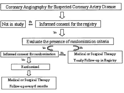
0f the eligible subjects, 4% declined to participate in the study and an additional 2% were flot enrolled for a variety of reasons, such as medical emergency or unavailabiiity of study personnel (Figure 2-1). The registry represents the population referred to each site for study and treatment of coronary artery disease and is the source from which candidates for randomization were identified. Clinical, laboratory and angiographic profiles were meticulously entered in a computer assisted medical database, which is available at the Montreal Heart Jnstitute. The registry at each participating center included ah patients whose primary indication for coronary angiography was suspected or proven CAD.
I
Cor Anoaphy for uspected Comnaîy ArtyDïseaseNot in study Inform consent forffieregstry
E1uate thepresence cfrandontion itena
C
IT.Jnfontied con5erf rranmizaon Media1 Suigical Tliarapy
‘-
.JJ.
Yea.lyfoUnw-up inRegistzyRanoitii
.L
Medicalor Suica1 TIrapy Follaw-upev1y6 nonilu
Figure 2-1. CASS patients flow diagram
Patients with normal coronary angiograms were also included in the registry. Patients studied because of suspicion of CAD who were diagnosed to have another form of heart disease were excluded from the registry. Sorne patients who underwent coronary angiography for evaluation of other conditions, such as valvular disease, cardiomyopathies and congenitat heart disease, were also excluded even if subsequent evidence showed that CAD was indeed a major clinical problem. These patients were flot sent by their referring physician due to suspected or proven CAD. This is clinically relevant, since the studied population consists of patients in whom there was a clinical suspicion of CAD, this being a reflection of current practice and rendering the resuÏts of CASS applicable to daily situations. Exclusion criteria for the registry consisted in the following: 1) inaccessibility for follow-up, 2) substantial language barrier, 3) referral to a CASS site expressly for surgery with coronary angiography performed elsewhere, 4) cardiomyopathy flot due to ischemic heart disease, 5) idiopathic hypertrophic subaortic stenosis and 6) significant
28
valvular heart disease. Patients with minimal regurgitation due to mitral valve prolapse were included in the registry.
2.1.2 Informed consent
Enrolment was contingent upon obtaining the patient’s signed informed consent and it was usually obtained before the initial index coronary angiographic study. Patients were told that data collected would be used to evaluate the natural history of CAD and that they wouid therefore be contacted annually for follow-up. Subjects have been assured that ah data are strictly confidentiai and that only CASS research personnel have access to their records. Data obtained for the present study was kept with the original encoding; therefore no personal information was obtained.
2.1.3 Baseline data collection and variables description
Basehine clinical and laboratory data on ail registry patients at participating clinical sites were recorded in printed forms. These forms were transmitted within 8 weeks to the coordinating center with strict quaiity control procedures. Variables included in this thesis wiil be in bold and explained. These forms were:
a) Patient information form. Mainly demographic data. Age and sex at time of enrolment. b) Health status questionnaire. A description of the type and severity of symptoms, including a subjective evaluation of functionai limitations.
c) Patient history form. A review of risk factors, prior cardiovascular ilinesses, and other medical problems. History of cigarette smoking was considered as presently smokes cigarettes (at enrolment and tbree months before or after), formerly smoked cigarettes and neyer smoked cigarettes. Recreational activity was assessed within the three months prior to enrolment and was divided as strenuous Qhysically demanding recreational activity involving competition or
endurance, including team efforts), moderate (activities performed for pleasure and relaxation but without competition or endurance), mild (recreation carried out for pleasure and relaxation and involving only slight physical activity) or sedentary. Any patient with a known past medical history of diabetes (DM) was considered as diabetic at entry of enrolment. Hypertension (HTN) was identified if there was a past medical history of hypertension, confirmed by a physician.
d) Present illness profile. This is an assessment of clinical manifestations ofCAD, with particular attention to the charactenstics and severity of angina pectoris and CHF and current medical therapy within two months of enrolment. Medications included among others -blockers (mainly propranolol), antiplatelets (aspirin or dipyridamole), digitalis, diuretics (furosemide, ethacrynic acid, thiazides or aldactone), antihypertensive agents (except diuretics), Iipid Iowering agents, and insulin therapy.
e) Physical examination and laboratory data form. These variables were vital signs, major cardiovascular findings and laboratory data. Resting heart rate was measured by radial pulse during 60 seconds with the patient in a sitting position. Body mass index (BMI) was calculated as weight in kg divided by the square of height in meters. The hospital laboratories of the cooperating clinics analyzed total cholestero] and triglyceride levels. Total cholesterol was expressed in milligrams per deciliters.
f) Exercise treadmili test (ETT). All patients in the randomized study were required to undergo a maximal ETT at the time of baseline evaluation, at 6 and 18 months afler actual or scheduled surgical treatment, and at 60 months afler enrolment. Although exercise testing was not mandatory for registry patients, it was encouraged and the resuits were recorded. They were performed in motor-driven treadmili, according to the Bmce protocol. Any patient suspected of having had a myocardial infarction within the preceding 6 weeks was routinely excluded from the
30
test. for most patients, testing was begun at stage 1 (1.7 mph at 10% siope). Reasons for discontinuing the test included the following: moderate or severe chest pain (3 or 4 on a scale of 4), classified as definitely angina, probably angina, probably not angina or definitely flot angina; unsteady gait; hypotension (a 20-mmHg drop in systolic blood pressure from the highest preceding value); ventricular arrhythmia, including ventricular tachycardia or ventricular fibrillation, coupled or frequent multifocal ventricular premature complexes or new ventricular bigeminy; rapid supraventricular arrhythrnias; symptoms (e.g, chest pain, dizziness, dyspnea, near syncope, lower extremity claudication, fatigue or weakness, and poor motivation). Patients were questioned at the time of ETT whether they were taking medication that might affect the test’s resuits. Among other medications, the ones affecting heart rate behavior were selected. It was registered whether the patient was either taking -b1ockers (within 24 hrs) or digitalis (within 7 days). Heart rate during exercise was recorded three times. Baseline heart rate was measured in a sitting position, before the start of ETT. Maximal heart rate was registered at maximal exercise with the patient in standing position. Post-exercise heart rate was measured 3 minutes afler ending of ETT with the patient in the sitting position, without any “cool-down” phase.
g) Chest x-ray and coronary angiography form. AIl sites measured the same variables in the same order and use the same nomenclature to describe left ventricular wall motion and coronary artery anatomy. A detailed description of the standard angiographic procedure has been published elsewhere (91). Coronary vasculature was divided in 18 numbered segments (Figure 2-2). Segments coded as anatomically absent were excluded. Dominance was considered in the assignment of individual segments to a particular major coronary artery distribution. The
maximum stenosis in % of lumen diameter was freely assigned (rather than fixed to a prespecified % stenosis range) by CASS-certified, clinical site angiographers in relation to an appropriate adjacent reference diameter. The estimated percentage of obstruction was derived from the angiographic view showing the greatest reduction in diameter for the vessel in question. Inter-reader variability in assessing segment stenosis in CASS had previously been reported, with a ic-statistic for between readers variability of 0.66 (0.33-0.86) for ah segments and 0.77 (0.65-0.86) when only proximal and mid segments ofthe major coronary vesseis were considered.
Figure 2-2. Terminology for coronary artery anatomy visualized at angiography utilized by cooperating sites in CASS. (1) proximal right; (2) mid-right; (3) distal right; (4) right posterior descending; (5) right posterior lateral segment; (6) first right posterior lateral; (7) second right posterior lateral; (8) third right posterior lateral; (9) inferior septal; (10) acute marginaI; (11) lefi main; (12) proximal left anterior descending; (13) mid-left anterior descending; (14) distal left antenor descending; (15) first diagonal; (16) second diagonal; (17) first septal; (18) proximal circumflex; (19) distal circumflex; (20) first obtuse marginal; (21) second obtuse marginal; (22) thfrd obtuse marginaI; (23) lefi atrio-ventricular; (24) first lefi posterior lateral; (25) second left posterior lateral; (26) third left posterior lateral; (27) left posterior descending. From National Heart, Lung, and Blood Insfitute Coronary Artery Surgery Study. A multicenter comparison of the effects of randomized medical and surgical treatment of mildly symptomatic patients with coronary artery disease, and a registry of consecutive patients undergoing coronary angiography. Circulation 1981 ;63(6 Pt 2) :11-81
The mean absolute deviation of coronary stenosis ranged from 1.8% to 9.4% in 870 randomly selected abnormal cine films (92). The criterion for chinically significant coronary artery obstruction is either 70% or more reduction in the internal diameter of the right coronary (RC),
32 lefi anterior descending (LAD) or left circumilex (LCx) coronary artery or 50% or more reduction in the internai diameter of the lefi main coronary artery (LMCA). The extent of CAD is defined as follows: In the presence of right dominant circulation, the three major branches involved in the one-, two- and three-vessel disease classification are the right anterior descending, LAD and LCx arteries respectively. In the case of a left-dominant circulation, they are the LAD, the proximal LCx artery and its marginal branches, and the distal LCx artery and its posterolateral branches. The nomenclature in the case of a baianced circulation is the same as that used to describe a right dominant circulation. A patient with 50% or greater obstruction of the lefi main coronary artery is classified as having two-vessei disease if the circulation is right-dominant and three-vessel disease if it is left-dominant.
h) Left ventriculography form. This was done with standardized procedure, projections and measurements. The left ventricular end-systolic volume (ESV), end-diastolic volume (EDV), and ejection fraction (EF) were caiculated using a single-plane adaptation of the area-iength method ofDodge and co-workers(93). Ef vas caiculated as
EDV-ESV
EDV
Ail these forms are available at the Montreal Heart Institute Coordination Center (MHICC).
2.1.4 Patients follow-up
The date of enrolment was that of the initial angiographic evaluation. Annual clinical follow-up was mandatory for ail patients in the registry. Additional information was obtained for ail patients in the registry who suffered a “coronary event”. CASS follow-up requirements for various situations designated as “coronary events” included the following:
(A) If a patient experienced a myocardial infarction, ail relevant information, including electrocardiograms (ECGs) and the resuits of enzyme studies, were obtained regardless of whether the patient was hospitalized.
(B) Detailed reports of hospitalizations for any cardiac event or stroke were collected if the period ofhospitalization exceeded 5 days.
(C) If a patient was hospitalized for coronary angiography or cardiac surgery, a specific description of the hospitalization and the procedures performed was obtained.
(D) If a patient died, a detailed report of the circumstances of death was fihled out.
Patients were followed aimually through 1982 and thereafter by a final mail survey between 198$ and 1991 to which 94% responded. Vital status among non-responders at last foilow-up was obtained through 1991 from the National Death Index and, in some cases, from next of km such that the status of 95.8% of ail CASS patients was known. Median duration of follow-up (and interquartile range) was 14.7 (9.0-16.1) years.
2.2 Statistical analysis
2.2.1 Variable selection and treatment
Variables for this thesis were chosen according to literature review, clinical relevance and data availability. Resting heart rate was analyzed both as a continuous and as a categoricai variable. Nonetheless, it seemed clinicaily more appropriate to present it divided by quintiles. These quintiles were based once again according to literature review and to our population’s distribution (94). It was flot the intention of this research project to find a threshold or cutoff variable separating “normal” from “abnormal” subjects. Ail heart rate measurements during ETT were kept as continuous variables, without any recoding. Solely for the purpose of baseline data
34
presentation and in an effort to harmonize with previous studies patients, were classified according to their chronotropic incompetence status. Chronotropic incompetence was defined as either failure to reach 85% of APMHR andlor < 80% HRRes, because these are the two most widely used definitions. Once again, it was not the purpose of this study to find normal values for heart rate measurements during ETT. The choice of a “normal” threshold in medical science is difficuit; it cannot be simply based on the 95% confidence interval of a normal distribution and much more complex statistical methods have been proposed (95, 96). Table 2-I displays in further detail the definition of each variable.
Table 2-I. List of variables.
Variable Definition
Age At baseline or at ETT Gender Male I Female
Resting heart rate Obtained manually from radial pulse during 60 seconds at baseline or at ETT with the patient in a sitting position
Maximal heart rate from ECG at peak exercise during ETT (HRMax)
Heart rate recovery Maximal heart rate during exercise minus post-exercise heart rate without cool-down (HRRec) period, 3 min after a modified Bmce protocol
% ofheart rate reserve ((Maximal heart rate —heart rate at rest) / (220— age —heart rate at rest)) x 100 (%HRRes)
Metabolic equivalents Fstimated by the total time compïeted in the final stage, speed and siope oftreadmill. (METS)
ST abnormality at peak 1 mm of horizontal or downsloping ST-segment depression> 80 ms after die J point or exercise if there vas I mm of additional ST-segment elevation in leads without pathologic Q
waves
Ef Single-plane area-length method. Ef: EDV—ESV
EDV Use of-blockers At baseline or at ETT
Hypertension History of hypertension, confirmed by a physician. Diabetes mellitus History ofdiabetes mellitus, confirmed by a physician Cholesterol level Expressed in milligrams per decilitres
BMI Weight in kg divided by the square ofheight in meters
Smoking status Within 3 months prior to or after enrolment. Presently, formerly or neyer smoked cigarettes
NCDV According to CAS S criteria
Recreational activity At baseline. Strenuous, moderate, mild or sedentary Antiplatelet therapy At baseline, mainly ASA or dipyridamole
Diuretics At baseline, mainly furosemide or hydrochiorothiazide Lipid-lowenng drugs At baseline
Total mortality Vital status obtained from FU forms, final survey and NDI records
CV mortality Cause of death if known, obtained from FU forms, fmal survey and NDI records. CV death included cardiac direct, cardiac confributrny and sudden unexplained death
Rehospitalizations due to Ever hospitalised for MI, angina, stroke, CHF, revascularization or rhythm disturbance CV cause
MI Ever hospitalised for MI, diagnosis based on ECG andJor enzyme analysis Angina Ever hospitalised for angina or chest pain for more than 5 days
Stroke Ever hospitalised for stroke or transient ischemic affack CHF Ever hospitalised for CHF for more than S days
ASA=aspirin; BMI=body mass index; CASS=Coronary Artery Surgery Study; CHf=congestive heart failure; CV=cardiovascutar; ECG=electrocardiogram EDV=end-diastolic volume; Efejection fraction; ESV=end-systolic volume;ETF=exercise treadmiil test; fU=follow
36
2.2.2 Descriptive statistics
for the purpose of data presentation bivariate analyses were performed in order to compare patients within different RHR quintiles and patients with or without clwonotropic incompetence. For categorical variables, a test was performed. When comparing continuous variables between patients with and without chronotropic incompetence, t-tests were used. Likewise, when comparing different RHR quintiles one-way ANOVA analysis wasperformed.
2.2.3 Survival analysis
In both articles, the dependent variable is time to an event (total or CV mortality, rehospitalization due to CV cause). Independent variables are variables at baseline, risk factors and different heart rate pararneters. Cox proportional hazards (PH) regression models were used. Correlations between independent variables were checked on a correlation matrix. When high correlation was found, clinical judgment was implemented to choose the most appropriate variable to be included in the model. To further select the variables to be incorporated in a multivariate model, univariate Cox PH regression analysis were first conducted and variables with a p value <0.25 were chosen to be included. Some variables were forced into the model, according to their clinical relevance. The proportional hazard assumption for the Cox model was tested graphically with a Ïog-log plot for categorical and Schoenfeld’s residuals plot for continuous variables. Linearity assumption was assessed by log transformation of each continuous variable and graphical testing against survival time. Results were expressed in hazard ratios for Cox PH model. Because of the large number of patients and variables included in the first article, we used two-tailed significance level of 0.01 and 99% confidence interval (CI). For the second article the significance level was set to 0.05 with 95% CI. For this same article, once Cox PH models that were most predictive of total and CV mortality were determined, different
exercise heart rate parameters and exercise capacity in METS were independently added to each Cox PH mode! to detemiine the incremental value of each parameter using the Wald test (97, 98). The parameter with the largest Wald test value was considered the one adding the most information to the model. Ail analyses were performed with SPSS v.12 for windows (SPSS 1nc, Chicago, Illinois). Further details about statistical analyses can be found in each article methods section.
o
CHAPTER 3
n
ORIGINAL ARTICLE NUMBER ONE
Long-term prognostic value of resting heart rate in
patients with suspected or proven coronary artery disease
Ariel Diazi, MD, Martial G. Bourassal, MD, Marie-Claude Guertin2, PhD and Jean-Claude Tardif, MD, FRCPC, FACCÏ
(1) Departrnent ofMedicine, Montreal Heart hstitute
and the (2) Montreal Heart Institute Coordinating Center (MHICC), Montreal, Canada
Published in the European Heart Journal., May 2005; 26: 967 —974