BLOOD CELL UNITS FOR USE IN THE RECIPIENT AFTER ALLOGENEIC
PBPC TRANSPLANTATION
Running head: Allogeneic RBC units and PBPC collection with rHuEpo
Brieuc SAUTOIS1, Etienne BAUDOUX2, Jean-Paul SALMON1, Sandrine MICHAUX1, Nicole SCHAAF-LAFONTAINE3, Maguy PEREIRA1, Jean-Michel PAULUS3, Georges FILLET1 and Yves BEGUIN1
1
Department of Medicine, Division of Hematology
2
Department of Transfusion Medicine
3
Department of Clinical Biology, Division of Laboratory Hematology University of Liège, Liège, Belgium
Y.B. is Research Director of the National Fund for Scientific Research (FNRS), Belgium. This work was partly supported by grants from the FNRS
Address for correspondence Yves BEGUIN, MD University of Liège Department of Hematology CHU Sart-Tilman B-4000 LIEGE BELGIUM Phone +32 4 3667201 Fax +32 4 3668855 E-Mail: [email protected]
ABSTRACT
Background and objectives: Reducing exposure of transplant recipients to homologous blood may be of interest. This may be achieved by procuring donor-derived RBC units, collecting more PBPC with a combination of G-CSF + rHuEpo and by administering rHuEpo posttransplant.
Design and methods: Eight ABO-compatible donors were treated with rHuEpo and IV iron to collect 12 RBC units for use in their recipient. PBPC were collected after mobilization with rHuEpo and G-CSF in the same donors. The recipients received G-CSF and rHuEpo posttransplant. A control group of 10 donor/recipient pairs received G-CSF alone for PBPC mobilization and posttransplant.
Results: Eighty-six out of 91 planned RBC units were collected in the donors without significant Hct decrease because of a 4-fold increase in RBC production despite functional iron deficiency. After 2 leukaphereses, the cumulative yields of NC and CFU-GM were lower in the study group while those of BFU-E, CFU-Mix and CD34+ cells were similar. However, erythroid recovery was significantly accelerated in the study group.
Interpretation and conclusions: (1) Collection of 12 units RBC within 6 weeks is feasible with rHuEpo and IV iron; (2) this strategy allows a dramatic reduction in recipient exposure to homologous blood; (3) rHuEpo has no synergistic effect with G-CSF for mobilization of PBPC in normal donors and may even be deleterious; and (4) rHuEpo in the recipient may enhance erythroid engraftment.
KEY WORDS
Allogeneic hematopoietic stem cell transplantation, peripheral blood stem cells, RBC transfusion, erythropoietin, iron.
INTRODUCTION
The first series of allogeneic transplantation with peripheral blood progenitor cells (allo-PBPCT) were reported in 1995 (1-3) and since then this procedure has become widely available. In comparison with allogeneic bone marrow transplantation (allo-BMT), allo-PBSCT is associated with faster hematological recovery, reduced transfusion requirements, shorter hospital stay, comparable incidence of acute graft-versus-host disease (aGVHD), transplant-related mortality and survival (4-6) although concerns have been raised about the trend towards an increased incidence of chronic GVHD (cGVHD) (5-6). Priming of PBPC in normal donors has been mainly achieved with filgrastim at a dose of 10 µ g/kg/day (7) but there are indications that lenograstim mobilizes progenitors more efficiently than filgrastim on a weight-by-weight basis (8,9).
Recombinant human erythropoietin (rHuEpo) used alone can induce a modest increase of progenitor cells in the peripheral blood of lymphoma patients (10) and autologous transplants have been performed with PBPC primed with rHuEpo alone although up to 14 leukaphereses were needed to reach the target of 6.5 x 10⁸ MNC/kg (11). Enhancement of progenitor mobilization was observed with the association of CSF + rHuEpo as compared to an historical group receiving G-CSF alone suggesting a synergy between the two growth factors in the autologous setting (12). The apparent synergism between G-CSF and rHuEpo has been confirmed in a randomized trial of PBPC mobilization in women with ovarian carcinoma (13). However, rHuEpo has never been used for allo-PBPC mobilization either alone or in association with G-CSF.
After allo-BMT, rHuEpo accelerates erythroid engraftment and produces some reduction in red blood cell (RBC) transfusion needs (14-20) but such an impact has not been shown after autologous transplantation whether rHuEpo is used alone (17,18,21) or in association with G- or GM-CSF (22-24). RHuEpo has never been used in the setting of allo-PBPCT.
Collection and storage of autologous RBC is feasible before elective orthopedic or cardiac surgery. Randomized placebo-controlled trials have shown that rHuEpo increases the ability of such patients to donate up to 6 U of autologous blood within 3 weeks (25) or up to 5 U within 2 weeks (26). Collection of up to 10 RBC units over a 5 week period has been attempted with rHuEpo stimulation in bone marrow donors but only 18 % of them were able to complete this program (27). Nevertheless, the availability of these donor-derived RBC units allowed to decrease homologous blood needs after allogeneic BMT. These results have been generally obtained with oral iron supplements, but in one study, IV iron has been shown to be superior to oral iron at least in relatively anemic patients (28). Such superiority of IV over oral iron has also been demonstrated in dialysis or pre-dialysis patients treated with rHuEpo (29-31). It is not known whether the use of IV iron would authorize an intensive phlebotomy program over several weeks.
Therefore, we designed a prospective study of rHuEpo therapy in pairs of PBPC donors and their recipients. RHuEpo was given to the donor to facilitate blood donation and to increase PBPC mobilization, and to their recipient to accelerate erythroid engraftment. The purposes of our study were: (i) to evaluate the feasibility of collecting up to 12 autologous RBC units within 6 weeks with the use of rHuEpo and IV iron supplementation; (ii) to investigate possible additive or synergistic effect of rHuEpo and G-CSF on the mobilization of PBPC in normal donors; (iii) to examine the possibility of avoiding posttransplant exposure of the recipient to homologous blood; and (iv) to study the effect of combined treatment with rHuEpo and G-CSF on engraftment of allogeneic PBPC and on the number of platelet and RBC transfusions.
PATIENTS AND METHODS
Patients and donors:
Patient and donor pairs were included into the study group if they fulfilled the following inclusion criteria: (i) patient eligible for an allogeneic transplant by generally accepted criteria; (ii) family donor eligible for stem cell donation by leukapheresis; (iii) donor older that 18 years; (iv) donor available for an intensive program of autologous blood donation; (v) patient and donor ABO-compatible for donor red cell transfusions; (vi) informed consent signed by donor and patient. The protocol was approved by the Ethical Committee of the University of Liège. Patients treated in the same period but not included into the protocol were offered a standard PBPCT (control group). Patient details are given in table 1 and donor characteristics are displayed in table 2. AML patients were in CR1 except 2 primary refractory patients in the study group.
Study design:
Donor treatment. In the study group (n=8), donors were administered rHuEpo (Eprex, kindly provided by Janssen-Cilag, Beerse, Belgium) subcutaneously at a dose of 600 U/kg twice weekly for 3 weeks before and 3 weeks after the transplant, except during the week of G-CSF administration (days –5 through –1) when the dose was 600 U/kg daily (total 15 doses). These donors also received 200 mg IV iron (Venofer, Vifor, St. Gallen, Switzerland), given in 500 ml saline to compensate for blood donation, twice weekly for 3 weeks before and 3 weeks after the transplant. RBC units were collected at a rate of 1 unit twice weekly for 3 weeks before and 3 weeks after the transplant (total 12 units) if the hematocrit was ≥ 33%. In the control group (n=10), no rHuEpo was given and no RBC collection was done. In both groups allogeneic PBPC were primed with glycosylated rHuG-CSF (Granocyte, kindly provided by Rhône-Poulenc-Rorer,
Montrouge, France) 10 µg/kg/day subcutaneously from day –5 through day –1. Donors were followed up for 3 weeks following cessation of all procedures.
Patient treatment. Patients in the study group were treated with 200 U/kg/d IV rHuEpo until
they reached an unsupported hematocrit ≥ 30% or for a maximum of 49 days. No iron supplements were given. Glycosylated rHuG-CSF (Granocyte) was also administered IV at a dose of 5µg/kg/d from day 1 until neutrophils were above 1 x 10⁹/L for 3 consecutive days or above 10 x 109/L for 1 day. Patients in the control group were given glycosylated rHuG-CSF but did not receive rHuEpo. For patients with myeloid malignancies, Granocyte was kindly provided by Rhône-Poulenc-Rorer.
Clinical care:
All blood products were irradiated. Single-donor platelet transfusions were given if platelet counts decreased below 15 x 109/L and packed red blood cells when the hemoglobin level decreased below 9 g/dl. GVHD prophylaxis was carried out with cyclosporine with (n=15) or without (n=3) short methotrexate. The diagnosis and grading of acute and chronic GVHD was established according to the Seattle criteria. Acute GVHD was treated by corticosteroids.
Leukaphereses:
PBPC were collected using the blood cell separators CS3000+ (Baxter-Fenwall Laboratories, Deerfield, IL, USA) or Spectra (Cobe BCT, Lakewood, CO, USA). Twelve liters of blood were processed per apheresis. Collections were performed on days -1 and 0. The harvest product obtained on day -1 was stored overnight at room temperature and both products were reinfused on day 0 through a double lumen Hickman catheter. In a few cases, 1 or 2 additional leukaphereses had to be performed and these cells were reinfused on the day of collection.
Laboratory analyses:
CFC assays. Hematopoietic progenitors were grown in 0.9% methylcellulose in a commercially
available medium (H4433, Terry Fox Laboratory, Vancouver, BC, Canada). Cultures were incubated at 37°C in a humidified 5% CO2 atmosphere. Formation of colony-forming
unit-granulocyte-macrophage (CFU-GM), burst-forming unit-erythroid (BFU-E) and colonies of multiple lineages (CFU-Mix) were scored on day 14.
Flow cytometry. Aliquots were incubated with phycoerythrin-conjugated monoclonal anti-CD34
(HPCA2, Becton-Dickinson, Palo Alto, CA, USA) for 20 minutes at 20°C, washed and fixed with 1% formaldehyde. A total of 1 x 10⁵ cells was analyzed using a FACScan analyzer (Becton-Dickinson). The percentage of CD34+ cells was defined with dot plot analysis using the whole nucleated cell population and the percentage of CD34+ cells in the isotype control was subtracted to give the final percentage of CD34+ cells. Data acquisition was performed with the Cellquest software (Becton-Dickinson).
Miscellaneous analyses. Complete blood counts were determined in a Technicon H3 cell counter
(Bayer, Tarrytown, NJ, USA). Serum iron, total iron binding capacity (TIBC), transferrin saturation and serum ferritin were measured by standard methods. Serum soluble transferrin receptor (sTfR), a quantitative assay of erythropoietic activity, was assessed by ELISA as previously described (32).
RBC production in the donor
The blood volume (BV) was estimated at baseline as the sum of the plasma volume (PV) and red cell mass (RCM) calculated from the following formulas based on the body surface area (BSA in m²) and age (in years): PV (ml) = 1578 x BSA in men or 1395 x BSA in women; RCM (ml) = (1486 x BSA) - 825 in men or (1.06 x age) + (822 x BSA) in women (33). The blood volume was assumed to remain constant and the RCM during treatment was calculated as follows: RCM (ml) =
BV x Hct (in %) x 0.92/100. The production of red cells between 2 visits was therefore derived from the following formula: RBC production (ml) = (RCM at visit 2 – RCM at visit 1) + RC volume removed at visit 1. The latter always included the volume of red cells in the collected unit plus the volume of blood drawn for laboratory analyses.
Statistical analyses:
Comparisons between the two groups were carried out with either Mann-Whitney U-tests or unpaired t-tests, then usually after log transformation because of skewed distribution of the data. Times to hematopoietic recovery were studied by life table analyses and Wilcoxon rank tests were used to compare the two groups. This included times for neutrophils to reach 0.5, 1 or 2 x 10⁹/L, platelets to reach 20, 25, 50, 100 or 150 x 109/L, hematocrit to reach 27%, 30% or a normal value, and reticulocytes to reach 0.5, 1 or 2%. Statistical analyses were done using Microsoft Excel (Microsoft Corp., Redmond, WA, USA) and Graphpad Prism (Graphpad Software, San Diego, CA, USA).
RESULTS
In the donor:
Tolerance and safety. Lenograstim injections were well tolerated with only mild bone pain in the
majority of the donors, which resolved after paracetamol administration. One migrainous donor complained of headache during lenograstim administration. RHuEpo administration did not generate any significant side effect, and in particular no hypertension was noted. No serious adverse event was reported during or after the study period. Platelets decreased to 253 ± 89 x 109/L in the study group and to 164 ± 42 x 109/L in the control group after PBPC collections (p=0.0158) but recovered within one week.
Red blood cell collections. This study was designed to collect 12 RBC units after stimulation with
rHuEpo and IV iron supplementation for use in the transplant recipient. In 4 donors this target was reached, 11 units were obtained in 1 donor, 10 units in 2 donors and 7 units in 1 donor. In this latter case, RBC collections were canceled on day 3 posttransplant because of a severe deterioration of the patient who died on day 4. Other reasons for canceling RBC collections were a low hematocrit (<33 %) (n=3), headache (n=1) and the need for a 4th PBPC collection (n=1). We therefore managed to collect 86 of the expected 96 RBC units, or 86 out of 91 (95%) if those expected for the patient who died on day 4 are not taken into account.
Erythropoiesis. After a slight initial drop, donor hematocrit remained fairly stable during RBC
collections and returned to baseline values within 3 weeks after the last phlebotomy (figure 1). This stability despite intensive phlebotomies was achieved through a considerable expansion of erythropoietic activity, as illustrated by a more than 4-fold elevation in sTfR levels. Reticulocytes
increased 5-fold during the first 10 days before decreasing progressively during the remaining course of rHuEpo therapy. After cessation of rHuEpo therapy, reticulocytes were even lower than at baseline, while sTfR levels were not yet normalized after 3 weeks. Daily RBC production was calculated as described above. Compared to baseline (15±5 ml/d), there was an almost 4-fold increase in red cell production (63±8 ml/d) during the first 3-week period and this higher rate was maintained during the entire stimulation period. The considerable expansion of erythropoietic activity produced an enormous demand for iron and transferrin saturation rapidly dropped below 20 % despite IV iron supplementation. Baseline ferritin ranged from 23 to 341 µg/L. Ferritin levels 3 weeks after cessation of rHuEpo and IV iron therapy (85±53 ng/ml) were not different from baseline values (97±106 ng/ml).
PBPC mobilization and collection. The administration of rHuEpo alone for 2.5 weeks was not
associated with any significant increase of circulating WBC, CD34+ cells or progenitor cell numbers. A steep rise of all these cell categories was observed after the start of G-CSF treatment. The concentrations of circulating CD34+ cells, CFU-GM, BFU-E or CFU-Mix on the days of PBPC collections were not significantly different in the study group and the control group. The number of CD34+ and progenitor cells in the PB tended to decrease below baseline (NS) 2 to 4 weeks after PBPC collections.
Leukaphereses were performed with the aim of collecting a minimum of 4 x 10⁶ CD34+ cells/kg of recipient body weight (b.w.). In the study group this target was reached after 1 leukapheresis in 1 donor, after 2 leukaphereses in 3 donors and after 3 leukaphereses in 2 donors. Four such procedures were performed in 2 subjects for whom total yields reached only 1.62 and 3.64 x 10⁶ cells/kg. In all but one donor at least 4 x 10⁶ CD34+ cells/kg donor b.w. were collected. In the control group the same goal was obtained after 1 leukapheresis in 1 donor, 2 leukaphereses in 7 donors, 3 leukaphereses in 1 donor and 4 leukaphereses in 1 donor. At least 4 x 10⁶ CD34+ cells/kg
donor b.w. were procured from all donors. We compared the yields of the first 2 leukaphereses per kg of donor b.w. in the study group and control group (Table 3). Significant differences were found in the total yields of NC, MNC and CFU-GM with higher numbers obtained after priming with G-CSF alone, while BFU-E, CFU-Mix and CD34+ cells yields were not statistically different.
In the recipient:
Hematological recovery posttransplant. WBC and platelet recoveries were not statistically different
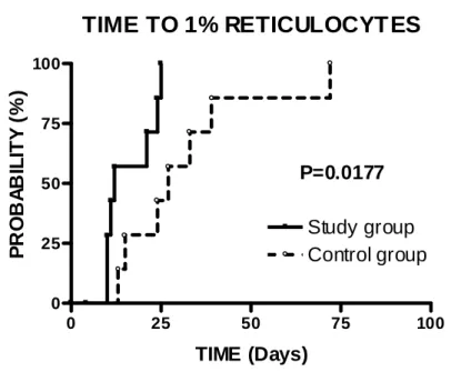
between the control and study groups. Engraftment to an absolute neutrophil count (ANC) of 0.5 x 10⁹/L was achieved after a median of 18 days (range 9-21) in the study group and after a median of 20 days (10-25) in the control group. Median (range) time to an unsupported platelet count of 20 x 109/L was 28 days (12-116) in the study group vs 30 (12-49) for controls. The patient with the latest platelet recovery (day 116) was diagnosed with posttransplant TTP. RBC recovery was accelerated after transplantation of G-CSF + rHuEpo mobilized PBPC and posttransplant rHuEpo compared with PBPC mobilized with G-CSF alone and no posttransplant rHuEpo. Median (range) times to ≥ 0.5 % reticulocytes were 10 (7-24) vs 21 (12-37) days (p = 0.0323) and to ≥ 1 % reticulocytes were 12 (10-25) vs 27 (13-72) days (p = 0.0177, figure 2A) in the study group and control group, respectively. After excluding 3 patients who had severe bleeding or TTP in the early posttransplant period, RBC transfusion independence was achieved after a median of 21 (7-29) days in the study group vs 40 (21-110) days in the control group (p = 0.0007, figure 2B).
Transfusion requirements. Comparative analysis of the 2 groups showed a trend towards fewer
platelet requirements in the study group. Between day 0 and day 30, a median (range) of 12 (4-31) vs 19 (3-24) platelet transfusions were required in the study and control groups, respectively (NS).
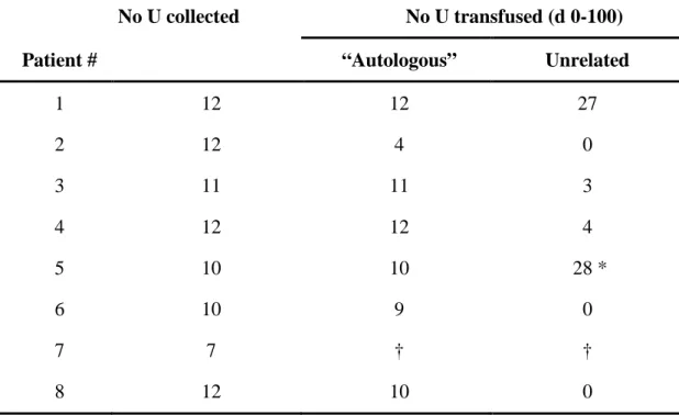
The total numbers of RBC units transfused were not significantly different between the 2 groups neither during nor after the period of rHuEpo treatment (table 4). However, the number of unrelated RBC units transfused was dramatically lower (2 vs 15, p = 0.026) in patients whose donors contributed to provide RBC. This difference was evident during the first 2 months posttransplant but the number of transfusions administered afterwards was low in both groups. Among 7 evaluable patients in the study group, 3 received exclusively “autologous” RBC units and 4 were also transfused with homologous blood, including 2 patients who received numerous transfusions because of prolonged severe GI bleeding (table 5). With the exception of these 2 patients, exposure to homologous blood was reduced by a median of 100 (73-100) %. Of the 86 units collected in the donors, 18 (21 %) were not used.
Patients’ outcome. In the study group, one patient developed TTP with subsequent delayed RBC
and platelet recoveries and another experienced severe GI bleeding because of grade IV acute GVHD. GVHD-associated severe GI bleeding was also encountered in one patient in the control group. Another patient in the control group developed TTP after full hematopoietic reconstitution. The incidence and severity of acute or chronic GVHD were not different among the two groups. Three patients in the study group died before day 100 of VOD and cardiac failure (day 4), acute GVHD (day 34) and lymphoma (day 67), respectively. Five control patients died before day 100. Causes of death were MOF (days 23 and 43), VOD (day 34), leukemia (day 59) and cerebral toxoplasmosis (day 81).
DISCUSSION
We evaluated the concomitant use of rHuEpo and rHuG-CSF to mobilize allogeneic PBPC. Contrary to a previous report in the autologous setting (11), we did not obtain progenitor mobilization with rHuEpo alone in our donor population. Also contrary to previous observations of PBPC mobilization after chemotherapy in cancer patients (12,13), we did not observe any synergistic effect between rHuEpo and glycosylated rHuG-CSF. Rather, the combination of growth factors yielded similar numbers of BFU-E but lower numbers of NC, MNC and CFU-GM than priming with G-CSF alone. This combination had never been tested for allogeneic PBPC mobilization but it is likely that somewhat different cell populations are mobilized when growth factors are used alone in normal subjects or combined with chemotherapy in cancer patients. However, it is also possible that the preceding use of large doses of rHuEpo could somehow impair the subsequent capacity of growth factors to mobilize PBPC. We have previously demonstrated that intensive treatment with rHuEpo is followed by a transient phase of profound inhibition of erythropoietic activity caused by erythroid marrow exhaustion (34). This was also accompanied by the reduction of marrow and spleen CFU-GM. Whether the omission of previous rHuEpo therapy would restore the synergism between rHuEpo and G-CSF remains to be demonstrated.
Recombinant human EPO increases the ability of patients to donate autologous RBC units (25,26) but the benefit is less evident in non-anemic patients (35) or with less intensive donation programs (36). We present here the most intensive phlebotomy program published so far, with 12 RBC unit donations scheduled over 6 weeks, and thus elicited to use a high dose of rHuEpo. While the utilization of rHuEpo in normal donors before bone marrow collection has been reported (37,38) and appears safe, Mitus et al. published the only study on the use of rHuEpo in allogeneic BMT donor/recipient pairs to facilitate collection of donor-derived RBC units available for use in the recipient after BMT (27). In their study, they administered rHuEpo at a dose of 1500U/kg/wk and
gave 300 mg elemental iron orally but were able to collect only a median of 6 units RBC out of the 10 scheduled over 5 weeks. Only 2/11 donors managed to donate all anticipated units while the remaining 9 gave between 4 and 8 U each, resulting in an overall success rate of only 65 %. Administering a slightly lower dose of rHuEpo with IV iron supplementation, we were able to collect a median of 12 U RBC and all donors procured at least 10 units, resulting in an overall success rate of 95 % and only 3 units not taken because of low Hct. Their lower RBC yield is in part attributable to blood losses associated with bone marrow donation but is in large part attributable to the relatively low iron status of several donors and to their use of oral instead of IV iron. Indeed functional or absolute iron deficiency is the main cause of low response to rHuEpo and in renal failure (29-31) as well as in autologous blood donations (28) is best overcome with administration of IV iron as compared to poorly absorbed oral iron. The efficiency of oral iron has even been questioned in autologous blood donors not receiving rHuEpo (39). An identical program of rHuEpo 600 U/kg twice a week and collection of 6 RBC units over 3 weeks but with oral iron resulted in a mean rate of additional red blood cell production of 34 ml/day compared to 22 ml/day with placebo (40). This 2.5-fold increase over basal erythropoiesis is much lower than the 4-fold increase over baseline and 48 ml/day of additional RBC production with our protocol. This implies that IV iron by itself has an impact that is comparable to that of rHuEpo in this setting.
Serum ferritin returned to baseline at the end of the period of observation, indicating that the dose of iron used (double that used in previous studies (28,36)) was appropriate for the additional demand for donated iron. In previous reports using oral (26) or IV (28,36) iron, ferritin was considerably decreased at the end of the iron supplementation period, so that it can be anticipated it would further decrease after cessation of iron therapy. Therefore, only the doses given in our study are capable of maintaining iron stores, yet are not excessive so as to produce relative iron loading. Despite the apparent efficacy of IV iron to support rHuEpo-driven erythropoietic activity, transferrin saturation constantly below 20 % is evidence that functional iron deficiency was limiting response to rHuEpo.
It remains to be determined whether higher doses or different schedules of iron administration, e.g. total dose infusion at the beginning of the program, would correct this phenomenon. Despite this limitation, the Hct remained relatively stable throughout the phlebotomy program. This is in contrast with previous studies where collection of only 6 units was associated with Hct decrements of 4 to 8 % (25,28,35). In a study of normal volunteers treated with oral iron and rHuEpo doses ranging from 750 to 3000 U/kg/wk for 4 weeks and donating up to 11 units within 4 weeks, the Hct dropped by 10 % and only 79 % of the scheduled units were collected (41).
Patients from both groups received G-CSF posttransplant and those in the study group were also treated with rHuEpo until an unsupported hematocrit ≥ 30% was reached. With the limitations of a small number of patients, we did not observe any difference in WBC and PLT recovery posttransplant but there was a trend towards fewer platelet transfusions in the study group. This is consistent with the observations of others who used rHuEpo without G-CSF after allogeneic BMT (17,19,20,42), with some non-randomized studies finding a favorable impact on platelet recovery and transfusion needs (43), particularly with the association of rHuEpo and G-CSF (44).
Erythroid recovery was accelerated in the study group, presumably in relation with posttransplant rHuEpo administration (progenitor cell yields were similar in the 2 groups). Impaired EPO response to anemia has been well documented in recipients of allogeneic BMT (45,46) and faster recovery of erythropoiesis and some reduction in RBC transfusion needs has been observed with the administration of rHuEpo (14,17,19,20,42,43). Although time to RBC transfusion independence was shorter in the study group this was not associated with reduced transfusion requirements. It should be emphasized that 2 patients in the study group had exceptionally high RBC product consumption due to prolonged severe bleeding. In addition, the trigger for RBC transfusions was relatively high compared to more recent practice. However, because of the large number of RBC units made available by the donors, exposure to homologous blood was significantly reduced in
patients receiving these donor-derived RBC units, with 50 % of evaluable patients not exposed to allogeneic RBC at all.
Our approach has several limitations. Exposing the donor to IV iron, SC rHuEpo and G-CSF could theoretically increase the risk of cell donation, but we did not observe any side effect related to erythropoietin or iron therapy. There are practical problems with keeping donors available for 6 weeks around the hospital and the inconvenience of almost 15 phlebotomy visits to the transfusion center is important. The logistical problems with organizing RBC collections in the donor may cause unwanted delays in the transplant procedure although this was not the case in our study. About 20% of RBC units collected were not used and this proportion could become even higher with the lower Hb cut-off values currently used for transfusing RBC. Therefore, the increased work load, cost and inconvenience to the donor may outweigh the benefits of the approach in practice.
REFERENCES
1. Bensinger WI, Weaver CH, Appelbaum FR, et al. Transplantation of allogeneic peripheral blood stem cells mobilized by recombinant human granulocyte colony-stimulating factor. Blood 1995;85:1655-8.
2. Schmitz N, Dreger P, Suttorp M, et al. Primary transplantation of allogeneic peripheral blood progenitor cells mobilized by filgrastim (granulocyte colony-stimulating factor). Blood 1995;85:1666-72.
3. Korbling M, Przepiorka D, Huh YO, et al. Allogeneic blood stem cell transplantation for refractory leukemia and lymphoma: potential advantage of blood over marrow allografts. Blood 1995;85:1659-65.
4. Van Hoef ME. HLA-identical sibling peripheral blood progenitor cell transplants (PBPCT). Bone Marrow Transplant 1999;24:707-14.
5. Blaise D, Kuentz M, Fortanier C, et al. Randomized trial of bone marrow versus lenograstim-primed blood cell allogeneic transplantation in patients with early-stage leukemia: a report from the Societe Francaise de Greffe de Moelle. J Clin Oncol 2000;18:537-46.
6. Champlin RE, Schmitz N, Horowitz MM, et al. Blood stem cells compared with bone marrow as a source of hematopoietic cells for allogeneic transplantation. IBMTR Histocompatibility and Stem Cell Sources Working Committee and the European Group for Blood and Marrow Transplantation (EBMT). Blood 2000;95:3702-9.
7. Grigg AP, Roberts AW, Raunow H, et al. Optimizing dose and scheduling of filgrastim (granulocyte colony- stimulating factor) for mobilization and collection of peripheral blood progenitor cells in normal volunteers. Blood 1995;86:4437-45.
8. Hoglund M, Smedmyr B, Bengtsson M, et al. Mobilization of CD34+ cells by glycosylated and nonglycosylated G-CSF in healthy volunteers--a comparative study. Eur J Haematol 1997;59:177-83.
9. Watts MJ, Addison I, Long SG, et al. Crossover study of the haematological effects and pharmacokinetics of glycosylated and non-glycosylated G-CSF in healthy volunteers. Br J Haematol 1997;98:474-9.
10. Pettengell R, Woll PJ, Chang J, et al. Effects of erythropoietin on mobilisation of haemopoietic progenitor cells. Bone Marrow Transplant 1994;14:125-30.
11. Kessinger A, Bishop MR, Jackson JD, et al. Erythropoietin for mobilization of circulating progenitor cells in patients with previously treated relapsed malignancies. Exp Hematol 1995;23:609-12.
12. Olivieri A, Offidani M, Cantori I, et al. Addition of erythropoietin to granulocyte colony-stimulating factor after priming chemotherapy enhances hemopoietic progenitor mobilization. Bone Marrow Transplant 1995;16:765-70.
13. Pierelli L, Perillo A, Greggi S, et al. Erythropoietin addition to granulocyte colony-stimulating factor abrogates life-threatening neutropenia and increases peripheral-blood progenitor-cell mobilization after epirubicin, paclitaxel, and cisplatin combination chemotherapy: results of a randomized comparison. J Clin Oncol 1999;17:1288.
14. Vannucchi AM, Bosi A, Grossi A, et al. Stimulation of erythroid engraftment by recombinant human erythropoietin in ABO-compatible, HLA-identical, allogeneic bone marrow transplant patients. Leukemia 1992;6:215-9.
15. Locatelli F, Zecca M, Beguin Y, et al. Accelerated erythroid repopulation with no stem-cell competition effect in children treated with recombinant human erythropoietin after allogeneic bone marrow transplantation. Br J Haematol 1993;84:752-4.
16. Link H, Brune T, Hubner G, et al. Effect of recombinant human erythropoietin after allogenic bone marrow transplantation. Ann Hematol 1993;67:169-73.
17. Link H, Boogaerts MA, Fauser AA, et al. A controlled trial of recombinant human erythropoietin after bone marrow transplantation. Blood 1994;84:3327-35.
18. Locatelli F, Zecca M, Pedrazzoli P, et al. Use of recombinant human erythropoietin after bone marrow transplantation in pediatric patients with acute leukemia: effect on erythroid repopulation in autologous versus allogeneic transplants. Bone Marrow Transplant 1994;13:403-10.
19. Klaesson S, Ringden O, Ljungman P, et al. Reduced blood transfusions requirements after allogeneic bone marrow transplantation: results of a randomised, double-blind study with high- dose erythropoietin. Bone Marrow Transplant 1994;13:397-402.
20. Biggs JC, Atkinson KA, Booker V, et al. Prospective randomised double-blind trial of the in vivo use of recombinant human erythropoietin in bone marrow transplantation from HLA-identical sibling donors. The Australian Bone Marrow Transplant Study Group. Bone Marrow Transplant 1995;15:129-34.
21. Ayash LJ, Elias A, Hunt M, et al. Recombinant human erythropoietin for the treatment of the anaemia associated with autologous bone marrow transplantation. Br J Haematol 1994;87:153-61.
22. Pene R, Appelbaum FR, Fisher L, et al. Use of granulocyte-macrophage colony-stimulating factor and erythropoietin in combination after autologous marrow transplantation. Bone Marrow Transplant 1993;11:219-22.
23. Chao NJ, Schriber JR, Long GD, et al. A randomized study of erythropoietin and granulocyte colony-stimulating factor (G-CSF) versus placebo and G-CSF for patients with Hodgkin's and non-Hodgkin's lymphoma undergoing autologous bone marrow transplantation. Blood 1994;83:2823-8.
24. Vannucchi AM, Bosi A, Ieri A, et al. Combination therapy with G-CSF and erythropoietin after autologous bone marrow transplantation for lymphoid malignancies: a randomized trial. Bone Marrow Transplant 1996;17:527-31.
25. Goodnough LT, Rudnick S, Price TH, et al. Increased preoperative collection of autologous blood with recombinant human erythropoietin therapy. N Engl J Med 1989;321:1163-8. 26. Cazenave JP, Irrmann C, Waller C, et al. Epoetin alfa facilitates presurgical autologous
blood donation in non- anaemic patients scheduled for orthopaedic or cardiovascular surgery. Eur J Anaesthesiol 1997;14:432-42.
27. Mitus AJ, Antin JH, Rutherford CJ, et al. Use of recombinant human erythropoietin in allogeneic bone marrow transplant donor/recipient pairs. Blood 1994;83:1952-7.
28. Mercuriali F, Zanella A, Barosi G, et al. Use of erythropoietin to increase the volume of autologous blood donated by orthopedic patients. Transfusion 1993;33:55-60.
29. Drueke TB, Barany P, Cazzola M, et al. Management of iron deficiency in renal anemia: guidelines for the optimal therapeutic approach in erythropoietin-treated patients. Clin Nephrol 1997;48:1-8.
30. Ahsan N. Intravenous infusion of total dose iron is superior to oral iron in treatment of anemia in peritoneal dialysis patients: a single center comparative study. J Am Soc Nephrol 1998;9:664-8.
31. Silverberg DS, Blum M, Agbaria Z, et al. Intravenous iron for the treatment of predialysis anemia. Kidney Int Suppl 1999;69:S79-S85.
32. Beguin Y, Clemons GK, Pootrakul P, Fillet G. Quantitative assessment of erythropoiesis and functional classification of anemia based on measurements of serum transferrin receptor and erythropoietin. Blood 1993;81:1067-76.
34. Piron M, Loo M, Gothot A, et al. Cessation of intensive treatment with recombinant human erythropoietin is followed by secondary anemia. Blood 2001;97:442-8.
35. Goodnough LT, Price TH, Friedman KD, et al. A phase III trial of recombinant human erythropoietin therapy in nonanemic orthopedic patients subjected to aggressive removal of blood for autologous use: dose, response, toxicity, and efficacy. Transfusion 1994;34:66-71. 36. de Pree C, Mermillod B, Hoffmeyer P, Beris P. Recombinant human erythropoietin as
adjuvant treatment for autologous blood donation in elective surgery with large blood needs (> or = 5 units): a randomized study. Transfusion 1997;37:708-14.
37. York A, Clift RA, Sanders JE, Buckner CD. Recombinant human erythropoietin (rh-Epo) administration to normal marrow donors. Bone Marrow Transplant 1992;10:415-7.
38. Martinez AM, Sastre A, Munoz A, et al. Recombinant human erythropoietin (rh-Epo) administration to normal child bone marrow donors. Bone Marrow Transplant 1998;22:137-8.
39. Biesma DH, Kraaijenhagen RJ, Poortman J, et al. The effect of oral iron supplementation on erythropoiesis in autologous blood donors. Transfusion 1992;32:162-5.
40. Goodnough LT, Price TH, Rudnick S, Soegiarso RW. Preoperative red cell production in patients undergoing aggressive autologous blood phlebotomy with and without erythropoietin therapy. Transfusion 1992;32:441-5.
41. Abraham PA, Halstenson CE, Macres MM, et al. Epoetin enhances erythropoiesis in normal men undergoing repeated phlebotomies. Clin Pharmacol Ther 1992;52:205-13.
42. Vannucchi AM, Bosi A, Linari S, et al. High doses of recombinant human erythropoietin fail to accelerate platelet reconstitution in allogeneic bone marrow transplantation. Results of a pilot study. Haematologica 1997;82:53-6.
43. Steegmann JL, Lopez J, Otero MJ, et al. Erythropoietin treatment in allogeneic BMT accelerates erythroid reconstitution: results of a prospective controlled randomized trial. Bone Marrow Transplant 1992;10:541-6.
44. Locatelli F, Zecca M, Ponchio L, et al. Pilot trial of combined administration of erythropoietin and granulocyte colony-stimulating factor to children undergoing allogeneic bone marrow transplantation. Bone Marrow Transplant 1994;14:929-35.
45. Beguin Y, Clemons GK, Oris R, Fillet G. Circulating erythropoietin levels after bone marrow transplantation: inappropriate response to anemia in allogeneic transplants. Blood 1991;77:868-73.
46. Beguin Y, Oris R, Fillet G. Dynamics of erythropoietic recovery following bone marrow transplantation: role of marrow proliferative capacity and erythropoietin production in autologous versus allogeneic transplants. Bone Marrow Transplant 1993;11:285-92.
Table 1. Patient characteristics
Study group Control group
Number of patients 8 10 Age (years) [median (range)] 45 (8-58) 34 (14-56) Male / Female 6 / 2 5/5 Diagnosis AML 5 1 CR1 3 1 refractory 2 0 MDS 0 3 RA-S - 1 RAEB - 2 CML (chronic phase) 1 2 NHL 1 2 CR2 0 1 PR1 1 0 PR2 0 1 SAA 1 1 Neuroblastoma 0 1
CR = complete remission; PR = partial remission; AML = acute myelogenous leukemia; CML = chronic myelogenous leukemia; MDS = myelodysplastic syndrome; RA-S = refractory anemia with ringed sideroblasts; RAEB = refractory anemia with excess of blasts; NHL = non-Hodgkin’s lymphoma; SAA = severe aplastic anemia
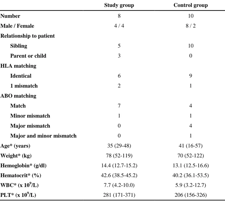
Table 2. Donor characteristics
Study group Control group
Number 8 10 Male / Female 4 / 4 8 / 2 Relationship to patient Sibling 5 10 Parent or child 3 0 HLA matching Identical 6 9 1 mismatch 2 1 ABO matching Match 7 4 Minor mismatch 1 1 Major mismatch 0 4
Major and minor mismatch 0 1
Age* (years) 35 (29-48) 41 (16-57) Weight* (kg) 78 (52-119) 70 (52-122) Hemoglobin* (g/dl) 14.4 (12.7-15.2) 13.1 (12.5-16.6) Hematocrit* (%) 42.6 (38.5-45.2) 40.2 (36.1-53.5) WBC* (x 109/L) 7.7 (4.2-10.0) 5.9 (3.2-12.7) PLT* (x 109/L) 281 (171-371) 206 (156-326)
* Values are given as median (range). There is no statistically significant difference between the two groups.
Table 3. Total PBPC collection yields after 2 leukaphereses (median [range])
Study group Control group p
NC x 108/kg donor b.w. 4.83 [2.41-9.78] 9.57 [5.21-17.34] 0.0006 MNC x 108/kg donor b.w. 4.80 [2.28-9.25] 9.37 [4.99-17.16] 0.0008 CFU-GM x 104/kg donor b.w. 31.99 [8.13-80.45] 80.90 [33.00-322.68] 0.0164 BFU-E x 104/kg donor b.w. 72.18 [21.35-138.69] 75.10 [10.78-242.39] NS CFU-Mix x 104/kg donor b.w. 1.79 [0-20.11] 10.06 [0-33.63] NS CD34 x 106/kg donor b.w. 4.79 [0.90-14.62] 10.75 [3.85-14.23] NS
Table 4. RBC transfusion requirements posttransplant (median [range])
Period (days) Study group Control group p
Any origin 0-56 12 [3-38] 13 [8-29] NS 57-100 1 [0-17] 3 [0-11] NS Total 12 [4-39] 15 [9-33] NS Unrelated origin 0-56 1 [0-28] 13 [8-29] 0.0185 57-100 0 [0-17] 3 [0-11] NS Total 2 [0-27] 15 [9-33] 0.026
Table 5. Origin of transfused red blood cells in the study group
No U collected No U transfused (d 0-100)
Patient # “Autologous” Unrelated
1 12 12 27 2 12 4 0 3 11 11 3 4 12 12 4 5 10 10 28 * 6 10 9 0 7 7 † † 8 12 10 0
* Until death on day 34.
FIGURE LEGENDS
Figure 1. Hematocrit (mean ± SD) in the donor during and after rHuEpo stimulation.
Figure 2. Erythroid engraftment in the recipient of the study group vs control group. A: Time to reach 1% reticulocytes.
Figure 1. 0 10 20 30 40 50 60 70 20 30 40 50
rHuEpo 600 U/kg biw RBC unit removal
Days
H
c
t
(%
)
Figure 2. A. B.
![Table 3. Total PBPC collection yields after 2 leukaphereses (median [range])](https://thumb-eu.123doks.com/thumbv2/123doknet/5511501.131492/25.892.102.796.192.480/table-total-pbpc-collection-yields-after-leukaphereses-median.webp)
![Table 4. RBC transfusion requirements posttransplant (median [range])](https://thumb-eu.123doks.com/thumbv2/123doknet/5511501.131492/26.892.96.788.189.553/table-rbc-transfusion-requirements-posttransplant-median-range.webp)