1
Université du Québec
Institut National de la Recherche Scientifique Centre Énergie, Matériaux et Télécommunications
Integrated multifunctional nanoplatforms with bioimaging and
therapeutic modalities
in the biological window
Par
Fan Yang
Thèse présentée pour l’obtention du grade de Philosophiæ doctor (Ph.D.) en sciences de l’énergie et des matériaux
Jury d’évaluation
Président du jury et examinateur interne
Jinyang Liang INRS-EMT
Examinateur externe Antonella Badia
Université de Montréal
Examinateur interne Hendrick W. de Haan
University of Ontario Institute of Technology
Directeur de recherche Dongling Ma
INRS-EMT
Codirecteur de recherche Fiorenzo Vetrone
INRS-EMT
Codirecteur de recherche Xinyu Liu
University of Toronto
2
ABSTRACT
Nowadays, with the rapid development of nanotechnology towards personalized and nano-medicine, engineering multifunctional nanoplatforms for the purpose of concurrent therapeutic and diagnostic (theranostic) modalities is critical for addressing challenging issues associated with cancers. Multifunctional nanoparticles (NPs), which integrate superparamagnetic and photoluminescent nanocomponents into a single particle, as an emerging class of nanomaterials, are extremely important for realizing this ultimate goal. Owing to their unique superparamagnetism, superparamagnetic NPs can be used to magnetically confine various biological species (DNAs, proteins, bacteria, cancer cells, etc.), thus allowing for ultra-sensitive biological detection. They can also serve as: i) magnetic resonance (MR) imaging contrast agents for the diagnosis of malignant tissues, ii) vehicles for carrying therapeutic payloads (anticancer drugs, small inhibitory RNA) to desired tumor sites in a target-specific manner, and iii) hyperthermia agents for cancer therapy under an alternating magnetic field. On the other hand, photoluminescent nanomaterials as contrast agents are widely used for the purposed of photoluminescence bioimaging, mainly for cells and tissues, thus allowing to acquire information of biological species and events.Therefore, multifunctional (superparamagnetic and photoluminescent) NPs which simultaneously possess both diagnostic and therapeutic functions, are expected to lead to a combined range of potential applications, such as bimodal imaging, photoluminescence monitored magnetic-driven drug delivery and simultaneous in vivo imaging and targeted hyperthermia therapy. However, the photoluminescent component in most studies regarding multifunctional NPs has so far been based on visible-emitting organic dyes, quantum dots (QDs) and upconverting nanoparticles (UCNPs). These multifunctional NPs exhibit low tissue penetration of excitation and emission light as a result of the considerable tissue absorption and low signal-to-noise ratio due to strong background autofluorescence from biological tissues, which restrict their use as contrast agents for in vivo imaging. To overcome this issue, the alternative contrast agents, whose absorption and emission wavelength are both in the so-called biological windows situated in the near-infrared (NIR) range (denoted as NIR-I: 700–950 nm; NIR-II: 1000–1350 nm) in which tissues are optically transparent, should be used. In this thesis, our work is mainly focused on the development of multifunctional magnetic and photoluminescent nanoplatforms in the NIR-I/II range with modalities for bioimaging and therapeutics.
In the first part, we developed a multifunctional core/shell/shell nanoplatform (Fe3O4@SiO2@NaYF4:Nd3+),
which consists of a superparamagnetic Fe3O4 core surrounded by an intermediate SiO2 shell and further
coated by an outer photoluminescent shell of NaYF4:Nd3+. Recently, Nd3+-doped NPs became one of the
3
compared with commonly used Yb3+-doped UCNPs. Most importantly, Nd3+-doped NPs can be efficiently
excited by laser at ca. 800 nm (NIR-I) and present three emission peaks at 900 (NIR-I), 1060 (NIR-II), and 1340 nm (NIR-II), respectively. Both excitation and emission wavelengths of Nd3+-doped NPs are located
within the optically transparent biological windows in the NIR. Owing to this unique NIR-to-NIR photoluminescence feature, the prepared Fe3O4@SiO2@NaYF4:Nd3+ NPs exhibit deep-tissue penetrated
optical properties with a high signal-to-noise ratio. Our NIR imaging experiment has demonstrated that the NIR photoluminescence signal of Fe3O4@SiO2@NaYF4:Nd3+ NPs can be transmitted across a tissue as
thick as 13 mm, about three times thicker than that can be achieved by similar core/shell/shell NPs containing the upconverting shell of Fe3O4@SiO2@NaYF4:Er3+,Yb3+ NPs. Meanwhile, these
multifunctional NPs possess excellent superparamagnetic properties due to Fe3O4 core inside, which result
in rapid magnetic response to an external magnetic field, making them suitable for magnetic-driven biological applications. Another important bio-medical application of Fe3O4@SiO2@NaYF4:Nd3+ NPs,
arising from superparamagnetic propriety, is their exploitation as T2 contrast agents for MR imaging. In
vivo MR imaging exhibits the significant darkening effect in T2-weighted images with the use of
Fe3O4@SiO2@NaYF4:Nd3+ NPs as contrast agents. Moreover, by designing this nanoplatform, the potential
toxicity of highly photoluminescent optical probes, such as QDs that usually contain Pb and/or Cd can be largely avoided, as demonstrated through cytotoxicity assay using HeLa cancer cells and human embryonic kidney (HEK 293T) cells. Therefore, this multifunctional nanoplatform is a promising candidate for high-resolution and deep-tissue bimodal (optical and MR) imaging in vivo.
The multifunctional nanoplatform in Part I shows low magnetization due to their single magnetic core feature, which is not suitable for magnetic-driven bioapplications and magnetothermal therapy. Engineering multifunctional nanoplatform containing multiple magnetic NPs is beneficial for realizing fast confinement bioapplications and achieving more effective magnetothermal therapy. Part II is thus focused on the development of novel multifunctional theranostic NPs that exploit multiple superparamagnetic Fe3O4 NPs
and interesting NIR-emitting PbS/CdS QDs, and their integration into a single nanoplatform. Self-assembly, as a powerful tool to design and fabricate functional nanomaterials for the purpose of rational control of the optical, electronic and magnetic pairing between distinct NPs, has attracted increasing research attention for their applications in biomedical diagnosis, plasmonics, and energy conversion. Self-assembled supernanoparticles (SPs) involving different types of NPs can possess not only the intrinsic physical and chemical characteristics of their individual NPs but also the collective properties of these NPs due to the coupling effect. In this part, the nanoplatform was specifically prepared by the self-assembling of superparamagnetic Fe3O4 NPs and photoluminescent PbS/CdS QDs with their emission in NIR-II and its
self-assembly formation mechanism was systematically studied. Due to their unique NIR photoluminescence feature, the self-assembled Fe3O4 and PbS/CdS (NIR-II) supernanoparticles [SASNs
4
(NIR-II)] exhibit outstanding deep-tissue penetration property as an optical imaging probe, allowing the NIR photoluminescence signal to be detected through a tissue as thick as 14 mm, about three times thicker than that can be achieved by their counterpart operating within the first biological window [SASNs (NIR-I)]. At the same time, clustered Fe3O4 NPs constituting SASNs (NIR-II) largely increase the magnetic field
inhomogeneity by the synergistic effect, resulting in a significantly enhanced T2 relaxivity (282 mM-1s-1,
ca. 4 times higher than that of free Fe3O4 NPs), as demonstrated by the remarkable darkening effect on in
vivo MR imaging. Regarding the potential nanomedicine-related therapeutic modalities, magnetothermal therapy suffers from the low heat conversion efficiency of currently studied magnetic NPs, while photothermal therapy is not suitable for deep-lying subcutaneous cancer cells due to the limitation of light penetration. More interestingly, the prepared SASNs (NIR-II) in our work possess the dual capacity to act as both magnetothermal and photothermal agents, overcoming the main drawbacks of each type of heating separately. When SASNs (NIR-II) were exposed to the dual-mode (magnetothermal and photothermal) heating set-up, the thermal energy transfer efficiency (specific loss power, SLP) was amplified 7-fold compared with magnetic heating alone. These results, in hand with the excellent photo and colloidal stability, and negligible cytotoxicity, demonstrate the potential use of SASNs (NIR-II) for deep-tissue bimodal (optical and MR) imaging in vivo, while simultaneously enabling SASNs (NIR-II) mediated dual-mode heating treatment for cancer therapy.
Although SASNs (NIR-II) possess excellent dual-mode heating therapeutic modality, polyvinylpyrrolidone (PVP) coating served as NPs surface stabilizer shows fair biocompatibility and difficulty of further versatile functionalization. In addition, we propose to explore drug delivery modality with our multifunctional nanoplatform. Previously published work has indicated mesoporous materials are extremely suitable for drug delivery. With this consideration, mesoporous silica (mSiO2) appears as a promising drug carrier
because it generally possesses a rigid mesostructured framework with high stability and ease of surface functionalization for linking drug molecules. In the third part, we specifically designed another type of multifunctional theranostic nanoplatform based on the large-pore mSiO2. To date, the work regarding the
preparation of uniform mSiO2 with large pore size (> 5 nm) is very limited. In this part,
relatively-large-pore (>10 nm) mSiO2 as matrix was deliberately synthesized by a biphase stratification continuous growth
approach, followed by a simple silane coupling reaction to form thiol-modified mSiO2. Owing to its unique
relatively-large-pore structure with high loading capacity, the nanoplatform (mSiO2@PbS/CdS-Fe3O4) was
then fabricated by coordination-driven embedding of superparamagnetic Fe3O4 NPs and PbS/CdS QDs of
suitable size into the mesoporous channels of mSiO2. In particular, the QDs were selected in such a way
that they could be excited by the light in NIR-I as well as emit in NIR-II. The excellent NIR deep-tissue optical and superparamagnetic behavior of mSiO2@PbS/CdS-Fe3O4 particles enables their use as bimodal
5
other hand, when the mSiO2@PbS/CdS-Fe3O4 nanoplatform was exposed to external physical stimuli of
magnetic field (MF) and/or a NIR laser, this nanoplatform produced strong local heating as a highly efficient magnetic hyperthermia therapy (MHT)/photothermal therapy (PTT) agent. At last, this nanoplatform also demonstrate great potential as a drug delivery carrier due to large-pore characteristics. Doxorubicin (DOX), a widely used clinical anticancer drug, was chosen as a model to study their drug release behavior. After being loaded with DOX, the release rate of DOX under multi-stimuli (pH/MF/NIR) was significantly enhanced at lower pH and higher temperatures, caused by magnethermal/photothermal effects. This nanoplatform thus yielded a synergistic effect from the integrated heating mode and multi-stimuli responsive drug release to achieve a high therapeutic efficacy.
6
ACKNOWLEDGEMENTS
Foremost I would like to express my deepest and sincere gratitude to my supervisor Prof. Dongling Ma, for accepting me in her group and giving me the chance to pursue my PhD degree. It has been a pleasure working with her who created a warm and stimulating research environment for us. The frequent and useful discussions with her at different stages of my research work largely contributed to the success of my research. I am forever and extremely grateful for her continual and dedicated guidance and constant encouragements. Her attitude towards academic research has greatly motivated me to be a good researcher. Meanwhile, I would like to acknowledge my co-supervisor Prof. Fiorenzo Vetrone. His endless effort in improving the quality of my research project by giving me some novel ideas makes me explore a fascinating scientific filed. He has always been a strong supporter when I need help throughout the duration of my PhD study.
Beside my supervisors in INRS, I would like to express my sincere thanks to another co-supervisor Prof. Xinyu Liu, from University of Toronto, for giving me invaluable suggestions and inspirations make me find the right direction of my research.
I am also grateful to Prof. John Oh from Concordia University for the collaboration of cytotoxicity assay in his group, and Prof. Sylvain Martel from Polytechnique Montréal for allowing me to carry out hyperthermia experiment in his lab. Their important comments have largely improved the quality of the articles I wrote for publication.
I would like to thank Dr. Fuqiang Ren who has contributed to my research work by carrying out some experiments involved in my project and discussing about the experiments and results with me. I also acknowledge Artiom Skripka, who gave me invaluable help and comments on my research and articles writing. I gratefully acknowledge the contribution to this work made by my collaborators, Dr. Antonio Benayas, Dr. Yue Huang, Xianke Dong, Sung Hwa Hong and Maryam Sadat Tabatabaei.
I am also grateful to all the group members for their help throughout the work. These people include: Dr. Hongyang Liang, Dr. Jianming Zhang, Dr. Mee Rahn Kim, Dr. Belete Atomsa Gonfa, Dr. Zhenghe Xu, Dr. Long Tan, Yanlong Liu, T. G. Deepak, Pandeng Li, Qingzhe Zhang, Shengyun Huang, Dr. Yannan Liu, Yong Wang and Ting Yu.
I would like to thank Jean-Philippe Masse for carrying out TEM measurements for me. I also thank departmental and technical staff at INRS-EMT, particularly Christophe Chabanier, for giving me training and technical help in using some equipment.
7
A massive thanks to all my friends: Chao Wang, Xin Chai, Xin Tong, Daling Cui, Wei Huang, Qiliang Wei, Xin Jin, Xiaohua Yang, et al.; without you I cannot have such wonderful memories these years.
I am eternally grateful to my parents, for their constant support and encouragement throughout all of my studies. I would like to thank my wife, Peijun Huang, who loves me deeply and supports me selflessly. Finally, I wish to acknowledge the following organizations for their financial support: The Natural Sciences and Engineering Research Council of Canada and Fonds de recherche du Québec-Nature et technologies for PhD Program.
8 CONTENTS CHAPTER 1 INTRODUCTION ... 19 1.1 Superparamagnetic nanoparticles ... 19 1.2 Fluorescent nanoparticles ... 21 1.2.1 Biological windows... 22 1.2.2 Quantum dots ... 23 1.2.2.1 PbS quantum dots... 24
1.2.2.2 Core/shell quantum dots ... 26
1.2.3 Lanthanide-doped upconverting nanoparticles ... 27
1.3 Research Objectives and Organization ... 30
1.3.1 Our objectives ... 30
1.3.2 Thesis organization ... 33
CHAPTER 2 LITERATURE REVIEW ON MUTIFUNCTIONAL FLUORESCENT-MAGNETIC HYBIRD NANOPARTICLES FOR BIOAPPLICATION ... 35
2.1 Introduction ... 35
2.2 Types and synthetic routes of multifunctional fluorescent-magnetic hybrid nanoparticles ... 37
2.2.1 Silica-based fluorescent-magnetic nanoparticles ... 37
2.2.1.1 Fluorescent silica shell coated magnetic nanoparticles ... 37
2.2.1.2 Magnetic and fluorescent nanoparticles co-embedded into silica matrix ... 39
2.2.1.3 Magnetic nanoparticles linked to fluorescent entity via silica spacer ... 40
2.2.2 Polymer based fluorescent-magnetic nanoparticles ... 42
2.2.2.1 Polymer assisted fluorescent-magnetic nanoparticles via coupling method ... 42
2.2.2.2 Magnetic and fluorescent nanoparticles co-embedded into polymer matrix ... 44
2.2.3 Fluorescent-magnetic nanoparticles by seed-mediated growth method... 46
2.2.4 Magnetically doped quantum dots ... 48
2.3 Bioapplication of multifunctional fluorescent-magnetic hybrid nanoparticles ... 50
2.3.1 Multimodal bioimaging... 50
2.3.2 Drug delivery ... 52
2.3.3 Cancer therapy ... 54
CHAPTER 3 EXPERIMENTS AND CHARACTERIZATIONS ... 56
3.1 Materials ... 56
3.2 Reaction setup ... 56
3.3 Synthesis of Fe3O4 nanoparticles, PbS and PbS/CdS quantum dots ... 57
3.3.1 Synthesis of Fe3O4 nanoparticles ... 57
9
3.3.3 Synthesis of smaller PbS quantum dots ... 58
3.3.4 Synthesis of PbS/CdS core/shell quantum dots ... 59
3.4 Synthesis of Fe3O4/SiO2 and Fe3O4/SiO2/NaYF4:Nd3+ nanoparticles ... 59
3.4.1 Synthesis of Fe3O4/SiO2 core/shell nanoparticles ... 59
3.4.2 Synthesis of core/shell/shell Fe3O4/SiO2/NaYF4:Nd3+ nanoparticles ... 59
3.5 Synthesis of self-assembled Fe3O4 and PbS/CdS supernanoparticles ... 59
3.6 Synthesis of Fe3O4 and PbS/CdS loaded mesoporous silica nanospheres ... 60
3.6.1 Synthesis of large-pore mesoporous silica nanospheres ... 60
3.6.2 Synthesis of thiol-modified mesoporous silica nanospheres ... 60
3.6.3 Synthesis of Fe3O4 and PbS/CdS mesoporous silica nanospheres ... 60
3.7 Characterization ... 60
3.7.1 Transmission electron microscopy and energy dispersive X-ray spectroscopy ... 60
3.7.2 X-ray diffraction ... 61
3.7.3 Absorption spectroscopy ... 61
3.7.4 Photoluminescence spectroscopy ... 61
3.7.5 Inductively coupled plasma optical emission spectroscopy ... 61
3.7.6 Fourier-transform infrared spectroscopy ... 62
3.7.7 Magnetic characterization ... 62
3.7.8 Viability assay ... 62
3.7.9 NIR and visible imaging ex vivo ... 62
3.7.10 T2 relaxivity measurements in vitro and MR imaging in vivo ... 63
CHAPTER 4 RESULTS ... 64
4.1 An Integrated Multifunctional Nanoplatform for Deep-tissue Dual-mode Imaging ... 64
4.2 Multifunctional Self-Assembled Supernanoparticles for Deep-Tissue Bimodal Imaging and Amplified Dual-Mode Heating Treatment ... 84
4.3 Magnetic-Photoluminescent Nanoplatform Built from Large-Pore Mesoporous Silica ... 117
CHAPTER 5 CONCLUSIONS AND PERSPECTIVES ... 142
5.1 Conclusions ... 142
5.2 Perspectives... 143
5.2.1 Synthetic procedures optimization ... 143
5.2.2 Photoluminescence efficiency optimization ... 144
5.2.3 Toxicity investigation ... 144
5.2.4 Surface modification ... 145
10
LIST OF FIGURES
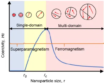
Figure 1.1 Schematic illustration of magnetic coercivity (Hc, magnetic field required to reduce the
magnetization to zero) behavior of a magnetic NP as a function of its size. When the size of NP gradually decreases to critical size (rc), the Hc shows an increase as the domain wall in NP disappears. If the size is
further decreased to r0 in which the thermal agitation energy is higher than magnetic anisotropy energy and
the magnetic moment of NP fluctuate freely, the NP enters the superparamagnetic regime and shows zero coercivity.
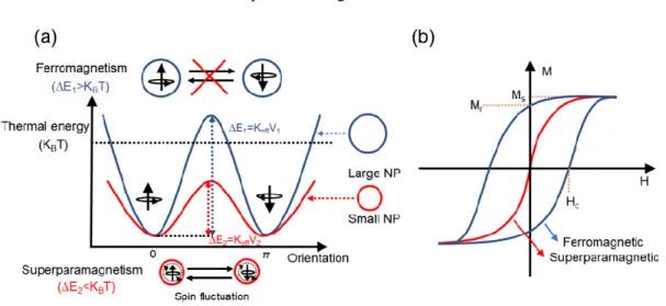
Figure 1.2 (a) Energy diagram of magnetic NPs with different magnetic spin alignment. Thermal energy
(KBT) represents the energy barrier to the rotation of the magnetization. The large NP shows the
ferromagnetism on the top and the small NP shows the superparamagnetism on the bottom. (b) The comparison of typical magnetic curves for ferromagnetic and superparamagnetic materials. Saturation magnetization (MS) is the maximum value of magnetic field; the remanence magnetization (Mr) stands for
the residual magnetization after removing external magnetic field; and the coercivity (Hc) is the external
field required to reduce the magnetization to zero.
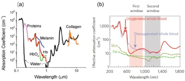
Figure 1.3 (a) Absorbance of various tissue and blood components from 200 nm to 10 μm. (b) Optical
windows in biological tissues. These plots of effective attenuation coefficient (on a log scale) versus wavelength show that absorption and scattering from oxygenated blood, deoxygenated blood, skin and fatty tissue is lowest in either the first (pink shaded area) or second (grey) near-infrared window. Figure 1.3a was taken from reference14 and Figure 1.3b was taken from reference7.
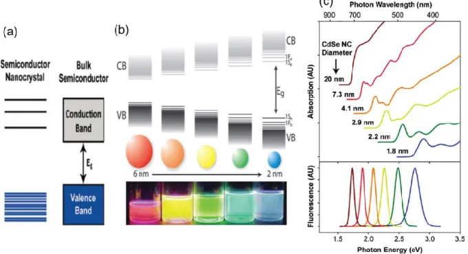
Figure 1.4 (a) Electronic energy states of a semiconductor in the transition from nanosized crystals to bulk
crystals. Blue shading denotes ground state electron occupation. (b) Schematic representation of the quantum confinement effect on the energy level structure of a semiconductor material. The lower panel shows colloidal suspensions of CdSe nanocrystals of different sizes under UV excitation. (c) Absorption (upper) and fluorescence (lower) spectra of CdSe semiconductor nanocrystals showing quantum confinement and size tunability. Figure 1.4a and c was taken from reference17 and Figure 1.4b was taken
from reference18.
Figure 1.5 (a) Absorption spectra spanning the range of tuneable sizes of PbS QDs. (b) Band-edge
absorption and photoluminescence peaks for PbS QDs 6.5 nm in diameter. (c) TEM image of colloidal PbS nanocrystals with an exciton absorption at 1440 nm. Figure 1.5 a~c was obtained from reference29.
11
Figure 1.6 (a) TEM images of CdSe plain core nanocrystals and the corresponding core/shell nanocrystals
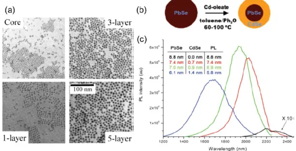
with different shell thickness from the same SILAR reaction. (b) Scheme of synthesis of PbSe/CdSe QDs by cation exchange method. (c) PL spectra from a series of aliquots during CdSe shell formation, corresponding to 15 min, 2 h, and 24 h of reaction time proceeding from red to blue. Each spectrum is corrected for variation in optical density as well as grating and detector efficiencies to reflect relative QY. Shown in black is the original PbSe core, magnified 10-fold so its shape is discernible. The inset lists the calculated core diameter (column “PbSe”) and shell thickness (“CdSe”) from elemental analysis. Also included is the approximate effective core size predicted by the PL peak position (“PL”). Figure 1.6a was obtained from reference32and Figure 1.6b~c was obtained from reference33.
Figure 1.7 (a) Energy diagrams for Ln3+ in a LaCl
3 lattice. (b) UC processes for lanthanide-doped crystals:
ESA and ETU. (c) Schematic illustration of UC nanoparticles composed of a crystalline host and lanthanide dopant ions (activator and sensitizer) embedded in the host lattice. Figure 1.7a was obtained from reference35.
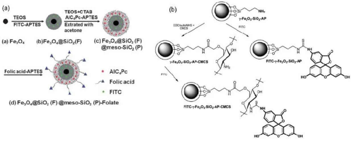
Figure 2.1 (a)Synthetic procedure of Fe3O4@SiO2(F)@meso-SiO2(P)-Folate nanoparticles. (b) Labeling
of g-Fe2O3-SiO2-AP and g-Fe2O3-SiO2-AP-CMCS nanoparticles with FITC. Figure 2.1a was taken from
reference50 and Figure 2.1b was taken from reference61.
Figure 2.2(a) Schematic illustration of the formation of NaYF4:Yb, Er/Tm@SiO2@Fe3O4 NPs. (b)
Bright-field TEM image and (c~e) EDX elemental mappings of NaYF4:Yb, Er/Tm@SiO2@Fe3O4, (f) HRTEM
image of NaYF4:Yb, Er/Tm@SiO2@Fe3O4 (inset: SAED image of NaYF4:Yb, Er) and (g) HRTEM image
of Fe3O4 (inset: FFT image of Fe3O4). (h) Schematic of fabrication of (CdTe/Fe3O4)@SiO2 FMNS with
different magnetic potentials: weak (WFMNS), moderate (M-FMNS), and strong (S-FMNS), Respectively. TEM images of (CdTe/Fe3O4)@SiO2 FMNS: (i) W-FMNS, (j) M-FMNS, and (k) S-FMNS. Figure 2.2a~g
was taken from reference66 and Figure 2.3h~k was taken from reference65.
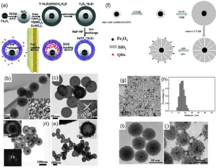
Figure 2.3 (a) Synthetic procedure for the drug-loaded Fe3O4@SiO2@α-NaYF/Yb, Er nanorattles
(DOX-MUC-F-NR). (b) TEM and SEM (the inset in (b)) images of Fe3O4@SiO2 nanospheres. (c) TEM image of
magnetic upconversion oxide nanospheres (MUC-O-NS); inset in (c) is a HRTEM image after growing the outer Y2O3/Yb, Er layer. (d) TEM image of the MUC-F-NR; insets are higher-magnification TEM image
of one (upper left) and the SAED image recorded on part of the α-NaYF4/Yb, Er shell (lower left). (e) TEM
image of Fe3O4@SiO2@α-NaYF/Yb, Er with SiO2 fully etched; inset is a higher-magnification TEM image
of a single hollow nanosphere. (f) Illustration of the preparation of multifunctional mesoporous Fe3O4/SiO2/CdTe nanoprobe. (g) TEM images of oleic acid-stabilized Fe3O4 nanoparticles and (h) their
12
Fe3O4/SiO2/CdTe nanoprobes. Figure 2.3a~e was taken from reference72 and Figure 2.3f~j was taken from
reference73.
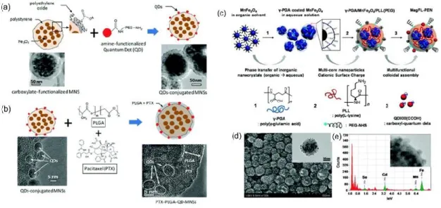
Figure 2.4 (a) Schematic diagram of the conjugation of amine-functionalized quantum dots (QDs) to the
surface of carboxylate-functionalized MNSs using conventional NHS/EDC coupling method; (b) Scheme of PTX loading using a thin PLGA coat on the surface of QD-MNSs. (c) Schematic illustration of the facile fabrication of MR/NIR multimodal imaging nanoprobes based on magnetofluorescent polyelectrolyte nanocomposites (MagFL-PEN) via electrostatic assembly between polyelectrolytes and functional colloidal nanoparticles. (d) SEM and TEM (inset) images of MagFL-PEN after adsorption of QD800(COOH). (e) EDX analysis of MagFL-PEN. Figure 2.4a~b was taken from reference81 and Figure 2.4c~e was taken from
reference85.
Figure 2.5 (a) The two-step hybrid formation. Step I: A) iron oxide nanocrystals (native ligands not shown),
B) iron oxide nanoparticles with the first polystyrene shell, C) nanohybrid after the deposition of QDQRs. Step II: D) nanohybrid after synthesizing a thin PS shell, E) nanohybrid after second emulsion polymerization, when dyads cluster owing to high styrene concentration. (b, c) Representative TEM images of the different hybrids achieved using different amounts of monomer during the second emulsion polymerization. The inset shows a three-dimensional histogram of the number of QDQRs n (QDQR) and the number of iron oxide n (Fe3O4) within one hybrid. Amounts used of each monomer: (b) 4 nmol and (c)
8 nmol. (d) Scheme for synthesis of MUCNBs; TEM images of 23 nm UCNPs (e), 15 nm IONPs (f), 6 nm UCNPs (g), and MUCNBs with 15 nm IONPs and 23 nm UCNPs (h) or 6 nm UCNPs (i). Figure 2.5a~c was taken from reference89 and Figure 2.6d~i was taken from reference94.
Figure 2.6 (a) Schematic representation of the synthetic routine of the water-soluble NaYF4:Yb3+,
Tm3+@Fe
xOy nanocrystals. (b) TEM image of NaYF4:Yb3+, Tm3+ nanocrystals and (c) NaYF4:Yb3+,
Tm3+@Fe
3O4 nanocrystals. (d) EDX spectrum of the NaYF4:Yb3+, Tm3+@Fe3O4 nanocrystals. (e)
Illustration of the multistep sequential synthesis producing the magneto-fluorescent hybrid structures. (f) TEM images of CdSe@CdS@hollow-Fe2O3 nanostructure. (g) Enlarged TEM image of f. Figure 2.6a~d
was taken from reference101 and Figure 2.6e~g was taken from reference102.
Figure 2.7 (a) Architecture of water-dispersible shell-doped mQDs (NAC-CdTe/30%Fe:ZnS) (A) and
core-doped mQDs (NAC-30%Fe-CdTe/ZnS (B). (b) Representative TEM image of shell-core-doped mQDs and the corresponding diffraction pattern (inset), indicating that the mQDs are crystalline. (c) The corresponding size distribution, where the average particle diameter was determined to be 2.9 ± 0.3 nm. Figure 2.7a~c was taken from reference104.
13
Figure 2.8 Dual-modal UCL/MR in vivo imaging. (a) The bright field, (b) UCL, and (c) merged images of
a KB tumor-bearing mouse one hour after intravenous injection of PEG–MFNP. Strong UCL signals were observed from the liver and tumor sites (arrow) of the mouse. (d) Ex vivo UCL imaging showing accumulation of MNFPs in the liver, spleen, tumor, bone, and lung of the injected mouse at 24 h post injection. UCL signals from other organs were barely detectable. T2-weighted images of KB-tumor bearing
nude mice with (e) and without (f) injection of MFNPs. Obvious darkening contrast was shown in the mouse liver and tumor. (g) Multimodal UCL and (h) MR imaging for in vivo lymphangiography mapping using MFNPs. MR images were taken before (left) and after (right) injection of MFNPs. Figure 2.8a~h was taken from reference117.
Figure 2.9 (a) Schematic illustration of targeting of DOX loaded multifunctional drug carrier to tumor cells
assisted by an externally applied magnetic field (MF). (b) Tumor location as defined by MUC-F-NR intensity increases with 1 h magnetic field treatment. Mice bearing H22 xenograft tumor were injected with DOX loaded MUC-F-NR (1 mg/kg) and subjected (+MF) or not subjected (-MF) to the magnetic field for 1 h. At 24 h postinjection, mice were imaged in vivo. (c) The luminescence signal was measured from the whole tumor in vivo and ex vivo. (Excitation was provided by the CW infrared laser at 980 nm and upconversion luminescence signals were collected at 650 ± 10 nm. Fluence rates for 980 nm excitation light were 80 mW/cm2.) (d) Tumor volume changes of saline-treated mice compared to mice treated with
MUC-F-NR, DOX, and DOX loaded MUC-F-NR over 21 d in the absence and presence of magnetic field. Data show mean ± SD (n = 5, *p ≤ 0.05). Figure 2.9a~d was taken from reference72.
Figure 2.10 (a) Schematic illustration of the preparation of the acetylated hyaluronic acid–pheophorbide-a
coated Fe3O4 magnetic nanoparticles (AHP@MNPs); amphiphilic and negatively charged AHP can interact
with positively charged MNP through multibinding interactions. (b) Schematic representation of multifunctional AHP@MNPs for tumor-targeted bimodal imaging and photodynamic/hyperthermia treatment when AHP@MNPs were irradiated with magnetic and near infrared lasers. Figure 2.10a~b was taken from reference120.
Figure 3.1 Schematic illustration of the setup for synthesis of (a) Fe3O4 NPs, (b) PbS QDs, (c) PbS/CdS
14
LIST OF CHEMICAL COMPOUNDS, ABBREVIATIONS AND SYMBOLS
Chemical compounds
FeCl3·6H2O iron chloride hexahydrate
NH3·H2O ammonium hydroxide solution
TEOS tetraethyl orthosilicate
Y(NO3)3·6H2O yttrium nitrate hexahydrate
Yb(NO3)3·5H2O ytterbium nitrate pentahydrate
Er(NO3)3·5H2O erbium nitrate pentahydrate
Nd(NO3)3·6H2O neodymium nitrate hexahydrate
PbCl2 lead chloride
Pb(OAc)2 lead acetate trihydrate
CdO cadmium oxide
S sulfur N2 nitrogen PbS lead sulfide CdS cadmium sulfide NH4NO3 ammonium nitrate Abbreviations OA oleic acid TDE 1-tetradecene ODE 1-octadecene OLA oleylamine (TMS)2S bis(trimethylsilyl) sulfide TEA triethanolamine THF tetrahydrofuran
15
TOP trioctylphosphine
PEI polyethylenimine
PBS phosphate buffered saline
PVP polyvinylpyrrolidone
EG ethylene glycol
DMSO dimethyl sulfoxide
CTAC cetyltrimethylammonium chloride
DTAB dodecyltrimethylammonium bromide
DOX doxorubicin hydrochloride
FBS fetal bovine serum
DMEM dulbecco’s modified Eagle’s medium
MTT 3-(4,5-dimethylthiazol-2-yl)-2,5-diphenyltetrazolium bromide
NPs nanoparticles
UCNPs upconverting nanoparticles
UV ultraviolet
NIR near-infrared
PL photoluminescence
QDs quantum dots
TEM transmission electron microscopy
SAED selected area electron diffraction
EDX energy dispersive x-ray spectroscopy
XRD x-ray diffraction
XPS x-ray photoelectron spectrometry
DLS dynamic light scattering
ICP-OES inductively coupled plasma-optical emission spectrometry FTIR fourier-transform infrared spectroscopy
16
VSM vibrating sample magnetometer
MR magnetic resonance BET Brunauer-Emmett-Teller FC field-cooled magnetization ZFC zero-field-cooled magnetization Symbols Tb blocking temperature
Oe Oersted (magnetic field strength unit)
M magnetization
emu/g mass magnetization
r relaxivity coefficient
H magnetic field strength
I applied current
V the volume of sample
17
LIST OF PUBLICATIONS AND CONFERENCE CONTRIBUTIONS
Journal Publications
1. Fan Yang, Artiom Skripka, Antonio Benayas, Xianke Dong, Sung Hwa Hong, Fuqiang Ren, Jung Kwon
Oh, Xinyu Liu, Fiorenzo Vetrone, and Dongling Ma, An Integrated Multifunctional Nanoplatform for Deep-tissue Dual-Mode Imaging. Adv. Funct. Mater., 28 (2018) 1706235.
2. Fan Yang, Artiom Skripka, Maryam Sadat Tabatabaei, Sung Hwa Hong, Fuqiang Ren, Antonio Benayas,
Jung Kwon Oh, Sylvain Martel, Xinyu Liu, Fiorenzo Vetrone, and Dongling Ma, Multifunctional self-assembled supernanoparticles for deep-tissue bimodal imaging and amplified dual-mode heating treatment. ACS Nano, 13 (2019) 408-420.
3. Fan Yang, Artiom Skripka, Maryam Sadat Tabatabaei, Sung Hwa Hong, Fuqiang Ren, Yue Huang, Jung Kwon Oh, Sylvain Martel, Xinyu Liu, Fiorenzo Vetrone, and Dongling Ma, Magnetic-Photoluminescent Nanoplatform Built from Large-Pore Mesoporous Silica. Chem. Mater., 31 (2019) 3201-3210.
4. Qingzhe Zhang, Fan Yang, Zhenhe Xu, Mohamed Chaker, and Dongling Ma, Are lanthanide-doped upconversion materials good candidates for photocatalysis? Nanoscale Horiz., 4 (2019) 579-591.
5. Fuqiang Ren, Shengyun Huang, Fan Yang, Aycan Yurtsever, Dongling Ma, Diameter dependent transparency changes of nanorod-based large-area flexible smart window devices. J. Mater. Chem. A, 6 (2018) 24157-24165.
6. Long Tan, Fan Yang, Mee Rahn Kim, Pandeng Li, Deepak Thrithamarassery Gangadharan, Joëlle Margot, Ricardo Izquierdo, Mohamed Chaker, and Dongling Ma, Enhanced Long-term and Thermal Stability of Polymer Solar Cells in Air at High Humidity with the Formation of Unusual Quantum Dot Networks. ACS Appl. Mater. Interfaces, 9 (2017) 26257-26267.
7. Fuqiang Ren, Blanca del Rosal, So Young An, Fan Yang, Elisa Carrasco, Antonio Benayas, Jung Kwon Oh, Daniel Jaque, Ángeles Juarranz de la Fuente, Fiorenzo Vetrone, and Dongling Ma, Development and Investigation of Ultrastable PbS/CdS/ZnS Quantum Dots for Near-Infrared Tumor Imaging. Part. Part. Syst. Charact., 34 (2017) 1600242.
8. Long Tan, Yufeng Zhou, Fuqiang Ren, Daniele Benetti, Fan Yang, Haiguang Zhao, Federico Rosei, Mohamed Chaker and Dongling Ma, Ultrasmall PbS quantum dots: a facile and greener synthetic route and their high performance in luminescent solar concentrators. J. Mater. Chem. A, 5 (2017) 10250-10260.
18
9. Fuqiang Ren, Sarah A. Lindley, Haiguang Zhao, Long Tan, Belete Atomsa Gonfa, Ying-Chih Pu, Fan
Yang, Xinyu Liu, François Vidal, Jin Z. Zhang, Fiorenzo Vetronead and Dongling Ma, Towards
understanding the unusual photoluminescence intensity variation of ultrasmall colloidal PbS quantum dots with the formation of a thin CdS shell. Phys. Chem. Chem. Phys., 18 (2016) 31828-31835.
10. Zhenhe Xu, Yanlong Liu, Fuqiang Ren, Fan Yang, Dongling Ma, Development of functional nanostructures and their applications in catalysis and solar cells. Coord. Chem. Rev., 320-321 (2016) 153-180. Invited
Conference Presentations
1. Fan Yang, Xinyu Liu, Fiorenzo Vetrone, Dongling Ma. Fluorescent-magnetic nanoparticles for bioapplications. International Conference on Energy, Materials and Photonics (EMP19). July 14-16, 2019, Shanghai, China (Oral presentation)
2. Fan Yang, Artiom Skripka, Antonio Benayas, Xianke Dong, Sung Hwa Hong, Fuqiang Ren, Jung Kwon Oh, Xinyu Liu, Fiorenzo Vetrone, Dongling Ma. An Integrated Multifunctional Nanoplatform Based on Superparamagnetism and Near-Infrared to Near-Infrared Photoluminescence for Deep-tissue Dual-mode Imaging. 256th ACS Fall National Meeting, August 19-23, 2018, Boston, USA (Oral presentation)
3. Fan Yang, Artiom Skripka, Antonio Benayas, Xianke Dong, Sung Hwa Hong, Fuqiang Ren, Jung Kwon Oh, Xinyu Liu, Fiorenzo Vetrone, Dongling Ma. A Dual-mode Nanoparticle Probe for Deep-tissue Optical and Magnetic Resonance Imaging. International Conference on Energy, Materials and Photonics (EMP18). Montreal, July 8-11, 2018 (Poster presentation)
4. Fan Yang, Artiom Skripka, Antonio Benayas, Xianke Dong, Sung Hwa Hong, Fuqiang Ren, Jung Kwon Oh, Xinyu Liu, Fiorenzo Vetrone, Dongling Ma. An Integrated Multifunctional Nanoplatform Based on Superparamagnetism and Near-Infrared to Near-Infrared Photoluminescent Nd3+-doped
NaYF4 Nanoparticles for Deep-tissue Dual-mode Imaging. 2nd annual meeting of Quebec Center for
Advanced Materials (QCAM), Montreal, May 3-4, 2018 (Oral presentation)
5. Fan Yang, Fuqiang Ren, Xinyu Liu, Fiorenzo Vetrone, Dongling Ma. Multifunctional (superparamagnetic and upconversion) core/shell/shell nanoparticles for biomedical applications. 9e
Colloque annuel du CQMF, Montreal, November 24 and 25, 2016 (Oral presentation)
6. Fan Yang, Fuqiang Ren, Xinyu Liu, Fiorenzo Vetrone, Dongling Ma. Synthesis and characterization of multifunctional (superparamagnetic and upconversion) core/shell/shell nanoparticles for biomedical applications. Materials Science & Technology, October 23-27, 2016 Salt Lake City, Utah USA (Oral presentation)
19
CHAPTER 1 INTRODUCTION
1.1 Superparamagnetic nanoparticles
Magnetic nanoparticles (NPs) exhibit unique magnetic property, which is totally different from their bulk counterparts. The bulk ferromagnetic materials usually consist of a large number of magnetic domains and each domain contains parallel magnetic moments that are separated by domain walls. The formation of domain walls is driven by the balance between the magnetostatic energy and domain-wall energy. The magnetostatic energy increases with the volume of materials proportionally while the domain-wall energy is responsible for the increased interfacial area between domains. If the size of ferromagnetic materials is reduced to the nanometer regime, there is a critical value below which a stable single domain NP can be formed since energy for creating domain walls is much higher than that of magnetostatic energy for the single domain state. When magnetostatic energy
Figure 1.1 Schematic illustration of magnetic coercivity (Hc, magnetic field required to reduce the magnetization to zero)
behavior of a magnetic NP as a function of its size. When the size of NP gradually decreases to critical size (rc), the Hc shows
an increase as the domain wall in NP disappears. If the size is further decreased to r0 in which the thermal agitation energy
is higher than magnetic anisotropy energy and the magnetic moment of NP fluctuates freely, the NP enters the superparamagnetic regime and shows zero coercivity.
is equal to domain-wall energy, with NP transferring from multiple domains to the single-domain state, as shown in Figure 1.1, the critical size (rc) of NP for can be expressed as the equation (1):1
𝑟𝑐 = 18. √𝐴.𝐾𝑒𝑓𝑓
20
where A is the exchange constant, Keff is the anisotropy constant, µ0 is the vacuum permeability and M is
the saturation magnetization. For the majority of magnetic NPs, the critical diameter typically lies in the range of 10~100 nm.
As mentioned above, the magnetic spins in a single domain of ferromagnetic NPs are coupled and parallel- aligned. The magnetic anisotropy energy (∆E) in single domain of ferromagnetic NPs can keep the magnetic moments along a certain direction, which is given by the equation (2):
∆𝐸 = 𝐾𝑒𝑓𝑓. 𝑉 (2)
where V is the volume of the NP, as exemplified in Figure 1.2a.
Figure 1.2 (a) Energy diagram of magnetic NPs with different magnetic spin alignment. Thermal energy (KBT) represents
the energy barrier to the rotation of the magnetization. The large NP shows the ferromagnetism on the top and the small NP shows the superparamagnetism on the bottom. (b) The comparison of typical magnetic curves for ferromagnetic and superparamagnetic materials. Saturation magnetization (MS) is the maximum value of magnetic field; the remanence
magnetization (Mr) stands for the residual magnetization after removing external magnetic field; and the coercivity (Hc) is
the external field required to reduce the magnetization to zero.
The relatively large single-domain ferromagnetic NPs have much larger magnetic anisotropy energy than the thermal energy (∆E > KBT, where KB is the Boltzmann constant and T is the temperature) (blue line in
Figure 1.2a). The thermal energy is not high enough to invert the magnetic spin-spin direction. When the size of single-domain ferromagnetic NPs is further decreased from rc to r0 (Figure 1.2a), the thermal energy
can overcome the magnetic anisotropy barrier (∆E < KBT) (red line in Figure 1.2a), which results in the
magnetic fluctuation of moment in the domain of NPs. Such magnetic fluctuation will no longer be stable and further leads to a zero net-magnetization, and this behavior is said to be superparamagnetism.2-3 The
comparison of typical magnetic curves for ferromagnetic and superparamagnetic NPs is shown in the Figure 1.2b. It can be seen that the superparamagnetic NPs are easily saturated under an external magnetic field and show similar saturation magnetization value (MS, maximum value of magnetic field) as that of
21
ferromagnetic NPs. Once the magnetic field is removed, the superparamagnetic NPs give a zero remanence (Mr, residual magnetization) and coercivity, which is different from ferromagnetic NPs.
Owing to their unique superparamagnetic properties, along with excellent biocompatibility and biodegradability, superparamagnetic NPs have great potential for use as inherent contrast agents for MR imaging. Additionally, after conjugation with additional functional targeting groups and therapeutic moieties, superparamagnetic NPs can be extended to other bioapplications beyond MR imaging contrast enhancer, including, but not limited to, cell separation and targeting, nanocarriers of therapeutic payloads (drugs, nucleic acids and proteins) and hyperthermia agents for treating cancers under an alternating magnetic field.
1.2 Fluorescent nanoparticles
The general definition of fluorescent NPs is nano-sized structure that can produce fluorescence light emission under suitable optical excitation. Their fluorescence feature makes them suitable for various biomedical applications. In the case of bioimaging, fluorescent NPs are frequently introduced to the portions of biological samples in vitro (cell/tissue level) and in vivo (the whole animal) being imaged in order to acquire better understanding of the cellular and physiological information. Over the past several decades different types of fluorescent NPs, including organic dye-doped silica NPs,4 organic polymer NPs,5
metallic NPs,6 carbon-based NPs (nanotubes and nanodots),7-8 quantum dots (QDs) and lanthanide-doped
upconverting nanoparticles (UCNPs),9-10 have emerged for bioimaging. Regarding the constituent of
fluorescent NPs used for bioimaging, they can be mainly classified into two groups: organic and inorganic NPs. As the general criteria for the selection of fluorescent probes for imaging, ideal fluorescent NPs should fulfill the following requirements: i) good stability under physiological conditions, ii) high brightness, iii) minimal cytotoxicity and minimal distortion of cellular functions, iv) large strokes shifts to avoid self-quenching and vi) high signal to background ratio.11 Although the organic-based NPs are the earliest and
classic contrast agents used for bioimaging, they suffer from several inherent drawbacks, such as low photostability (susceptible photobleaching), small Stokes shifts (difficulty in separating the excitation and emission signals). In addition, in general they have short fluorescence lifetime ca. 10-9 s, which is too short
for efficient discrimination of short-lived fluorescence interference from scattered excitation light.12
Nowadays inorganic-based NPs are attracting the interest of the largest scientific community, so in this chapter we mainly focus on inorganic-based QDs and lanthanide-doped UCNPs, which are also the luminescent nanomaterials explored in this thesis.
22
1.2.1 Biological windows
As we described above, in biological and preclinical studies, fluorescent NPs are utilized to localize biospecies, to elucidate the cellular and tissue structures or to monitor the internal dynamic processes of interaction with the biological media in cells or living organisms. For in vitro imaging experiments, the medium between cells and the microscope objectives is only composed of water-based phosphate-buffered saline or cell growth medium, which has a thickness of several hundred micrometers or less. Therefore, even the emission of fluorescent NPs is partially absorbed by the cell medium, they still can be appropriate for in vitro imaging, as the reduced thickness of cell medium has negligible effect on the attenuation of fluorescence intensity.
However, regarding in vivo experiments, the situation becomes more complicated as the medium between fluorescent NPs and the microscope imaging system is a highly inhomogeneous tissue containing different absorbing and scattering components, unlike the thin water-like layer in in vitro imaging study. Both the excited/emitted photons can interact with tissues, depending on their optical properties. The sum of the light absorption and scattering by tissues is termed as optical extinction (attenuation), which can be expressed as the equation (3):13
𝛼𝑒𝑥𝑡= 𝛼𝑎𝑏𝑠+ 𝛼𝑠𝑐𝑡 (3)
The overall value of extinction determines the penetration depth of light (both excitation and emission) in living tissue and the relevant extinction coefficient of given tissue strongly depends on wavelengths.
Figure 1.3 (a) Absorbance of various tissue and blood components from 200 nm to 10 μm. (b) Optical windows in biological tissues. These plots of effective attenuation coefficient (on a log scale) versus wavelength show that absorption and scattering from oxygenated blood, deoxygenated blood, skin and fatty tissue is lowest in either the first (pink shaded area) or second (grey) near-infrared window. Figure 1.3a was taken from reference14 and Figure 1.3b was taken from reference7.
23
Normally, the absorbance of the blood component (water, proteins, melanin and hemoglobin-Hb) is quite high in the range of 200~650 nm, which almost covers the whole visible range, as can be seen in Figure 1.3a.14 In this wavelength range study of tissue by fluorescence techniques can only be roughly assessed as
the light has a small penetration depth due to the heavy tissue absorbance. This is a major barrier for in vivo imaging. In order to overcome this issue, it is highly desired to perform fluorescence imaging with the wavelength region above 650 nm. Figure 1.3b shows the extinction coefficient of main components in tissue. In the visible range tissue extinction shows the similar trend as absorbance of the blood component, mainly from the oxygenated and deoxygenated blood.7 For wavelength larger than 1400 nm in the NIR range, the
oxygenated/deoxygenated blood-induced extinction remains at a high level. Thus, the overall extinction coefficient can be minimized in the two wavelength ranges (650-950 nm and 1000-1350 nm), the so-called first and second biological windows (NIR-I and NIR-II), as shown in Figure 1.3b. In addition, autofluorescence, which comes from the natural light emission by biological tissues and cells under the excitation light, is reduced since light absorbed by tissue is negligible in NIR-I/II. Initially most of the in vivo imaging studies are performed in the visible range, however, with the objective of deep-tissue in vivo imaging, researchers have shown great interest in moving into the NIR-I/II.15
1.2.2 Quantum dots
QDs are tiny semiconductor nanocrystals with their diameters varying from 1 nm to about 20 nm, normally composed of II-VI, III-V, and IV-VI elements of the periodic table.16 Unlike their bulk semiconductors, the
most striking characteristic of QDs is the unique size-dependent optical property which arises from the so-called quantum confinement effect. When the size of the semiconductor material is smaller than the Bohr exciton radius (known as the distance in an electron-hole pair), the exciton is spatially confined and the electrons of QDs are quantized to certain energies presented as discrete energy levels, rather than “bands” of energies in the bulk material (Figure 1.4a).17 The band energy gap of QDs can be regarded as the sum of
the energy gap of the bulk material, the confinement energy of exciton and the Coulomb attraction energy between the electron and hole in a spherical QD, as described in the equation (4):
𝐸𝑔(𝑄𝐷𝑠)= 𝐸𝑔(𝑏𝑢𝑙𝑘)+ ℎ2 8𝑅2( 1 𝑚𝑒∗+ 1 𝑚ℎ∗) − 1.8𝑒2 4𝜋𝜀𝜀0𝑅 (4)
where 𝐸𝑔(𝑏𝑢𝑙𝑘)is the band gap energy of the bulk material, h is the Plank constant, R is the radius of QDs, 𝑚𝑒∗ and 𝑚ℎ∗are the effective mass of electron and hole, 𝜀0 and 𝜀 are the vacuum permittivity and permittivity
of QDs. The second term and the third term are the confinement and Coulomb attraction energy of exciton, respectively.
From the equation (4) above, it can be concluded that the QDs show size-dependent band gap when the dimension of semiconductor nanocrystal is smaller than the exciton Bohr radius. That is to say that the
24
bandgap of QDs decreases as the size of QDs increases because more and more atoms are bound together which makes the discrete energy levels gradually merge into energy bands, as shown in Figure 1.4b.18
Meanwhile, the optical properties of the QDs become size-dependent, which allows their absorption and photoluminescence (PL) tuned through a wide spectral range by varying their size. For example, the PL emission wavelength of CdSe QDs (Eg = 1.76 eV for the bulk, Bohr radius = 9.6 nm) can be tuned within the visible range from 450 nm to 750 nm by controlling their size, as shown in Figure 1.4c17. The unique
size-dependent optical properties of QDs make them extremely useful for bioimaging, diagnostics and various types of optoelectronic devices.
Figure 1.4 (a) Electronic energy states of a semiconductor in the transition from nanosized crystals to bulk crystals. Blue shading denotes ground state electron occupation. (b) Schematic representation of the quantum confinement effect on the energy level structure of a semiconductor material. The lower panel shows colloidal suspensions of CdSe nanocrystals of different sizes under UV excitation. (c) Absorption (upper) and fluorescence (lower) spectra of CdSe semiconductor nanocrystals showing quantum confinement and size tunability. Figure 1.4a and c was taken from reference17 and Figure
1.4b was taken from reference18.
1.2.2.1 PbS quantum dots
NIR-emitting QDs, with their wavelengths tuned from 750~3700 nm, are of great interest in the past two decades due to their potential applications in the field of solar cells, telecommunications, quantum computing and biomedicine.19 Of all these applications, NIR QDs appear as a very powerful and crucial
tool for in vivo bioimaging, diagnostics and possible therapeutics since they exhibit deep-tissue penetration of excitation and emission light through thick tissue and reduced autofluorescence background in the
NIR-25
I/II, as we mentioned in the chapter 1.2.1. A variety of NIR QDs including, InX (X= As, P, III-V),20-21 PbX
(X= S, Se, Te, IV-VI),22-24 Ag
2X (X=S, Se, I-VI) 25-26 and CuInX2 (X= S, Se, I-III-VI2),27-28 have been
reported for bio-related applications.
Among these NIR QDs, PbS QDs with a direct band gap of 0.41 eV at room temperature and large exciton
Figure 1.5 (a) Absorption spectra spanning the range of tuneable sizes of PbS QDs. (b) Band-edge absorption and photoluminescence peaks for PbS QDs 6.5 nm in diameter. (c) TEM image of colloidal PbS nanocrystals with an exciton absorption at 1440 nm. Figure 1.5 a~ c was obtained from reference29.
Bohr radius of 18 nm, providing tunable fluorescence emission in the NIR regions (825-1750 nm), have gained increasing attention for bioimaging application. There are various synthetic routes for the synthesis of PbS QDs, including gas phase, solid-state synthesis and wet chemistry.29 However, the gas phase and
solid-state synthesis are limited as both are difficult to achieve tunable yet highly uniform particle size. The wet chemistry method, normally based on hot-injection of sulfur source to lead organometallic precursors, can solve this problem as an efficient low-cost approach to obtain high-quality PbS QDs because the nucleation and growth stages of PbS QDs are effectively separated by precisely controlling temperature during the hot-injection process. The initial well-known hot-injection synthesis for the production of monodisperse PbS QDs using lead oxide and bis(trimethylsilyl) sulfide [(TMS)2S] was reported by Hines
and Scholes.29 The PbS QDs show first-excitonic absorption peaks tuned from 800 nm to 1800 nm (Figure
1.5a) and narrow size dispersion (15-20%, Figure 1.5c) with full width at half maximum (FWHM) of PL peak about 100 meV without any post-synthesis size-selective precipitation (Figure 1.5b). Currently, most of the studies regarding synthesis of PbS QDs follow this or slightly modified method. However, toxic chemical (TMS)2S was involved as the sulfur source in the QDs synthesis and the obligatory manipulation
of air and moisture-sensitive (TMS)2S in a glove box made this synthesis complicated and inconvenient for
large-scale industrial production. Another route, green hot-injection, in which (TMS)2S was replaced with
high-26
quality PbS QDs (with FWHM of PL peak ca. 52 meV) could be produced in multigram-scale quantities. However, the synthetic reaction was achieved in viscous solution with high concentration of lead precursors and molar ratio of (Pb/S), which could not easily operate under certain circumstances. Recently, Owen et al.31 reported a fast and reproducible method of preparing PbS QDs with comparatively small sizes
(first-excitonic absorption peak tuned down to 850 nm) and narrow size distribution using thiourea derivatives as sulfur source, which could be produced at industrially relevant reaction scales. Although other sulfur source can be used, complicated procedure was required to synthesize the thiourea precursors. Among these previously reported studies, Ozin’s method is considered as relatively green and less costly due to the use of elemental sulfur. In our work, we made our effort to synthesize PbS QDs in a simpler and greener “non-viscous” oleylamine (OLA) system.
1.2.2.2 Core/shell quantum dots
Normally, QDs synthesized by the wet chemistry method are capped by organic ligands, which are sensitive to the surface state due to the large surface-to-volume ratio. The introduction of trap states on the surface of QDs increases the probability of nonradiative decay, thus decreasing the fluorescence quantum yield (QY). They also make QDs unstable during manipulation. It is thus essential to eliminate the surface trap by surface engineering to obtain more stable QDs with higher QY. To this end, a few groups have tried to grow an inorganic shell of larger band gap over the QDs to form a core/shell structure.32-33 After coating
with a robust inorganic shell, the core QDs are not only protected by shell against the surrounding environment, but also passivated with reduced surface trap sites. Therefore, the stability and fluorescence QY of core/shell QDs can be largely enhanced compared to the initial core QDs. For example, the cadmium-based QDs have been passivated by growing a CdS or ZnS shell using classical epitaxial growth. Peng et al.32 firstly applied an extremely efficient technique named successive ion layer adsorption and reaction
(SILAR) to grow a passivating shell of CdS over CdSe core (Figure 1.6a), in which the increase of QDs’ size observed by TEM correlates well with the numbers of CdS monolayers estimated from the injected amount of Cd or S precursors. The synthesis of core/shell CdSe/CdS QDs with PLQY of 20~40% can be readily performed on a multigram scale and their size distribution was maintained even after five monolayers of CdS shell were grown onto the core CdSe QDs. The SILAR method is based on the formation of monolayer of shell at one time by alternating injections of cationic and anionic precursors into the reaction mixture of core QDs. The most attractive feature of SILAR technique is the precise thickness control without homogeneous nucleation of the shell QDs. This technique has been extended to NIR core/shell QDs, such as PbSe/PbS QDs.34
Recently, another less used approach, named cation exchange, has been reported to synthesize high-quality core/shell QDs. In this approach, the cationic precursor of shell materials is introduced during the reaction
27
and the shell growth proceeds at the expense of core cations; they are replaced by newly introduced cations and the anion sublattice remains basically undisturbed. Hollingsworth et al.33 firstly demonstrated the
controllable synthesis of PbSe/CdSe core/shell QDs via partially cation exchange method [Figure 1.6b and c]. The observed PL peak shifts to shorter wavelength as the effective size of PbSe core decreases resulting from sacrificial replacement of Pb with Cd during cation exchange. In addition, the PbSe/CdSe QDs exhibit enhanced PLQY compared to bare PbSe QDs due to the surface passivation of CdSe shell. Although a large number of visible-emitting core/shell QDs have been studied, relevant publications regarding NIR-emitting lead chalcogenide-based core/shell QDs, especially PbS-based core/shell QDs, are limited.
Figure 1.6 (a) TEM images of CdSe plain core nanocrystals and the corresponding core/shell nanocrystals with different shell thickness from the same SILAR reaction. (b) Scheme of synthesis of PbSe/CdSe QDs by cation exchange method. (c) PL spectra from a series of aliquots during CdSe shell formation, corresponding to 15 min, 2 h, and 24 h of reaction time proceeding from red to blue. Each spectrum is corrected for variation in optical density as well as grating and detector efficiencies to reflect relative QY. Shown in black is the original PbSe core, magnified 10-fold so its shape is discernible. The inset lists the calculated core diameter (column “PbSe”) and shell thickness (“CdSe”) from elemental analysis. Also included is the approximate effective core size predicted by the PL peak position (“PL”). Figure 1.6a was obtained from reference32 and Figure 1.6b~c was obtained from reference33.
1.2.3 Lanthanide-doped upconverting nanoparticles
Lanthanides (Ln) refer to the series of metallic chemical elements with atomic numbers 57 through 71 from lanthanum (La) to lutetium (Lu). Owing to their similar electron configuration [Ln]4fn5d0-16s2, Ln exhibits
similar physical properties. Ln ions, normally exist in their most stable oxidation state as trivalent ions (Ln3+), which are characterized by their unique 4fn (0<n<14) electron configuration. Because of the diverse
28
originating from the Coulombic interaction and the spin-orbit coupling between f electrons,35 as shown in
Figure 1.7a. These arrangements endow Ln3+ capability of emitting photons in the UV-visible-NIR range
through 4f-4f intra-configurational transitions. The radiative transition between the energy levels depends on the selected Ln3+ ions. It is obvious that a less radiative process occurs if the transition happens between
a larger energy gap. For example, the radiation of Gd3+ locates at shorter wavelength in the UV region due
to lack of adequate intermediate states while the transition of Sm3+ (4f5), Eu3+ (4f6), Tb3+ (4f8), Dy3+ (4f9)
and Ho3+ (4f10) can give out strong emission in the visible resulting from their adequate energy gaps. In
addition, compared with those five ions mentioned above, Nd3+ (4f3), Er3+ (4f11) and Tm3+ (4f12) possess
smaller energy gaps, which usually leads to the NIR emission. With the rapid developments on lanthanide luminescence and related materials, especially on upconversion luminescence, the versatile lanthanide-doped UCNPs have gained tremendous attraction for bioimaging, diagnostics and therapeutics.
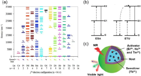
Figure 1.7 (a) Energy diagrams for Ln3+ in a LaCl3 lattice. (b) UC processes for lanthanide-doped crystals: ESA and ETU.
(c) Schematic illustration of UC nanoparticles composed of a crystalline host and lanthanide dopant ions (activator and sensitizer) embedded in the host lattice. Figure 1.7a was obtained from reference35.
Upconversion is a phenomenon by the conversion of low energy of long-wavelength radiation (NIR excitation), to higher energy short-wavelength emission, usually in the UV and visible range via the anti-Stokes optical process. In this process, upconversion is achieved by absorbing two or more excitation photons to generate one emission photon, which can be explained by two main mechanisms: excited state absorption (ESA) and energy transfer upconversion (ETU), as shown in Figure 1.7b. In a typical ESA process, the emitting ions in the ground state sequentially absorb at least two photons of suitable energy to promote to the higher excited state. The UC emission occurs when the electron returns back to the ground state in a radiative manner. The requirements for ESA are rigorous as the adequate population of the
29
intermediate state is required to capture the second photon. Unlike ESA, ETU process usually involves two types of luminescent centers (sensitizer and activator). In ETU, one photon of energy is absorbed by the activator, but the absorbed sequential photon energy transferred from the neighboring ions (sensitizer) result in the population of higher excited states of emitting ions. Radiative emission could be observed as the activator drops back to the ground state. ETU is the most frequently observed in the lanthanide-doped UCNPs since it is easier to achieve compared to ESA. The UC process benefits from the long lifetime of intermediate excited states of excited Ln3+ ions, typically in the μs to ms range. These long lifetimes allow
Ln3+ ions to be promoted to higher energy states by step-wise absorption of photons before returning to the
ground state, followed by the generation of higher energy photon.
Basically, the lanthanide-doped UCNPs consist of inorganic crystalline host and doped ions (Ln3+ ions)
acting as activators/sensitizers (Figure 1.7c). In addition to acting as a host matrix, the lattice has a strong influence on the UC of doped ions because it determines the distance between the doped ions, their coordination numbers and the anions surrounding the dopant. In order to achieve efficient UC, the optimal host should have low lattice phonon energy in order to reduce nonradiative losses, as well as high chemical stability. Of the available host materials for UC Ln3+ ions pairs, fluoride materials can fulfill these
requirements, which possess low phonon energies (ca. 350 cm-1) of the crystal lattice accompanied with the
long lifetimes of the excited states of Ln3+ ions.36 Among fluoride hosts, hexagonal (β-) phase NaYF 4 and
NaGdF4 are extensively studied as the most efficient UC host materials to date. It is believed that the high
emission intensity of Ln3+ ions in β-phase NaYF
4/NaGdF4 results from the interaction of doped ions located
at two different lattice sites.37 Regarding doped ions (Figure 1.7c), Yb3+ is normally selected as the sensitizer
in the ETU system due to its unique and simple energy level with only one excited level of 2F
5/2 (Figure
1.7a), which is in resonance with the energy of excited 980 nm photons; While Er3+, Tm3+ and Ho3+ are
typically employed as activators in UC system owing to the long lifetime of excited states and excellent resonance energy transfer with transition (2F
30
1.3 Research Objectives and Organization
1.3.1 Our objectivesThis thesis is divided into two parts with two highly related objectives. Specifically, two different main types of multifunctional nanoplatforms will be designed based on magnetic core. The first type is a core/shell structure with a fluorescence shell around a single magnetic core while the other multifunctional nanoplatform contains randomly distributed hundreds of magnetic and fluorescent NPs.
Part I: Multifunctional (superparamagnetic and NIR-to-NIR photoluminescent) nanocomposites with single magnetic core for dual-mode bioimaging.
So far, multifunctional (superparamagnetic and photoluminescent) NPs have been shown as extremely useful tools for bioapplications. One of the most interesting bioapplications is dual-mode imaging. It is well known that optical imaging shows great potential to translate into the clinic due to its high sensitivity at the subcellular level and the relatively low cost of imaging facilities; whereas MR imaging is considered as a superior technique for obtaining anatomical, physiological and functional images with high 3D spatial resolution. The integration of optical and MR imaging for dual-mode imaging is clearly advantageous as it will allow retrieving both macroscopic and subcellular information of biological species, thus leading to improved diagnostic accuracy. Thus, it is of great importance to engineer new, high-sensitivity multifunctional (superparamagnetic and photoluminescent) NPs for dual-mode imaging. However, multifunctional (superparamagnetic and photoluminescent) NPs in most cases are based on visible emitting organic dyes, QDs and UCNPs, which show low tissue penetration and severe autofluorescence from biological tissue, thereby limiting their use as bioprobe for in vivo imaging. To address this issue, the ideal photoluminescent components with their absorption and emission wavelengths in the optically transparent biological windows (NIR-I/II) are highly desired. This can ensure that both the excitation and emission signals are less attenuated for acquiring deep-tissue imaging with higher signal-to-noise ratio.
Therefore, the objectives for this part are as follows:
1. Preparing and characterizing novel multifunctional (superparamagnetic and photoluminescent) core/shell/shell Fe3O4/SiO2/NaYF4:Nd3+ NPs.
2. Investigating the NIR-to-NIR photoluminescence properties of Fe3O4/SiO2/NaYF4:Nd3+ NPs.
3. Comparing the deep-tissue penetration properties of NIR-to-NIR Fe3O4/SiO2/NaYF4:Nd3+ NPs
with visible-emitting upconverting Fe3O4/SiO2/NaYF4:Er3+, Yb3+ NPs by ex vivo optical imaging.
31
Part II: Multifunctional nanoplatforms based on multiple magnetic NPs and NIR QDs with therapeutic modality
The development of advanced multifunctional theranostic nanoplatforms is critical for addressing the challenges with cancers treatment, aiming to realize the ultimate goals of personalized medicine. These systems are expected to simultaneously carry both diagnostic and therapeutic functions to get high-quality images identifying the tumor site/morphology and to achieve the enhanced cure rate of cancers through specific therapeutic strategies. The survival rate can be largely increased using these multifunctional theranostic nanoplatforms. Specifically, compared with optical imaging and MR imaging alone, as individual diagnostic tools, bimodal imaging has shown to be particularly attractive because they can provide complementary information, as we mentioned above. Regarding the nanoplatform-related therapeutic modalities, magnetothermal therapy is hindered by poor thermal energy transfer efficiency of currently reported magnetic NPs, while photothermal therapy is not suitable for cancer cells in distant organs due to the limited penetration depth of light into the tissue. Thus, the integration of magnetothermal and photothermal therapy into a single nanoplatform may provide a dual-mode therapeutic approach to achieve high-efficiency and deep-tissue cancer treatment. In addition, conventional chemotherapy against cancers often suffers from the low therapeutic efficacy (since the mono-therapy approach cannot cure cancer in a synergistic or an additive manner) and severe side effects due to uncontrollable drug release characteristics. Multifunctional (superparamagnetic and photoluminescent) nanoplatform with drug-delivery modality can improve the performance of chemotherapeutic agents and reduce the overall toxicity by enhancing the specificity of the drug delivery through tumor targeting in the presence of an applied magnetic field. Furthermore, designing external stimulus-responsive nanoplatform for remotely controlled cancer treatment can potentially avoid drug overdose and reduce side effects.
Therefore, the objectives for this part are as follows:
1. Preparing and characterizing self-assembled Fe3O4 and PbS/CdS (NIR-II) supernanoparticles
[SASNs (NIR-II)], and studying their self-assembly formation mechanism.
2. Investigating the application of SASNs (NIR-II) for deep-tissue bimodal (optical and MR) imaging. 3. Studying the dual-mode (magnetothermal and photothermal therapy) heating treatment using
SASNs (NIR-II).
4. Synthesizing and characterizing large-pore mSiO2 and mSiO2 based multifunctional
(superparamagnetic and photoluminescent) nanoplatform (mSiO2@PbS/CdS-Fe3O4).
5. Monitoring the multi-stimuli (pH/MF/NIR) responsive drug release behavior of mSiO2
32
6. Studying the synergistic effect from the integrated heating mode and multi-stimuli responsive drug release of mSiO2@PbS/CdS-Fe3O4.