WHEN IS ‘‘BRAINSTEM DEATH” BRAIN DEATH? THE CASE FOR ANCILLARY
TESTING IN PRIMARY INFRATENTORIAL BRAIN LESION
Uwe Walter a,b, José Luis Fernández-Torre c,d, Timo Kirschstein a,e, Steven Laureys f
a Department of Neurology, University of Rostock, Rostock, Germany
b Center for Transdisciplinary Neurosciences Rostock (CTNR), University of Rostock, Rostock, Germany c Department of Clinical Neurophysiology, Marqués de Valdecilla University Hospital, Santander, Spain d Biomedical Research Institute (IDIVAL), Santander, Spain
e Oscar Langendorff Institute of Physiology, University of Rostock, Rostock, Germany
f Coma Science Group, GIGA-Consciousness and Neurology Department, University and University Hospital of Liège, Liège, Belgium
KEYWORDS: Brain death, Infratentorial brain lesion, Locked-in syndrome, Reticular formation, Electroencephalography, Cerebral circulatory arrest
HIGHLIGHTS : The ‘‘brainstem death” syndrome is clinically indistinguishable from apneic total locked-in syndrome. - For primary infratentorial brain lesions ancillary testing is mandatory for diagnosis of brain death. - For this, proof of either, electro-cortical inactivity or cerebral circulatory arrest, is suggested.
ABSTRACT
The widely accepted concept of brain death (BD) comprises the demonstration of irreversible coma in combination with the loss of brainstem reflexes and irreversible apnea. In some countries the combined clinical finding of coma, apnea, and loss of all tested brainstem reflexes (‘‘brainstem death”) is sufficient for diagnosing BD irrespective of the primary location of brain lesion. The present article aims to substantiate the need for ancillary testing in patients with primary infratentorial brain lesions. Anatomically, the ‘‘brainstem-death” syndrome can theoretically occur without relevant lesion of the mesopontine tegmental reticular formation (MPT-RF). Thus, a brainstem lesion may cause an apneic total locked-in syndrome, a rare syndrome with preserved capability for consciousness, mimicking ‘‘brainstem death”. Findings in animals and humans have shown that alpha- or alpha/theta-EEG patterns in case of isolated brainstem lesion indicate intactness of relevant parts of the MPT-RF. In such patients the presence of irreversible coma has to be doubted, and the potential capacity for some degree of consciousness cannot be excluded as long as the EEG activity persists. Consequently the demonstration of either ancillary finding, electrocortical inactivity or, preferably, cerebral circulatory arrest, is mandatory for diagnosing BD in patients with a primary infratentorial brain lesion.
Introduction
The biological concept of the irreversible cessation of brain function (brain death, BD) occurring while cardiorespiratory function is maintained has been issued already by M. F. Xavier Bichat (1800) and Harvey Cushing (1902), and was systematically developed since the 19500 s (Mollaret et al., 1959; Wertheimer et al., 1959; Beecher, 1968, Wijdicks et al., 2010). The primary goal was to establish medico-legal criteria for the termination of mechanical ventilation and intensive medical care in severely brain damaged patients with irreversible coma and fatal outcome. Later the criteria of BD were also introduced as a pre-condition to be fulfilled in heart-beating donors before organ harvesting. The meanwhile widely accepted concept of BD comprises two major criteria: i) the demonstration of the irreversible loss of the capacity for consciousness (irreversible coma), and ii) the demonstration of irreversible loss of the ability of breathing, i.e. the central drive to breathe (irreversible apnea) (Shemie et al., 2014). To substantiate the irreversibility of (i) and (ii), the clear diagnosis of the underlying severe brain damage as well as the complete absence of distinct brainstem reflexes are to be documented medically (Laureys and Fins, 2008). If the two criteria (i, ii) are fulfilled regardless of the artificial maintenance of cardiorespiratory function, such a human corpse can be regarded being dead from the biological point of view (Pallis, 1990; Shemie et al., 2014). Since the brain is the only irreplaceable body part to constitute the human personality (Alcaro et al., 2017; Lee, 2016), the equation of BD and death can be accepted also from an anthropological perspective (Bonelli et al., 2009; Moschella, 2016). The medically lege artis diagnosed BD is associated with irreversible cardiac arrest that occurs in less than an hour when artificial means of support are removed (López-Navidad, 1999). If intensive medical care is maintained after the diagnosis of BD, spontaneous cardiac arrest classically occurs within a few days and autopsy classically shows a liquefied ‘‘respirator brain” (Walker et al., 1975). In a large study in the United Kingdom (UK) asystole was found to occur in 78% of BD cases within 72 h (Jennett et al., 1981). The latency between BD diagnosis and asystole can be prolonged by increasing medical support (Yoshioka et al., 1986), and may in rare cases (e.g., pregnancy) extend to several weeks or months – or exceptionally even years (Shewmon, 1998). Even though the concept of BD is uniform worldwide the practical codes for the determination of BD differ between countries. A part of these differences are related to organisatory/authoritative issues that could be harmonized at European or international levels (Citerio et al., 2014; Shemie et al., 2014; Wahlster et al., 2015). However, dissent exists over the need of ancillary testing, using electrophysiological or cerebrovascular imaging techniques, to prove the loss of forebrain function in an individual with a primary infratentorial brain lesion fulfilling the clinical criteria of BD. Here, the question is whether the primary location of the brain lesion (supratentorial vs. infratentorial) matters, or not (Fig. 1). Historically, this dissent developed in Europe in the 1980s after the medico-legal acceptance of the ‘‘brainstem death” (BSD) concept in the UK (Department of Health and Social Security Great Britain, 1979). The BSD is diagnosed irrespective of the primary location of the brain lesion, at the presence of deep coma and proven apnea along with the absence of all tested brainstem reflexes, and ancillary testing is normally not required. Reports of cases with infratentorial brain lesions showing a near normal electroencephalogram (EEG) in combination with well-preserved cortical
visual evoked potentials after the diagnosis of BSD lead to stricter BD codes in other European countries (Frowein et al., 1987; Haupt and Rudolf, 1999; Bohatyrewicz et al., 2009). These stricter BD codes include the obligatory demonstration of electro-cortical silence (on EEG) and/or cerebral circulatory arrest (on cerebral angiography, perfusion scintigraphy, or Doppler sonography) in patients with primary infratentorial lesion. In the USA, guidelines emphasizing the importance of EEG (the ‘Harvard criteria’) and others de-emphasizing it (the ‘Minnesota criteria’) existed in parallel for many years (Beecher, 1968; Mohandas and Chou, 1971). However, the practice guidelines of the American Academy of Neurology (AAN) issued in 1995, confirmed in 2010, are close to the UK BSD criteria, especially in that there is no discrimination of a primary infratentorial from a primary supratentorial brain lesion, and no recommendation of ancillary testing if the clinical BSD criteria are fulfilled (Wijdicks, 1995, 2010, 2012; Wijdicks et al., 2010). On the other hand, alpha- or alpha-/theta-EEG activity persisted for at least 20 hours on EEG in 1.4% of 289 cases lege artis diagnosed with BSD, and was only seen following primary infratentorial brain lesions (Fernández-Torre et al., 2013). This raised the question what may be the motivation of EEG testing in cases of BSD (van Dijk, 2013). The present article addresses this question and aims to substantiate the need for ancillary testing in patients with primary infratentorial brain lesion. The following questions are discussed:
1. Which brain structures are minimally affected with the clinical diagnosis of BSD ? 2. Which brain structures seem essential for some degree of consciousness in humans ? 3. Can clinically diagnosed BSD in a patient with a primary infratentorial brain lesion be
differentiated clinically from a complete locked-in syndrome ? 4. Which brainstem structures influence global EEG activity ?
5. What does alpha-EEG activity indicate in patients with isolated brainstem lesion ?
6. Why are there caveats against ancillary testing using EEG, and how are these issues resolved ?
Figure 1. The three types of brain lesions that may lead to loss of brainstem function (the ‘‘brainstem death” syndrome) and potentially brain death (BD).
A) A primary supratentorial brain lesion (e.g. hemorrhage, ischemia, trauma) is so severe and widespread that it gradually leads to transtentorial herniation with compression of the upper brainstem, followed eventually by complete rostrocaudal herniation and medullary function loss, including breathing.
B) A primary infratentorial brain lesion (e.g. hemorrhage, ischemia, trauma) is so severe and widespread that it leads to loss of brainstem function and occlusion of the aqueduct with progressive supratentorial hydrocephalus and gradual loss of supratentorial brain function.
C) A secondary brain lesion (e.g. hypoxia after cardiac arrest) leads to loss of function of both supratentorial brain and brainstem at the same time, without necessarily developing massive cerebral edema and central herniation. In the two situations (A) and (C) both supratentorial and infratentorial function are lost, and both ‘‘brainstem death” and BD criteria are met. In the situation (B) an apneic total locked-in syndrome cannot be discriminated from BD with clinical investigation only; ancillary testing is necessary to prove the loss of supratentorial brain function.
Which brain structures are minimally affected with the
clinical diagnosis of BSD ?
The clinical diagnosis of BSD is established, after the exclusion of potential confounders (e.g. hypothermia, endocrine or metabolic causes, intoxication, sedation, relaxation, neuromuscular disorders), by demonstrating the presence of irreversible coma with complete absence of brainstem reflexes and formal apnea testing showing the absence of any spontaneous respiratory response to increasing hypercapnia for 5 minutes after disconnection from respirator (Academy of the Medical Royal Colleges, 2008; Pallis, 1982). The waiting period between the time of the acquired acute brain lesion and the clinical diagnosis of BSD is at the discretion of the diagnosing physician according to the UK code (Academy of the Medical Royal Colleges, 2008), while in the AAN code a waiting period of ‘‘usually several hours” is stated (Wijdicks et al., 2010). One clinical investigation is regarded being sufficient irrespective of the primary location of the brain lesion in the UK, and in most US states. This differs from the BD codes of many European countries where either ancillary testing is obligatory irrespective of the primary location of the brain lesion, or ancillary testing is obligatory in primary infratentorial brain lesion (Haupt and Rudolf, 1999; Bohatyrewicz et al., 2009). Therefore, this section deals with the brain structures minimally affected with prolonged coma, with a state clinically mimicking coma, with loss of the tested brainstem reflexes, and with apnea in the case of a primary infratentorial brain lesion. In a case series including 47 patients with isolated brainstem lesions of whom 9 had coma, long-duration coma has been associated with bilateral lesion of either the upper pontine tegmentum or both, the upper pontine and mesencephalic tegmentum (Parvizi and Damasio, 2003). The lesions of the upper pontine tegmentum involve the raphe complex, locus coeruleus and subcoeruleus (LC), laterodorsal tegmental nucleus (LDT), pedunculopontine tegmental nucleus (PPT), nucleus pontis oralis (PO) and the lateral and medial parabrachial nuclei (LPB, MPB). The local connections between these nuclei and the ascending central tegmental tract were also severed. The maximum lesion overlap area in patients with coma coincided with the location of the nuclei PO, LC, raphe, LDT and LPB/MPB (Parvizi and Damasio, 2003) (Fig. 2) – of note, most of the reported patients later regained consciousness. However, the findings of a recent voxel-based lesion-symptom mapping study confirm the obligatory involvement of rostral dorsolateral pontine tegmentum in brainstem coma (Fischer et al., 2016). The BD (or BSD) codes in most countries worldwide including the USA, UK and continental European countries require the testing of the following
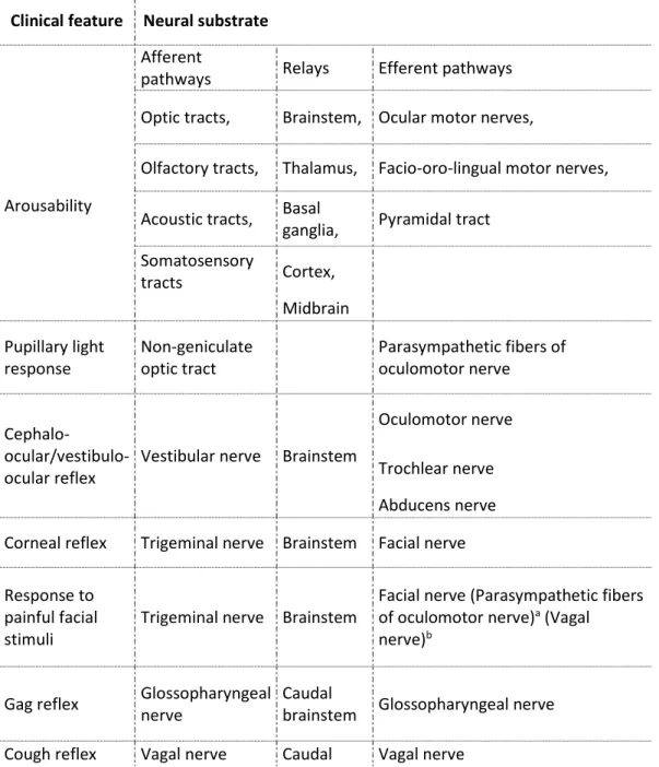
brainstem reflexes that need to be absent bilaterally: pupillary response to bright light, cephalocular reflex/vestibulo-ocular reflex, corneal reflex, grimace to painful stimuli in the face (trigeminal nerve), gag reflex, and cough reflex, along with the prove of apnea (Table 1) (Academy of the Medical Royal Colleges, 2008; Wahlster et al., 2015; Wijdicks et al., 2010). Fig. 2 shows the functional neuroanatomy of these brainstem reflexes including those related to the central drive to breathe (Afifi and Bergman, 1998; Blanco et al., 2013; Horn-Bochtler and Büttner-Ennever, 2011; Parvizi and Damasio, 2003; Zec and Kinney, 2001). It is evident that a lesion affecting all of these brainstem reflexes does not necessarily involve the mesopontine dorsolateral tegmental reticular formation or their connections to the supratentorial brain regions (Fig. 3).
Figure 2. Principal anatomy of brainstem functions assessed clinically for the determination of ‘‘brainstem death” and brain death.
Note the topographical dissociation between the tested brainstem reflexes and the mesopontine tegmental reticular formation. The anatomy of the tested brainstem reflexes is shown with distinct colours: pupillary light response – yellow (light, ellipse: pretectal nucleus; light, line: afferent visual pathway; dark, ellipse and line: Edinger-Westphal nucleus of oculomotor nerve with efferent pathway); cephalo-ocular/vestibulo-ocular reflex – green (light, ellipse: vestibular nuclei; light, line: afferent and internuclear pathway including the medial longitudinal fascicle; medium, ellipse and line: abducens nucleus with efferent pathway; dark: somatic motor nucleus of oculomotor nerve with efferent pathway; turquois: trochlear nucleus with efferent pathway); corneal reflex and facial motor response to trigeminal pain – red (light: main sensory and spinal nucleus of trigeminal nerve with afferent and internuclear pathways; dark: motor nucleus of facial nerve with efferent pathway); gag reflex – blue (light, bar and line: solitary nucleus with afferent and internuclear pathway; dark, bar and line: ventral motor nucleus of vagal nerve with efferent pathway); cough reflex and drive to breathe – blue/purple (light blue, bar: solitary nucleus; light purple, ellipse: dorsal respiratory group; light purple, line:
afferent and internuclear pathways; dark blue, bar and line: ventral motor nucleus of vagal nerve with efferent pathway; dark purple, ellipse: ventral respiratory group including chemosensitive neurons in the retrotrapezoid nucleus; dark purple, line: efferent pathway to cervical motor neurons). The black circles indicate the origins of the involved cranial nerves at the surface of the brainstem. A) Ventral aspect. Medium grey: mesopontine reticular formation; dark grey: neuromodulatory nuclei of the reticular activating system (LC = locus coeruleus and subcoeruleus; LDT = laterodorsal tegmental n., LPB = lateral parabrachial n., MPB = medial parabrachial n., PO = n. pontis oralis; PPT = pedunculopontine tegmental n.). B) Lateral aspect. Light grey: periaqueductal grey matter.
Table 1 : Brain functions that are to be absent on clinical assessment as a prerequisite for diagnosing brain death with the either concept, ‘‘brainstem death” and brain death.
Clinical feature Neural substrate
Afferent
pathways Relays Efferent pathways
Arousability
Optic tracts, Brainstem, Ocular motor nerves,
Olfactory tracts, Thalamus, Facio-oro-lingual motor nerves,
Acoustic tracts, Basal
ganglia, Pyramidal tract Somatosensory tracts Cortex, Midbrain Pupillary light response Non-geniculate optic tract Parasympathetic fibers of oculomotor nerve Cephalo- ocular/vestibulo-ocular reflex
Vestibular nerve Brainstem
Oculomotor nerve
Trochlear nerve Abducens nerve Corneal reflex Trigeminal nerve Brainstem Facial nerve
Response to painful facial stimuli
Trigeminal nerve Brainstem
Facial nerve (Parasympathetic fibers of oculomotor nerve)a (Vagal nerve)b
Gag reflex Glossopharyngeal nerve
Caudal
brainstem Glossopharyngeal nerve Cough reflex Vagal nerve Caudal Vagal nerve
brainstem Bulbospinal axons of brainstem respiratory neurons (via ventral and lateral spinal funicles)
Breathing drive Brainstem chemoreceptors, Vagal nerve (?) Caudal brainstem
Bulbospinal axons of brainstem respiratory neurons (via ventral and lateral spinal funicles)
aTrigemino-pupillary reflex (not obligatorily assessed). bTrigemino-autonomic reflex (not obligatorily assessed).
Box 1 : The loss of all brainstem reflexes in combination with apnea, tetraplegia, anarthria, bilateral ptosis and complete ophthalmoplegia can theoretically occur from an anatomic point of view without complete destruction of the mesopontine tegmental reticular formation.
Which brain structures are essential for some degree of
consciousness in humans ?
This challenging question has been addressed in many studies and review articles (e.g., Giacino et al., 2014; Gosseries et al., 2014). The evidence summarized in a recent review article suggests that the brainstem reticular formation, paramedian thalamus and parts of the temporo-parieto-occipital associative cortices provide the background conditions for conscious awareness (Koch et al., 2016). Voxel-based analyses between brain metabolic scans obtained in awake yet unaware patients compared to healthy controls (between-subject) or comparisons with recovery of awareness (within-subject) pointed towards the critical role of a widespread fronto-temporo-parietal associative cortical network in the recovery of conscious awareness (Thibaut et al., 2012). Based on a review of functional brain imaging data in patients with disorders of consciousness it has been suggested that the ‘external awareness’ network encompasses lateral fronto-temporo-parietal cortices bilaterally, and the ‘internal awareness’ network includes midline anterior cingulate/mesiofrontal and posterior cingulate/precuneal cortices (Demertzi et al., 2013). While the exact cortical regions required for conscious awareness are still under debate the data available from multimodal neuroimaging studies have substantiated the essential involvement of associative cerebral cortices in the generation of any conscious percept. In turn, a number of animal experiments as well as clinical studies on extended patient series have shown that a healthy cerebral cortex cannot by itself (i.e. without out any input from thalamus and brainstem) maintain the conscious state (Cairns, 1952). A recent quantitative EEG study in 83 patients with severe subarachnoid hemorrhage who were in different stages of coma (Glasgow Coma Scale score 5–8), with accompanying brainstem lesions in only three of them, underpinned the idea that impaired connectivity of cortex with both central thalamus and basal forebrain underlies decreasing levels of consciousness (Claassen et al., 2016). In the context of propofol-induced
sedation of humans the breakdown of pallido-cortical connectivity within the cortico-basal ganglia-thalamo-cortical loop was the main factor characterizing the absence of consciousness (Crone et al., 2017). A continuous isoelectric EEG, irrespective of its cause (e.g. brain disorder, pharmacological effect), reflects an interruption of endogenously-generated activity in cortical networks and systematically results in a complete dissolution of conscious processes (Altwegg-Boussac et al., 2017). Persistent electro-cortical inactivity has been classified as the most severe grade of coma (Synek, 1988) and is, together with a lack of responsiveness to various stimuli, considered the classical electrical sign of BD (Laureys, 2005a). Functional MRI studies, assessing the functional connectivity among default network areas by correlations of spatially separated bloodoxygen-level-dependent (BOLD) signals at a given time, confirmed a preserved cortico-cortical and cortico-thalamic connectivity in four patients with minimally conscious state, a preserved corticocortical but absent cortico-thalamic connectivity in five unconscious patients in an unresponsive wakefulness syndrome (previously coined vegetative state), a severely reduced cortico-cortical connectivity in five coma patients and the absence of any connectivity in a BD patient with a primary supratentorial hemorrhage (Boly et al., 2009; Vanhaudenhuyse et al., 2010). Even though the functional MRI technique does not directly measure neuronal activity (unlike EEG), these studies, among others, underpin the notion that cortico-thalamic connectivity needs to be present in order to establish the ability of being conscious, or being capable of having any percept. The overall level of activity in the thalamo-cortical system, in turn, is thought to be regulated by the ascending arousal system. It is known since Moruzzi and Magoun (1949) that extended brainstem lesions typically cause immediate coma by damaging the reticular activating system. Subsequent studies showed that lesions at the level of the rostral pons, but not in the midpons or more caudally, could cause coma both in animals and in humans (Batini et al., 1958, 1959; Parvizi and Damasio, 2003). Two parallel pathways have been identified to arise from mesopontine nuclei to activate the thalamus and cerebral cortex (Steriade, 2003; Brown et al., 2012). First, the dorsal pathway comprising reticular formation glutamatergic and cholinergic neurons in the PPT and LDT nuclei innervates the midline and intralaminar (nonspecific) thalamocortical neurons that, in turn, release glutamate at the cortical level. Second, the ventral path comprising fibers of the medial forebrain bundle passes through the midbrain, posterior/lateral hypothalamus, and basal forebrain on the way to the cortex. In rodents, cell-specific lesions of parabrachial (LPB/MBP) but not neighboring brainstem neurons caused a coma-like state, suggesting that this nucleus may be particularly important in maintaining wakefulness (Fuller et al., 2011). The role of the parabrachial complex in the generation of wakefulness drive and influence on respiratory control is increasingly recognized (Martelli et al., 2013; Ikeda et al., 2017). Taken together, current evidence supports the view that the reticular neurons located in the lateral tegmentum (Fig. 2) are key players in the transition from arousability to nonarousability (coma). On the other hand, neurological patients with a severely damaged cortex, but with relatively spared brainstem function, typically remain in an unresponsive/vegetative state. This suggests that brainstem activity without cortical activity is insufficient to sustain consciousness in a clinical sense. Based on a comparison of animal and human brain functional anatomy (Merker, 2007), or the observation of emotional-like behavior in decorticate animals (Solms, 2017), it has been suggested that a non-declarative, primitive (affective) form of consciousness is possible in humans without any cortical function, operated by the centrencephalic (mesodiencephalic) system, comprising the thalamus/
hypothalamus, the periaqueductal gray, and the superior colliculus. This suggestion, however, has been refused by others (e.g., Doesburg and Ward, 2007; Edelman, 2007). Especially, the exemplary patients proposed by Merker and others to represent consciousness despite ‘‘decortication” all had some preserved functioning cerebral cortex (Aleman and Merker, 2014; Shewmon et al., 1999). It has been shown that unawareness induced by anesthesia correlates more closely with the impairment in cortical function than with thalamic activity (Boly et al., 2012). In turn, vivid sleep-onset hallucinations are still experienced when the thalamus is already deactivated (Magnin et al., 2010). And finally, in patients with disorders of consciousness, the ability of being at least minimally conscious is indicated specifically by cortically mediated behaviours (Naccache, 2018).
Figure 3. Schematic illustration of a primary infratentorial (isolated brainstem) lesion leading to an apneic total locked-in syndrome clinically mimicking the ‘‘brainstem death” syndrome.
This lesion is indicated be rosé color. Such a lesion would cause complete ophthalmoplegia, anarthria and tetraplegia, loss of all tested brainstem reflexes, and apnea, mimicking coma (‘‘complete unresponsivity”). Since however large parts of the mesopontine reticular formation (shown in grey colour) and their connections to the supratentorial brain structures are largely preserved this individual is likely to be conscious.
Box 2 : Mandatory structures to establish some degree of consciousness in humans are critical parts of the mesopontine reticular formation, its dorsal pathway to the thalamo-cortical and/or its ventral pathway to the corticocortical projection systems, and parts of the associative cerebral cortices.
Can clinically diagnosed BSD in a patient with a primary
infratentorial brain lesion always be differentiated
clinically from a complete locked-in syndrome ?
In patients with coma, testing brainstem reflexes is used as a proxy for brainstem function, while the capacity for breathing is tested using apnea testing. In the presence of a primary supratentorial brain lesion (Fig. 1A) or a secondary brain lesion (Fig. 1C) a severe forebrain lesion is combined with either the subsequent gradual loss of brainstem function due to rostrocaudal transtentorial brain herniation, or the brainstem is along with the forebrain severely affected by the secondary brain lesion (e.g. cerebral hypoxia). In these situations the clinical BSD criteria are reliable since the forebrain is severely damaged (Jørgensen, 1981). There is a minority of patients, however, who have a massive primary infratentorial brain lesion, for example, from basilar artery thrombosis or large brainstem or cerebellar bleed (Fig. 1B). These patients may clinically meet BSD criteria. They may, however, retain blood flow and even EEG activity (Ferbert et al., 1985, 1986a, 1986b, 1989; Fernández-Torre et al., 2013; Roth and Ferbert, 2016; Wagner et al., 1993; Zwarts et al., 2001), which may arguably be surrogate markers of potential residual consciousness in specific cases. So far, there are no identified patients who fulfilled BSD criteria but survived (Wijdicks, 2012). All this says is, in fact, that patients fulfilling the BSD criteria have a lethal prognosis. However, this does not rule out the possibility that a patient with a primary infratentorial brain lesion fulfilling the BSD criteria might still be conscious to some degree for some time. Especially, but not only, the two well-documented cases showing slow alpha-EEG activity after the diagnosis of BSD who survived for more than 48 hours give rise for such a concern (Ferbert et al., 1986a; Fernández-Torre et al., 2013). Varelas (2016) speculates that it remains possible, in theory, with modern intensive care unit support that some patients with primary brainstem destruction might recover consciousness despite fulfilling criteria for BSD, even though there are no such cases identified or reported to date. Especially, earlier reviews did not address reports suggesting the potential possibility of a total locked-in syndrome possibly being mistaken for BSD. The term locked-in syndrome (LIS) traditionally refers to patients who are totally paralyzed (ophthalmoplegia, anarthria, tetraplegia) but fully conscious and able to use vertical eye movements or blinking in order to communicate (Posner et al., 2007). LIS is not a disorder of consciousness, but can be (and often is) mistaken for one (Laureys, 2005a, 2005b; Bernat, 2006; Bruno et al., 2008; Schnakers et al., 2009). One the other hand, patients with LIS due to acute brainstem lesion may initially be comatose and gradually wake up during the following days, but remaining paralyzed and voiceless, superficially resembling patients in an unresponsive wakefulness syndrome (previously coined vegetative state) (Laureys et al., 2005). It has been shown that patients with LIS do not necessarily rate their quality of life as unmeaningful (Bruno et al., 2008, 2011; Linse et al., 2017; Lulé et al., 2009), which questions the preconceived idea of ‘‘dying is always better than living with LIS”. On the other hand, LIS patients fully experience the feelings of ‘‘hunger for air” if ventilator support is withdrawn (Heywood et al., 1996). Therefore, it is important to discriminate the LIS from coma in order to allow every LIS patient to tell whether she/he wants to live or die (Bruno et al.,
2008). Detecting signs of consciousness is particularly challenging in patients with a total LIS (t-LIS) which consists of complete immobility including all eye movements combined with preserved consciousness (Bauer et al., 1979; Schnakers et al., 2009). Usually, the pupillary reflex is preserved in LIS and t-LIS, and may be the only clinical brainstem reflex preserved (Ueyama et al., 1996). There are, however, single case reports of LIS in association with fixed dilated pupils (Meienberg et al., 1979), and a likely t-LIS in association with fixed pupils (Burns et al., 2008). LIS patients may also be apneic with permanent or transient failure of triggering the ventilator (Kotagal et al., 1984; Wijdicks and Scott, 1996). As behavioral tools may not always be sufficient for discriminating LIS from coma or even from BSD in rare cases (Table 2), documenting cerebral circulatory arrest to prove BD, or the use of electrophysiological tools such as the detection of EEG reactivity or cortical event-related potentials in order to facilitate the detection of signs of consciousness have been recommended (Hawkes and Bryan-Smyth, 1974; Schnakers et al., 2009; Laureys, 2005a). In principle, also quantitative EEG, quantitative high-density EEG and functional brain imaging (functional MRI, positron emission tomography) can be applied to discriminate patients with LIS or minimally conscious state from unconscious patients, however these methods are not widely available and may be logistically complicated in mechanically ventilated patients (Vanhaudenhuyse et al., 2010; Stender et al., 2014). Of note, a number of LIS patients have been reported who exhibited an unreactive alpha pattern on standard EEG (Jacome and Morilla-Pastor, 1990; Gütling et al., 1996), indicating that the neural substrates producing alpha reactivity might be dissociated from those responsible for arousability or awareness. A more recent study on 13 patients with LIS due to brainstem lesions and 15 control subjects showed a lowered power of cortical sources of alpha-rhythms in LIS (Babiloni et al., 2010). There is, however, no report of cortical inactivity on standard EEG in patients with LIS. Evidently, the detection of electro-cortical inactivity on EEG (provided the correct technique and the exclusion of confounders such as sedative medication or hypothermia) excludes the presence of LIS in an individual with the BSD syndrome.
Box 3: It is important to discriminate the LIS from coma in order to allow the LIS patient to tell whether she/he wants to live or die. There are reports of t-LIS with fixed pupils, complete ophthalmoplegia with bilateral ptosis, apnea, or unreactive alpha EEG pattern. This underlines the notion that the clinical BSD syndrome in patients with a primary infratentorial brain lesion cannot with 100% certainty be discriminated from t-LIS without ancillary testing. On the basis of existing evidence either finding, electro-cortical inactivity on EEG (after exclusion of confounders) and cerebral circulatory arrest (which is always associated with electrocortical inactivity), excludes the presence of a LIS.
Which brainstem structures influence global EEG
activity?
Alpha rhythms result from an interaction of thalamic and neocortical circuitry, together with a moderate level of brainstem cholinergic input (Brown et al., 2012). For the occipital alpha rhythm, local lateral geniculate GABAergic interneurons excited by highthreshold bursting thalamocortical neurons are important in periodically silencing thalamocortical neurons (Hughes and Crunelli,
2005; Lopes da Silva et al., 1980). The electro-cortical activity measured on scalp EEG reflects the summed excitatory postsynaptic potentials of pyramidal neurons generated by thalamocortical afferents that form synapses with the pyramidal neurons in the cerebral cortex. Non-specific thalamocortical afferents terminate at pyramidal neurons predominantly in the superficial cortical layers I to III, whereas specific thalamocortical afferents mostly terminate in the deep layers IV to VI (Kirschstein and Köhling, 2009). It has been shown that severe and extended anoxic lesions of the neocortical layers IV, V, and VI do not preclude the generation of dominant alpha EEG in comatose patients without gross pathologic changes of the thalamus and the brainstem (Vignaendra et al., 1974). This implies that unspecific thalamic inputs to the cortical layers I to III, driven by brainstem cholinergic input, are sufficient to sustain widespread cortical alpha activity. In patients with postanoxic alpha, theta and alpha-theta coma the probability of clinical recovery has been related to the presence of EEG reactivity, pointing towards the prognostic relevance of the capability of frequency shifting (Fernández-Torre et al., 2018). Mesopontine (pedunculopontine and laterodorsal tegmental) cholinergic reticular neurons are operational in shifting the state of thalamocortical neurons from EEG-synchronized sleep to EEG-desynchronization associated with wakefulness and REM sleep (Steriade et al., 1990; Hughes and Crunelli, 2005). Efferents from the pedunculopontine tegmentum exert widespread control over neocortical activity and aid in maintaining high-frequency (4– 40 Hz) electro-cortical activation, involving relays in both the basal forebrain and central thalamus. Animal studies suggest a predominant role of the basal forebrain which also receives a substantial input from glutamatergic neurons in the parabrachial nucleus and adjacent pre-coeruleus area (Dringenberg and Olmstead, 2003; Fuller et al., 2011). The cholinergic brainstem neurons involved in regulating the sleep-wakefulness states are largely located in the peribrachial regions of the caudal mesencephalic and rostral pontine tegmentum (Sakai et al., 1990). Selective lesions of the parabrachial-precoeruleus complex in rats produced behavioral unresponsiveness and a monotonous sub-1 Hz cortical EEG whereas extensive thalamic lesions had little effect on EEG or behavioral measures of wakefulness (Fuller et al., 2011). Taken together, the presence of widespread alpha-EEG activity requires a critical function of mesopontine reticular neurons and a critical, at least unspecific, activity of linked thalamic and neocortical neurons. The contribution of neuronal input from the brainstem on the function of the cerebral hemispheres has been investigated using the cerveau isolé (Bremer, 1935) animal model and several preparations of the encephale isolé (Lindsley et al., 1949). Arduini and Moruzzi (1953) demonstrated an olfactory EEG arousal reaction whereas visual stimuli failed to produce an arousal reaction in the cerveau isolé cat despite the presence of visually evoked potentials in the area striata. Batsel (1960) found in the EEG of the chronic cerveau isolé dog not only slow waves and spindles as in the original experiment of Bremer (1935) but also a desynchronized pattern similar to the spectrum of the normal waking dog which reappeared only many days after the lesioning. Of note, in this model olfactory arousal was observed only with incomplete midbrain transection, and then the arousal did not persist beyond the duration of stimulation (Batsel, 1960). In cats, lesion of the caudal brainstem as well as selective destruction of the medial and lateral lemnisci and spinothalamic tracts, on each side of the midbrain, did not diminish the electro-cortical activation pattern (10–50 Hz) of the encephale isolé (Lindsley et al., 1949). By contrast,
mesencephalic lesions which destroyed large areas of the tegmentum, reduced or abolished the 10–50 Hz electro-cortical pattern of activation and precipitated recurring bursts or spindles in the same model. Similarly, lesions destroying the sub- and hypothalamus, from the optic chiasma to the mammillary bodies or anterior midbrain, reduced or abolished EEG activation and the electro-cortical activity of the preparation subsequently consisted of bursts or spindles of high voltage slow waves, recurring rapidly and interminably throughout observation periods of several hours (Lindsley et al., 1949). Studies in the feline model showed that an electrolytic transection of the pons, performed just rostrally to the trigeminal rootlets (‘‘midpontine pretrigeminal” preparation), is followed by a low voltage fast electro-cortical activity in the beta band (around 40 Hz). This EEG pattern, which was very similar to the one characteristic of alert behavior in the normal cat, persisted invariably for most of the day (about 80 per cent of the time, as shown by continuous EEG recording), during the whole survival time of the animal (several days) (Batini et al., 1958, 1959). These animals were able to follow with vertical eye movements an object entering its visual field and sometimes reacted with pupillary dilatation to an emotional stimulus, such as the presentation of a dog or a mouse, indicating some degree of consciousness (Batini et al., 1959). Of note, such eye movements were absent whenever, sometimes occurring, EEG sleep patterns were being recorded (Batini et al. 1958). An opposite alteration of the sleepwakefulness rhythm was induced by slightly more rostral transections of the pons (pretrigeminal rostropontine preparations), which were followed by lasting patterns (up to 7 days) of EEG synchrony, associated with the usual ocular signs of sleep (such as myosis and lack of purposeful eye movements). Fig. 4 illustrates the typical EEG patterns related to brainstem lesion levels in the animal model.
Box 4 : The data obtained in animal studies suggest that near-normal (i.e. predominant alpha-, beta-, or theta-) EEG activity is unlikely to occur if significant amounts of the tegmental reticular neurons at the ponto-mesencephalic junction are destroyed.
What does alpha-EEG activity indicate in patients with
isolated brainstem lesion ?
In a recent study of 289 cases with clinically proven BSD with supra- and/or infratentorial brain lesion in ten (3.5 %) of these cases cortical neuronal activity was documented on EEG, most often in the alpha- or alpha-/theta-band, which persisted at least for several hours (Fernández-Torre et al., 2013). All of these patients had either a primary infratentorial brain lesion (n = 7) or a spontaneous subarachnoid hemorrhage (n = 3), which can be regarded as a combined primary infra- and supratentorial brain lesion. In four of the patients with a pure infratentorial lesion (one with basilar artery thrombosis, the other three with cerebellar hematomas) predominant alpha- or alpha/theta-EEG activity was found that persisted for longer than 48 hours in one of them. In the same study, 28 patients with primary infratentorial brain lesions had electro-cortical silence documented on the first EEG examination. These data suggest that in about 10% of the patients
with isolated infratentorial brain lesion, in whom BSD is clinically diagnosed, well-preserved alpha- or alpha-/theta-EEG activity is present. A number of similar cases, partly with documented BSD, are listed in Table 3 (Britt et al., 1980; Chatrian et al., 1964; Deliyannakis et al., 1975; Ferbert et al., 1985, 1986a, 1986b, 1989; Grigg et al., 1987; Hayashi et al., 1996; Kaukinen et al., 1995; Kuwabara et al., 1982; Loeb, 1958; Loeb and Poggio 1953; Lundervold et al., 1956; Obeso et al., 1980; Rodin et al., 1985; Roth and Ferbert, 2016; Steudel et al., 1979; Uldry et al., 1991; Wagner et al., 1993; Westmoreland et al., 1975; Wilkus et al., 1971; Zwarts et al., 2001). Some of these cases deserve more detailed consideration. It has been reported already in the 1950s that upper pontine lesions associated with deep coma can show diffuse 8–12 Hz alpha EEG activity with well-preserved amplitudes (Loeb, 1958; Loeb and Poggio, 1953). This condition has later been referred to as ‘‘alpha coma” EEG. Interestingly, revisiting the case reports of infratentorial brain lesions associated with alpha EEG, it turns out that none of these cases would have been diagnosed with BSD today. The first case reported by Loeb and Poggio (1953) with ‘‘deep coma” still had a bilaterally positive bulbomimetic sign, characterized by contraction of the facial muscles of expression caused by pressure on the bulbus oculi, engaging the same neurological tracts as the corneal reflex. In addition, in this case a spontaneous stertorous respiration, anisocoria (R > L), generalized flaccidity, hyporeflexia of the arms, relative hyperreflexia of the legs, bilateral Babinski sign, and right spinal synergia were noted. Other details on the presence or absence of brainstem reflexes, or on the progression of the clinical signs were not given. The EEG, 6 hours after the onset of coma, showed a rather regular rhythm at 8–9 Hz, 100 mV, with some low voltage fast activity especially bifrontal, and occasionally diffuse bursts at 3–4 Hz, and negative diphasic spikes. Also, bilateral driving of the cerebral rhythms by visual flicker stimulation could be demonstrated. This patient, not being artificially respirated, died only 36 hours later. Post-mortem anatomical examination showed a large hemorrhage in the central portion of the pons, extending to the most caudal portion of the mesencephalon. Another case was reported by Lundervold et al. (1956) with a vascular ischemic lesion of large parts of lower and mid pontine brainstem including the bilateral pyramidal tracts and the medial lemnisci. The lesion however spared many parts of the pontomesencephalic tegmentum. This patient was found clinically to be ‘‘unconscious”, with hypotonic quadriplegia and without reaction to pain stimuli, neither from face nor from rest of body. However, the pupillary reaction to light and the corneal reflexes were preserved. This patient, after initial tracheostomy, survived and stayed apparently unconscious for one year and a half, and showed for several weeks a predominant 8-Hz alpha EEG. The EEG activity was, on repeated testing over several weeks, unresponsive to pain, acoustic or smell (camphor, peppermint) stimulation while on some recordings photic stimulation caused changes in frequency but no photic driving effect. This patient would not be diagnosed with BSD today, but rather with a t-LIS, given the preserved pupillary reaction to light and the EEG reactivity to visual stimuli. A case with brainstem infarction reported by Westmoreland et al. (1975) presented initially with a tetraplegic LIS when he could still respond to questions by blinking his eyes and by vertical eye movements. Four days later, his clinical state deteriorated and he became completely unresponsive (apparently comatose) with non-reactive pupils and absent oculocephalic and caloric reflexes. At this stage, well-preserved alpha-EEG activity during the day, a sleep EEG pattern
during the night, followed by alpha EEG on the subsequent day was documented. This patient died three days later; on postmortem pathologic investigation bilateral lesion of large parts of the rostral pontine tegmentum but no pathology of the mesencephalic tegmentum was found. It may be speculated that this patient might have had a t-LIS finally mistaken for coma. In a well-documented case of brainstem hematoma, with unilateral involvement of the mesencephalic tegmentum, the EEG 7 hours after onset of the coma showed a slow alpha activity while the EEG at the 7th day was dominated by generalized delta activity (Steudel et al., 1979). One may speculate that reactive inflammation/edema near the hematoma affected the function also of the contralateral mesencephalic tegmentum, thereby leading to slowing of EEG activity over time. There is similarity to the case with brainstem hemorrhage reported by Hayashi et al. (1996) in whom the bilateral pontine tegmentum but not the mesencephalic tegmentum was affected initially. This patient had a ‘‘severe impairment of consciousness”, flaccid tetraplegia and decerebrate posturing after painful stimulation, but preserved brainstem reflexes. EEG showed predominant 8- to 9-Hz alpha activity; photic stimulation elicited a normal driving response on EEG. These findings could be repeated after 7 days. On the subsequent day the clinical condition deteriorated markedly, which was related to a massive enlargement of the brainstem hematoma, involving the bilateral rostropontine and mesencephalic tegmentum. Now a SPECT investigation showed preserved cortical and subcortical perfusion indicating preserved thalamocortical circuits, and synchronized, delta activity dominated the EEG. In another case series of primary pontine hemorrhages a prolonged state of coma or LIS was associated with lesion of the bilateral pontine tegmentum (Kuwabara et al., 1982). In two out of these patients who remained severely disabled in prolonged coma or the LIS, EEG in the first days showed alpha- or even beta activity that changed, after 2–3 weeks, into low-voltage theta activity. In a study on 15 patients with spontaneous brainstem hemorrhages involving the pontine tegmentum but mostly sparing the mesencephalic tegmentum slow alpha or mixed alpha-theta activity unreactive to flicker stimulation was typically found (Ferbert et al., 1990). In some cases, serial recordings showed a progressive slowing of the EEG activity. The findings of these cases suggest that predominant alpha- or alpha-/theta-EEG activity can be present with partial lesion of the bilateral rostral pontine tegmentum but not with bilaterally severe damage of the rostropontine and mesencephalic tegmentum. Of note, none of the cases described here in detail had a documented clinical BSD syndrome. Even though there are a number of case reports of clinical BSD in association with primary infratentorial brain lesion in whom alpha- or fast theta-EEG activity persisted for one or more days (Table 3), the details on the degree of pathology of the pontine and mesencephalic tegmentum are missing.
Figure 4. Schematic illustration of the relationship between complete transection of the brainstem at different levels and the cortical EEG pattern found in the feline and the canine animal model (Batini et al., 1958, 1959; Batsel, 1960; Lindsley et al., 1949).
The corresponding anatomy has been adapted here to the human anatomy of brainstem (lateral view). The mesopontine tegmental reticular formation is indicated in medium grey, and the periaqueductal grey matter in light grey colour. Note that ‘‘desynchronized” here refers to fast-wave, awake-like EEG patterns (>4 Hz), while ‘‘synchronized” refers to slow-wave (<4 Hz), sleep-like EEG patterns.
Table 3. Clinical and EEG findings in reported cases with localized brainstem lesions causing coma and/or the clinical ‘‘brainstem death” syndrome. Reference; case No. a Age; gender [y; M/F] Lesion etiology Lesion location Ms/P/Md PoTg lesion +/(+)/− MsTg lesion +/(+)/− Coma duration b [h] GCS score PRL +/− COR/VOR +/− CR +/− TPR +/− GR/TR +/− SDB +/− BSD +/− EEG frequ. [Hz; uV] Post-EEG survival d [d] Loeb (1953); 1 57; F Hemorrhage Ms/P ? ? 6 ? ? ? + ? ? + − 8–9; 100 1e Lundervold et al. (1956); 1 63; M Ischemia Ms/P (+) (+) 24 3 + ? + − ? ? − 8; 20 550 Loeb (1958); 14 60; M Hemorrhage P + − 4 3 ? ? − − ? + − 9; 30 ?e Loeb (1958); 18 57; F Hemorrhage Ms/P + (+) 3 3 ? ? − − ? + − 7–8; 20 ?e Chatrian et al. (1964); 1 59; M Ischemia Ms/P/M + (+) 140 4 + − ? − ? ? − 8–9; 25 8 Wilkus et al. (1971); 1 33; M Ischemia P (+) − 310 4 + − + + + + − 8–9; 20 51 Deliyannakis
et al. (1975); 1 21; M Bacterial meningitis ?f ?f ?f 48 3 − ? ? − ? ? ? 1–2; 20 13
Westmoreland
et al. (1975); 1 56; M BA thrombosis Ms/P + − ∼18 g 3 − − ? − ? ? ? 8–9; 30 3
Steudel et al.
Britt et al.
(1980); 1 14; M Trauma/hemorrhage Ms ? + 330 4 + + + + + + − 0.5–1.0 >4
Obeso et al.
(1980); 1 72; F BA thrombosis P (+) − ? 3 + − ? ? ? + − alpha 10
Kuwabara et
al. (1982); 11 45; M Hemorrhage P + − 24 3 + − ? ? ? + − beta; 10 >365i
Kuwabara et
al. (1982); 13 39; M Hemorrhage P + − 20 3 + − ? − ? + − 8–9; 20 >300i
Ferbert et al. (1985); 1 52; M BA thrombosis Ms/P ? ? 3 3 − − − − − − + theta 1 Rodin et al. (1985); 1 71; F Hemorrhage Ms/P + ? 12 3 − − − − ? − j ? 5–6; 50 15k Ferbert et al. (1986a); 1 46; M Hemorrhage P ? ? 6 3 − − − − − − + 8–9; 50 3 Ferbert et al. (1986b); 1 55; M BA thrombosis Ms/P ? ? 14 3 − − − − − − j + 5–6; 20 1 Ferbert et al. (1986b); 2 47; M BA thrombosis Ms/P ? ? 2 3 − − − − − − + 7–8; 20 1
Grigg et al. (1987); 9 71;? Ischemia Ms/P ? ? 7 3 − − − − − − j ? 4–5; 30 2 Grigg et al. (1987); 10 63; M Ischemia Ms/P ? ? 168 3 − − − − − − j ? 4–5; 30 7 Ferbert et al. (1989); 1 70; F BA thrombosis Ms/P ? ? 49 3 − − − − − − + 5–6; 70 2 Uldry et al. (1991); 18 60; M Hemorrhage Ms/P ? ? 5 4 ? ? ? ? ? ? - 8; 30 1 Wagner et al. (1993); 1 67; M CB hemorrhage Ms/P/Md ? ? 2 3 − − − − − − + “activity” 2l Kaukinen et al. (1995); 1 46; M Trauma/hemorrhage Ms/P/Md ? ? 4 3 − − ? − ? − ? 8–9; 40 2k Hayashi et al. (1996); 1 54; F Hemorrhage Ms/P + + ∼1 3 − ? ? ? ? + − 3–4; 100 6e Zwarts et al. (2001); 1 31; M Hemorrhage Ms/P + ? ? 3 − − − − − − j + 7–8; 20 <1k Roth and Ferbert (2016); 1 41; M Infratentorial SAH Ms/P/Md ? ? 48 3 − − - - - - + 8–9; >10 1l
BA indicates basilar artery; BSD, clinical syndrome of brain stem death; CB, cerebellar; COR, cephalo-ocular reflex; CR, corneal reflex; EEG, electroencephalogram; GCS, Glasgow Coma Scale; GR, gag reflex; Md, medulla oblongata; Ms, mesencephalon; MsTg, mesencepehalic tegmentum; P, pons; PoTg, pontine tegmentum; PRL, pupillary reaction to light; SAH,
subarachnoid hemorrhage; SDB, spontaneous drive to breathe at arterial paCO2 >60 mmHg; TPR, mimic reaction to trigeminal pain; TR, tracheal cough reflex; +, present/major; (+), minor; -, absent.
a Numbered as 1 if only one case reported, otherwise the case number given in the referring article. b Duration of the deepest coma stage found clinically it this case until recording of the index EEG.
c Dominating EEG frequency with respective amplitude in the central-posterior region; note that fast theta-rhythm may (in some cases) actually be a slowed alpha-rhythm. d Time to spontaneous cardiac arrest under mechanical respiration (if not indicated otherwise) after recording of the index EEG.
e Spontaneous cardiac arrest without mechanical respiration. f Brainstem liquefied at post-mortem anatomical investigation.
g Time since the progression from initial locked-in syndrome to (pseudo-?) coma.
h Mesencephalic tegmentum unilaterally involved by the hematoma; EEG at the 7th day showed 1- to 2-Hz, 70- to 100-uV activity. i Survival without mechanical respiration.
j Arterial paCO2 for apnea testing not reported.
k Spontaneous cardiac arrest after termination of life support systems. l Declaration of brain death after ancillary testing.
Box 5 : The finding of predominant alpha- or alpha/theta-(as opposed to slower, less than 4-Hz) EEG pattern in non-sedated patients with isolated brainstem lesions indicates that
relevant parts of the reticular activating system of the rostral pons and midbrain are intact. Therefore, in such patients the presence of an irreversible coma has to be doubted, and the
Why are there caveats against ancillary testing using
EEG, and how are these issues resolved ?
First, it has been argued that the outcome of the clinical BSD syndrome is independent from EEG findings. Indeed, there is no scientific evidence so far showing a case with neurologic recovery after the clinical diagnosis of BD has been established using the criteria given in the 1995 AAN practice parameter that are close to the UK BSD criteria (Wijdicks, 1995, 2012; Wijdicks et al., 2010). As mentioned above, the prognosis after diagnosing BSD is lethal with spontaneous asystole occurring mostly within a few days despite sustained intensive medical care. This means however no more than that the BSD criteria are predictive for the outcome. In princi- ple, this result could even have been influenced by the medical procedures immediately after diagnosing BSD, often with removing the artificial means of support, or proceeding to organ harvesting. In neuropathological studies of patients with the BSD syndrome, various degrees of brain pathology have been reported. One early study investigated 25 cases with severe head injury in most instances, two of whom had no apparent macroscopic or microscopic lesions of the brainstem (Mohandas and Chou, 1971). These patients had only 1 day or less of mechanical respiration, and no information was given by the authors whether any possible influence of sedatives had been ruled before clinical testing. Also the apnea testing was not performed at the level of today’s standard, i.e. apnea was considered present if there was ‘‘no spontaneous respiration when tested for a period of 4 minutes at a time.” None of their patients had a primary infratentorial brain lesion. Still Mohandas and Chou (1971) made the suggestion that ‘‘in patients with known but irreparable intracranial lesions” the ‘‘state of irreversible damage to the brainstem” was the ‘‘point of no return.” Since the EEG showed low-voltage activity in some of their cases several hours prior to declaration of death by clinical judgement, and in view of earlier reports of false-positive EEG findings under sedatives, the authors emphasized that the EEG was not mandatory for the diagnosis. Of note, in defending the concept of BSD Pallis (1982) emphasized that ‘‘very low voltage residual electroencephalographic activity - seen in a few cases - did not influence the outcome.” Similar to the findings of Mohandas and Chou (1971), after the diagnosis of BD following the practice guidelines of the AAN, that are close to the UK BSD criteria (Wijdicks, 1995, 2012), in about half of the 41 samples on post-mortem examination only mild neuronal ischemic changes of the brainstem were found (Wijdicks and Pfeifer, 2008). Of note, in this study BD was declared within 24 hours after the initial brain injury in two thirds of the patients, and the time to fixation of the brain from declaration of BD was within 0 to 12 hours for 29% of cases. The Collaborative Study on Cerebral Survival on 226 autopsied cases of initially apneic coma patients showed that those surviving for 3 to 4 days more often showed persistent EEG activity (no specified pattern) compared to those with shorter survival time (Walker et al., 1975). Postmortem examination of these cases showed that generalized swelling and softening of the brain along with necrosis of many parts of the brain, particularly the cerebellar folia, the brainstem, and the cerebral cortex - a condition referred by the authors to as the ‘respirator brain’ - was clearly related to electro-cortical
inactivity on the last EEG prior to the spontaneous cardiac arrest. In turn, the patients showing persistent EEG activity prior to death typically had no generalized brain swelling and no ‘respirator brain’ (Walker et al., 1975). These findings suggest that (i) with the clinical BSD syndrome the pathologic changes of the brainstem are often not massive, and (ii) persisting EEG activity is related to less severe lesions of the forebrain and brainstem.
Box 6 :Persistent EEG activity (irrespective of its pattern) has been associated with longer survival and less severe brainstem damage, and may therefore be related to outcome in patients with a primary infratentorial brain lesion. Second, it has been put forward that the EEG documentation can yield inconclusive, false-negative, and false-positive findings in diagnosing BD, and may therefore cause uncertainty (Wijdicks, 2010). Indeed, inconclusive EEG findings potentially prolong the time until BD is diagnosed which may cause confusion for the medical team and the patient’s relatives, and may reduce the chance of organ donation because of evolving hemodynamic instability, cardio-circulatory arrest, or the relatives’ withdrawal of consent (Fernández-Torre et al., 2013; van der Hoeven et al., 2003). It is well known that the sensitivity and the specificity of the EEG are limited mainly due to technical problems and sometimes difficult or even impossible discrimination of artifacts from electrocortical activity (Buchner and Schuchardt, 1990). False-negative or inconclusive EEG findings can be due to electromyographic activity mistaken as electrocortical activity, but also due to mechanical or electromagnetic artifacts simulating electrocortical activity (Tatum et al., 2011; Venizelos and Bleck, 2013; Wee, 1986). False positive findings, i.e. apparent electro-cortical inactivity on scalp EEG despite the preserved ability for generating electro-cortical activity may occur with the influence of sedative drugs, during the first hours after successful cardio-pulmonary resuscitation, and with hypo- or hyperthermia (Altwegg-Boussac et al., 2017; Hofmeijer and van Putten, 2016; Jørgensen, 1974; Jørgensen and Malchow-Møller, 1981; Reilly, 1981) (Table 4). The reader is referred to the international uniform guidelines for the conduction of EEG when diagnosing BD (Stecker et al., 2016; Szurhaj et al., 2015; Walter et al., 2016a, 2018). It is of the utmost importance to stress that seeing an iso-electric EEG per se (without having examined the patient nor having carefully excluded confounding factors such as hypothermia, sedation or intoxication) can never be equated with BD, and that for the diagnosis of BD in the case of a primary infratentorial brain lesion both the clinical findings of BD and the supportive findings with appropriate ancillary methods are needed (Heckmann et al., 2003). Novel digital EEG analysis tools have been proposed for the improved discrimination of deep-coma EEG from electro-cortical inactivity however are not yet ready for clinical use (Cao et al., 2013). Also, transcranial magnetic stimulation-EEG (TMS-EEG) has been applied to evaluate cerebral excitability in patients with disorders of consciousness, however its use in the context of BD determination remains to be explored (Bai et al., 2017; Sarasso et al., 2014).
Box 7 :Nowadays the possible causes and the handling of false-negative and false-positive EEG findings in the setting of BD diagnostics are well defined and widely adopted.
Third, it has been argued that EEG yields information of primarily the cortex (Wijdicks, 2010), and therefore is generally not directly sensitive to deep brain activity. Indeed, electro-cortical inactivity on EEG does not prove that there is complete failure of all brain functions since EEG does not examine the subcortical brain nor does the (classically employed low-density) EEG cover the brain’s complete cortical surface. However, as discussed above (cf. Section 3 of this review), the cortex seems critical for any conscious percept. The main goal of performing ancillary testing in a patient with a primary infratentorial brain lesion and the BSD syndrome is to exclude an t-LIS as well as the capability for conscious perception. This goal is met with the documentation of either cerebral circulatory arrest (by means of angiography, transcranial Doppler sonography or radionuclide cerebral imaging) or electrocortical silence (by means of EEG performed according to international guidelines). It has been pointed out that there may be disparate findings between EEG and other ancillary methods used for BD diagnostics (Wijdicks, 2010). Indeed there can be irreversible electro-cortical inactivity despite preserved cerebral circulation (e.g. after severe toxic, hypoxic, or inflammatory brain lesion without development of severe brain edema); this type of disparate finding is, if present along with the clinical BD criteria and after the careful exclusion of potential confounders, compatible with the diagnosis of BD. However, with documented irreversible cerebral circulatory arrest there is no persisting EEG activity since it irreversibly disappears within a few minutes after the onset of cerebral circulatory arrest (Misis et al., 2008; Norton et al., 2017; Pana et al., 2016). In turn, persistent EEG activity always indicates preserved cerebral circulation. Consequently, if one method fails due to technical or anatomical reasons, EEG and the methods approved for the determination of cerebral circulatory arrest can be replaced by the other one since these approaches are equally valid when carefully performed and interpreted according to established guidelines (Cabrer et al., 2003; Escudero et al., 2015; Garrett et al., 2018; Medlock et al., 1993; Vicenzini et al., 2010; Walter et al., 2016b, 2018; Welschehold et al., 2012, 2013). Because of the high technical and procedural demands on EEG in BD diagnostics (Stecker et al., 2016; Szurhaj et al., 2015; Walter et al., 2018), the use of a method approved for the demonstration of cerebral circulatory arrest may be preferable, even though EEG can be applied today with high sensitivity and specificity provided the sufficient experience, especially in discriminating artifacts from electro-cortical activity (Fernández-Torre et al. 2013; Tatum et al., 2011). Doppler sonography represents a robust, non-invasive method for the demonstration of cerebral circulatory arrest if conducted according to the guidelines by an adequately trained physician (Ducrocq et al., 1998; Chang et al., 2016; Walter et al., 2016b). Computed tomography angiography is increasingly employed for this purpose in adults, however, requires a strictly standardized protocol to ensure validity (Taylor et al., 2014; Welschehold et al., 2013). In general, the physicians conducting these ancillary methods for BD diagnostics need to be sufficiently qualified and well-informed about the potential confounders and limitations.
Box 8 : In primary infratentorial brain lesions with the clinical BSD syndrome either ancillary finding, electro-cortical inactivity or cerebral circulatory arrest, is required for documenting both the absence of consciousness and the irreversibility of BD.
Table 4 : Causes of false-negative/inconclusive and false-positive EEG findings and resolving measures in the diagnosis of brain death.
Cause Resolving measures
False-negative/inconclusive EEG a
Extrinsic sources of electromagnetic artifacts (50/60 Hz,
electronics, electrodes, etc.) Identification and deactivation/isolation of source during EEG recording if possible, or Use of a method for proving
cerebral circulatory arrest Intrinsic sources of electromagnetic artifacts
(pacemakers, pumps, intravenous drips, etc.) Identification and deactivation of source during EEG
recording if possible, or Use of a method for proving cerebral circulatory arrest
Intrinsic sources of physiologic artifacts
Electrocardiographic activity Identification by simultaneous ECG recording Arterial pulse artifact Identification by simultaneous ECG recording
Change of position of affected EEG electrodes if possible
Electromyographic activity IV application of muscle relaxant
Mechanical ventilation Identification of source during EEG recording
Vibrating fluid in the respiration tube Identification and removal of source during EEG recording False-positive EEGb
Sedative drugs (barbiturates, propofol, benzodiazepines, etc.)
Laboratory analyses and waiting period until subtherapeutic serum levels are proven, or
IV application of antagonizing drug if available, or
Use of a method for proving cerebral circulatory arrest
First 12 hours after successful CPR Check of brainstem reflexes usually recovering before EEG
Follow-up EEG recording
Hypothermia Re-warming to a core body temperature >33 °C, or
Use of a method for proving cerebral circulatory arrest
Hyperthermia Antipyresis/cooling to a core body temperature <40 °C, or
Use of a method for proving cerebral circulatory arrest
CPR: cardio-pulmonary resuscitation; ECG: electrocardiogram; EEG: electroencephalogram.
a EEG activity emerging from sources other than the brain that cannot always be discriminated from brain activity. b Reversible electro-cortical inactivity caused by confounders.
Conclusions and outlook on future developments
International harmonization of BD criteria has been repeatedly called for (Citerio et al., 2014; Shemie et al., 2014; Wahlster et al., 2015). However, beside religious and philosophical reservations (e.g., Accad, 2015; Kahn, 2016; Rady and Verheijde, 2018; Shewmon, 2010), there exists even an international dissent in the medical criteria which is most relevant in cases with primary infratentorial brain lesions diagnosed clinically with BSD. In an earlier opinion article dealing with