J. Clin. Endocrinol. Metab. 2011 96: 894-904, doi: 10.1210/jc.2010-1048 Mary Lee Vance
Pamela U. Freda, Albert M. Beckers, Laurence Katznelson, Mark E. Molitch, Victor M. Montori, Kalmon D. Post and
Society please go to: http://jcem.endojournals.org//subscriptions/
or any of the other journals published by The Endocrine
Journal of Clinical Endocrinology & Metabolism
To subscribe to
Pituitary Incidentaloma: An Endocrine Society Clinical
Practice Guideline
Pamela U. Freda, Albert M. Beckers, Laurence Katznelson, Mark E. Molitch, Victor M. Montori, Kalmon D. Post, and Mary Lee Vance
Columbia College of Physicians & Surgeons (P.U.F.), New York, New York 10032; Centre Hospitalier Universitaire de Lie`ge (A.M.B.), University of Lie`ge Domaine Universitaire du Sart-Tilman, 4020 Lie`ge, Belgium; Stanford University (L.K.) Stanford, California 94305; Northwestern University Feinberg School of Medicine (M.E.M.) Chicago, Illinois 60611; Mayo Clinic Rochester (V.M.M.), Rochester, Minnesota 55905; Mount Sinai Medical Center (K.D.P.) New York, New York 10029; and University of Virginia Health Science Center (M.L.V.) Charlottesville, Virginia 22903
Objective: The aim was to formulate practice guidelines for endocrine evaluation and treatment of pituitary incidentalomas.
Consensus Process: Consensus was guided by systematic reviews of evidence and discussions through a series of conference calls and e-mails and one in-person meeting.
Conclusions: We recommend that patients with a pituitary incidentaloma undergo a complete history and physical examination, laboratory evaluations screening for hormone hypersecretion and for hypopituitarism, and a visual field examination if the lesion abuts the optic nerves or chiasm. We recommend that patients with incidentalomas not meeting criteria for surgical removal be followed with clinical assessments, neuroimaging (magnetic resonance imaging at 6 months for macroincidentalomas, 1 yr for a microincidentaloma, and thereafter progressively less frequently if unchanged in size), visual field examinations for incidentalomas that abut or compress the optic nerve and chiasm (6 months and yearly), and endocrine testing for macroincidentalomas (6 months and yearly) after the initial evaluations. We recommend that patients with a pituitary inci-dentaloma be referred for surgery if they have a visual field deficit; signs of compression by the tumor leading to other visual abnormalities, such as ophthalmoplegia, or neurological com-promise due to compression by the lesion; a lesion abutting the optic nerves or chiasm; pituitary apoplexy with visual disturbance; or if the incidentaloma is a hypersecreting tumor other than a prolactinoma. (J Clin Endocrinol Metab 96: 894 –904, 2011)
Summary of Recommendations
1.0 Initial evaluation of a patient with a pituitary incidentaloma
1.1 We recommend that patients presenting with a pi-tuitary incidentaloma undergo a complete history and physical examination that includes evaluations for evi-dence of hypopituitarism and a hormone hypersecretion syndrome. Patients with evidence of either of these
con-ditions should undergo an appropriately directed bio-chemical evaluation:
1.1.1 We recommend that all patients with a pituitary incidentaloma, including those without symptoms, un-dergo clinical and laboratory evaluations for hormone hy-persecretion (1ⱍQQQE).
1.1.2 We recommend that patients with a pituitary inci-dentaloma with or without symptoms also undergo clinical and laboratory evaluations for hypopituitarism (1ⱍQQQE). 1.1.3 We recommend that all patients presenting with a pituitary incidentaloma abutting the optic nerves or chi-asm on magnetic resonance imaging (MRI) undergo a for-mal visual field (VF) examination (1ⱍQQQQ).
ISSN Print 0021-972X ISSN Online 1945-7197 Printed in U.S.A.
Copyright © 2011 by The Endocrine Society
doi: 10.1210/jc.2010-1048 Received May 6, 2010. Accepted December 7, 2010.
For editorial see page 939
Abbreviations: CT, Computed tomography; GHD, GH deficiency; MRI, magnetic resonance imaging; VF, visual field.
C l i n i c a l P r a c t i c e G u i d e l i n e
1.1.4 We recommend that all patients have a MRI scan, if possible, to evaluate the pituitary incidentaloma [if the incidentaloma was initially only diagnosed by computed tomography (CT) scan] to better delineate the nature and extent of the incidentaloma (1ⱍQQQQ).
2.0 Follow-up testing of the pituitary incidentaloma
2.1 Patients with incidentalomas who do not meet cri-teria for surgical removal of the tumor should receive non-surgical follow-up (2ⱍQQEE) with clinical assessments and the following tests:
2.1.1 MRI scan of the pituitary 6 months after the ini-tial scan if the incidentaloma is a macroincidentaloma and 1 yr after the initial scan if it is a microincidentaloma (1ⱍQQEE). In patients whose incidentaloma does not change in size, we suggest repeating the MRI every year for macroincidentalomas and every 1–2 yr in microinciden-talomas for the following 3 yr, and gradually less fre-quently thereafter (2ⱍQQEE).
2.1.2 VF testing in patients with a pituitary incidentaloma that enlarges to abut or compress the optic nerves or chiasm on a follow-up imaging study (1ⱍQQQQ). We suggest that clinicians do not need to test VF in patients whose inciden-talomas are not close to the chiasm and who have no new symptoms and are being followed closely by MRI (2ⱍQEEE).
2.1.3 Clinical and biochemical evaluations for hypop-ituitarism 6 months after the initial testing and yearly thereafter in patients with a pituitary macroincidenta-loma, although typically hypopituitarism develops with the finding of an increase in size of the incidentaloma (1ⱍQQEE). We suggest that clinicians do not need to test for hypopituitarism in patients with pituitary microinci-dentalomas whose clinical picture, history, and MRI do not change over time (2ⱍQQEE).
2.2 Patients who develop any signs or symptoms po-tentially related to the incidentaloma or who show an in-crease in size of the incidentaloma on MRI should undergo more frequent or detailed evaluations as indicated clini-cally (1ⱍQQEE).
3.0 Indications for surgical therapy of the pituitary incidentaloma
3.1 We recommend that patients with a pituitary inci-dentaloma be referred for surgery if they have the follow-ing (1ⱍQQQQ):
• A VF deficit due to the lesion.
• Other visual abnormalities, such as ophthalmoplegia or neurological compromise due to compression by the lesion.
• Lesion abutting or compressing the optic nerves or chiasm on MRI.
• Pituitary apoplexy with visual disturbance.
• Hypersecreting tumors other than prolactinomas as recommended by other guidelines of The Endocrine Society and The Pituitary Society.
3.2 We suggest that surgery be considered for patients with a pituitary incidentaloma if they have the following (2ⱍQQEE):
• Clinically significant growth of the pituitary incidentaloma.
• Loss of endocrinological function.
• A lesion close to the optic chiasm and a plan to be-come pregnant.
• Unremitting headache.
Method of Development of Evidence-Based Clinical Practice Guidelines
The Clinical Guidelines Subcommittee of The Endocrine Society deemed the subject of pituitary incidentalomas a priority area in need of practice guidelines and appointed a Task Force to formulate evidence-based recommenda-tions. Consensus was guided by systematic reviews of ev-idence and discussions through a series of conference calls and e-mails and one in-person meeting. An initial draft guideline was prepared by the Task Force, with the help of a medical writer, and reviewed and commented on by members of The Endocrine Society and the European So-ciety of Endocrinology. A second draft was reviewed and approved by The Endocrine Society Council. At each stage of review, the Task Force received written comments and incorporated substantive changes. The evidence was de-veloped using the Grading of Recommendations, Assess-ment, DevelopAssess-ment, and Evaluation (GRADE) system to describe the strength of recommendations and the quality of evidence, which was low or very low. The GRADE group is an international group with expertise in devel-opment and implementation of evidence-based guidelines (1). A detailed description of the grading scheme has been published elsewhere (2). The Task Force used the best available research evidence identified and one commis-sioned systematic review to develop some of the recom-mendations (3). The Task Force also used consistent lan-guage and graphical descriptions of both the strength of a recommendation and the quality of evidence. In terms of the strength of the recommendation, strong recommen-dations use the phrase “we recommend” and the number 1, and weak recommendations use the phrase “we sug-gest” and the number 2. Cross-filled circles indicate the quality of the evidence, such that QEEE denotes very low
quality evidence; QQEE, low quality; QQQE, moderate quality; and QQQQ, high quality. The final category may include circumstances in which there is a consistent ob-servation of uniformly poor serious outcomes that will not reverse spontaneously, but when treated, often through surgical means, may dramatically improve or be cured. The Task Force has confidence that persons who receive care according to the strong recommendations will derive, on average, more good than harm. Weak recommenda-tions require more careful consideration of the person’s circumstances, values, and preferences to determine the best course of action. Linked to each recommendation is a description of the evidence and the values that panelists considered in making the recommendation; in some in-stances, there are remarks, a section in which panelists offer technical suggestions for testing conditions, dosing, and monitoring. These technical comments reflect the best available evidence applied to a typical person being treated. Often this evidence comes from the unsystem-atic observations of the panelists and their values and preferences; therefore, these remarks should be consid-ered suggestions.
Definition, etiology, and epidemiology of pituitary incidentalomas
Definition of pituitary incidentaloma
A pituitary incidentaloma is a previously unsuspected pituitary lesion that is discovered on an imaging study performed for an unrelated reason. By definition, the im-aging study is not done for a symptom specifically related to the lesion, such as visual loss, or a clinical manifestation of hypopituitarism or hormone excess, but rather for the evaluation of symptoms such as headache, or other head or neck neurological or central nervous system complaints or head trauma (4 –9). Studies reviewed for these guide-lines vary, however, in their definition of a “pituitary in-cidentaloma.” For example, some studies exclude cystic lesions and include only those that fulfill radiographic criteria for a pituitary adenoma (4, 5), whereas others include all lesions (6 –9). The guidelines presented here are relevant to all pituitary incidentalomas, those that have the appearance typical of a pituitary adenoma as well as cystic lesions. By convention, microincidenta-lomas are less than 1 cm and macroincidentamicroincidenta-lomas are at least 1 cm in size.
Etiology of pituitary incidentalomas
Because incidentalomas infrequently come to surgery, the true pathological diagnoses of most are unknown. In a series of sellar masses that required surgery, 91% were pituitary adenomas and about 9% were nonpituitary in origin, of which most were craniopharyngiomas and
Rathke’s cleft cysts (10). It is unknown whether the etiologies of incidentalomas are similar to this surgical cohort, but in one series of 29 incidentalomas that did come to surgery, 23 were found to be pituitary adeno-mas, four were Rathke’s cleft cysts, and two were cra-niopharyngiomas (6, 7, 9). Immunohistochemical anal-ysis of 20 of these adenomas was reported as negative in 50%, plurihormonal in 20%, gonadotroph positive in 15%, and GH positive in 10% (6, 7, 9). In another series of 139 mass lesions without overt symptoms, 73 had a cystic appearance on imaging study (5). Cystic lesions are most likely to be Rathke’s cleft cysts, which often present incidentally, or craniopharyngiomas (11, 12). Of the non-cystic-appearing incidentalomas, nearly all are likely to be pituitary adenomas, and most clinically nonfunctioning pituitary adenomas are of gonadotrope origin as determined by immunocytochemical studies (13–15). However, the differential diagnosis of sellar incidentalomas is broad and should include other pos-sibilities (10).
Epidemiology of pituitary incidentalomas
The prevalence of pituitary incidentalomas has been estimated from data on pituitary adenomas found at au-topsy and from imaging in patients who underwent a CT or MRI of the head for reasons other than pituitary dis-ease. Because most pituitary incidentalomas are adeno-mas, autopsy data on adenomas may provide information relevant to incidentaloma detected during life. In com-bined autopsy data, the average frequency of a pituitary adenoma was 10.6% (16). The tumors were distributed equally across genders and the adult age range, and nearly all of these incidentalomas were microadenomas (16). In adults who underwent cranial imaging studies for reasons other than pituitary disease, microincidentalomas were seen on CT in 4 –20% (17–19) or on MRI in 10 –38% (20) of patients. Macroincidentalomas were found in 0.2% of patients who underwent CT scans for central nervous sys-tem symptoms (21) and by MRI in 0.16% of a population study cohort (22). In pooled data from 10 series of pitu-itary incidentalomas, 160 of 353 (45%) were macroinci-dentalomas (4 –9, 23–26), a larger percentage than has been found on autopsy studies and the other screening studies. This suggests that these patients may in fact have had some symptom that was not readily apparent or re-ported but that led to the imaging study or that microin-cidentalomas were not referred to these centers for evaluation.
Data are not available on pituitary incidentalomas in the pediatric population; these guidelines are limited to adults.
1.0 Initial evaluation of a patient with a pituitary incidentaloma
Recommendations
1.1 We recommend that patients presenting with a pi-tuitary incidentaloma undergo a complete history and physical examination that includes evaluations for evi-dence of hypopituitarism and a hormone hypersecretion syndrome. Patients with evidence of either of these con-ditions should undergo an appropriately directed bio-chemical evaluation.
1.1.1 We recommend that all patients with a pituitary incidentaloma, including those without symptoms, un-dergo clinical and laboratory evaluations for hormone hy-persecretion (1ⱍQQQE).
1.1–1.1.1 Evidence
The goals of the endocrine evaluation of pituitary in-cidentalomas are to identify hormone hypersecretion and hypopituitarism. The recommendations for evaluation of pituitary function considered the likelihood of an abnor-mality in a given patient. However, valid estimates of the pretest probability of an abnormal test of pituitary func-tion could not be definitively determined because litera-ture on this topic is sparse. Therefore, recommendations for the evaluation relied heavily on clinical experience.
Data on the prevalence of hormone hypersecretion in patients with an incidentaloma are available from small observational studies (most retrospective) and estimated from autopsy data. Screening for hypersecretion is impor-tant to perform because the prevalence of clinically evident pituitary adenomas has recently been appreciated to be as high as 1/1000 in a Belgian population (27), and 0.776/ 1000 (of which 0.542/1000 were hormone secreting) in a region of the United Kingdom (28), or as low as 0.04/1000 in Finland (29). The incidence of incidentally discovered pituitary adenomas was recently reported as 0.016/1000 in a retrospective review from Finland (29).
The evaluation for hypersecretion should include an assessment for prolactin, GH, and ACTH hypersecretion. Evidence is strongest for the need to measure a serum pro-lactin level in all patients presenting with an incidenta-loma. Ideally, for patients with large macroincidentalo-mas, the laboratory should measure prolactin levels in diluted serum to ensure that levels are not falsely lowered by a hook effect in the assay. Hyperprolactinemia was found in five of 42 patients with microincidentalomas at initial evaluation (4), but in other studies none of 22 de-veloped a prolactin elevation on prospective follow-up (8, 9). In other studies, prolactinomas were detected in seven of 46 patients with incidentalomas (micro and macro com-bined) (7). In macroincidentalomas, prolactin levels were elevated in two of 16 (9). In a large autopsy study, 39.5%
of the adenomas detected (most microadenomas) were found to stain positive for prolactin (30). These data might suggest that prolactinomas are very common among pi-tuitary incidentalomas, which is contrary to the literature. Autopsy data should cautiously be considered represen-tative of incidentalomas presenting in life because the au-topsy studies lack clinical data, and prolactin staining may not have been associated with clinically relevant circulat-ing hyperprolactinemia. Patients with hyperprolactinemia could receive a trial of dopamine agonist therapy so long as it is recognized that mild/moderate elevations may be due to stalk compression from a lesion other than a pro-lactinoma. In these patients, tumor shrinkage is unlikely, and growth of the incidentaloma is still possible, so con-tinued follow-up with repeat imaging is warranted. The treatment of hyperprolactinemia has been recently re-viewed (31).
Although silent somatotroph-secreting tumors are rare, evaluation for the possibility is recommended. In a pro-spective study, one of 11 macroincidentalomas were found to have an elevated IGF-I consistent with subclinical GH excess (8), and in another study, two of 13 inciden-talomas that were surgically removed were positive on immunohistochemistry for GH (7). One series of 3048 autopsies reported 334 pituitary adenomas, of which 1.8% stained positive for GH (30). Because the initial treatment for a secreting tumor is surgery and GH-secreting microadenomas can be cured surgically in almost all cases, screening for a GH-secreting tumor by measure-ment of an IGF-I level is warranted. If this is elevated, further evaluation for GH excess is suggested.
Screening for glucocorticoid excess due to a possible corticotroph tumor may also be considered when this is suspected clinically. In the series of 3048 autopsies, 13.8% of 334 pituitary adenomas stained positive for ACTH (30). No systematic screening of incidentalomas for sub-clinical glucocorticoid excess has been reported (8, 9). However, patients with adrenal incidentalomas may have Cushing’s syndrome-associated morbidities such as dia-betes mellitus, hypertension, obesity, and osteoporosis (32), so in a patient with a pituitary incidentaloma sub-clinical Cushing’s disease could also be associated with these morbidities (16). In patients with a clinical suspicion for glucocorticoid excess, laboratory screening is there-fore suggested. Detection of subclinical hypercortisolism should be followed by evaluation for possible Cushing’s disease. The screening and evaluation of Cushing’s dis-ease has been reviewed in the “Diagnosis of Cushing’s Syndrome: an Endocrine Society Clinical Practice Guideline” (33).
The Task Force does not recommend the routine mea-surement of plasma ACTH levels in patients with
inciden-talomas. However, some of the Task Force members often measure ACTH because a small percentage of inciden-talomas may be silent corticotroph tumors (34), and occasionally plasma ACTH levels are elevated in pa-tients harboring these tumors despite the lack of clinical manifestations of cortisol excess (34).
In patients whose personal or family history suggests the possibility of a multiple endocrine neoplasia syn-drome, additional screening and follow-up as appropriate to the suspected syndrome should be undertaken. 1.1–1.1.1 Values and preferences
The recommendations for screening for hypersecretion needed to balance the potential benefits of early detection with the relatively low likelihood of finding certain ab-normalities in a given patient and the costs and burden of potentially unnecessary testing.
Screening for certain hormone hypersecretion syn-dromes was considered important, even if the patient was asymptomatic or if the abnormality was unlikely in the patient population. Screening for hyperprolactinemia was considered essential because of the potential for successful treatment with an oral dopamine agonist. Screening for GH excess with a serum IGF-I level was recommended because early detection of a GH-secreting tumor, which would likely be asymptomatic, could reduce long-term morbidity and increase the likelihood of surgical cure. Some consider screening also for glucocorticoid excess in all patients, but others may limit screening to patients for whom there is a clinical suspicion, due to the high false-positive rate and low rate of true-false-positive testing in the former group of patients.
1.1–1.1.1 Remarks
The pros and cons of detailed vs. limited screening for hypersecretion syndromes (other than for prolactinoma) were debated, and the Task Force was split on this point. The quality of evidence for or against one particular test-ing strategy was weak.
Recommendation
1.1.2 We recommend that patients with a pituitary in-cidentaloma with or without symptoms also undergo clinical and laboratory evaluations for hypopituitarism (1ⱍQQQE).
1.1.2 Evidence
Evidence to support screening for hypopituitarism in patients with an incidentaloma also comes from small ob-servational studies. In combined data in micro- and mac-roincidentalomas, hypopituitarism was present in seven of 66 (5) and 19 of 46 patients (7). Deficits of gonadotropins
(not associated with hyperprolactinemia) were detected in up to 30% of patients (4, 7, 8), of the ACTH/cortisol axis in up to 18% (7, 8), thyroid axis in up to 28% (7, 8), and GH axis in up to 8% (4).
Different approaches can be taken to the initial evalu-ation of the patient for hypopituitarism. Some Task Force members recommend a minimal screening with the mea-surement of free T4, morning cortisol, and testosterone
levels, whereas others recommend that the initial evalua-tion should also include the measurement of TSH, LH, and FSH and IGF-I. A broad initial approach to this testing is favored by some to avoid the repeated blood sampling that would be needed to confirm a central origin of target organ deficiencies should they be detected. Low gonadotropin levels in postmenopausal women provide evidence of hy-popituitarism and in men exclude primary hypogonadism when testosterone levels are low. Similarly, normal or low TSH levels help distinguish a pituitary etiology of hypo-thyroidism when the free T4is low. Gonadal function can
be assessed in premenopausal women by history and ex-amination. If baseline testing suggests hypopituitarism, further stimulation tests of the pituitary-adrenal or GH-IGF-I axis should be performed.
1.1.2 Values and preferences
The size of the incidentaloma may also be relevant to the risk of hypopituitarism, so the Task Force debated whether the size should factor into the decision to screen or not to screen for hypopituitarism. The Task Force more strongly favored routine testing for hypopituitarism in macroincidentalomas and larger microincidentalomas, for example 6 –9 mm, and not necessarily in smaller mi-croincidentalomas because asymptomatic pituitary hor-mone deficits can be detected and larger lesions seem more likely to be associated with hypopituitarism. Although there are no specific data on the prevalence of hypopitu-itarism in larger vs. smaller microincidentalomas, in the Task Force’s experience some larger microincidentalomas may behave more like macroincidentalomas, which leads some endocrinologists to routinely evaluate larger micro-incidentalomas for hypopituitarism. Routine screening for hypopituitarism among smaller microincidentalomas may not be necessary because the rate of hypopituitarism among them appears to be very low, although the number of patients reported is very small (4, 8, 9). However, recent data do show that hypopituitarism can occur in microad-enomas (35). The Task Force recognized that there is a continuum of size of pituitary incidentalomas, so a 1-cm size cutoff may be arbitrary in evaluating the risk of hy-popituitarism and other anatomic characteristics of the incidentaloma need to be considered. The evidence was downgraded because only observational data from
rela-tively small studies are available, and some decisions had to be made based on the clinical experiences of the Task Force members rather than rigorous studies.
1.1.2 Remarks
Our recommendations include routine screening for GH deficiency (GHD) with an IGF-I level in all patients, but the Task Force recognized that this alone is insufficient evidence for or against GHD in adults. In patients with a clinical suspicion of GHD, in particular if the IGF-I is low, further testing should be undertaken as recommended in the “Evaluation and Treatment of Adult Growth Hor-mone Deficiency: an Endocrine Society Clinical Practice Guideline” (36). The authors of this guideline also recog-nized that the approach taken to the testing for and treat-ment of GHD varies among endocrinologists and that some would only consider performing the testing if the clinical setting suggested the potential for a benefit of GH therapy.
Recommendation
1.1.3 We recommend that all patients presenting with a pituitary incidentaloma abutting or compressing the
op-tic nerves or chiasm on MRI undergo a formal VF examination (1ⱍQQQQ). 1.1.3 Evidence
Baseline VF testing is recom-mended for all patients with an inci-dentaloma abutting the optic nerves or chiasm, even without visual symp-toms (Fig. 1). In one prospective study of 11 macroincidentalomas, one pa-tient had VF abnormalities, and two had compression of the optic chiasm (8). In other studies, 15% (32) and 5% (4) of patients had unrecognized VF abnormalities at presentation. Recommendation
1.1.4 We recommend that all pa-tients have a MRI scan, if possible, to evaluate the pituitary incidentaloma (if the incidentaloma was initially only di-agnosed by CT scan) to better delineate the nature and extent of the incidenta-loma (1ⱍQQQQ).
1.1.4 Remarks
For the MRI, a specific pituitary pro-tocol should be done that includes fine cuts thorough the sella with and with-out the administration of the contrast agent gadolinium. Guidelines for the evaluation of renal function before administration of gadolinium should be followed.
2.0 Follow-up testing of the pituitary incidentaloma
Recommendations
2.1 Patients with incidentalomas who do not meet cri-teria for surgical removal of the tumor should receive non-surgical follow-up (2ⱍQQEE), with clinical assessments and the following tests:
2.1.1 MRI scan of the pituitary 6 months after the ini-tial scan if the incidentaloma is a macroincidentaloma and 1 yr after the initial scan if it is a microincidentaloma (1ⱍQQEE). In patients whose incidentaloma does not change in size, we suggest repeating the MRI every year for macroincidentalomas and every 1–2 yr in microinciden-talomas for the following 3 yr and gradually less fre-quently thereafter (2ⱍQQEE).
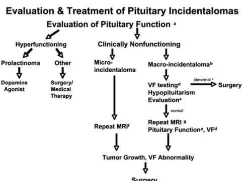
2.1.2 VF testing in patients with a pituitary incidenta-loma that enlarges to abut or compress the optic nerves or chiasm on a follow-up imaging study (1ⱍQQQQ). We sug-FIG. 1. Flow diagram for the evaluation and treatment of pituitary incidentalomas. a, Baseline
evaluation in all patients should include a history and physical exam evaluating for signs and symptoms of hyperfunction and hypopituitarism and a laboratory evaluation for hypersecretion. b, This group may also include large microlesions (see Section 2.1 Evidence). c, The recommendation for surgery includes the presence of abnormalities of VF or vision and signs of tumor compression (Section 3.1); surgery is also suggested for other findings (see Section 3.2). d, VF testing is recommended for patients with lesions abutting or compressing the optic nerves or chiasm at the initial evaluation and during follow-up. e, Evaluation for hypopituitarism is recommended for the baseline evaluation and during follow-up evaluations. This is most strongly recommended for macrolesions and larger microlesions (see Section 1.3). f, Repeat MRI in 1 yr, yearly for 3 yr, and then less frequently thereafter if no change in lesion size. g, Repeat the MRI in 6 months, yearly for 3 yr, and then less frequently if no change in lesion size. [Modified from Molitch ME: J Clin
gest that clinicians do not need to test VF in patients whose incidentalomas are not close to the chiasm and who have no new symptoms and are being followed closely by MRI (2ⱍQEEE).
2.1.3 Clinical and biochemical evaluations for hypop-ituitarism 6 months after the initial testing and yearly thereafter in patients with a pituitary macroincidenta-loma, although typically hypopituitarism develops with the finding of an increase in size of the incidentaloma (1ⱍQQEE). We suggest that clinicians do not need to test for hypopituitarism in patients with pituitary microinci-dentalomas whose clinical picture, history, and MRI do not change over time (2ⱍQQEE).
2.1 Evidence
The options for treatment of patients with asymptomatic, clinically nonfunctioning pituitary incidentalomas are con-servative follow-up without surgery (Fig. 1) or immediate surgery despite the lack of indications for this. Conservative follow-up was recommended with the recognition that the proper algorithm for this and its appropriateness and safety have not been tested prospectively. Few data are available for or against a nonsurgical approach to management of asymp-tomatic pituitary incidentalomas. Therefore, evidence in support of these guidelines also relied on the clinical experi-ences of the Task Force members.
The proper algorithm for endocrine testing during this follow-up has not been tested as prospectively conducted endocrine testing of patients with pituitary incidentalo-mas who were followed without surgery has only been reported in 49 patients (8, 9). Follow-up endocrine testing is recommended for patients with macroincidentalomas because they are at risk of developing hypopituitarism. Of the macroincidentalomas followed prospectively, wors-ening hypopituitarism developed in one of seven (8) and three of 28 (25) patients, all of whom had enlargement of their tumors. Hypopituitarism also developed in four of 37 (5) and one of 248 (6) patients who developed apoplexy on follow-up. Follow-up endocrine testing was recom-mended despite the paucity of data because of the potential high risk to the patient of untreated hypopituitarism. In a meta-analysis of incidentaloma studies, new endocrine dysfunction developed overall in 2.4% of patients per year (3). It is unclear how often new hypopituitarism develops in the absence of tumor growth. Rapid growth may in-crease the risk of new hypopituitarism. Routine follow-up endocrine testing is not required for microincidentalomas whose clinical picture does not change because the risk of developing new hypopituitarism is extremely low. In pre-vious studies, none of the pituitary microincidentalomas followed prospectively was reported to be associated with changes in pituitary function (4 –9).
The proposed algorithms for the MRI follow-up of pi-tuitary incidentalomas took into account those adopted in prior studies. However, those algorithms varied consid-erably from study to study (4 –9), and none was validated. As a result, the imaging follow-up proposed in this guide-line incorporated the experiences of the Task Force’s members. Follow-up MRI scans were recommended for macroincidentalomas because it has been demonstrated that although generally these lesions grow slowly, some do enlarge and become symptomatic. In data combined from a number of studies, macroincidentalomas enlarged in 85 of 353 (24%) patients (4 –9, 23–26). VF abnormalities developed in 28 (8%) patients over time, which demon-strated that the enlargement adversely affected the pa-tient’s health. Pituitary apoplexy developed in seven of 353 (2%) patients, of whom most developed permanent hypopituitarism and one had permanent vision impair-ment (5). In a meta-analysis of these studies, 8.2% of in-cidentalomas enlarged per year with a follow-up of 472 person-years (3). Less frequent surveillance of microinci-dentalomas was recommended because their rate of en-largement was low, being reported in 17 of 160 patients (10.6%) followed from 2.3 to 7 yr (5–9, 23–25). In the meta-analysis, 1.7% of microincidentalomas enlarged per year (3). Importantly, none of the patients with these mi-croincidentalomas developed new VF abnormalities that would have necessitated surgery.
Overall, the Task Force considered that repeat scanning within the first year was warranted for all patients because, although most incidentalomas grow slowly, some do en-large, and the true proliferative nature of the incidentaloma is unknown (Fig. 1). If no growth is detected, then the interval between MRI scans can be increased. Evidence does not sup-port one particular algorithm for the frequency of follow-up imaging, but we recommend repeating the MRI every year in macroincidentalomas, every 1–2 yr in microincidentalomas for the next 3 yr, and then every other year for the next 6 yr and gradually less frequently indefinitely so long as the lesion continues not to threaten the patient’s health. Some Task Force members would continue imaging every 5 yr. Uncer-tainty as to the optimal interval and duration of long-term follow-up imaging can be shared with the patient, and the scheme followed can be individualized to balance the phy-sician’s assessment of the risk that the lesion poses to the patient’s health with the burden to the patient of surveillance testing.
2.1 Values and preferences
The decision to proceed with conservative nonsurgical management of pituitary incidentalomas not meeting cri-teria for surgery (see Section 3.0) puts a relatively high value on avoiding surgical intervention and its associated
morbidity and cost, and a relatively low value in avoiding apoplexy. The evidence that most incidentalomas do not progress over time to cause visual or other disturbances supports this value judgment. On the other hand, apo-plexy developed in seven of 248 patients with incidenta-lomas (4 – 6, 8, 9, 26). Large-scale prospective studies of conservative nonsurgical management of pituitary inci-dentalomas are needed to further inform this judgment. 2.1 Remarks
The Task Force recognized that a continuum of size of incidentalomas exists, and some large microincidentalo-mas (6 –9 mm) may behave more like macroincidentalo-mas. Therefore, some Task Force members would also perform follow-up MRIs and hypopituitarism evaluations in larger microincidentalomas as they would for macro-incidentalomas. Some also considered that follow-up eval-uations for hypopituitarism were needed only for patients with enlarging incidentalomas because new hypopituitar-ism would be very unlikely to develop without tumor growth. However, no data are available to support one particular size threshold for the increase in size that should trigger this evaluation. The Task Force was split on these points.
Recommendation
2.2 Patients who develop any signs or symptoms po-tentially related to the incidentaloma or who show an in-crease in size of the incidentaloma on MRI should undergo more frequent or detailed evaluations as indicated clini-cally (1ⱍQQEE).
3.0 Indications for surgical therapy of the pituitary incidentaloma
Recommendation
3.1 We recommend that patients with a pituitary inci-dentaloma be referred for surgery if they have the follow-ing (1ⱍQQQQ):
• A VF deficit due to the lesion.
• Other visual abnormalities, such as ophthalmoplegia or neurological compromise due to compression by the lesion.
• Lesion abutting or compressing the optic nerves or chiasm on MRI.
• Pituitary apoplexy with visual disturbance.
• Hypersecreting tumors other than prolactinomas as recommended by other guidelines of The Endocrine Society and The Pituitary Society (31, 33).
3.1 Evidence
The decision to recommend surgery as initial therapy for a patient with a pituitary incidentaloma needs to be
individ-ualized (Fig. 1). Few data are available that specifically ex-amine the outcome of surgery for incidentalomas. Some of the evidence considered in developing the recommended cri-teria for surgery was from transsphenoidal surgical series of symptomatic and often large pituitary lesions. From these data and based on the clinical experiences of the Task Force members, it is clear that substantial evidence supports the need for surgery for pituitary incidentalomas causing visual or neurological compromise. The presence of visual or neu-rological abnormalities due to compression of the optic nerves or chiasm by the incidentaloma is the strongest indi-cation for surgery. Although the risks of not performing sur-gery on tumors that abut the chiasm or for those within cer-tain proximity of the chiasm but where no VF abnormalities are present have not been quantified, in the Task Force’s experience significant risk of future vision disturbance in these patients exists to warrant surgery. The age of the patient is also an important consideration. Surgery may be favored in younger vs. older patients given the higher lifetime prob-ability of tumor enlargement in the former and the greater risks of surgical intervention in the latter group of patients. Some may elect to follow elderly patients with significant risks to surgery conservatively and closely for any deterio-ration in vision. The decision to recommend surgery should also consider whether future fertility is of concern to the pa-tient. Difficult cases could also be discussed at a pituitary multidisciplinary team meeting when this is available.
Surgery is indicated in patients with apoplexy and visual disturbance. In a retrospective review of 30 sub-jects with pituitary apoplexy, the 20 who were followed conservatively had a similar long-term risk of hypopi-tuitarism as those treated surgically (37). Therefore, patients with apoplexy and without visual compromise may be followed conservatively with serial imaging and hormonal assessments.
3.1 Values and preferences
Our recommendations for surgery for incidentalomas put a high value on alleviating or preventing visual or neurolog-ical compromise. Although the benefits of surgery in pre-venting future visual abnormalities are not known, a rela-tively higher value was put on the prevention of VF abnormalities than on avoiding the morbidity (e.g. diabetes insipidus, hypopituitarism) and the cost of surgery. New hy-popituitarism as a result of transsphenoidal surgery is rare in the Task Force’s experience, but the risk should be consid-ered in the clinical context of the particular patient. 3.1 Remarks
The recommendations for surgical therapy of a pitu-itary incidentaloma were based on the Task Force’s ex-pectations for outcome and improvements in vision and
endocrine function. These expectations were based on known literature and Task Force members’ clinical expe-rience with surgery performed by a surgeon expeexpe-rienced in transsphenoidal pituitary surgery. The success of surgery for hormone-secreting tumors is highly dependent on the expertise, skill, and case volume of a pituitary surgeon supported by an experienced team (38, 39). This is likely to also be true for pituitary surgery of other types of le-sions. The availability of such a pituitary surgeon needs to be considered when following these guidelines.
Recommendation
3.2 We suggest that surgery be considered for patients with a pituitary incidentaloma if they have the following (2ⱍQQEE):
• Clinically significant growth of the pituitary incidentaloma.
• Loss of endocrinological function.
• A lesion close to the optic chiasm and a plan to be-come pregnant.
• Unremitting headache. 3.2 Evidence
The evidence for or against a recommendation for sur-gery because of growth of a pituitary incidentaloma is limited. Surgery was suggested for incidentalomas that demonstrate clinically significant growth on follow-up imaging studies, which is growth that could pose a health risk to the patient such as to their vision. Such growing incidentalomas typically continue to grow, and surgery is most effective for smaller lesions. A specific size cutoff for the incidentaloma was not considered a requirement for surgery because some large incidentalomas are predomi-nantly intra- and infrasellar. Although there are no estab-lished changes in size or growth rate that would automat-ically trigger the need for surgery, the pattern of growth of the incidentaloma was considered more important. For example, a 1-mm enlargement within the sella of a 5-mm intrasellar microadenoma would not be clinically signifi-cant, but a 1-mm enlargement toward the chiasm in a lesion only 3 mm from the chiasm would be significant. Therefore, incidentalomas exhibiting significant growth (which includes growth that is rapid, occurring over a 1-to 2-yr period, and/or that is 1-toward the optic chiasm and if continued could threaten vision in the near future) should be considered for surgery before the incidentaloma progresses to abut the chiasm or produce visual deficits. A consideration of the clinical characteristics of the patient including their age and other risks for surgery needs to be incorporated into the decision to proceed to surgery.
The evidence for or against a recommendation for sur-gery because of the presence of hypopituitarism is also
limited. Although some surgical series of symptomatic in-cidentalomas show that hypopituitarism can improve with surgery (40, 41), these data may not be applicable to the incidentaloma, and as a result hypopituitarism was considered only a relative indication for surgery. Adequate replacement therapy should be instituted whether or not surgery is recommended. Some patients planning preg-nancy may benefit from surgery if their tumor is close to the optic chiasm because there is a small risk that lac-totroph hyperplasia in the normal gland may lead to tumor compression of the optic nerve or chiasm, and closer fol-low-up in such patients should be undertaken. Headache may or may not improve with transsphenoidal removal of the tumor, so it can only be suggested as an indication for surgery, and the quality of evidence is low.
Medical therapy of the pituitary incidentaloma
In patients with incidentalomas and hyperprolactine-mia that may be due to tumoral compression of the hy-pothalamic-pituitary stalk, symptomatic hyperprolactine-mia may be treated with a dopamine agonist. However, incidentalomas other than a prolactinoma will rarely shrink, and dopamine agonists cannot be relied upon for this purpose. Therefore, continued monitoring of lesion size is necessary, regardless of changes in prolactin levels. Medical therapy of pituitary incidentalomas has not been systematically studied. Some parallels to the effects of medical therapy on pathologically confirmed nonfunc-tioning pituitary tumors are possible, but this connection needs to be made cautiously. The reported efficacy of do-pamine agonist therapy of nonfunctioning pituitary ade-nomas varies widely. With cabergoline or bromocriptine treatment for patients with residual tumor after surgery, some degree of tumor shrinkage was detected in 8 – 45% (42– 44), and the amount of shrinkage varied from 10 – 62% (43, 44) or 3–14 mm (42). Somatostatin analogs have also been tried. With no more than 1 yr of octreotide ther-apy, shrinkage was reported in approximately 5–25%, increase in 12%, and stabilization in 83% of tumors (45– 48). Insufficient data are available to suggest the routine use of medical therapy of pituitary incidentalomas.
Acknowledgments
The members of the Task Force thank The Endocrine Society’s Clinical Guidelines Subcommittee, Clinical Affairs Core Com-mittee, and Council for their careful, critical review of earlier versions of this manuscript and their helpful comments and sug-gestions. We also thank the leadership of the European Society of Endocrinology for their review and comments. In addition, we thank the many members of The Endocrine Society who re-viewed the draft version of this manuscript when it was posted
on the Society’s web site and who sent a great number of addi-tional comments and suggestions, most of which were incorpo-rated into the final version of the manuscript. Finally, we thank the staff at the Society office for their helpful support during the development of this guideline.
Address all correspondence and requests for reprints to: The Endocrine Society, 8401 Connecticut Avenue, Suite 900, Chevy Chase, MD 20815. E-mail: govt-prof@endo-society.org, Tele-phone: 301-941-0200. Address all commercial reprint requests for orders 101 and more to: Walchli Tauber Group Inc., E-mail: Karen.burkhardt@wt-group.com. Address all reprint requests for orders for 100 or fewer to Society Services, Telephone: 301-941-0210, E-mail: societyservices@endo-society.org, or Fax: 301-941-0257.
Cosponsoring Associations: European Society of Endocrinology. Financial Disclosures of the Task Force: Pamela U. Freda, M.D.*—Financial or Business/Organizational Interests: No-vartis, Ipsen, and Pfizer; Significant Financial Interest or Lead-ership Position: none declared. Albert M. Beckers, M.D., D.Sc., Ph.D.—Financial or Business/Organizational Interests: No-vartis, Ipsen, and Pfizer; Significant Financial Interest or Lead-ership Position: none declared. Laurence Katznelson, M.D.— Financial or Business/Organizational Interests: Novartis, Ipsen, and Novo Nordisk; Significant Financial Interest or Leadership Position: none declared. Mark E. Molitch, M.D.—Financial or Business/Organizational Interests: Tercica/Ipsen, Novartis; Sig-nificant Financial Interest or Leadership Position: none declared. Victor M. Montori, M.D.*—Financial or Business/Organiza-tional Interests: KER Unit (Mayo Clinic); Significant Financial Interest or Leadership Position: none declared. Kalmon D. Post, M.D. —Financial or Business/Organizational Interests: none de-clared; Significant Financial Interest or Leadership Position: none declared. Mary Lee Vance, M.D.—Financial or Business/ Organizational Interests: Novartis; Significant Financial Interest or Leadership Position: none declared.
*Evidence-based reviews for this guideline were prepared un-der contract with The Endocrine Society.
References
1. Atkins D, Best D, Briss PA, Eccles M, Falck-Ytter Y, Flottorp S, Guyatt GH, Harbour RT, Haugh MC, Henry D, Hill S, Jaeschke R, Leng G, Liberati A, Magrini N, Mason J, Middleton P, Mrukowicz J, O’Connell D, Oxman AD, Phillips B, Schu¨nemann HJ, Edejer TT, Varonen H, Vist GE, Williams Jr JW, Zaza S 2004 Grading quality of evidence and strength of recommendations. BMJ 328:1490 2. Swiglo BA, Murad MH, Schu¨nemann HJ, Kunz R, Vigersky RA,
Guyatt GH, Montori VM 2008 A case for clarity, consistency, and helpfulness: state-of-the-art clinical practice guidelines in endocri-nology using the grading of recommendations, assessment, devel-opment, and evaluation system. J Clin Endocrinol Metab 93:666 – 673
3. Fernandez-Balsells M MM, Barwise A, Gallegos-Orozco J, Paul A, Lane M, Carpio I, Perestelo-Perez LI, Ponce de Leon Lovaton P, Erwin, P, Carey J, Montori VM 2010 The natural history of pituitary incidentalomas: a systematic review and meta-analysis. J Clin Endocrinol Metab (In press)
4. Feldkamp J, Santen R, Harms E, Aulich A, Mo¨dder U, Scherbaum WA 1999 Incidentally discovered pituitary lesions: high frequency of macroadenomas and hormone-secreting adenomas—results of a prospective study. Clin Endocrinol (Oxf) 51:109 –113
5. Arita K, Tominaga A, Sugiyama K, Eguchi K, Iida K, Sumida M, Migita K, Kurisu K 2006 Natural course of incidentally found non-functioning pituitary adenoma, with special reference to pituitary apoplexy during follow-up examination. J Neurosurg 104:884 – 891 6. Sanno N, Oyama K, Tahara S, Teramoto A, Kato Y 2003 A survey of pituitary incidentaloma in Japan. Eur J Endocrinol 149:123–127 7. Fainstein Day P, Guitelman M, Artese R, Fiszledjer L, Chervin A, Vitale NM, Stalldecker G, De Miguel V, Cornalo´ D, Alfieri A, Su-sana M, Gil M 2004 Retrospective multicentric study of pituitary incidentalomas. Pituitary 7:145–148
8. Reincke M, Allolio B, Saeger W, Menzel J, Winkelmann W 1990 The ‘incidentaloma’ of the pituitary gland. Is neurosurgery required? JAMA 263:2772–2776
9. Donovan LE, Corenblum B 1995 The natural history of the pituitary incidentaloma. Arch Intern Med 155:181–183
10. Freda PU, Post KD 1999 Differential diagnosis of sellar masses. Endocrinol Metab Clin North Am 28:81–117, vi
11. Zada G, Lin N, Ojerholm E, Ramkissoon S, Laws ER Craniophar-yngioma and other cystic epithelial lesions of the sellar region: a review of clinical, imaging, and histopathological relationships. Neurosurg Focus 28:E4
12. Kanter AS, Sansur CA, Jane Jr JA, Laws Jr ER 2006 Rathke’s cleft cysts. Front Horm Res 34:127–157
13. Black PM, Hsu DW, Klibanski A, Kliman B, Jameson JL, Ridgway EC, Hedley-Whyte ET, Zervas NT 1987 Hormone production in clinically nonfunctioning pituitary adenomas. J Neurosurg 66:244 – 250
14. Esiri MM, Adams CB, Burke C, Underdown R 1983 Pituitary ad-enomas: immunohistology and ultrastructural analysis of 118 tu-mors. Acta Neuropathol 62:1–14
15. Al-Shraim M, Asa SL 2006 The 2004 World Health Organization classification of pituitary tumors: what is new? Acta Neuropathol 111:1–7
16. Molitch ME 2008 Nonfunctioning pituitary tumors and pituitary incidentalomas. Endocrinol Metab Clin North Am 37:151–171, xi 17. Wolpert SM, Molitch ME, Goldman JA, Wood JB 1984 Size, shape, and appearance of the normal female pituitary gland. AJR Am J Roentgenol 143:377–381
18. Chambers EF, Turski PA, LaMasters D, Newton TH 1982 Regions of low density in the contrast-enhanced pituitary gland: normal and pathologic processes. Radiology 144:109 –113
19. Peyster RG, Adler LP, Viscarello RR, Hoover ED, Skarzynski J 1986 CT of the normal pituitary gland. Neuroradiology 28:161–165 20. Hall WA, Luciano MG, Doppman JL, Patronas NJ, Oldfield EH
1994 Pituitary magnetic resonance imaging in normal human vol-unteers: occult adenomas in the general population. Ann Intern Med 120:817– 820
21. Nammour GM, Ybarra J, Naheedy MH, Romeo JH, Aron DC 1997 Incidental pituitary macroadenoma: a population-based study. Am J Med Sci 314:287–291
22. Yue NC, Longstreth Jr WT, Elster AD, Jungreis CA, O’Leary DH, Poirier VC 1997 Clinically serious abnormalities found incidentally at MR imaging of the brain: data from the Cardiovascular Health Study. Radiology 202:41– 46
23. Igarashi T, Saeki N, Yamaura A 1999 Long-term magnetic reso-nance imaging follow-up of asymptomatic sellar tumors—their nat-ural history and surgical indications. Neurol Med Chir (Tokyo) 39:592–598; discussion 598 –599
24. Karavitaki N, Collison K, Halliday J, Byrne JV, Price P, Cudlip S, Wass JA 2007 What is the natural history of nonoperated nonfunc-tioning pituitary adenomas? Clin Endocrinol (Oxf) 67:938 –943 25. Dekkers OM, Hammer S, de Keizer RJ, Roelfsema F, Schutte PJ,
Smit JW, Romijn JA, Pereira AM 2007 The natural course of non-functioning pituitary macroadenomas. Eur J Endocrinol 156:217– 224
26. Nishizawa S, Ohta S, Yokoyama T, Uemura K 1998 Therapeutic strategy for incidentally found pituitary tumors (“pituitary inciden-talomas”). Neurosurgery 43:1344 –1348; discussion 1348 –1350
27. Daly AF, Rixhon M, Adam C, Dempegioti A, Tichomirowa MA, Beckers A 2006 High prevalence of pituitary adenomas: a cross-sectional study in the province of Liege, Belgium. J Clin Endocrinol Metab 91:4769 – 4775
28. Fernandez A, Karavitaki N, Wass JA Prevalence of pituitary ade-nomas: a community-based, cross-sectional study in Banbury (Ox-fordshire, UK). Clin Endocrinol (Oxf) 72:377–382
29. Raappana A, Koivukangas J, Ebeling T, Pirila¨ T 2010 Incidence of pituitary adenomas in Northern Finland in 1992–2007. J Clin En-docrinol Metab 95:4268 – 4275
30. Buurman H, Saeger W 2006 Subclinical adenomas in postmortem pituitaries: classification and correlations to clinical data. Eur J En-docrinol 154:753–758
31. Casanueva FF, Molitch ME, Schlechte JA, Abs R, Bonert V, Bron-stein MD, Brue T, Cappabianca P, Colao A, Fahlbusch R, Fideleff H, Hadani M, Kelly P, Kleinberg D, Laws E, Marek J, Scanlon M, Sobrinho LG, Wass JA, Giustina A 2006 Guidelines of the Pituitary Society for the diagnosis and management of prolactinomas. Clin Endocrinol (Oxf) 65:265–273
32. Angeli A, Terzolo M 2002 Adrenal incidentaloma—a modern dis-ease with old complications. J Clin Endocrinol Metab 87:4869 – 4871
33. Nieman LK, Biller BM, Findling JW, Newell-Price J, Savage MO, Stewart PM, Montori VM 2008 The diagnosis of Cushing’s syn-drome: an Endocrine Society Clinical Practice Guideline. J Clin En-docrinol Metab 93:1526 –1540
34. Karavitaki N, Ansorge O, Wass JA 2007 Silent corticotroph ade-nomas. Arq Bras Endocrinol Metabol 51:1314 –1318
35. Yuen KC, Cook DM, Sahasranam P, Patel P, Ghods DE, Shahinian HK, Friedman TC 2008 Prevalence of GH and other anterior pitu-itary hormone deficiencies in adults with nonsecreting pitupitu-itary mi-croadenomas and normal serum IGF-1 levels. Clin Endocrinol (Oxf) 69:292–298
36. Molitch ME, Clemmons DR, Malozowski S, Merriam GR, Shalet SM, Vance ML, Stephens PA 2006 Evaluation and treatment of adult growth hormone deficiency: an Endocrine Society Clinical Practice Guideline. J Clin Endocrinol Metab 91:1621–1634 37. Gruber A, Clayton J, Kumar S, Robertson I, Howlett TA, Mansell
P 2006 Pituitary apoplexy: retrospective review of 30 patients—is surgical intervention always necessary? Br J Neurosurg 20:379 –385 38. Barker 2nd FG, Klibanski A, Swearingen B 2003 Transsphenoidal surgery for pituitary tumors in the United States, 1996 –2000: mor-tality, morbidity, and the effects of hospital and surgeon volume. J Clin Endocrinol Metab 88:4709 – 4719
39. Gittoes NJ, Sheppard MC, Johnson AP, Stewart PM 1999 Outcome of surgery for acromegaly—the experience of a dedicated pituitary surgeon. QJM 92:741–745
40. Arafah BM, Kailani SH, Nekl KE, Gold RS, Selman WR 1994 Im-mediate recovery of pituitary function after transsphenoidal resec-tion of pituitary macroadenomas. J Clin Endocrinol Metab 79:348 – 354
41. Arafah BM 1986 Reversible hypopituitarism in patients with large nonfunctioning pituitary adenomas. J Clin Endocrinol Metab 62: 1173–1179
42. Greenman Y, Tordjman K, Osher E, Veshchev I, Shenkerman G, Reider-Groswasser II, Segev Y, Ouaknine G, Stern N 2005 Postop-erative treatment of clinically nonfunctioning pituitary adenomas with dopamine agonists decreases tumour remnant growth. Clin Endocrinol (Oxf) 63:39 – 44
43. Lohmann T, Trantakis C, Biesold M, Prothmann S, Guenzel S, Schober R, Paschke R 2001 Minor tumour shrinkage in nonfunc-tioning pituitary adenomas by long-term treatment with the dopa-mine agonist cabergoline. Pituitary 4:173–178
44. Pivonello R, Matrone C, Filippella M, Cavallo LM, Di Somma C, Cappabianca P, Colao A, Annunziato L, Lombardi G 2004 Dopa-mine receptor expression and function in clinically nonfunctioning pituitary tumors: comparison with the effectiveness of cabergoline treatment. J Clin Endocrinol Metab 89:1674 –1683
45. Shomali ME, Katznelson L 2002 Medical therapy of gonadotro-pin-producing and nonfunctioning pituitary adenomas. Pituitary 5:89 –98
46. Merola B, Colao A, Ferone D, Selleri A, Di Sarno A, Marzullo P, Biondi B, Spaziante R, Rossi E, Lombardi G 1993 Effects of a chronic treatment with octreotide in patients with functionless pi-tuitary adenomas. Horm Res 40:149 –155
47. de Bruin TW, Kwekkeboom DJ, Van’t Verlaat JW, Reubi JC, Kren-ning EP, Lamberts SW, Croughs RJ 1992 Clinically nonfunctioKren-ning pituitary adenoma and octreotide response to long term high dose treatment, and studies in vitro. J Clin Endocrinol Metab 75:1310 – 1317
48. Colao A, Di Somma C, Pivonello R, Faggiano A, Lombardi G, Savas-tano S 2008 Medical therapy for clinically non-functioning pituitary adenomas. Endocr Relat Cancer 15:905–915
49. Molitch ME 1995 Clinical review 65. Evaluation and treatment of the patient with a pituitary incidentaloma. J Clin Endocrinol Metab 80:3– 6