Damien Lesenfants,
PhD*
Dina Habbal, PhD*
Camille Chatelle, PhD
Caroline Schnakers, PhD
Steven Laureys, MD,
PhD
Quentin Noirhomme,
PhD
Correspondence to Dr. Lesenfants: damien_lesenfants@brown.edu Supplemental data at Neurology.orgElectromyographic decoding of response
to command in disorders of consciousness
ABSTRACT
Objective:
To propose a new methodology based on single-trial analysis for detecting residual
response to command with EMG in patients with disorders of consciousness (DOC), overcoming
the issue of trial dependency and decreasing the influence of a patient’s fluctuation of vigilance or
arousal over time on diagnostic accuracy.
Methods:
Forty-five patients with DOC (18 with vegetative/unresponsive wakefulness syndrome
[VS/UWS], 22 in a minimally conscious state [MCS], 3 who emerged from MCS [EMCS], and 2 with
locked-in syndrome [LIS]) and 20 healthy controls were included in the study. Patients were
ran-domly instructed to either move their left or right hand or listen to a control command (“It is a sunny
day”) while EMG activity was recorded on both arms.
Results:
Differential EMG activity was detected in all MCS cases displaying reproducible
response to command at bedside on multiple assessments, even though only 6 of the 14
individ-uals presented a behavioral response to command on the day of the EMG assessment. An EMG
response was also detected in all EMCS and LIS patients, and 2 MCS patients showing
nonreflex-ive movements without command following at the bedside. None of the VS/UWS presented
a response to command with this method.
Conclusions:
This method allowed us to reliably distinguish between different levels of
conscious-ness and could potentially help decrease diagnostic errors in patients with motor impairment but
presenting residual motor activity.
Neurology®2016;87:2099–2107GLOSSARY
CRS-R5 Coma Recovery Scale–Revised; DOC 5 disorders of consciousness; EMCS 5 emergence from minimally conscious state; LIS5 locked-in syndrome; MCS 5 minimally conscious state; RMS 5 root mean square; VS/UWS 5 vegetative state/ unresponsive wakefulness syndrome.
Keystones in the diagnosis of patients recovering from coma are the acquisition of voluntary
responses such as command following, distinguishing patients in a vegetative state/unresponsive
wakefulness syndrome (VS/UWS; characterized by the recovery of eye opening without
aware-ness of self and environment
1–3) from patients in a minimally conscious state (MCS;
character-ized by inconsistent, fluctuating but reproducible signs of consciousness
4). However, patients
with disorders of consciousness (DOC) have limited neuromuscular abilities,
5,6challenging the
detection of behavioral response to command based on visual and tactile feedback, as used in
clinical gold standard behavioral scales. An additional limitation of behavioral assessment is its
dependence on an examiner’s experience and subjectivity.
7Recent neuroimaging studies have
suggested that 11%–33% of patients behaviorally diagnosed as unresponsive using behavioral
scales may actually present brain-related signs of consciousness,
8highlighting the need to
develop more objective and observer-independent diagnostic tools for this population. In
par-ticular, EMG has been proposed for the detection of micromovements that often go unnoticed
by an observer at a patient’s bedside, but results have been mixed.
9,10In the current study, we
*These authors contributed equally to this work.From the School of Engineering and Institute for Brain Science (D.L.), Brown University, Providence, RI; Coma Science Group (D.H., S.L.), GIGA-Research, CHU University Hospital of Liège, Belgium; Laboratory for NeuroImaging of Coma and Consciousness (C.C.), Massachusetts General Hospital, Boston; Department of Neurosurgery (C.S.), UCLA, Los Angeles, CA; and Brain Innovation B.V. and Maastricht University (Q.N.), the Netherlands.
aimed to improve the detection of residual
mus-cular activity related to command following
using a novel EMG method with single-trial
level analysis. Given high non-stationarities in
the EMG signal (e.g., artifact) and fluctuations
in the level of consciousness or arousal over
time, we hypothesized that removing
depen-dence on intertrial consistency in this
popula-tion could improve detecpopula-tion of volipopula-tional
response to command.
METHODS Participants.Among all patients admitted to the University Hospital of Liège between 2013 and 2014, 45 patients were included in this study (mean age 406 15 years; 30 male). Patients were subcategorized according to the following diagno-ses: MCS2 encompasses patients without signs of language pres-ervation (i.e., showing only visual pursuit or fixation, object localization or manipulation, localization of noxious stimulation, automatic motor response, or smiling/crying in response to exter-nal stimuli) whereas MCS1 includes patients showing behavioral responses suggesting language preservation such as command fol-lowing or intelligible words.11–13Emergence from MCS (EMCS)
is characterized by the recovery of functional communication or functional object use.4The locked-in syndrome (LIS), on the
other hand, is a state in which the patient is paralyzed but awake and fully conscious.14
In our study, 17 patients were diagnosed as being in VS/ UWS, 7 in MCS2, 14 in MCS1, 5 in EMCS, and 2 in LIS. Patient LIS1 was able to perform horizontal head movements and slight movements of the arms. Patient LIS2 had a left hemi-plegia but could move his right arm within a normal range of motion. Both showed very little spasticity. Inclusion criteria were (1) at least 28 days postinjury, (2) preserved auditory evoked potentials or presence of auditory startle, and (3) no neuromus-cular function blockers and no sedation within the prior 24 hours. Exclusion criteria were (1) a documented history of prior brain injury, (2) a premorbid history of developmental, psychi-atric, or neurologic illness resulting in documented functional disability up to the time of the injury, (3) a premorbid history of uncorrected hearing impairments, (4) flaccidity in response to noxious stimulation, and (5) acute illness. Four of these patients were evaluated twice (see table 1). Twenty-three patients had traumatic and 22 patients had nontraumatic etiologies (i.e., stroke, hemorrhage, cardiac arrest, infection, or metabolic dis-orders). Average duration since insult was 386 48 months (range 1 month–18 years; median 14 months). Table 1 sum-marizes patients’ demographic and clinical data. We also included 20 healthy controls (mean age 346 13 years; 11 male; see table 2). For this group, exclusion criteria were (1) uncor-rected hearing impairments, (2) muscle disease or muscle dys-function due to an injury, and (3) developmental, psychiatric, or neurologic illness. Spasticity of the upper limbs was evaluated using the Modified Ashworth Scale by a trained physiologist and is reported in table e-1 at Neurology.org along with antispastic medications.
Standard protocol approvals, registrations, and patient consents.The study was approved by the Ethics Committee of the University Hospital of Liège. Each healthy control and each patient’s legal representative provided written informed consent.
Behavioral assessment and final diagnosis.Patients’ level of consciousness was assessed by a trained examiner using the Coma
Recovery Scale–Revised (CRS-R) on the day of the EMG recording and several times during the week to increase diagnostic accuracy.15The best score obtained during the week was used as
the final diagnosis.
Paradigm.Three different instructions (recorded using a neutral male voice) were presented to the participants: 2 target instruc-tions (i.e.,“Move your left hand” and “Move your right hand”) and 1 control instruction (i.e.,“It is a sunny day”). Each instruc-tion was presented 3 times in a row within a trial. Each trial lasted 21 seconds, including the instructions (3 seconds). A block of stimulation consisted of 3 minutes of rest followed by 5 trials of each instruction randomly presented with an intertrial interval of 10 seconds (about 10 minutes in total) (figure 1). Each par-ticipant completed a total of 3 blocks with breaks of varied dura-tion, depending on level of fatigue.
Signal acquisition.Left and right upper limb electrical activity of the abductor policis brevis muscle (channel“Hand”) and the flexor digitorum superficialis muscle (channel“Arm”) was re-corded at the bedside with 8 Ag/AgCl self-adhesive surface electrodes, placed in a bipolar derivation with an interelectrode distance of 20 mm, sampled at 500 Hz.16,17 Electrodes were
connected to a portable BrainVision vAmp amplifier. Data were acquired and auditory instructions presented using a laptop running the general-purpose software platform BCI2000.18 Data analysis.The EMG signals were filtered with a zero-phase fourth-order bandpass Butterworth filter (IIR, fc5 20–120 Hz) and a second-order notch filter (IIR, fc5 50 Hz, Q 5 35). We then computed the root mean square (RMS) of 1-second overlapping (90% overlap) windows, occurring between the beginning of the 2-second and end of the 3-second following the presentation of each instruction within a trial (see gray area in figure 1), resulting in 33 windows for each trial and each location. For each location, we then extracted the difference (Δactive)
between averaged RMS value during the trial and the preceding intertrial interval. The difference (Δrest) between averaged RMS
value was also evaluated on consecutive overlapping windows during baseline (1-second window, 90% overlap; interwindow distance and length were chosen to match those used forΔactive).
Mean (mΔrest) and SD (sΔrest) of the RMS difference during
baseline were then used to set the threshold equal tomΔrest 12.6 sΔrest, which corresponded to detecting an unexpected event with a p value of 0.01 if the data were normally distributed. We considered a positive activation during a trial if at least 1 of the 2 ipsilateral locations exceeded the respective threshold, i.e., Δactive_arm_ipsilateral. Tarm_ipsilateralorΔactive_hand_ipsilateral. T han-d_ipsilateral. We considered a control trial (placebo) as positive if
Δactiveat one of the 4 locations exceeded threshold.
We hypothesized that an increase in EMG activity during commands“Move your right hand” and “Move your left hand” could be observed in conscious patients while absent in uncon-scious VS/UWS patients. Because of the patients’ clinical condi-tion, we did not expect an EMG response to all commands, but hypothesized a difference in the ratio between response to com-mands“Move your right/left hand” and control command “It is a sunny day”; this ratio could be used to distinguish volitional response to command from reflexive, spastic, or involuntary movements. We computed an EMG score defined by (L1R)/ (C11), with L and R being the number of positive activations detected during left and right command, respectively, and C being the number of wrongly positive activations during the control condition. By including the control condition, the score takes into account the number of false-positives observed. We then defined a threshold for response (vs no response) to
Table 1 Demographic, clinical, and task-related data of the patient sample
Final diagnosis Sex Age, y Etiology TSI, mo
Behavioral assessment EMG assessment
Diagnosis CRS-R L R P Score Above threshold score
VS/UWS1 M 27 Trauma 1 VS 1-1-1-0-1-1 5 4 8 1.0 VS/UWS2 F 26 Trauma 7 VS 1-1-2-1-0-0 0 3 5 0.5 VS/UWS3 M 41 Trauma 10 VS 1-0-2-1-0-2 6 6 8 1.3 VS/UWS4 M 41 Trauma 14 VS 0-1-1-1-0-2 2 4 4 1.2 VS/UWS5 M 55 Trauma 19 VS 1-0-1-1-0-1 1 3 2 1.3 VS/UWS6 M 32 Trauma 48 VS 0-0-1-1-0-1 2 6 6 1.1 VS/UWS7 M 28 SAH 3 VS 1-1-1-1-0-2 4 6 10 0.9 VS/UWS8 F 60 Infection 4 VS 2-0-2-1-0-2 6 3 5 1.5 VS/UWS9a M 43 Anoxia 6 VS 1-1-2-2-0-2 2 6 5 1.3 VS/UWS10 F 66 SAH 7 VS 0-0-1-1-0-1 7 5 7 1.5 VS/UWS11b M 57 SAH 9 VS 1-0-2-1-0-2 5 6 7 1.4
VS/UWS12 M 42 Cardiac arrest 10 VS 1-0-2-0-0-2 1 1 1 1.0
VS/UWS13 F 66 Hypoglycemia 11 VS 1-1-1-1-0-1 4 5 5 1.5 VS/UWS14 M 47 AVC 48 VS 1-0-1-1-0-1 1 2 1 1.5 VS/UWS15 M 7 Anoxia 64 VS 1-1-1-1-0-1 2 3 3 1.3 MCS21 M 29 Trauma 6 VS 0-1-1-1-0-1 3 3 5 1.0 MCS22 F 51 Trauma 7 VS 0-0-1-1-0-1 8 3 7 1.4 MCS23 F 25 Trauma 11 MCS2 2-3-2-1-0-2 4 6 6 1.4 MCS24 F 40 Trauma 42 MCS2 2-1-2-2-0-1 5 3 6 1.1 MCS25 F 20 Trauma 43 VS 1-0-1-1-0-1 7 7 2 4.7 X MCS26 F 33 Trauma 46 MCS2 0-0-1-2-0-1 3 8 6 1.6 X MCS27 M 26 Trauma 145 MCS2 2-3-1-1-0-2 4 3 4 1.4 MCS28a M 44 Anoxic 20 VS 1-1-1-2-0-2 7 2 5 1.5 MCS11 M 55 Trauma 1 VS 1-0-2-1-0-1 7 10 7 2.1 X MCS12 M 25 Trauma 18 MCS1 3-3-2-1-0-1 6 6 6 1.7 X MCS13 M 32 Trauma 35 VS 1-0-1-1-0-1 6 2 2 2.7 X MCS14 M 28 Trauma 61 MCS1 3-4-5-2-0-2 2 6 3 2.0 X MCS15 F 32 Trauma 154 MCS1 2-4-5-1-0-2 6 5 6 1.6 X MCS16 M 39 SAH 8 MCS1 4-1-2-2-1-1 2 11 5 2.2 X 3 7 3 2.5 X MCS17 F 70 SAH 10 MCS1 3-4-5-2-0-2 10 7 7 2.1 X MCS18 M 29 Anoxia 13 MCS- 1-4-5-2-0-2 4 8 3 3.0 X MCS19b M 59 SAH 25 VS 1-0-1-1-0-2 3 4 2 2.3 X MCS110 M 55 Cardiac arrest 68 MCS1 3-3-5-1-0-1 6 9 3 3.8 X MCS111 M 38 Infection 88 MCS2 3-1-1-1-0-1 5 5 4 2.0 X MCS112 F 70 Stroke 101 VS 1-1-1-1-0-1 4 3 1 3.5 X MCS113 M 43 Cardiac arrest 107 VS 1-1-1-1-0-2 4 3 3 1.8 X MCS114 M 36 Infection 144 MCS2 1-3-1-1-0-2 0 3 0 3.0 X
EMCS1 M 25 Trauma 5 EMCS 4-5-5-3-2-3 10 10 7 2.5 X
EMCS2 M 38 Trauma 213 EMCS 4-5-5-1-2-2 12 12 5 4.0 X
EMCS3 M 58 SAH 31 EMCS 4-5-6-1-2-3 11 12 3 5.8 X
command using a leave-one-out cross-validation analysis. Since we first wanted to validate this technique on patients with a more stable diagnosis/level of consciousness, VS/UWS and MCS2 in an acute/subacute stage (,1 year post insult) were excluded for this analysis. The defined threshold was nevertheless used after-wards to detect response to command in this excluded group.
Highest bin count of threshold histogram was selected as the best threshold. In the following, a score higher than 1.5 was consid-ered to be representative of a response to command.
RESULTS
From an initial cohort of 45 patients with
DOC, 5 were excluded due to high levels of agitation
throughout the evaluation, fluctuation in signal due
to poor electrode contact, or highly noisy signal in more
than a third of the signal. The final cohort consisted of
40 patients (mean age 41
6 15 years; 27 male): 15 VS/
UWS, 7 MCS
2, 13 MCS1, 3 EMCS, and 2 LIS.
Behavioral evaluation of response to command.A
repro-ducible response to command was detected in 6/14
MCS1, 3/3 EMCS, and 2/2 LIS with the CRS-R
performed on the day of the EMG evaluation. No
response to command was detected on the day of the
EMG assessment with the CRS-R in the VS/UWS
and the MCS2 groups.
EMG-based evaluation of response to command.
EMG
allowed us to detect a response to command in all
healthy controls at a single-subject level (see table
2). Mean detected command was 14.8 (left), 14.6
(right), and 2.0 (control) out of 15, corresponding
to a mean EMG score of 14.
At a single-subject level, the method could detect
a response to command in 14/14 MCS
1, 3/3
EMCS, and 2/2 LIS. The RMS signal of patient
EMCS3 is shown in figure 2A. Two out of the 8
MCS
2 patients also illustrated a response to
com-mand with the EMG at a single-subject level (see
table 1). No reproducible response to command
was detectable behaviorally, based on the weekly
CRS-R evaluation performed in these patients. The
RMS signal of patient MCS
25 is shown in figure 2B.
At a group level, an activation was detected on
average: for the VS/UWS patients, 3.2 (left), 4.2
(right), and 5.1 (control), corresponding to a mean
EMG score of 1.2
6 0.3; for the MCS2 patients,
Table 1 Continued
Final diagnosis Sex Age, y Etiology TSI, mo
Behavioral assessment EMG assessment
Diagnosis CRS-R L R P Score Above threshold score
LIS1 F 36 Stroke 37 LIS NA 9 12 5 3.5 X
10 15 3 6.3 X
LIS2 M 52 BAO 5 LIS NA 5 12 3 4.3 X
Abbreviations: BAO5 basilar artery thrombosis; CRS-R 5 Coma Recovery Scale–Revised; EMCS 5 emergence from minimally conscious state; LIS 5 locked-in syndrome; MCS5 minimally conscious state; SAH 5 subarachnoid hemorrhage; TSI 5 time since insult; VS/UWS 5 vegetative state/unresponsive wakefulness syndrome.
Behavioral assessment columns indicate the CRS-R subscores at the day of the EMG assessment for auditory, visual, motor, verbal, communication, and arousal functions, respectively, and related diagnosis. EMG assessment columns illustrate the number of positive activations during“Move your left hand” (column L),“Move your right hand” (column R), and “It is a sunny day” (column P) commands. Column score indicates the EMG score. The last column indicates EMG scores above threshold, illustrating a detected response to command with EMG.
a,bFour of these patients were evaluated twice.
Table 2 Demographic and task-related data of the healthy control sample
Sex Age, y EMG assessment L R P Score Above threshold score HV1 M 27 15 15 1 15.0 X HV2 F 36 15 15 0 30.0 X HV3 F 31 15 15 2 10.0 X HV4 M 29 15 13 2 9.3 X HV5 M 26 15 13 1 14.0 X HV6 M 36 15 15 5 5.0 X HV7 F 14 15 15 1 15.0 X HV8 M 56 15 14 4 5.8 X HV9 M 33 15 14 1 14.5 X HV10 F 23 15 15 4 6.0 X HV11 M 39 15 15 2 10.0 X HV12 M 72 15 15 6 4.3 X HV13 F 29 15 15 0 30.0 X HV14 F 24 15 15 4 6.0 X HV15 F 26 15 15 0 30.0 X HV16 F 31 11 13 6 3.4 X HV17 M 29 15 15 2 10.0 X HV18 M 45 15 14 1 14.5 X HV19 F 37 15 15 1 15.0 X HV20 M 29 15 15 0 30.0 X
EMG assessment columns illustrate the number of positive activations during“Move your left hand” (column L), “Move your right hand” (column R), and “It is a sunny day” (column P) commands. Column score indicates the EMG score. The last column indicates EMG scores above threshold, illustrating a detected response to command with EMG.
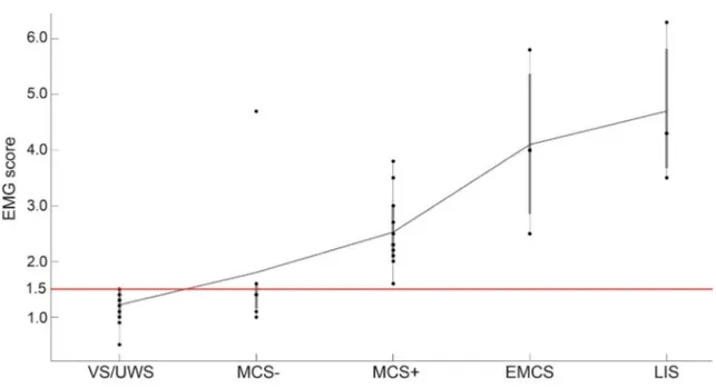
5.1 (left), 4.4 (right), and 5.1 (control),
correspond-ing to a mean EMG score of 1.8
6 1.1; for the
MCS1 patients, 4.5 (left), 5.9 (right), and 3.7
(con-trol), corresponding to a mean EMG score of 2.4
6
0.6; for the EMCS patients, 11.0 (left), 11.3 (right),
and 5.0 (control), corresponding to a mean EMG
score of 4.1
6 1.3; for the LIS patients, 8.0 (left),
13.0 (right), and 3.7 (control), corresponding to
a mean EMG score of 4.7
6 1.2. Figure 3 illustrates
the boxplot of the different groups.
Robustness and diagnosis evolution.
Four of the 40
pa-tients were assessed twice. LIS1 was evaluated twice
the same day (morning/afternoon) and showed
a response to command in both sessions. VS/
UWS11, VS/UWS9, and MCS
16 were evaluated,
respectively, 14, 16, and 11 months after the first
eval-uation. MCS
16 was MCS1 during the 2 evaluations,
and this was correctly detected by EMG at each
eval-uation. VS/UWS11 evolved into an MCS
1 (see
MCS
19 in table 1). His EMG score increased from
1.4 to 2.3 with this change in level of consciousness,
and a response to command was detected by EMG on
his second evaluation, while the CRS-R evaluation was
not able to detect a response to command the day of the
assessment. VS/UWS9 evolved into an MCS2 (see
MCS28 in table 1). The EMG score was below
threshold during both evaluations.
DISCUSSION
The present study confirms the
inter-est in EMG for the detection of responses to command
in severely brain-injured patients. The proposed
methodology allowed detection of a response to
command in all MCS
1 (n 5 14) patients included
in this study, while the behavioral evaluation
performed on the day of the EMG assessment only
allowed detection in 6 out of the 14 MCS1
patients. All EMCS (n
5 3) and LIS (n 5 2)
patients also presented a response to command as
assessed by EMG. It is important to note that LIS
patients in our study were in an incomplete LIS,
meaning they showed residual motor abilities.
Patients in a classical or complete LIS, with complete
cerebromedullospinal
disconnection,
would
not
present a response to command with our method.
Previous EMG studies were tested on a limited
number of MCS patients
9(n
5 2) or illustrated a high
false-negative rate
10(3 detections of response to
command out of 20 MCS
1 patients; 85%).
False-negatives have also been observed in several
neuroi-maging (range 50%
–67%)
19,20and electrophysiology
studies (range 22%
–100%),
21–25using imagery or
top-down modulation of attention (for a review, see Ref.
8,26). On the contrary, our paradigm is less cognitively
demanding and easier to perform. Indeed, the
partic-ipant is instructed to perform a movement, not to
imagine a movement
19,20,22,27or pay attention to
a sound.
21,23–25In addition, in comparison to previous
EMG studies,
9,10the increased number of trials and the
evaluation of the response to the command on each
side (left and right) gives more power to detect
repro-ducible willful motor response and to exclude any
ran-dom motor activity in this population with severe
motor impairments and vigilance fluctuations.
28,29No patients with VS/UWS (n
5 15) but 2 patients
in MCS
2 (n 5 8) presented a response to command
with the EMG. While volitional brain activity has
pre-viously been found in patients considered in VS/UWS
or MCS
2,
19,20,23,26,30,31we do not pretend that the
detection of response to command with our EMG
paradigm in behaviorally nonresponsive patients
re-flects a higher level of consciousness. They may be
false-positives. Patient MCS25 only showed
inconsis-tent behavioral signs of consciousness (i.e., visual
pur-suit during 1 out of 5 behavioral evaluations, the
remaining assessments concluding to a VS/UWS).
Figure 1 The experimental paradigm
The session was composed of 3 blocks, and each block consisted of 3 minutes recording at rest (baseline) followed by 15 trials. Each trial began with auditory presentation of the task instructions. Then, the EMG response to the command/control was collected. The instruction was repeated 3 times within a trial. Rest EMG activity was recorded during the 10-second intertrial interval (ITI).
MRI and fluorodeoxyglucose–PET confirmed the
diagnosis of MCS. The patient returned to her home
country and did not show much improvement
ac-cording to her treating physician. Patient MCS
26
died of a cardiopulmonary arrest 8 days following the
EMG evaluation. In our study, the EMG score
thresh-old determination was based on leave-one-out
cross-validation on the patients with a more stable
diagnosis/level of consciousness (
.1 year postinjury).
A receiver operating characteristic curve analysis led to
the determination of the same threshold (area
under the curve 1). Using the whole dataset led to
a slightly higher threshold of 1.6 (area under the curve
0.96), removing patients MCS26 and MCS15 from
responders’ cohort. Multiple patient testing on an
extended cohort would better assert the reliability of
the used threshold and results.
Evaluating the presence of a response to command
on a single trial basis allows to test the performance
and signal fluctuation across time, particularly
rele-vant in this population presenting nonstationarities
in brain response (e.g., fluctuation of arousal and
con-sciousness) and signal (e.g., artifact, noise).
Differen-tial EMG response on spaDifferen-tially close recording
locations and on temporally close period of time (trial
vs pretrial), as well as use of baseline activity as a
refer-ence, also allow to reduce the effect of
nonstationar-ities. However, the proposed approach detected
responses to the control instruction (“It is a sunny
day”) at a single-trial level in majority of the patients.
Figure 2 Evolution of root mean square (RMS) EMG signal (lower) andΔactive (upper), i.e., the difference between averaged RMS value during the trial and the preceding intertrial interval, within a block at right arm for patient EMCS3 and at right hand for patient MCS25
(A) Patient EMCS3. (B) Patient MCS25. Light gray represents control trials; medium and dark gray represent right and left target commands, respectively. An asterisk above a trial illustrates a positive activation at the corresponding location, i.e., the difference between the EMG activity during the trial and the previous intertrial interval is significantly higher (threshold set at p 5 0.01) than baseline fluctuations. Note the positive activation of all 5 right target trials and none of the control trial for patient EMCS3 and 6 target trials (3“right” and 3 “left”) and none of the control trial for patient MCS25.
These may be due to patients
’ spasticity, which is
common in this population and could make EMG
assessment and interpretation challenging. It is
important to note that the 3 MCS
1 patients with
an EMG score lower than 2 illustrated the higher
spasticity scores (table e-1), which could explain the
difficulty of our methodology to detect an answer. A
better model of EMG at rest could improve the single
trial detection and enable the translation to
EMG-based real-time communication.
Although the results illustrate the interest of our
method and suggest that these tools may provide
bed-side detection of command following, several
limita-tions could hamper its successful applicability in this
clinical setting. First, the preservation of some residual
voluntary muscle is a condicio sine qua non,
prevent-ing its use with patients with complete paralysis.
Motor-independent active paradigms relying on
func-tional neuroimaging (e.g., brain-computer interfaces)
could represent an interesting alternative in these
spe-cific cases. As an illustration, 30 out of the 40 patients
were selected to test a motor imagery fMRI-based
par-adigm
19but only one of them illustrated a response to
command with this paradigm (VS/UWS3); 25 of them
presented head movement preventing interpretable
data acquisition (see table e-1). The PET examination
of patient VS/UWS3 also illustrated active brain
re-gions similar to an MCS patient. The patient, however,
did not respond to command with our EMG
para-digm. This could be due to motor paralysis or lack
of awareness at the time of the test. Alternatively,
patient LIS2 tested an EEG-based motor imagery
par-adigm during her stay in our hospital and obtained
85% accuracy. Future studies should also evaluate
the effect of neuromuscular weakness on the
perfor-mance of the proposed method and compare
classifi-cation obtained during motor-based active task using
a multimodal EMG, fMRI, or EEG approach. Second,
the success of this paradigm relies on the patient
’s
understanding of the instructions, ability to follow
the command and motivation, which might be
decreased in case of language or memory
impair-ments,
32dysexecutive syndrome such as akinetic
mut-ism
33or perseveration, posttraumatic agitation (often
associated with delirium),
34hypoarousal cause by
sedating medication,
4or loss of motivation.
35,36The proposed EMG-based paradigm allows
a 40-minute (which is around the time of a CRS-R
assessment) bedside evaluation of response to
com-mand using only a few EMG electrodes, an amplifier,
and a computer to present the stimuli and record and
analyze the signal. Moreover, the paradigm is
inde-pendent of the examiner
’s experience or subjectivity.
7The results presented in this article were obtained
using a single session and may benefit from repetitive
evaluation within the week, as is the case with the
CRS-R. The potential use of the presented system as
a communication tool in the severely brain-injured
population should be investigated in the future.
AUTHOR CONTRIBUTIONSD.L. and D.H. obtained data and wrote the manuscript. D.L. and Q.N. analyzed and interpreted the data. D.L., D.H., C.C., C.S., S.L., and Q.N. designed the protocol. C.C., C.S., Q.N., and S.L. contributed to the writing of the manuscript. D.L., Q.N., and S.L. were the main inves-tigators. All authors were involved in editing the paper and approved the final text.
Figure 3 Boxplot of the different patient groups
Each dot represents a patient from the respective group. The dashed black line represents the mean EMG score. The horizontal line illustrates the threshold. Note the linear increase of EMG score with an increased level of consciousness and the presence of a response to command in all minimally conscious state plus (MCS1), emergence from minimally conscious state (EMCS), and locked-in syndrome (LIS), as well as in 2 of 8 MCS2. VS/UWS 5 vegetative state/unresponsive wakefulness syndrome.
ACKNOWLEDGMENT
The authors thank the Neurology Department staff of the University Hos-pital of Liège and the participants and their families for their collaboration.
STUDY FUNDING
Supported by the National Funds for Scientific Research (FNRS), European ICT Programme Projects FP7-247919 DECODER, James McDonnell Foundation, French Speaking Community Concerted Research Action, and University of Liege. The funders had no role in study design, data collection and analysis, decision to publish, or prepa-ration of the manuscript. D.L., D.H., C.C., C.S., S.L., and Q.N. had full access to all the data in the study, and the corresponding author had final responsibility for the decision to submit for publication.
DISCLOSURE
The authors report no disclosures relevant to the manuscript. Go to Neurology.org for full disclosures.
Received February 10, 2016. Accepted in final form July 29, 2016.
REFERENCES
1. The Multi-Society Task Force on PVS. Medical aspects of the persistent vegetative state (1). N Engl J Med 1994; 330:1499–1508.
2. Laureys S, Celesia G, Cohadon F, et al. Unresponsive wakefulness syndrome: a new name for the vegetative state or apallic syndrome. BMC Med 2010;8:68.
3. von Wild K, Laureys S, Gerstenbrand F, Dolce G, Onose G. The vegetative state: a syndrome in search of a name. J Med Life 2012;5:3–15.
4. Giacino JT, Ashwal S, Childs N, et al. The minimally conscious state: definition and diagnostic criteria. Neurology 2002;58:349–353.
5. Laureys S, Perrin F, Schnakers C, Boly M, Majerus S. Residual cognitive function in comatose, vegetative and minimally conscious states. Curr Opin Neurol 2005;18:726–733.
6. Laureys S, Schiff ND. Coma and consciousness: para-digms (re)framed by neuroimaging. Neuroimage 2012; 61:478–491.
7. Lovstad M, Froslie KF, Giacino JT, Skandsen T, Anke A, Schanke AK. Reliability and diagnostic characteristics of the JFK coma recovery scale-revised: exploring the influ-ence of rater’s level of experience. J Head Trauma Rehabil 2010;25:349–356.
8. Noirhomme Q, Brecheisen R, Lesenfants D, Antonopou-los G, Laureys S.“Look at my classifier’s result”: disentan-gling unresponsive from (minimally) conscious patients. Neuroimage 2015;8119:01119–1122.
9. Bekinschtein TA, Coleman MR, Niklison J, Pickard JD, Manes FF. Can electromyography objectively detect vol-untary movement in disorders of consciousness? J Neurol Neurosurg Psychiatry 2008;79:826–828.
10. Habbal D, Gosseries O, Noirhomme Q, et al. Volitional electromyographic responses in disorders of consciousness. Brain Inj 2014;28:1171–1179.
11. Bruno MA, Vanhaudenhuyse A, Thibaut A, Moonen G, Laureys S. From unresponsive wakefulness to minimally conscious plus and functional locked-in syndromes: recent advances in our understanding of disorders of conscious-ness. J Neurol 2011;258:1373–1384.
12. Bruno MA, Majerus S, Boly M, et al. Functional neu-roanatomy underlying the clinical subcategorization of minimally conscious state patients. J Neurol 2012; 259:1087–1098.
13. Giacino JT, Edlow B, Chatelle C, Schnakers C. The minimally conscious state: clinical features, pathophys-iology and therapeutic implications. In: Laureys S, Tononi G, Gosseries O, eds. The Neurology of Consciousness, 2nd ed. Cambridge, MA: Academic Press; 2016.
14. Laureys S, Pellas F, van Eeckhout P, et al. The locked-in syndrome: what is it like to be conscious but paralyzed and voiceless? Prog Brain Res 2005;150:495–511.
15. Giacino JT, Kalmar K, Whyte J. The JFK Coma Recovery Scale–Revised: measurement characteristics and diagnostic utility. Arch Phys Med Rehabil 2004;85:2020–2029. 16. Hugger S, Schindler HJ, Kordass B, Hugger A. Clinical
relevance of surface EMG of the masticatory muscles (part 1): resting activity, maximal and submaximal voluntary contraction, symmetry of EMG activity. Int J Comput Dent 2012;15:297–314.
17. Nöjd N, Hannula M, Narra N, Hyttinen J. Electrode posi-tion optimizaposi-tion for facial EMG measurements for human-computer interface. Methods Inf Med 2008;47:192–197. 18. Schalk G, McFarland DJ, Hinterberger T, Birbaumer N,
Wolpaw JR. BCI2000: a general-purpose brain-computer interface (BCI) system. IEEE Trans Biomed Eng 2004;51: 1034–1043.
19. Monti MM, Vanhaudenhuyse A, Coleman MR, et al. Willful modulation of brain activity in disorders of con-sciousness. N Engl J Med 2010;362:579–589.
20. Stender J, Gosseries O, Bruno MA, et al. Diagnostic pre-cision of PET imaging and functional MRI in disorders of consciousness: a clinical validation study. Lancet 2014; 384:514–522.
21. Goldfine AM, Victor JD, Conte MM, Bardin JC, Schiff ND. Determination of awareness in patients with severe brain injury using EEG power spectral analysis. Clin Neu-rophysiol 2011;122:2157–2168.
22. Cruse D, Chennu S, Chatelle C, et al. Relationship between etiology and covert cognition in the minimally conscious state. Neurology 2012;78:816–822.
23. King JR, Faugeras F, Gramfort A, et al. Single-trial decod-ing of auditory novelty responses facilitates the detection of residual consciousness. Neuroimage 2013;83:726–738. 24. Lulé D, Noirhomme Q, Kleih SC, et al. Probing
com-mand following in patients with disorders of consciousness using a brain–computer interface. Clin Neurophysiol 2013;124:101–106.
25. Pokorny C, Klobassa DS, Pichler G, et al. The auditory P300-based single-switch brain–computer interface: para-digm transition from healthy subjects to minimally con-scious patients. Artif Intell Med 2013;59:81–90. 26. Chatelle C, Lesenfants D, Guller Y, Laureys S,
Noir-homme Q. Brain-computer interface for assessing con-sciousness in severely brain-injured patients. In: Rossetti AO, Laureys S, eds. Clinical Neurophysiology in Disorders of Consciousness. Vienna: Springer-Verlag Wien; 2015: 133–148.
27. Cruse D, Chennu S, Chatelle C, et al. Bedside detection of awareness in the vegetative state: a cohort study. Lancet 2011;378:2088–2094.
28. Thibaut A, Chatelle C, Ziegler E, Bruno MA, Laureys S, Gosseries O. Spasticity after stroke: physiology, assessment and treatment. Brain Inj 2013;27:1093–1105.
29. Nakase-Richardson R, Yablon SA, Sherer M, Nick TG, Evans CC. Emergence from minimally conscious state: insights from evaluation of post-traumatic confusion. Neurology 2009;73:1120–1126.
30. Schnakers C, Perrin F, Schabus M, et al. Detecting con-sciousness in a total locked-in syndrome: an active event-related paradigm. Neurocase 2009;15:271–277. 31. Chennu S, Finoia P, Kamau E, et al. Dissociable
endog-enous and exogendog-enous attention in disorders of conscious-ness. Neuroimage Clin 2013;3:450–461.
32. Majerus S, Bruno MA, Schnakers C, Giacino JT, Laureys S. The problem of aphasia in the assessment of consciousness in brain-damaged patients. Prog Brain Res 2009;177:49–61. 33. Giacino JT. Disorders of consciousness: differential
diag-nosis and neuropathologic features. Semin Neurol 1997; 17:105–111.
34. Sandel ME, Mysiw WJ. The agitated brain injured patient: part 1: definitions, differential diagnosis, and assessment. Arch Phys Med Rehabil 1996;77:617–623.
35. Kleih SC, Nijboer F, Halder S, Kübler A. Motivation modulates the P300 amplitude during brain-computer interface use. Clin Neurophysiol 2010;121:1023– 1031.
36. Nijboer F, Birbaumer N, Kübler A. The influence of psychological state and motivation on brain-computer interface performance in patients with amyotrophic lat-eral sclerosis: a longitudinal study. Front Neurosci 2010;4:55.