HAL Id: hal-02512446
https://hal.archives-ouvertes.fr/hal-02512446
Submitted on 23 Mar 2020HAL is a multi-disciplinary open access archive for the deposit and dissemination of sci-entific research documents, whether they are pub-lished or not. The documents may come from teaching and research institutions in France or abroad, or from public or private research centers.
L’archive ouverte pluridisciplinaire HAL, est destinée au dépôt et à la diffusion de documents scientifiques de niveau recherche, publiés ou non, émanant des établissements d’enseignement et de recherche français ou étrangers, des laboratoires publics ou privés.
Econazole imprinted textiles with antifungal activity
Mirza Hossain, Fabrice Pagniez, Aicha Benhaddou, Martine Raymond, Karine
Théberge, Patrice Le Pape, Pierre Simard, Jeanne Leblond, Augustine Lalloz
To cite this version:
Mirza Hossain, Fabrice Pagniez, Aicha Benhaddou, Martine Raymond, Karine Théberge, et al.. Econa-zole imprinted textiles with antifungal activity. European Journal of Pharmaceutics and Biopharma-ceutics, Elsevier, 2016, �10.1016/j.ejpb.2016.02.003�. �hal-02512446�
Econazole Imprinted Textiles with Antifungal Activity
1
Mirza Akram Hossaina, Augustine Lalloza, Aicha Benhaddoub, Fabrice
2
Pagniezc, Martine Raymondd, Patrice Le Papec, Pierre Simardb, Karine
3 Thébergeb, and Jeanne Leblonda 4 5 a Faculty of Pharmacy, Université de Montréal, PO Box 6128 Station Centre-6 Ville, Montreal, QC, H3C 3J7, Canada. 7 b Biomod Concepts Inc, Sainte-Julie, QC, J3E 1Y6, Canada. 8 c Département de Parasitologie et de Mycologie Médicale, Université de 9 Nantes, Nantes Atlantique Universités, EA 1155 - IICiMed, Faculté de 10 Pharmacie, Nantes, France ; Laboratoire de Parasitologie-Mycologie, CHU de 11 Nantes, Nantes, France 12 d Institute for Research in Immunology and Cancer and Department of 13 Biochemistry and Molecular Medicine, Université de Montréal, Montreal, QC, 14 H3C 3J7, Canada 15 16 Corresponding author: [email protected] ; + (1) 514-343-17 6455, Faculty of Pharmacy, Université de Montréal, PO Box 6128 Station 18 Centre-Ville, Montreal, QC, H3C 3J7, Canada. 19 Keywords 20
Econazole nitrate; fungal infection, topical administration; thermo-responsive 21 formulation; lipid microparticles; candida. 22 Abstract 23
In this work, we propose pharmaceutical textiles imprinted with lipid
24
microparticles of Econazole nitrate (ECN) as a mean to improve patient
25
compliance while maintaining drug activity. Lipid microparticles were
26
prepared and characterized by laser diffraction (3.5±0.1 μm). Using an
27
optimized screen-printing method, microparticles were deposited on textiles,
28
as observed by Scanning Electron Microscopy. The drug content of textiles
29
(97±3 μg/cm2) was reproducible and stable up to 4 months storage at
30
25°C/65% Relative Humidity. Imprinted textiles exhibited a thermosensitive
31
behavior, as witnessed by a fusion temperature of 34.8°C, which enabled a
32
larger drug release at 32°C (temperature of the skin) than at room
33
temperature. In vitro antifungal activity of ECN textiles was compared to
34
commercial 1% (w/w) ECN cream Pevaryl®. ECN textiles maintained their
35
antifungal activity against a broad range of Candida species as well as major
36
dermatophyte species. In vivo, ECN textiles also preserved the antifungal
37
efficacy of ECN on cutaneous candidiasis infection in mice. Ex vivo
38
percutaneous absorption studies demonstrated that ECN released from
39
pharmaceutical textiles concentrated more in the upper skin layers, where the
40
fungal infections develop, as compared to dermal absorption of Pevaryl®.
Overall, these results showed that this technology is promising to develop
42
pharmaceutical garments textiles for the treatment of superficial fungal
43 infections. 44 45 1. Introduction 46 Textile is a material that has been purposed to clothing for centuries. In 47
recent years, the combined efforts of chemists, textile engineers and
48
cosmetologists resulted in the development of biofunctional textiles that bring
49
additional functions to garments than simple warmth and body protection.
50
Also called cosmetotextiles, such textiles are defined as textile items containing
51
substance or mixture that release their active compounds when in contact with
52
the human body [1]. Firstly focused on improved comfort, cosmetotextiles
53
have since then been developed for slimming, moisturizing, and perfuming [2].
54
Innovative technologies have been incorporated into such fabrics, such as
55 microencapsulated substances [1, 3] or phase change materials that help the 56 thermoregulation of the body [4]. Rapidly, various biofunctional textiles have 57 been envisioned for the delivery of topical bioactive molecules, since the close 58 and prolonged contact of fabric with the skin could make cloth an easy drug 59
delivery system. Silver nanoparticles [5] and chitosan [6] were used as
60
preservatives for antibacterial clothing. Fabrics with antioxidant properties
were developed by incorporation of vitamin E [7] or gallic acid [8]. Some
62
clinical indications have also been examined, such as venous insufficiency
63
using aescin supported textiles [9] and atopic dermatitis with zinc oxide
64
functionalized textiles [10]. Such examples show the evolution of
cosmeto-65
textiles to pharmaceutical textiles, offering more than an improved comfort,
66
but also a treatment for various skin diseases.
67
In particular, superficial fungal diseases are common worldwide and
68 their incidence continues to increase. In 2010, they were the 4th most prevalent 69 disease in the world, affecting more than 948 million people worldwide [11]. 70 As compared to bacteria, fungal topical infections are longer in duration and 71 require weeks and even months of fastidious treatment. Patient compliance 72
would be greatly improved if a regular piece of textile (such as bandage or
73 socks) could be used instead of applying a cream daily. Antifungal textiles have 74 been prepared by soaking the fabric into a solution of antifungals [12, 13], and 75 promising clinical results have been obtained from a sock prototype to treat 76
tinea pedis [14]. However, fabrication technology and controlled release of
77
antifungal agents still need to be improved.
78
Econazole Nitrate (ECN) is currently marketed for the treatment of
79
vaginal candidiasis and topical fungal infections as a cream formulation [15,
80
16]. It has demonstrated antifungal activity against Candida and
81
dermatophytes species [15-17]. Encapsulation of ECN in lipid particles [18],
microspheres [19], and micelles [20] has been reported to improve cutaneous
83
efficacy of ECN. More precisely, comparing micro- and nano- solid lipid
84
particles, nanoparticles were shown to improve transdermal administration
85
whereas microparticles enhanced skin deposition [21]. Moreover, the lipid
86
composition favored a good biocompatibility of the particles and improved
87 skin penetration of the drug [22]. 88 In this work, a novel ECN formulation on textile support was tested as a 89 proof of concept for the treatment of topical fungal infections. The formulation 90
is based on proprietary lipid microparticles exhibiting thermosensitivity in
91
order to release the drug on contact with the skin [23]. Deposition of the
92
microparticles on textile is achieved using an in-house modified
screen-93 printing technique. The latter is a simple method where the microparticles are 94 passed through a mesh with predefined openings to control the amount and 95 the topology of the deposit [24, 25]. This method allows for a physical uniform 96 deposit of the microparticles at specific areas on textiles without addition of 97
chemical binders. The solid microparticles (Dermotex®) and deposition
98
method (On2TM) are technologies proprietary to Biomod Concepts Inc., and
99
have been used by the company to produce intelligent cosmetic textiles [23].
100
The objective of this study is to evaluate the potential of a pharmaceutical
101
textile, namely a microparticle formulation of ECN deposited on textile. Its in
102
vitro antifungal activity, percutaneous absorption, and in vivo pharmaceutical
efficacy on a superficial fungal infection were compared to the commercial 1% 104 (w/w) ECN cream Pevaryl®. 105 106 2. Experimental methods 107 2.1. Materials 108 ECN-loaded microparticles on textile and all placebo textile formulations were 109
provided by Biomod Concepts Inc. (Ste-Julie, QC, Canada) and prepared
110
according to their patented technology [23]. LayaTM textiles were provided by
111
Biomod Concepts (Sainte-Julie, QC, Canada). ECN was purchased from AK
112
Scientific (Union City, CA, USA, Lot# TC24717). Pevaryl® 1% (w/w) ECN
113
formulation was purchased from Johnson & Johnson (France, Lot # DDB3400)
114
and its generic version from Mylan Pharmaceuticals (Saint-Priest, France).
115
Miconazole Nitrate was purchased from AK Scientific (Union City, CA, USA,
116
Lot# TC25782). ECN standard disks were purchased from Rosco
(Neo-117
sensitabs 10 μg disks, Denmark, Lot #1201-1). Prednisolone acetate was
118
purchased from Sanofi Aventis (Paris, France) Polyethylene Glycol 400 (PEG-119
400) was purchased from Medisca Inc. (Montreal, QC, Canada). Sodium dodecyl
120
sulfate (SDS) and semi-permeable polycarbonate membranes (Nucleopore
121 Track-Etch Membrane, pores of 0.6 μm, 25 mm in diameter) were purchased 122 from Sigma-Aldrich (Oakville, ON, Canada). Tape used for tape stripping was 123 purchased from 3M tape (St-Paul, MN, USA). All samples were filtered using 124
PTFE filters purchased from Fisher Scientific (EMD Millipore Millex, pores 0.45 125 μm, 13 mm in diameter, Ottawa, ON, Canada). All solvents (HPLC grade) were 126 bought from Fisher Scientific (Ottawa, ON, Canada). 127 2.2 Organisms 128
Candida albicans strain SC5314 was originally isolated from a patient with
129 disseminated candidiasis, and served as reference for the C. albicans genome 130 sequencing project [26, 27]. Thirteen clinical isolates of Candida spp. and C. 131 albicans (CAAL93, CAAL121, CAAL123, CAAL124, CAAL294), C. kefyr (CAKE3, 132
CAKE4), C. krusei (CAKR1, CAKR3), C. glabrata (CAGL1,CAGL5), and C.
133
lusitaniae (CALU1, CALU2) were obtained from the Department of Parasitology
134
and Medical Mycology, EA1155, at the University of Nantes, France.
135 Trichophyton rubrum (n=2) and T. mentagrophytes (n=2) were obtained from 136 the Laboratory of Parasitology and Medical Mycology at the Centre Hospitalier 137 Universitaire of Nantes. 138 2.3. Preparation of ECN textiles 139 Intelligent textiles imprinted with ECN-loaded microparticles were prepared 140
by Biomod Concepts Inc. using their patented technology [23]. Briefly, ECN
141
lipid microparticles (1% w/w) were prepared under high shear using
FDA-142
approved ingredients. The microparticles formulation was then applied onto
143
textile surface using an adapted screen-printing method optimized for the
144
microparticles deposition. A stencil with openings of more than 400 µm was
used to apply the microparticles on 21.6 x 27.9 cm pieces of a polyester non-146
woven textile provided by Biomod Concepts Inc. ECN imprinted textiles were
147
kept at 22°C in sealed aluminum/acrylonitrile-coated packagings until
148
analysis.
149
2.4. Characterization of microparticles
150
One hundred milligram (100 mg) of the ECN-loaded microparticles
151
preparation used for screen-printing was diluted in 5 mL of milliQ water and
152
analyzed for article size distribution at 22°C by laser diffraction (LS 13 320,
153
Beckman Coulter, Mississauga, ON, Canada). Pevaryl® particle size was
154
measured by dynamic light scattering (Zetasizer Nano ZS, Malvern,
155
Worcestershire, UK) using the automatic algorithm mode. Samples were
156
prepared by diluting 100 mg of Pevaryl® in 5 mL of MilliQ water, position 4.65
157
and attenuator at 8. Measurements were recorded 3 times for each
158 formulation. 159 Fusion temperature of the microparticles imprinted on textile was measured 160 using thermal analysis based on heat-leak-modulus (TA-HLM) [28]. With TA-161
HLM, textile samples are wrapped around a sensor probe and heated. The
162 samples of ECN-loaded textile (2.5 x 5 cm) were analyzed at a heating rate of 163 0.8°C per second and heated from 0°C to 100°C. Measurement was repeated 3 164 times. 165 2.5 HPLC-UV analysis 166
High-performance liquid chromatography (HPLC) with ultraviolet (UV)-167 analysis was used for stability and quantification of samples. 168 The HPLC-UV system (Agilent 1100 Series, Mississauga, ON, Canada) consisted 169 in a degasser, dual pumps, auto-sampler, column heater and photo-diode array 170 detector. A C18 column (25 x 4.6 mm, 5 μm packing, Zorbax-C18, Agilent, Santa 171 Clara, CA, USA) was used with a matching pre-column (Agilent Zorbax C18). 172 Mobile phase was composed of methanol and water using the gradient detailed 173 in Table 1. 174 The flow rate was 1.4 mL per minute. The column temperature was set to 35°C. 175 The injection volume was 20 μL. ECN was analyzed at 220 nm. ECN retention 176 time was 8.7 minutes. The limit of quantification with this method is 9 μg/mL. 177 178 Table 1: Gradient of solvents in the HPLC-UV system 179
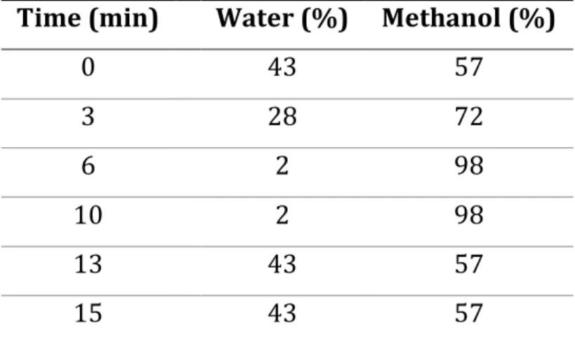
Time (min) Water (%) Methanol (%)
0 43 57 3 28 72 6 2 98 10 2 98 13 43 57 15 43 57 180 2.6 HPLC-MS/MS method 181
HPLC-Mass spectrometry (MS)/MS was used for in vitro release and ex vivo 182 experiments on pig skin, which presented lower concentrations of ECN than 183 the limit of quantification (LOQ) of HPLC-UV method. An Agilent 1100 series 184 HPLC (Mississauga, ON, Canada) was coupled to a 4000Q TRAPTM (AB Sciex, 185
Concord, ON, Canada) hybrid triple-quadrupole/linear ion trap MS. All the
186 parameters can be found in Table 2. Each sample was injected twice. HPLC-187 MS/MS method was developed and validated for ECN, using miconazole nitrate 188 as an internal standard. No matrix effect was found with any components of 189 the skin. 190 191 Table 2: LC-MS/MS parameters 192 HPLC Agilent 1100 series MS/MS AB Sciex 4000 Qtrap Software Analyst® (version 1.6.2) Ionisation Turbo electrospray, positive ionization (ESI) Scan mode Multiple reaction monitoring (MRM) Analyte
parameters Compounds DP (V) MRM CE (eV) Test molecule Econazole 90 381 > 125 40 Internal Standard Miconazole 90 417 > 161 40 Source parameters Gas temp (°C) 650 Gas flow (L/min) 50 Curtain gas (psi) 25 Capillary (V) 5500 Mobile phase Composition A: 0.1% Formic Acid (FA)+ H2O B: 0.1 % Formic Acid + Acetonitrile: Isopropanol (80/20) Gradient 15 to 97% of phase B in 1.5 min, then stay at 97% until 2.2 min, decrease to
15% at 2.3 min and stay at 15% of phase B until 3.5 min Flow rate 0.7 mL.min-1 Column temperature 45°C Injection volume 2 µL Injection temperature 5°C
Column Luna C8 column (30 x 2.0 mm, 5 µm, Phenomenex, Torrance, CA, USA) 193 2.7 Quantification of ECN Textile 194 One-cm2 pieces of ECN imprinted textile were sampled from the center region 195 of randomly selected textile sheets for a good statistical analysis. Ten out of the 196 30 sheets imprinted for this study were sampled and analyzed. To extract ECN 197 from the fabric, textile samples were suspended in 1 mL of methanol, sonicated 198 for 30 minutes and heated at 55°C for 4 hours. After cooling down, 500 μL of 199 the extraction solution was sampled, filtered, and quantified by HPLC-UV. 200 2.8 Stability 201 Three 15 x 15 cm sheets of imprinted ECN textile were placed in a stability 202 chamber (25°C/65% relative humidity (RH)). At each time point, three 1-cm2 203 pieces were samples per sheet from the center area of the textile for analysis. 204 The sample preparation and quantification methods were as described above 205 (section 2.7). The stability was monitored up to 4 months. 206 2.9 In vitro release 207
Disks of 0.79 cm2 (10 mm diameter, containing 71 μg of ECN) were cut out of 208 the imprinted textile. In vitro release was performed using Franz cells from 209 PermGear Inc. (Hellertown, PA, USA) with an opening of 9 mm in diameter, 5 210
mL receptor size and a thermostated jacket. Diffusion tests were carried on
211
semi-permeable polycarbonate membranes of 0.6 μm in pore size. The
212
receptor fluid composition was optimized to ensure ECN diffusion was not
213 limited by ECN solubility. Although ECN was not soluble at pH 7.4, its solubility 214 was improved in 10 mM phosphate buffer solution (PBS) with 1.37 mM of NaCl 215 at pH 4.5 with PEG-400 (70:30 v/v) [29]. Indeed, Pevaryl diffusion (20 mg) on 216 Frantz cells using a semi-permeable polycarbonate membranes of 0.6 μm in 217
pore size after 6h at 32°C, ECN solution recovery was 49.5%±1.3 and
218 13.5%±1.3 with and without PEG, respectively (n=3 for each condition, data 219 not shown). The receptor fluid (10 mM phosphate buffer solution (PBS) with 220 1.37 mM of NaCl at pH 4.5 with PEG-400 (70:30 v/v)) was thermostated at 22°C 221
or 32°C and was constantly stirred at 100 rpm. Samples of 400 μL were
222 withdrawn at 30 minutes, 1, 2, 3, 4, and 6 hours, filtered and replaced with the 223 same volume of receptor fluid. Samples were diluted 1:50 in a mixture of 20% 224 acetonitrile: 80% H2O with 0.1% FA before quantification by HPLC-MS/MS. 225 2.10 Antifungal Disk Diffusion tests 226 C. albicans SC5314 and Candida strains from Nantes were routinely grown at 227 30°C in yeast peptone dextrose (YPD; 1% yeast extract, 2% Bacto peptone, 2% 228
dextrose plus 2% agar for solid medium) and Sabouraud (SB) culture medium 229 respectively. C. albicans SC5314 were suspended in liquid YPD medium to an 230 OD600 of 0.1, and 150 μL of the cell suspension were spread on YPD Petri dishes 231 (10 cm diameter). Disks of 0.79 cm2 (10 mm diameter, containing 71 μg of ECN) 232 were cut out of the imprinted textile, and equivalent quantity of Pevaryl® (7.1 233
mg Pevaryl®, 71 μg ECN) was weighed on a filter paper disk (10 mm in
234
diameter). Placebo textiles imprinted with drug-free microparticles and
235
standard 10-μg ECN disks were used as controls. All disks were placed on the
236
YPD Petri plates. The plates were incubated at 32°C, and growth inhibition
237
diameters were measured at 18 h. This was replicated 9 times for each
238 formulation and repeated 3 times independently. 239 Other Candida spp isolates were suspended in sterile saline (0.85% NaCl) to 240 achieve 1 x 10⁶ cells per mL, which were deposited onto SB Petri dishes (10 cm 241 in diameter). Disks of 0.50 cm2 (8 mm in diameter, containing 50 μg of ECN) 242 were cut out of the imprinted textile, and equivalent quantity of Pevaryl® (5.0 243
mg Pevaryl®, 50 μg ECN) was weighed on a filter paper disk (8 mm in
244
diameter). Placebo textiles (8 mm in diameter) and standard 10-μg ECN disks
245
were used as controls. All disks were placed on the SB Petri plates. The plates
246
were incubated at 35°C and growth inhibition diameters were measured at
247
18h. This was replicated 4 times for each formulation and repeated 2 times
248
independently.
Trichophyton rubrum and T. mentagrophytes were grown on SB culture 250 medium. Trichophyton species were suspended in sterile saline (0.85% NaCl) 251 with 0.1% SDS to achieve 1 x 10⁶ fungal cells per mL, and the cells deposited 252 on SB petri dishes (10 cm in diameter). The plates were incubated at 25°C, and 253 growth inhibition diameters were measured after 4 days for T. mentagrophytes 254 and after 7 days for T. rubrum. This was replicated 3 times for each formulation 255 and species. This was repeated 3 times independently for T. mentagrophytes 256 and once for T. rubrum. 257 2.11 Ex vivo diffusion test on pig ear skin 258
Pig ears were kindly provided by Dr. Fairbrother from the Veterinary
259
Department of Université de Montréal (Saint-Hyacinthe, QC, Canada). Ears
260
were washed with water and 1% SDS and shaved using a razor. The skin was
261
gently peeled off from the dorsal region of the ears, washed again with 1% SDS
262
and water and stored at -20°C until the next day. Skin diffusion tests were
263
performed using Franz cells as described above. The receptor fluid was
264
identical to in vitro release studies. It contained 10 mM PBS at pH 4.5 with 30%
265
PEG-400, to ensure ECN solubility, and was stirred at 100 rpm [29]. The
266 receptor compartments were heated to 37°C to help maintain the surface of 267 the skin at 32°C. Disks of 0.79 cm2 (10 mm in diameter, containing 71 μg of 268 ECN) were cut out of the imprinted textile, and equivalent quantity of Pevaryl® 269 (7.1 mg, 71 μg ECN) was weighted on a filter paper disk (10 mm in diameter). 270
The disks were applied upside down on the surface of the skin, so that the
271
formulation was in direct contact with the skin. A small weight (5 g) was
272
applied in order to ensure contact between the formulation and the skin
273
surface. The system was dismounted after 24 hours and all the receptor fluid
274
was collected. The used textiles and filter paper disks were collected for ECN
275
quantification. The skin surface was washed with 8 mL of ethanol/water
276
(50:50), which was collected for ECN quantification. The skin was separated in
277
3 layers: stratum corneum (SC), epidermis and dermis. The SC was removed
278
using 20 strips of 1 cm2 3M tape, which was extracted with 20 mL of
279
acetonitrile. Epidermis was peeled off from the dermis after heating at 80°C for
280
10 seconds. Both epidermis and dermis were cut into pieces and were
281 suspended in 1 mL acetonitrile. All samples were sonicated for 30 min then 282 heated at 55°C for 4 hours. Liquid layers were filtered and diluted with 20% 283 acetonitrile with 0.1% FA. Samples were diluted (1:20 for epidermis, 1:200 for 284 residual formulation on filter paper disks and textiles, and 1:10 for washing, 285 SC, dermis, and receptor fluid) and quantified by HPLC-MS/MS. The Overall 286 recovery of ECN (sum of residual textile, washing, SC, epidermis, dermis) was 287 97.7% ± 5.7 (n = 9). Skins from 3 different pig ears were tested in triplicate 288 each (n = 9). 289 2.12 Cutaneous candidiasis model in immunosuppressed mice 290
Female mice were treated with prednisolone acetate on the day before and on 291 the day after inoculation. Hairs on the back of anesthetized mice were plucked 292 by hand to make a hairless square. The skin was then slightly abraded using 293
sandpaper and Candida albicans inoculum (25 µL at 3. 109 yeast/mL) was
294 applied. Mice were then randomly distributed into 4 groups of 6 animals. At 295 day 3 post-infection, treatment was applied topically on the skin lesion once 296 daily during 5 consecutive days. A first group was treated with the reference 297 drug, Mylan ECN cream at a dose of 50 µg of ECN per lesion, a second one with 298 a disk of ECN textile at a dose of 50 µg of ECN per lesion, a third one with a 299 placebo textile, and a fourth one was untreated and served as control group of 300 the disease. 301
In order to evaluate the infection level, microbiological studies were
302
undertaken. Skin specimens from infected locus were taken with a biopsy
303
punch at day 9 post-infection. Each sample (half of the biopsy) was
304
homogenized in saline solution with a tissue grinder. Dilutions were inoculated
305
on Sabouraud-chloramphenicol-gentamicin agar plates. After a 48-h
306
incubation time at 35°C, the number of yeast colonies was counted.
307
The procedure was approved by the ethical committee of Pays de la Loire,
308
France with the agreement D44015 for the Unité Thérapeutique
309
Experimentale, Faculté de médecine, Nantes.
310
2.13 Statistical Analysis
Statistical analysis was executed by means of Graph Pad® 6.0c (Prism 312 Software, San Diego, CA, USA). Multiple t-test was used with corrected p-value 313 using the sidak-bonferroni assuming unequal variance method for ex vivo pig 314 skin diffusion tests. All p-values ≤ 0.05 were considered to be significant. 315 316 3. Results and discussion 317 318 3.1 Physical properties of pharmaceutical textiles 319 320 ECN-loaded lipid microparticles (1% w/w) were prepared under high shear 321
and deposited on textile using a screen-printing method as previously
322 described [23]. The pharmaceutical textiles were first characterized for their 323 ECN content. ECN was extracted from textile samples and analyzed by HPLC. 324 ECN content was measured to be 90±19 μg per cm2. The uniformity and the 325 homogeneity of deposition of ECN-loaded microparticles were assessed from 326
10 different sheets of textile and 3 different areas per sheet and was
327
determined to be less than 10% of the mean ECN value (data not shown).
328
Moreover, the ECN content was monitored as indicator of the chemical stability
329
of the pharmaceutical textiles. Stability was monitored each week during 6
330 weeks and again after 16 weeks using a stability chamber at 25C°C/65% RH 331 (Table 3). After 4 months, ECN content was still 97 μg/cm2, which represents 332 108% of the initial content. Altogether, the reproducibility of the production 333
process and the stability over several months demonstrated the potential of 334 the pharmaceutical textiles as new therapeutic products. 335 336 Table 3: ECN content of pharmaceutical textiles upon storage at 25°C/65%RH. 337 T0: After screen-printing. (n = 9) 338 Time in weeks T0 T1 T2 T3 T4 T6 T16 ECN Textile content (μg/cm2) 90±19 99±10 105±11 100±20 96±9 84±2 97±3 339
In a second step, physicochemical properties of the microparticles were
340 examined. Microparticle size was evaluated before deposition on textile using 341 laser diffraction (Figure 1). Particles exhibited micro-range diameter (3.5±0.1 342 μm). Pevaryl® particle size was also measured by dynamic light scattering and 343
indicated a mean particle size of 348 nm with a polydispersity of 0.3. After
344 screen-printing, ECN textiles were observed by scanning electron microscopy 345 for their size and morphology. Figure 2 shows the presence of microparticles 346 deposited on the textile fibers along with a film surrounding the microparticles. 347
The film could be microparticles that partially melted or fused during the
348
deposition process, probably due to the low fusion temperature of
349
microparticles.
350
352 353 Figure 1: ECN microparticles size distribution by laser diffraction (n=9). 354 355 356
Figure 2: Scanning electron microscopy image of ECN pharmaceutical textile. 357 Scale = 20 µm. 358 359
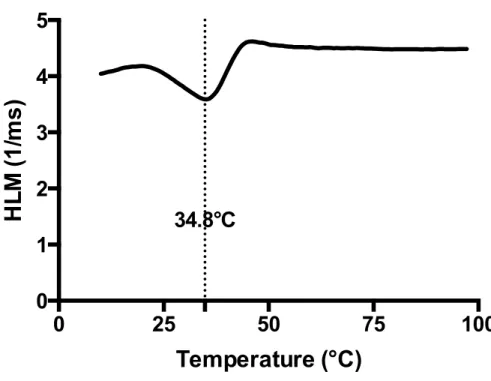
Lipid microparticles deposited on textile exhibited a fusion temperature of
360
34.8°C, as determined by TA-HLM, which confirmed their solid character at
361
room temperature (Figure 3). The latter is a method similar to differential
362
scanning calorimetry, which allows for the measurement of fusion
363 temperature of microparticles deposited on a textile surface [28]. This fusion 364 temperature value is crucial to allow triggered release upon contact with the 365 skin, which is at approximately 32°C, while maintaining a good stability upon 366 storage at room temperature. 367 368 369 Figure 3: TA-HLM of ECN textile. 370
0
25
50
75
100
34.8°C
0
1
2
3
4
5
Temperature (°C)
HL
M
(1
/ms
)
371
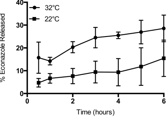
The thermo-sensitive behavior of the pharmaceutical textiles was further
372 confirmed by the drug release profiles obtained at room temperature and at 373 32°C in Franz diffusion cells (Figure 4). As expected, release at 32°C was higher 374 and faster than at room temperature. Indeed, the textiles released about twice 375 the amount of ECN at all time points (although no significant difference was 376 seen at 6 hours due to larger error bars). Raising the temperature to 32°C, close 377
to the fusion temperature of the microparticles, initiated the
378 fluidification/fusion of the microparticles and allowed ECN diffusion from the 379 textile. It can be noted that no increase in ECN concentration in receptor fluid 380 was observed after 24h (data not shown), indicating that the maximum release 381 was reached within 6 hours. This suggested the textiles served as a reservoir 382 for ECN, since less than 29±6% of the total ECN was released. 383
384 385 Figure 4: ECN release from pharmaceutical textiles at 22°C (black squares) and 386
32°C (black circles) on Franz diffusion cells through polycarbonate
387 membranes. Mean ± standard error bars (n=4). 388 389 3.2 Antifungal activity of pharmaceutical textiles 390 Once their thermo-sensitive behavior was verified, the intelligent textiles were 391 examined to ensure that the developed technology was able to preserve the 392
pharmaceutical activity of the drug. Antifungal activity of ECN textiles was
393
compared to Pevaryl®, a commercial formulation of ECN. Pevaryl® is a 1%
394
(w/w) ECN cream indicated to treat Candida and dermatophytes superficial
395 infections [15, 16]. 396
0
2
4
6
0
10
20
30
40
Time (hours)
%
E
co
na
zo
le
R
el
ea
se
d
32°C
22°C
397 Figure 5: Antifungal disk diffusion test on C. albicans SC5314 containing textile 398 placebo (imprinted with blank formulation) (A), Pevaryl® formulation (71 μg 399 ECN) on a filter paper (B), soluble ECN 10 μg standard disks (C), and ECN textile 400 formulation (71 μg ECN) (D). 401 402
Experimental conditions were first optimized with the C. albicans SC5314
403
strain using the antifungal disk diffusion test [30]. Figure 5 shows a
404
representative setup for the experiment; ECN imprinted textiles were
405
compared to ECN standard disks, Pevaryl® deposited on filter paper, and
406
placebo composed of textiles imprinted with a blank formulation (i.e. same
407
composition, without ECN). The textile formulation demonstrated an
408
inhibition zone corresponding to 81% of that of the commercial formulation
albicans strains and the ECN textiles exhibited an inhibition of 93% (Table 4). 411 These results showed that ECN maintained a roughly comparable activity on 412 textile as compared to the cream formulation. The slightly lower activity may 413 be due to the lower diffusion of ECN from the textile support. Pevaryl® might 414
also exhibit a slightly better efficacy because of its nanometer particle size
415
range, which increases surface area and facilitates diffusion, resulting in
416
increased drug activity [30]. These results were confirmed by assessing other
417
Candida species. ECN activity of ECN textiles reached 91%, 84% and 91% of
418
the activity of the commercial formulation on C. lusitaniae, C. kefyr and C.
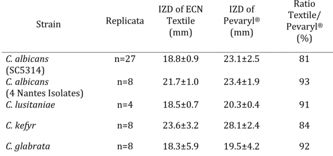
419 glabrata, respectively (Table 4). The activity was less important for C. krusei, 420 which often demonstrates an intrinsic resistance to azole drugs [31]. 421 422 Table 4: Inhibition zone diameter (IZD) of ECN textile and Pevaryl® on 423 Candida spp and Trichophyton species after 18 hours at 32°C 424 Strain Replicata IZD of ECN Textile (mm) IZD of Pevaryl® (mm) Ratio Textile/ Pevaryl® (%) C. albicans (SC5314) n=27 18.8±0.9 23.1±2.5 81 C. albicans (4 Nantes Isolates) n=8 21.7±1.0 23.4±1.9 93 C. lusitaniae n=4 18.5±0.7 20.3±0.4 91 C. kefyr n=8 23.6±3.2 28.1±2.4 84 C. glabrata n=8 18.3±5.9 19.5±4.2 92
T. mentagrophytes n=14 24.8±0.7a 39.8±3.2a 62a T. rubrum n=3 29±1.7b 47±4.2b 62b a IZD of textile formulation on T. mentagrophytes were measured after 4 days 425 at 25°C 426 b IZD of textile formulation on T. rubrum were measured after 7 days at 25°C. 427 428
Finally, inhibition of dermatophytes growth was examined on the two most
429
prevalent species in fungal skin infections, namely Trichophyton
430
mentagrophytes and T. rubrum [12, 13] (Table 3). In these latter tests, the
431
activity of ECN reached 62% of the commercial formulation activity. This lower
432
result could be explained by the temperature (25°C) at which the experiment
433
was conducted. This temperature was selected for dermatophytes to grow
434
several days in vitro, but is lower than the fusion temperature of the
435
microparticles. At this temperature, ECN has a limited release, as shown in
436
figure 3, so that the effective concentration of ECN might have been lower than
437
the commercial formulation. Although the experimental conditions were not
438
adapted for the pharmaceutical textiles, similar results were obtained by
439 Hammer’s et al. They concluded that T. mentagrophytes and T. rubrum were 440 less susceptible than C. albicans when exposed to antifungal textiles [13]. 441 Overall, these results demonstrate that ECN textiles maintain their antifungal 442 activity against all azole-susceptible Candida species tested in vitro. Although 443 in vitro disk diffusion tests are not designed to test the long-term efficacy of 444
controlled release products like the ECN textiles but rather immediate release, 445 this newly developed technology of imprinted textiles was shown to preserve 446 the pharmaceutical activity of the antifungal drug. 447 448 3.3 Percutaneous absorption of ECN 449 The impact of the lipid microparticle formulation on percutaneous absorption 450
was examined ex vivo by comparing the ECN textile to the commercial
451
formulation. Pig skin has been reported as a good model for skin percutaneous
452
absorption [32]. Commercial formulation was weighed on a filter paper to
453
provide the same ECN amount as ECN textile. Both ECN formulations were
454 applied upside down on pig skin for 24 h with the receptor fluid temperature 455 maintained at 37°C in the Franz diffusion cells. ECN was quantified by HPLC-456 MS/MS in the different layers of the skin: SC, epidermis, dermis, and receptor 457 compartment (Figure 6). Overall percutaneous absorption was similar for both 458
formulations, showing that both systems diffused similar quantities of ECN.
459
This was in agreement with the comparable antifungal activity of both
460
treatments on Candida species (Table 4). Pevaryl’s diffusion may be slightly
461
underestimated due to some absorption of ECN on the paper support.
462
Nevertheless, this partition would not have significantly impacted the total
463
release of econazole, which was less than 1% of the ECN loading. Other stimuli,
464
like friction, might further enhance drug delivery upon contact with the skin.
Nevertheless, this reservoir effect of the textile was already observed for other 466 cosmetotextiles [8]. 467 468 469 Figure 6: ECN content after 24 hour in Franz cells diffusion test on pig skin 470
using ECN textile (black bars) and Pevaryl® on filter paper (grey bars).
471
Receptor compartment was set at 37°C. ECN quantification was done by LC-472
MS/MS. Mean ± standard deviation (n=4). Asterisks indicate statistically
473 significant values (p < 0.05). 474 475
Total Absorption
SC
Epidermis
Dermis
Receptor
0.0
0.5
1.0
1.5
Ec
on
az
ol
e
qu
an
tit
y
(µ
g/
cm
2)
ECN Textile
Pevaryl
*
*
*
ECN skin distribution was slightly different between the ECN textile and the 476 commercial cream on filter paper. ECN from textiles was mainly distributed in 477 the SC and penetrated less in the epidermis and dermis, whereas ECN from the 478
commercial formulation was mainly found in the dermis and was even
479
quantifiable in the receptor compartment (Figure 6). This difference might be
480
attributed to the particle size of both formulations. Indeed, Pevaryl® was
481 determined to be a nano suspension whereas the particles deposited on the 482 textiles were in the micron range (Figures 1&2). Nanometer formulations have 483 been reported to penetrate deeply into the dermis, and have also been used for 484 transcutaneous absorption of drugs [21]. In our case, the micrometer particles 485 were designed to concentrate into the upper layer of the skin, where the fungal 486
infections develop. Moreover, their lipid composition is thought to improve
487 their affinity for the skin tissues, resulting in a higher concentration of ECN in 488 the SC than the commercial formulation. This suggests that the pharmaceutical 489 textiles allowed for targeted delivery of active drug in the upper skin layers, as 490
observed for cosmetotextiles using microspheres [8, 21]. No ECN was
491 measured in the receptor compartment with the ECN textile, indicating that 492 systemic exposure was limited as compared to Pevaryl®, which helps limiting 493 undesirable effects. 494 495 3.4 Activity in a cutaneous candidiasis murine model 496
The antifungal activity of ECN textiles was evaluated by challenging animals
497
with superficial fungal infection induced by C. albicans. The clinical strain used
498
for the experimental murine model was confirmed to be responsive to
499
econazole by the diffusion test (IZD = 25 mm and 31 mm for ECN textile and
500
commercial cream, respectively). ECN cream was applied daily, and textiles
501
(placebo or ECN) were replaced daily for 5 days. After 5 days of treatment,
502
effective infection by C. albicans was observed in the control group by
503
retroculture of biopsy samples (Figure 7). In contrast, the treatment with the
504
ECN textiles led to an important reduction of the yeast cutaneous burden,
505
which was also observed in the group treated with the ECN cream. This study
506
also highlighted that the textiles alone helped reducing of the burden.
507
However, no statistical significance could be concluded from this experiment,
508
since few mice from the control group were cured without any treatment,
509
probably because of incomplete immunosuppression. Nevertheless, wound
510 observation before and after treatment revealed that ECN textiles were well 511 tolerated and helped reducing the fungal burden without causing any irritation 512 (Figure 8). 513 514
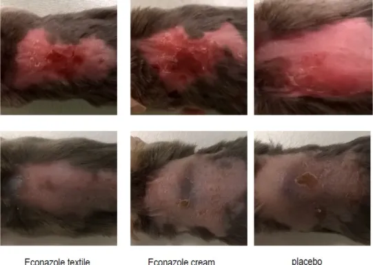
515
Figure 7. In vivo antifungal efficacy of pharmaceutical textiles, commercial
516 cream and placebo textiles. Evaluated by quantification of the fungal burden 517 (CFU) in skin biopsy samples. Mean ± standard deviation (n=6). 518 519 520 521
522 Figure 8: Representative cutaneous pathology pictures before (upper pannel) 523 and after treatment with ECN textiles, ECN cream or placebo textile (bottom 524 pannel). 525 526 4. Conclusion 527
This study was aimed at assessing pharmaceutical textiles using ECN as a
528
model drug for skin diseases. The technology of pharmaceutical textiles lies in
529
a dual innovation. Firstly, the thermo-sensitive microparticle formulation
530
ensured stability during storage and triggered thermo-sensitive release upon
531
contact with skin. The lipid microparticles allowed skin diffusion and drug
532
distribution within the upper layers of the skin, which is optimal to treat
Secondly, the adapted screen-printing method adapted to the microparticle 535 drug formulation was able preserved the pharmaceutical activity of the drug. 536 ECN efficacy was maintained in vitro on a broad range of fungi strains and in 537
vivo, ECN textiles enabled high therapeutic efficacy against cutaneous
538
candidiasis in mice. Overall, these results revealed the potential this
539
technology to develop pharmaceutical textiles for the treatment of superficial
540
infections. Such textiles could be developed in bandages or socks, which,
541 through ease of use, would improve patient compliance. 542 Acknowledgements 543 The authors thank the Natural Sciences and Engineering Research Council of 544 Canada, Fonds de Recherche du Québec - Nature et technologies and Biomod 545 Concepts Inc. for their financial support. We thank Dr Fairbrother for providing 546 pig ears and Martin Jutras for developing the HPLC-MS/MS method. 547 Conflict of interest. 548 This work has been partially founded by Biomod Concepts Inc. The patented 549
technology belongs to Karine Théberge and Biomod Concepts Inc. All the
550
analyses and characterizations have been performed at the University of
551 Montreal or Nantes, independently from the company. 552 553 554
References 555 556 [1] L. Ripoll, C. Bordes, S. Etheve, A. Elaissari, H. Fessi, Cosmeto-textile 557 from formulation to characterization: an overview, E-Polymers, (2010). 558 [2] M.K. Singh, V.K. Varun, B.K. Behera, Cosmetotextiles: State of Art, 559 Fibres Text. East Eur., 19 (2011) 27-33. 560 [3] G. Nelson, Application of microencapsulation in textiles, Int. J. 561 Pharm., 242 (2002) 55-62. 562 [4] M. Karthikeyan, T. Ramachandran, O.L.S. Sundaram, 563 Nanoencapsulated phase change materials based on polyethylene glycol 564 for creating thermoregulating cotton, J. Ind. Text., 44 (2014) 130-146. 565 [5] M. Ibanescu, V. Musat, T. Textor, V. Badilita, B. Mahltig, 566 Photocatalytic and antimicrobial Ag/ZnO nanocomposites for 567 functionalization of textile fabrics, J. Alloys Compd., 610 (2014) 244-568 249. 569 [6] J. Liu, C. Liu, Y. Liu, M. Chen, Y. Hu, Z. Yang, Study on the grafting of 570 chitosan-gelatin microcapsules onto cotton fabrics and its antibacterial 571 effect, Colloids Surf. B. Biointerfaces, 109 (2013) 103-108. 572 [7] C. Alonso, M. Marti, V. Martinez, L. Rubio, J.L. Parra, L. Coderch, 573 Antioxidant cosmeto-textiles: Skin assessment, Eur. J. Pharm. Biopharm., 574 84 (2013) 192-199. 575 [8] M. Marti, V. Martinez, N. Carreras, C. Alonso, M.J. Lis, J.L. Parra, L. 576 Coderch, Textiles with gallic acid microspheres: in vitro release 577 characteristics, J. Microencaps., 31 (2014) 535-541. 578 [9] G. Cravotto, L. Beltramo, S. Sapino, A. Binello, M.E. Carlotti, A new 579 cyclodextrin-grafted viscose loaded with aescin formulations for a 580 cosmeto-textile approach to chronic venous insufficiency, J. Mater. Sci. - 581 Mater. Med., 22 (2011) 2387-2395. 582 [10] C. Wiegand, U.C. Hipler, S. Boldt, J. Strehle, U. Wollina, Skin-583 protective effects of a zinc oxide-functionalized textile and its relevance 584 for atopic dermatitis, Clin. Cosmetic Invest. Dermatol., 6 (2013) 115-585 121. 586 [11] R.J. Hay, N.E. Johns, H.C. Williams, I.W. Bolliger, R.P. Dellavalle, D.J. 587 Margolis, R. Marks, L. Naldi, M.A. Weinstock, S.K. Wulf, C. Michaud, J.L.M. 588 C, M. Naghavi, The global burden of skin disease in 2010: an analysis of 589 the prevalence and impact of skin conditions, J. Invest. Dermatol., 134 590 (2014) 1527-1534. 591
[12] P. Vltavska, V. Kasparkova, R. Janis, L. Bunkova, Antifungal and 592 antibacterial effects of 1-monocaprylin on textile materials, Eur. J. Lipid 593 Sci. Technol., 114 (2012) 849-856. 594 [13] T.R. Hammer, H. Mucha, D. Hoefer, Dermatophyte susceptibility 595 varies towards antimicrobial textiles, Mycoses, 55 (2012) 344-351. 596 [14] C.W.M. Yuen, J. Yip, H.C. Cheung, L.W. Liu, C.H. Luk, W.C. Wai, 597 Treatment of interdigital-type tinea pedis with a 2-week regimen of 598 wearing hygienic socks loaded with antifungal microcapsules: A 599 randomized, double-blind, placebo-controlled study, J. Am. Acad. 600 Dermatol., 69 (2013) 495-496. 601 [15] R.C. Heel, R.N. Brogden, T.M. Speight, G.S. Avery, Econazole - Review 602 of its antifungal activity and therapeutic efficacy, Drugs, 16 (1978) 177-603 201. 604 [16] A. Brayfield, Martindale: The Complete Drug Reference, in: P. Press 605 (Ed.), MedicineComplete, 2014. 606 [17] M. Alsterholm, N. Karami, J. Faergemann, Antimicrobial activity of 607 topical skin pharmaceuticals - an in vitro study, Acta Derm. Venereol., 608 90 (2010) 239-245. 609 [18] M. Gupta, S.P. Vyas, Development, characterization and in vivo 610 assessment of effective lipidic nanoparticles for dermal delivery of 611 fluconazole against cutaneous candidiasis, Chem. Phys. Lipids, 165 612 (2012) 454-461. 613 [19] B. Albertini, N. Passerini, M. Di Sabatino, B. Vitali, P. Brigidi, L. 614 Rodriguez, Polymer-lipid based mucoadhesive microspheres prepared 615 by spray-congealing for the vaginal delivery of econazole nitrate, Eur. J. 616 Pharm. Sci., 36 (2009) 591-601. 617 [20] Y.G. Bachhav, K. Mondon, Y.N. Kalia, R. Gurny, M. Moller, Novel 618 micelle formulations to increase cutaneous bioavailability of azole 619 antifungals, J. Control. Release, 153 (2011) 126-132. 620 [21] N. Passerini, E. Gavini, B. Albertini, G. Rassu, M. Di Sabatino, V. 621 Sanna, P. Giunchedi, L. Rodriguez, Evaluation of solid lipid 622 microparticles produced by spray congealing for topical application of 623 econazole nitrate, J. Pharm. Pharmacol, 61 (2009) 559-567. 624 [22] M. Gupta, S. Tiwari, S.P. Vyas, Influence of various lipid core on 625 characteristics of SLNs designed for topical delivery of fluconazole 626 against cutaneous candidiasis, Pharm. Dev. Technol., 18 (2013) 550-627 559. 628 [23] K. Theberge, J. Goudreault, F. Quirion, G. Perron, Articles of 629 manufacture releasing an active ingredient, International Intellectual 630
Properties Patent no. 20100305209, (filed in US, Canada, Europe, China, 631 India, Australia, New Zealand, Brazil, Russia, Israel, Corea and South 632 Africa). 633 [24] I. Kazani, C. Hertleer, G. De Mey, A. Schwarz, G. Guxho, L. Van 634 Langenhove, Electrical Conductive Textiles Obtained by Screen Printing, 635 Fibres Text. East Eur., 20 (2012) 57-63. 636 [25] E. Skrzetuska, M. Puchalski, I. Krucinska, Chemically driven printed 637 textile sensors based on graphene and carbon nanotubes, Sensors 638 (Basel), 14 (2014) 16816-16828. 639 [26] T. Jones, N.A. Federspiel, H. Chibana, J. Dungan, S. Kalman, B.B. 640 Magee, G. Newport, Y.R. Thorstenson, N. Agabian, P.T. Magee, R.W. Davis, 641 S. Scherer, The diploid genome sequence of Candida albicans, Proc. Natl. 642 Acad. Sci. U S A, 101 (2004) 7329-7334. 643 [27] A.M. Gillum, E.Y. Tsay, D.R. Kirsch, Isolation of the Candida albicans 644 gene for orotidine-5'-phosphate decarboxylase by complementation of 645 S. cerevisiae ura3 and E. coli pyrF mutations, Mol. Gen. Genet., 198 646 (1984) 179-182. 647 [28] F. Quirion, D. Lambert, G. Perron, The Hlm method - a simple way to 648 get the solid liquid-phase diagrams and enthalpies of transition of pure 649 components and mixtures, Can. J. Chem., 70 (1992) 2745-2750. 650 [29] K. Kovacs, G. Stampf, I. Klebovich, I. Antal, K. Ludanyi, Aqueous 651 solvent system for the solubilization of azole compounds, Eur. J. Pharm. 652 Sci., 36 (2009) 352-358. 653 [30] A. Melkoumov, M. Goupil, F. Louhichi, M. Raymond, L. de 654 Repentigny, G. Leclair, Nystatin nanosizing enhances in vitro and in vivo 655 antifungal activity against Candida albicans, J. Antimicrob. Chemother., 656 68 (2013) 2099-2105. 657 [31] S.S. Richter, R.P. Galask, S.A. Messer, R.J. Hollis, D.J. Diekema, M.A. 658 Pfaller, Antifungal susceptibilities of Candida species causing 659 vulvovaginitis and epidemiology of recurrent cases, J. Clin. Microbiol., 43 660 (2005) 2155-2162. 661 [32] C. Herkenne, A. Naik, Y.N. Kalia, J. Hadgraft, R.H. Guy, Pig ear skin ex 662 vivo as a model for in vivo dermatopharmacokinetic studies in man, 663 Pharm. Res., 23 (2006) 1850-1856. 664 665 666 667