DOCTORAT DE L'UNIVERSITÉ DE TOULOUSE
Délivré par :
Institut National Polytechnique de Toulouse (Toulouse INP)
Discipline ou spécialité :
Pathologie, Toxicologie, Génétique et Nutrition
Présentée et soutenue par :
Mme CHRISTELLE EL HAJJ ASSAF le lundi 17 décembre 2018
Titre :
Unité de recherche : Ecole doctorale :
Patulin, main mycotoxin of the apple industry: regulation of its biosynthetic
pathway and influence of processing factors in cloudy apple juice
production
Sciences Ecologiques, Vétérinaires, Agronomiques et Bioingénieries (SEVAB) Toxicologie Alimentaire (ToxAlim)
Directeur(s) de Thèse : M. OLIVIER PUEL MME ELS VAN PAMEL
Rapporteurs :
M. BART HENRI HUYBRECHTS, SCIENSANO Mme NADIA PONTS, INRA BORDEAUX
Membre(s) du jury :
Mme ISABELLE OSWALD, INRA TOULOUSE, Président
M. JEAN-DENIS BAILLY, ECOLE NATIONALE VETERINAIRE DE TOULOUSE, Membre Mme ELS VAN PAMEL, ILVO, Membre
Mme SABINE GALINDO-SCHORR, UNIVERSITE MONTPELLIER 2, Membre
i
"Research is to see what everybody has seen and think what nobody
has thought."
Albert Szent-Györgyi (1893-1986)
Received the Nobel Prize for discovering vitamin C
iii
I dedicate the fruit of these long years of study
To my parents,
The reason of what I turned into today.
Thank you for your great support and continuous care.
To my brother and sister,
I am really grateful to both of you.
You have been my soul mates and my inspiration.
Acknowledgments
This thesis work was done at the UMR1331 Toxalim (Research Centre in Food
Toxicology), INRA of Toulouse and Flanders Research Institute for Agricultural, Fisheries and
Food (ILVO) in Melle-Belgium. I consider it as my best investment, so far. It was an enriching
experience for me on all levels: scientific, personal or cultural ones. It developed in me a bigger
curiosity and passion to the mycotoxin and food toxicology world. The last three years made me
more patient, sociable and practical. As for the cultural aspect, my main focus was definitely on food.
In addition to the French cuisine that I absolutely adore, the Belgian frietjes managed to occupy a
special place in my heart. And now, in order to end this chapter as it is right, I would like to thank
all the people contributed in making this experience so rewarding. I will try not to forget anyone,
but just in case I do, please excuse me.
I would like to start by thanking la Région Occitanie under Grant 15050427 for supporting
and funding this doctoral fellowship. The work was funded by CASDAR AAP RT 2015 under
Grant 1508 and by French National Research Agency under Grant ANR-17CE21-0008
PATRISK. I would like to thank the Belgian Research Institute for Agriculture, Fisheries and Food
for supporting and funding this research work as well.
At first, a big thank you goes to the rapporteurs Mrs. Nadia Ponts and Mr. Bart Henri
Huybrechts, as well as all the members of the jury Mrs. Isabelle Oswald, Mrs. Sabine Galindo, Mrs.
Els Van Pamel, Mr. Jean-Denis Bailly and Mrs. Sophie Lorber for doing me the honour of reviewing
and examining this doctoral dissertation.
Then, I would like to thank Mr. Bernard Salles, director of UMR 1331 Toxalim of INRA
Toulouse and Mrs. Lieve Herman, head of Department of Technology and Nutrition and the
Technology and Food Sciences Unit, for allowing me to conduct my thesis work in their laboratories.
I would also like to express my sincere gratitude to Mrs. Isabelle Oswald, director of the Biosynthesis
and Toxicity of Mycotoxins Team, who welcomed me in her team and gave me the confidence to
vi
accomplish my thesis work. I highly appreciate and respect the scientist and director you are. Your
skills, your critical eye and your valuable ideas have taught me a lot! I will never forget March 27th,
2015, the special day when you, Mrs. Herman and Mr. Puel sat with me around that round table in
Lebanon and offered me this opportunity. I think I have never been as thrilled as that moment in
my life. Thank you all for that! Be sure that I did everything to ensure that the trust you gave me was
in place.
To my thesis director, Mr. Olivier Puel. Words can never be enough to express how grateful
I am! I would like to thank you for presenting me such an interesting thesis topic to work on and
for guiding me with your invaluable suggestions all along. Each meeting with you, whether in your
office, around a coffee or in the HPLC room, added priceless aspects to the implementation and
enlarged my perspectives. Thank you Olivier for sharing your scientific knowledge and for
transmitting me your enthusiasm for research as well as your passion for patulin ... Your precious
and continuous ideas regarding the project always pushed me to think more quickly and more
critically, to select my problems and solve them. I would also like to thank you for the congresses
that you gave me the opportunity to attend. And finally, I thank you for the time you spent to read
and correct my papers and my manuscript.
I would like to address special thanks to Mrs. Sophie Lorber. Your presence was crucial to
my thesis and essential for me on a personal and professional level. I would like to thank you first
for your smile and uplifting spirit, it has always given me positive energy. Thank you for giving me
the feeling that I am still behind my desk at INRA when I was already in Belgium. Thank you for
your emails that have always boosted me up and encouraged me to work harder during the week
AND the weekends. Thank you for sharing with me every information I should know about and
every letter I receive. And thank you for all the work and corrections you did for my article and my
thesis.
And to Els Van Pamel, literally the super scientist, supermom, and super woman I have
known. I still do not know how you manage to do it all!! Too many things to thank you for, really,
I do not know where to start: spreading love and positive vibes around me, helping me settle in
Ghent, making sure I am ok, bringing me gifts, feeding me... You introduced me to Sinterklaas and
made sure I get my chocolate! Seriously, you made all my colleagues jealous! And when it comes to
science, thank you for leading me through these 18 months, and for all the discussions and ideas you
shared with me concerning my project. Thank you for answering all my questions, for contributing
in pushing my project forward and for correcting my papers. We managed to have a very interesting
story at the end! Els Van Coillie and Geertrui Vlaemynch I would first like to thank you for your
smiley faces and good spirits! It was amazing to work with people like you. Thank you for your
precious ideas and remarks! The discussions we had during our meetings were very fruitful. You
taught me how to select practical ideas and develop them. I would also like to thank you for all the
time you spent reading and correcting my papers. Olivier, Sophie, Els VP, Els VC and Geertrui, it
was a great pleasure to work with you. YOU were the best choice I could have ever made for directors
and advisors... Sometimes, we are just (or incredibly) lucky!!!
I would also like to thank Mr. Jean-Denis Bailly and Mrs. Sylviane Bailly for always being
part of my thesis committees and for comforting me and supporting me after every presentation. I
appreciate all the advices you gave me, and I learnt a lot from you. Sylviane, a big thank as well for
sharing with me your expertise related to the morphological studies and development of the fungus.
That was very instructive. I would like to show all my gratitude to Mr. Domien de Paepe. We made
a lot of good (and toxic) cloudy apple juice together! Thank you for sharing your knowledge and
experience with me. Thank you also for all the time that you dedicated to me and my work. I still
owe you a pasta invitation! And finally, thanks to Mr. Christof Van Poucke for helping me out when
all my peaks were interfering on the HPLC.
I would also like to thank Selma and Nikki. Working with you, Selma, during the first few
months of my PhD was a big help. Thank you for showing me around, for explaining how the lab
functions and for your help with the mutant. My thesis has quickly progressed after that. And Nikki,
thank you for showing me how it works at ILVO, for your presence and ideas in the meetings. And
thank you for the nice discussions we had at work or around a drink. I would like to thank Soraya
viii
as well for her presence, her kindness and for the help she provided when I left for Belgium. I also
want to express my gratitude to Joëlle. You are a good example of how organised and ambitious
people are. Thank you for your lovely smile and for helping me with all the orders.
I am also deeply grateful for the opportunity to know many incredible people who were part
of the team E05; with whom I shared and learnt many things. Thank you Immourana (for all these
remarks, tips and great moments spent in Poland), Philippe (for your kindness and the great time in
Berlin), Laura S., Chloé and Manon (for the coffee breaks), Tala (for your sweetness), Chrystiane
(for the review), Aurora, Sylvie, Anne-Marie, Arlette, Delphine, Gisella, Su, Ophélie, Tarek, Abdula.
I am also grateful to every person who helped me in any way at ILVO, who showed me
around, who sacrificed their time to help me with my experiments. Thank you Mariejke (for all the
quality tests you have done), Kathleen (for organising the time of my experiments), Ann (for your
smile first and for having an answer to every problem), Nathalie (for the AA test and others), Jari
and all the food pilot people (for the juice experiment), and last but not least to the QACL people:
Stijn, Daan, Niels and Pétra.
I would also like to present all my gratitude to some people who helped either with their
scientific expertise: Yanick and Claire (for sharing your qPCR expertise), Emilien and Jean-François
(for the MS and analysis); administrative one: Jeannette, Joelle, Sarah; computer one: Vincent; or
culinary one: Pierre, Aurore and Lolita (God knows how much I missed your delicious meals in
Belgium!).
I will be failing in my duty if I do not acknowledge some of my friends with whom I have
shared my research experiences, my happy moments and definitely some not-so-happy ones as well.
It was a joy having you by my side and having each other’s back. I will start with you Amaranta.
Honestly, I find myself speechless here! You made my thesis looks easy and my problems small! We
definitely shared more than the office! Should I start counting? Let’s see: Lebanon together? My first
congress? And my second one? And Ghent? And all the drinks in between?? Ô Boudu Pont has heard
a lot of our stories and “cochonneries”! For all that, I say thank you! See now why I forgive you for
ruining my French? This thesis made me gain a friend for life! A bientôt ma belle! Rhoda, my friend
for almost 11 years now!! Crazy how time flies. Remember the CA days? I knew back then that we
will always have each other’s back! #myprestou for life! Thank you for being the friend you are and
thank you for all the moments we shared! Am sure we will have a lot more in the future! And Isaura,
I should start by thanking you for giving me my first tequila shot and informing me that Chiclets
wasn’t Lebanese!!! You did break my heart first but then I was quickly over it! I could not ask for a
better friend to share the office with! Thank you for always bringing joy wherever you are! Alix,
Laura C., Joya, Sandy and Brankica, how beautiful are you! Each one on her own unique way! Thank
you for all the amazing moments we shared, for the Halloween party Alix, for the amazing wedding
Laura, for the one of a kind spirit strong Joya, for the drunkly ever after times on the pont neuf/
the endless waiting moments and the best laughs in the world Sandy and Brankica for saying YES
(that’s the first thing I thought of)! I can’t wait for our trip to Serbia girl!! I wasn’t only lucky for
having the best directors but also for having you my chicas!
I would also like to thank the friends I made at ILVO. Maria-Eletta (or OH Maria, Maria ♬), we
shared the non-speaking Dutch quality first and then an amazing friendship! Thank you for the
amazing time spent in Belgium and Milano. Tina, the first person who approached me when I got
to Belgium! Thank you for being sociable and friendly!! It is the end for both of us, we can do it
girl!! Evelyne, am grateful for the time we spent. Thank you being my confident ;)! I Am also very
thankful to all the women I met in this journey, shared memorable moments and good laughs: Sayu,
Jennifer, Judith, Karolien, Kathleen, Anna and Hang.
I would like to thank some people who, even though were not part of these last three years,
had a role in me being here and influenced my career. Thank you to Mr. André El Khoury and Mr.
Ali Atoui for connecting me with INRA Toulouse and for believing in me and in my scientific
potential. You gave me an opportunity to follow my passion for research after that I let go my first
one. I will always be grateful to you and to the support you provided during all these academic years.
I would also like to thank Mr. Philippe Lecomte. During my internships you taught me that to be a
good researcher, I do not only need to do good experiments, I should know how to analyse them
x
and discuss the results. Since then, I worked on that point and here I am defending my thesis! Thank
you for pushing me to become the thinker I am today! And finally, Mr. Thierry Langin. I always
believed that having the opportunity to pursue my PhD has something to do with you. Thank you
for believing in me when I was just a young 20 years old scientist doing her internships under your
direction. Thank you for seeing the potential in me. I can assure you that it is back then, in 2012,
that I started developing my real scientific thinking. I appreciate you as a researcher and respect you
more as a human. Thank you for advising me to enrol in my second masters and for being such a
support every time I needed you or asked you for something.
To my friends Cynthia, Samar, Stephanie M, Laury, Carla, Romy, Rana, Sarah N, Johnny,
Kamal, Sarah R, Stephanie H, Ramona, Hiba K, Jamal … too many to list here but you know who
you are. Thank you for providing the friendship that I needed, supporting me during my thesis and
encouraging me to strive towards my goal. Cynthia, the only one I can rely on to think at my place
when I need a neutral/objective opinion!! Your presence is crucial to my everyday life, so you can
imagine how it was during my PhD. Thank you for always understanding, for never judging and for
being there at my hardest times. You are an example of what a best friend could be. And Samar, my
partner in all crimes and my #ForeverFriend!! Thank you for being you!! You always get all the
craziness out of me! Thank you for making me slow down my work rhythm when I had to and for
always being on #ChristyTeam (whether I am right or wrong). A PhD without people like all of
you would be quite hard. You were always my recharge battery. THANK YOU!
I would also like to thank my téta and my jeddo, my Antoun and my 3amto; you loved me
so much, you prayed for me a lot, you believed in me, you wished the best for me and I know how
real was all that! Thank you for your phone calls, your video calls, your continuous messages! How
lucky am I to have you in my life. You were always my boost of energy!
To Martine, Luc, Stefanie, Christiane and Pedro. You were and will always be my family
XL. Thank you for your kindness and love. Thank you for your care and for making me feel home
when I was far away from home. Belgium would have never been the same without you. Thank you
for the amazing memories that we made in Ghent, Lebanon, the coast… and of course for all your
delicious meals Martine and Luc. Cheers to many more!
I would like to express appreciation to my Michi, my gift from God. I know I was not easy
to handle, especially at the end of this journey. But you managed! You supported me in my worse so
you definitely deserve me in my best! Thank you for being there, for helping me think logically/
strategically, set priorities and mostly for believing in me and my work! Thank you for all these rides
from and to Ghent AND for your amazing home-cooked dishes. Thank you for giving me all the
love in the world!
The endless thanks go to my family. Words cannot express how grateful I am to my mother
and my father for all of the sacrifices that you have made in my behalf. Your prayers for me were
what sustained me thus far. Thank you for your advice, your patience and your faith! Thank you for
always understanding! Kiko, the day I decided to get back to France and start my PhD, you were
there, packed and ready! Ready to support me and take care of me just like you always did! You got
me to Toulouse and helped me get settled. You made sure to bring the smile back to my face before
heading back to Lebanon. Thank you for that! For your advice and opinions in everything I do. For
standing by my side in my ups and downs! Karrouni, my princess, considering me your idol was
always what pushed me forward to accomplish more!! But that’s what sisters are for, right?? Getting
the best out of each other!! Even though we were separated for 10 or 11 months/year, our bond
kept on getting stronger! Thank you for being there for me and especially for supporting all my
nagging. And finally Carroul, the new amazing addition to my family! Thank you for being a sister
and a close friend. Thank you for visiting me and cooking for me. Honestly, after writing these
acknowledgments, I realised that I thanked a lot of people just for feeding me!!! Apparently, that is
the way to my heart! Thank you my precious people!
Mum, Karrouni, Kiko, Carla, Samar, Sarah, Elsa, Maan, Rana, Joe, Johnny, Nathalie, Rhoda,
Maria-Eletta … our trips together during these 3 years of thesis and your visits whether to Toulouse or
xii
Belgium were the most memorable. Every small escape made me gain a lot of energy and pursue my
work! You made my cold nights warmer! Thank you for being there!
Finally, I thank my God, my good Father, for letting me through all the difficulties. I have
experienced your guidance day by day. You are the one who let me see the good in everything, helped
me adapt every time to a new situation, country, and system. You gave me the strength to finish my
degree, the best way possible. You blessed me with a lot! I will keep on trusting You forever. Thank
you, Lord.
All of you made it possible for me to reach this last stage of my endeavour.
Thank You from the bottom of my heart.
List of Publications
and Scientific
Communications
Publications
▪ El Hajj Assaf, C., Snini, S., Tadrist, S., Bailly, S., Naylies, C., Oswald, I.P., Lorber, S., Puel, O.
Impact of veA on the development, aggressiveness, dissemination and secondary metabolism of Penicillium expansum. Molecular Plant Pathology 19:1971–1983. doi: 10.1111/mpp.12673
▪ El Hajj Assaf, C., De Clercq, N., Vlaemynck, G., Van Coillie, E., Puel, O., Van Pamel, E. (XXXX)
Fate of patulin in apple juices with special emphasis on the impact of ascorbic acid. Mycotoxin research journal (Submitted review)
▪ El Hajj Assaf, C., De Clercq, N., Van Poucke, C., Vlaemynck, G., Van Coillie, E., Van Pamel, E.
(XXXX) Impact of ascorbic acid on the patulin concentration in aqueous solution and cloudy apple juice. Mycotoxin research journal (Submitted article)
▪ El Hajj Assaf, C., De Paepe, D., De Clercq, N., Vlaemynck, G., Van Coillie, E., Van Pamel, E.
(XXXX) Effect of ascorbic acid, oxygen and storage on patulin in cloudy apple juice produced under low oxygen conditions. (Written article_to be submitted)
▪ El Hajj Assaf, C., Zettina, C., Oswald, I.P., Lorber, S., Puel, O. Secondary metabolism of
Penicillium spp. and its regulation. (Review_Manuscript in preparation)
▪ El Hajj Assaf, C., Zettina, C., Tadrist, S., Jamin, E.,Martin, J.F., Oswald, I.P., Lorber, S., Puel, O. Effect of veA on the metabolome of Penicillium expansum (Article_Manuscript in preparation)
xvi
Oral communications
▪ Christelle El Hajj Assaf, Nikki De Clercq, Geertrui Vlaemynck, Els Van Coillie, Els Van Pamel.
Influence of ascorbic acid on patulin concentration in cloudy apple juice. 40th Mycotoxin
Workshop, June 11th-13th 2018, Munich, Germany. (Oral)
▪ Christelle El Hajj Assaf, Sylviane Bailly, Isabelle Oswald, Sophie Lorber, Olivier Puel, Els Van
Pamel, Els Van Coillie, Geertrui Vlaemynck. Patulin, main mycotoxin in apple products: Elucidation of mechanisms involved in its biosynthetic pathway and development of preventive approaches.
PhD challenge. ILVO (Melle, Belgium), November 21st, 2017. (Oral)
▪ Christelle El Hajj Assaf, Selma Snini, Sylviane Bailly, Yannick Lippi, Emilien Jamin, Jean-François
Martin, Isabelle Oswald, Sophie Lorber and Olivier Puel*. The velvet complex in blue mould fungus Penicillium expansum: impact of veA on development, aggressiveness and secondary metabolism.
1st Mycokey Conference, September 11th-14th 2017, Ghent, Belgium. (Oral)
▪ Christelle El Hajj Assaf, Selma Snini, Sylviane Bailly, Yannick Lippi, Emilien Jamin, Jean-François
Martin, Isabelle Oswald, Sophie Lorber and Olivier Puel*. Impact of veA on development, aggressiveness and secondary metabolism of Penicillium expansum, principal agent of blue mould disease. 39th Mycotoxin Workshop, June 19th-21st 2017, Bydgoszcz, Poland. (Oral)
▪ Christelle El Hajj Assaf, Selma Snini, Sylviane Bailly, Yannick Lippi, Emilien Jamin, Jean-François
Martin, Isabelle Oswald, Sophie Lorber and Olivier Puel*. A veA mutant strain of Penicillium expansum: Study of its virulence, morphology and expression of secondary metabolite clusters.
Poster
▪ Christelle El Hajj Assaf, Nikki De Clercq, Christof Van Poucke, Els Van Pamel. Optimization of the
methodology for the detection/quantification of patulin and breakdown products. 40th
xix
xxi
A
a* CIELAB (1976) colour space coordinate representing the red/green component
A. flavus Aspergillus flavus
AA Ascorbic acid
AcA Acetic acid
ACN Acetonitrile
AcOEt Ethyl acetate
AFB1, AFB2, AFG1, AFG2 and AFM1
Aflatoxins
ANOVA Analysis of variance
AOAC Association of Analytical Communities APAM Apple-based apple puree agar medium
ASC-E Ascladiol-E
ASC-E/Z Ascladiol-(E/Z)
ASC-Z Ascladiol-Z
ATA Trichothecene Toxic Food Aleucie
ATP Adenosine triphosphate
aw Water activity
α Alpha
B
b* CIELAB (1976) colour space coordinate representing the yellow/blue component
b-Tubulin beta-Tubulin
bw/day Body weight per day
β Beta
C
CE Capillary Electrophoresis
°C Celsius degree
C* CIELCH colour space coordinate representing the chroma or colour purity
xxii
C.E.H.A. Christelle El Hajj Assaf C6H8O6 Ascorbic acid (vitamin C)
CAJ Cloudy apple juice
cAMP Cyclic adenosine monophosphate
CAT Catalase
CCP Critical control points
cDNA Complementary Deoxyribonucleic acid
CGA Chlorogenic acid
CGA Czapek glucose agar
CIT Citrinin
cm/day centimetre per day
Co-60 Cobalt-60
cond (1, 2, 3) Condition number
CY Czapek yeast extract liquid medium
CYA Czapek yeast extract agar
CzA Czapek dox agar
D
DHA dehydroascorbic acid
DKGA 2,3-diketogulonic acid
DMATS Dimethyl-allyltryptophan synthase
dNTP Deoxynucleoside triphosphate
DON Deoxynivalenol
DPA Desoxypatulinic acid
dpi Days post-inoculation
Δa* Difference in redness
Δb* Difference in yellowness
ΔC* Chroma difference
ΔE* Extent of colour difference
ΔL* Difference in lightness
E
e.g. For example (exempli gratia)
EFSA European Food Safety Authority
ESI Electrospray ionization
et al. Et alii
etc. Et cetera
ETPs Epipolythiodioxopiperazines
EU European Union
F
FAO Food and Agriculture Organization
FB1, FB2 Fumonisins
Fig. Figure
G
GAP Good agricultural practices
GC-MS Gas Chromatography Mass Spectrometry GMP
GLA GOX2 γ
Good manufacturing practices D-Gluconic acid
Glucose oxidase Gamma
H
HHP High hydrostatic pressure
HACCP Hazard Analysis Critical Control Points system
HMF 5-hydroxymethylfurfural
HOG High osmolarity glycerol
HPLC High Performance Liquid Chromatography
HPLC-DAD High-performance liquid chromatography with a diode array detector HPLC-MS Liquid chromatography coupled with mass spectrometry
HPLC-MS/MS Tandem Mass Spectrometry
HPLC-UV High Performance Liquid Chromatography coupled to UltraViolet detection
I
IARC International Agency for Research on Cancer
xxiv
ILVO Flanders Research Institute for Agricultural, Fisheries and Food
J
JECFA Joint Food and Agricultural Organization/World Health Organization Expert Committee on Food Additives
K
KCl Potassium chloride
L
L* CIELAB (1976) colour space coordinate representing the luminance component
L-AA L-ascorbic acid
LAB Lactic acid bacteria
LC-MS Liquid Chromatography - MASS Spectrometry
M
mg/kg Milligrams to kilograms
µg/kg Microgram per kilogram
1-MCP 1-methylcyclopropene
MAPK Mitogen-activated protein kinase
MC Moisture concentration
MEA Malt extract agar
min Minute
MIP Molecularly imprinted polymer
mM Millimolar
N
NPS Nano-porous silicon
n Number of replicates
Na2HPO4 Disodium phosphate
NaCl Sodium chloride
NADPH Nicotinamide adenine dinucleotide phosphate
nd Refractometer index
ND Not detectable
NOEL No-Observed Effect Level
NRPS Non-ribosomal peptide synthases NRPSs Non-ribosomal peptide synthetases NTU Nephelometric Turbidity Units
O
O2 Oxygen O3 Ozone OTA Ochratoxin A Ox – Anaerobic conditions Ox + Aerobic conditionsP
P. citrinum Penicillium citrinum P. expansum Penicillium expansum
PAT Patulin
PCR Polymerase chain reaction
PDA Potato Dextrose Agar
PE Polyethylene
Pe∆veA Mutated in the veA gene Pe∆veA:veA Complemented in the veA gene
pH Potential of hydrogen
p-HHP pulsed-HHP
PIT Photonics Immobilization Technique
PKA Protein kinase A
PKS Polyketide synthases
PMTDI Provisional Maximum Tolerable Daily Intake
ppb Parts per billion
ppm Parts per million
PPO Polyphenoloxidase
PTWI Provisional Tolerable Weekly Intake p-values Probability values
xxvi
Q
QCM Quartz crystal microbalance
qPCR Quantitative real-time PCR
R
RF Radio frequency
RM 40 Refractometry
RNA Ribonucleic acid
RNAi Ribonucleic acid interference
ROS Reactive oxygen species
RP-HPLC method Reversed-Phase High-Performance Liquid Chromatography
rpm Revolutions per minute
rt Retention time
RT-PCR Reverse transcription polymerase chain reaction
RT-qPCR Quantitative reverse transcription-polymerase chain reaction
S
S (1, 2, 3, 4) Sampling step number S. cerevisiae Saccharomyces cerevisiae
SEM Standard error of the mean
SM Secondary metabolites
SO2 Sulphur dioxide
SOD Superoxide dismutase
SPE-LC-MS Solid-phase extraction-liquid chromatography-mass spectrometry
T
TFs Transcription Factors
TLC Thin-Layer Chromatography
TOR Target of Rapamycin
TS Terpene synthases
TSS Total soluble solids
TTO Tea tree oil
U
UHPLC Ultra-high-performance liquid chromatography ULO Ultra-low oxygen atmospheric condition
UV Ultraviolet
UV-VIS detector Ultraviolet/Visible HPLC Detectors
V
v/v Volume/volume
W
w/v Weight/volume
WHO World Health Organization
WT Wild type
Y
YPD Yeast extract peptone dextrose
Z
ZEA Zearalenone
xxix
LIST OF FIGURES
Introduction
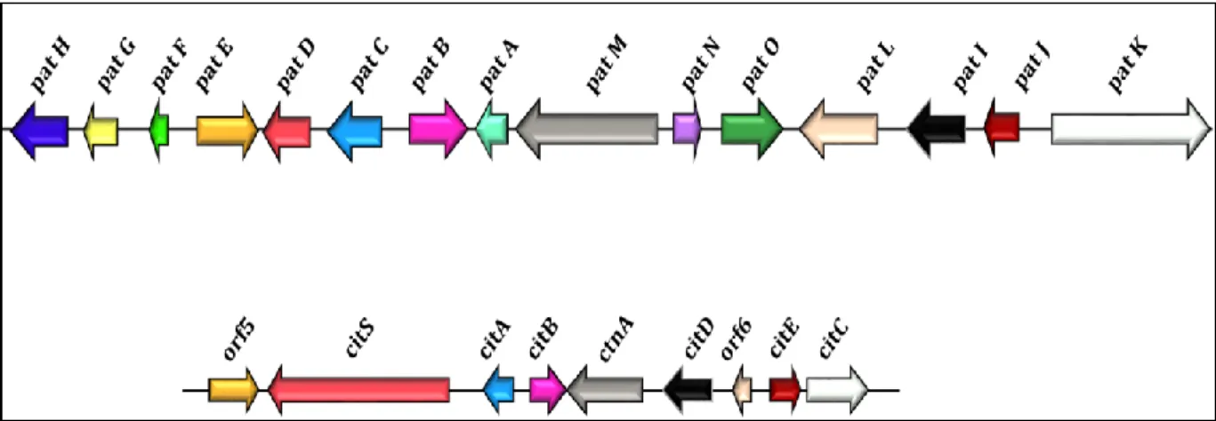
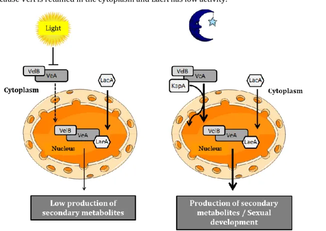
Figure 1 General overview of the PhD thesis --- 5 Figure 2 Risks impacting food safety and security throughout the business supply chain --- 7 Figure 3 Art as evidence of mycotoxin contamination --- 10 Figure 4 The chemical structures of major mycotoxins--- 11 Figure 5 Biosynthetic pathways of secondary metabolites --- 18 Figure 6 Gene clusters of the patulin and citrinin biosynthesis pathways --- 19 Figure 7Global regulatory proteins involved in the regulation of gene clusters involved in the
production of various fungal secondary metabolites --- 23
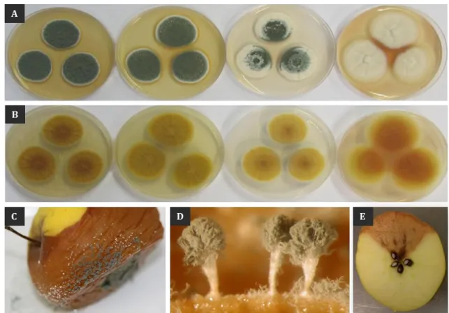
Figure 8 Operating model of the velvet complex in Aspergillus nidulans --- 26 Figure 9 Microscopic appearance of conidiophores and conidia of Penicillium expansum --- 34 Figure 10 Macroscopic observations of Penicillium expansum and its rot --- 50 Figure 11 Patulin biosynthesis pathway in P. expansum --- 55 Figure 12 Chemical structure of patulin --- 59 Figure 13 Apple juice processing steps --- 70 Figure 14 Chemical structure of ascorbic acid --- 74
Chapter 1_Part 1: Characterization of veA gene: study of its impact on development,
aggressiveness and metabolome of Penicillium expansum
Figure 1 Morphological appearance of the wild-type NRRL 35695, null mutant and complemented
strains --- 101
Figure 2 Growth curves of the rotten spots and rot volume in Golden Delicious apples--- 102 Figure 3 Implication of veA in the development and dissemination of Penicillium expansum on Golden
Delicious apples--- 103
Figure 4 Patulin production on Golden Delicious apples--- 103 Figure 5 Patulin and citrinin production on synthetic media by the wild-type NRRL 35695, null
mutant and complemented strains--- 104
Figure 6 Fold change expression of genes belonging to the clusters involved in citrinin and patulin
biosynthesis in the wild-type NRRL 35695 and null mutant strains--- 104
Figure 7 Relative expression of 35 genes encoding backbone enzymes involved in secondary
metabolite biosynthesis--- 105
Figure S1 Construction of the null mutant Pe∆veA--- 114 Figure S2 Polymerase chain reaction analyses of genomic DNA of the wild-type NRRL 35695, null
mutant and complemented strains--- 117
Figure S3 Southern blot analyses of genomic DNA of the wild-type NRRL 35695, null mutant and
complemented strains--- 118
Figure S4 Expression of the veA gene on Malt Extract Agar medium and Potato Dextrose Agar
medium--- 119
Figure S5 Morphological appearance of the wild-type NRRL 35695, null mutant and complemented
xxx
Figure S6 Influence of light on the morphological appearance of the wild-type NRRL 35695, null
mutant and complemented strains--- 121
Figure S7 Relative gene expression of the patulin and citrinin biosynthesis genes on Malt Extract
Agar medium--- 123
Figure S8 Relative gene expression of the patulin and citrinin biosynthesis genes on Potato Dextrose
Agar medium--- 124
Figure S9 Relative expression of 35 genes encoding backbone enzymes involved in secondary
metabolite biosynthesis--- 127
Figure S10 Relative expression of the laeA and velB genes on synthetic media--- 128
Chapter 1_Part 2: Impact of veA on the metabolome of Penicillium expansum
Figure 1 Venn diagrams showing the number of significant ions differentiating the null mutant strain
from each of the wild type and complemented ones--- 140
Figure 2 Total ion current chromatograms--- 142 Figure 3 Graphs showing the relative abundance of metabolites produced by the WT strain NRRL
35695 of Penicillium expansum and the null mutant PeΔveA, in the medium--- 145
Figure 4 Graphs showing the relative abundance of metabolites produced by the WT strain of
Penicillium expansum, the null mutant PeΔveA and complemented PeΔveA:veA ones, in apples---- 147
Chapter 1_Part 3: Impact of veA gene on Penicillium expansum development and
patulin production in presence of different sugar sources
Figure 1 Impact of different sugar concentrations on fungal development and PAT production--- 160 Figure 2 Comparison of the development and production of PAT of the WT, null mutant and
complemented strains--- 161
Figure S1 Development of the WT, null mutant and complemented strains--- 162 Figure S2 Concentration of PAT produced by the WT, null mutant and complemented strains---- 163
Chapter 2_Part 1: Impact of ascorbic acid on the patulin concentration in aqueous
solution and cloudy apple juice
Figure 1 UHPLC chromatograms showing the interference of the UHPLC-UV signal of the secondary
metabolites, degradation and reaction products--- 177
Figure 2 UHPLC chromatogram of PAT, its breakdown products and reaction products--- 178 Figure 3 Patulin concentration of PAT-contaminated cloudy apple juice--- 180 Figure 4 Patulin concentration in absence or presence of ascorbic acid in PAT-contaminated cloudy
apple juice--- 181
Figure 5 Compound X formation in an aqueous solution artificially contaminated with pure patulin
standard--- 182
Figure 6 Ascladiol-Z formation in a patulin-contaminated apple juice--- 183 Figure 7 Dehydroascorbic acid formation in an aqueous solution artificially contaminated with pure
Chapter 2_Part 2: Effect of ascorbic acid, oxygen and storage on patulin in cloudy apple
juice produced on a semi-industrial scale
Figure 1 Monitoring of patulin concentration in samples taken at different sampling steps along the
cloudy apple juice production process--- 207
Figure 2 Monitoring of patulin concentration in γ-radiated bags containing cloudy apple juices-- 209 Figure 3 Dehydroascorbic acid formation in γ-radiated bags containing cloudy apple juices--- 211 Figure 4 5-hydroxymethylfurfural formation in samples taken from different sampling steps all
along the cloudy apple juice process--- 212
Figure 5 5-hydroxymethylfurfural formation containing cloudy apple juices--- 213
General discussion
Figure 1 Hypothetical diagram of the spatial organization of the patulin biosynthesis pathway within
the fungal cell--- 226
LIST OF TABLES
Introduction
Table 1 Examples of identified Zn(II)2Cys6 transcription factors involvement in secondary
metabolism in fungi adapted and updated from Yin and Keller --- 21
Table 2 Secondary metabolites of P. expansum reported in the literature --- 49 Table 3 Reports regarding worldwide contamination of apple juice by patulin --- 81 Table 4 Reports regarding patulin occurrence in fresh and processed food products --- 89
Chapter 1_Part 1: Characterization of veA gene: study of its impact on development,
aggressiveness and metabolome of Penicillium expansum
Table S1 Primers used in the construction and the validation of null mutant Pe∆veA and
complemented Pe∆veA:veA--- 115
Table S2 Primers used to analyze the expression of the citrinin cluster --- 122 Table S3 Primers used to analyze the expression of other backbone enzyme genes--- 125 Table S4 Primers used to analyze the expression of the laeA and velB genes--- 128
Chapter 1_Part 2: Impact of veA on the metabolome of Penicillium expansum
Table 1 Secondary metabolites detected from Penicillium expansum--- 142 Table 2 List of the secondary metabolites detected in spores of Penicillium expansum --- 149
Chapter 2_Part 2: Effect of ascorbic acid, oxygen and storage on patulin in cloudy apple
juice produced on a semi-industrial scale
Table 1 Specification of the four conditions of cloudy apple juice tested--- 202 Table 2 Juice quality parameters of the four cloudy apple juices produced--- 206
TABLE OF CONTENT
List of publications and scientific communications --- xiii
List of abreviations --- xix
List of figures --- xxix
List of tables---
xxxiii
Introduction --- 1
Study context and objectives --- 3
Bibliographic review --- 6
A.Moulds and mycotoxins---7
A.1 Fungi: microscopic filamentous fungi --- 7 A.2 Filamentous fungi and their secondary metabolites --- 8 A.3 Mycotoxins and their impact on human health --- 9
a.Historical overview --- 9
b.Definition and characteristics --- 10
c.Health effects --- 12
d.Regulations --- 12
B.Secondary metabolism of Penicillium spp. and its regulation---15
B.1 Introduction --- 17 B.2 Regulation of secondary metabolism --- 19a. Specific transcription factors / cluster specific regulators --- 19 b. Global regulators --- 23 c. Signal transduction pathways --- 29 d. Epigenetic regulation --- 31
B.3 Conclusion --- 32
C.Study of the virulence and control of P. expansum---34
C.1 Effect of SM on virulence of P. expansum --- 50 C.2 Controls of P. expansum --- 53 C.3 Conclusion --- 56
D.Fate of patulin in apple juices with special emphasis on the impact of ascorbic
acid--- 57
D.1 Introduction --- 59 D.2 Determination of PAT in apple products --- 61
a.Chromatographic methods --- 61
xxxvi
D.3 Techniques decreasing PAT concentration and its degradation products --- 63
a.Strategies preventing PAT contamination of the raw material for apple juice --- 64
b.Strategies degrading PAT in apple juice --- 65
i.Physical treatments--- 65 ii.Chemical treatments --- 66 iii.Biological treatments--- 67 c.Degradation products of PAT --- 68
D.4 PAT control steps during apple juice processing --- 69 D.5 Ascorbic acid --- 73
a.Identity of AA --- 73
b.Methods for the detection and determination of AA --- 74
c.Factors impacting the stability of AA---75 i.Effect of heating--- 75 ii.Effect of storage--- 76 d.PAT degradation in presence of AA --- 77
D.6 Conclusion---77
Experimental Work --- 91
Chapter 1
Characterization of veA gene
--- 93Chapter 1_Part 1
Summary of the study---97 Characterization of veA gene: study of its impact on development, aggressiveness and metabolome of Penicillium expansum (article 1) --- 95
Chapter 1_Part 2
Impact of veA on the metabolome of Penicillium expansum --- 131
1.2.1. Background--- 133 1.2.2. Material and methods---135 1.2.3. Results and discussion---138 1.2.4. Conclusion--- 149
Chapter 1_Part 3
Impact of veA gene on Penicillium expansum development and patulin production in presence of different sugar sources --- 151
1.3.1. Background--- 153 1.3.2. Material and methods---155 1.3.3. Results and discussion---156 1.3.4. Conclusion--- 159
Chapter 2
Impact of ascorbic acid on the patulin concentration on a laboratory and
semi-industrial scales
--- 165Chapter 2_Part 1
Summary of the study---169 Impact of ascorbic acid on the patulin concentration in aqueous solution and cloudy apple juice (article 2) --- 171
Chapter 2_Part 2
Summary of the study---195 Effect of ascorbic acid, oxygen and storage on patulin in cloudy apple juice produced on a semi-industrial scale (article 3)--- 197
General discussion --- 221
General discussion and perspectives---223 Conclusions--- 238
References --- 239
Abstract---281
Résumé--- 283
STUDY CONTEXT AND OBJECTIVES
Food-related health concerns are and have been of major importance all around the world for ages. Their repercussions on the global economy are huge and most likely even underestimated. They can be caused by various agents, particularly pathogenic microorganisms.
In addition to viruses and pathogenic bacteria, toxigenic fungi are considered a real threat to humans and animals health either due to their presence or to their secretion of highly toxic substances called mycotoxins. The latter are widespread at all stages of the food and feed chains. As a consequence of their toxicity, regulations have been set to protect consumers from the damaging effects of mycotoxins that can contaminate foodstuffs and to ensure the appliance of correct practices in the food trade. Levels of mycotoxins harmful for health should not be exceeded. While some mycotoxins (e.g. aflatoxins) have been studied intensively since the beginning of their discovery, others (e.g. patulin (PAT)) have remained in the shade for a while but have been fully studied and recently regulated. For mycotoxins such as HT-2 and T-2, the recommendations apply while others are not yet regulated (e.g. Alternaria toxins).
PAT, the mycotoxin of interest in this study, was first discovered by Waksman et al. (1942) and was considered as a promising antibiotic, effective against nasal congestion and colds. It was only few years after that it was proven not only toxic to fungi but also to animals and higher plants (Moake et al. 2005). From then one, many researchers were particularly interested in PAT to determine its physicochemical properties, elucidate its biosynthetic pathway, isolate and characterize the fungal producing species, evaluate its toxic potential based on in vitro and in vivo studies. Apple juices as well as other food products are sometimes heavily contaminated by PAT, exceeding the maximum regulatory limits. Thus, more studies and efforts are needed to minimize the contamination of food products by this mycotoxin.
However, controlling the occurrence of PAT in food products require a better understanding of Penicillium expansum, the main producer of this toxin. In addition, elucidation of the biochemical and molecular mechanisms that induce PAT production could help regulate its biosynthesis. The elucidation of the biosynthetic cluster of PAT by Tannous et al. in 2014 evidenced specific factors that regulate its biosynthesis.
The full understanding of the mechanisms involved in the toxin biosynthesis is complicated and the big economic losses that occur due to the food products already contaminated by PAT should be taken into account. In this context, many questions arose.
▪ What are the factors that affect PAT biosynthesis outside the genes of its cluster and more generally, what is the impact of global regulators on the secondary metabolism of P. expansum?
4
▪ What are the processing factors that could have an effect on PAT concentration in contaminated food products? Are there any degradation products generated and if so, are they less toxic than PAT?
This multidisciplinary research aims to understand the role of the veA gene involved in PAT biosynthesis and to determine the role of the chemical agent ascorbic acid (AA), which affects PAT
(Figure 1). The first objective focuses on the elucidation of the roles of veA and the study of its
impact on the development, aggressiveness, dissemination and secondary metabolism of P. expansum, with emphasis on PAT biosynthesis. A null mutant Pe∆veA strain and a complemented Pe∆veA:veA strain were generated using the wild type strain of P. expansum (NRRL 35695). A morphological study was conducted and pathogenicity on apples was studied. Expression of genes belonging to the two corresponding clusters of PAT and citrinin, another mycotoxin produced by the fungus, as well as genes that encode backbone enzymes of secondary metabolites found in the genome of P. expansum was analysed.
The second objective of this thesis aims to better understand the fate of PAT during the production of cloudy apple juice, more specifically during pressing of apples (using the new spiral filter press technique characterized by low-oxygen pressing conditions) and storage. The influence of AA, known as vitamin C, was examined throughout the process. At first, optimal conditions of action of AA were identified in the presence/absence of oxygen and different storage temperatures. This experiment was performed both on pure PAT standard in aqueous solution and on PAT-contaminated cloudy apple juice (CAJ). An in-house HPLC methodology was optimized leading to a good separation and detection of PAT and degradation and reaction products. Then, the evaluation of the effect of the antioxidant AA on PAT concentration during cloudy apple juice processing and storage was examined (using a VaculIQ 1000 machine). Four conditions of processing CAJ were tested. The effect of AA, oxygen and storage on PAT in the CAJ produced were evaluated.
To conclude, the results obtained in this study will result in a deeper knowledge of the regulation of the secondary metabolism of P. expansum by veA with a special emphasis on PAT and its fate in CAJ when AA is added. They will allow investigating whether veA and AA affect the biosynthesis and degradation of this toxin, respectively, leading to possible processing factors affecting the presence of PAT in apples and by-products.
6
BIBLIOGRAPHIC REVIEW
The bibliographic study carried out during this thesis concerns a critical analysis of the literature and highlights the main issues dealt with later on in this manuscript. This bibliographic review is structured in three main parts. First, it will consist of an overview of the main filamentous fungi and their secondary metabolites with particular interest in mycotoxins and their impact on human health. The second and third part addresses the different levels of regulation of secondary metabolites in Penicillium sp. with a special focus on P. expansum. In addition, it discusses how the secondary metabolites of P. expansum contribute to its virulence and pathogenicity. Finally, it exposes the different control treatments applied for P. expansum in order to inhibit its growth, decrease diseases incidence and mycotoxin production. In the last part of this bibliographical summary, an overview on patulin (PAT) mitigation research performed so far is given. It summarizes the current knowledge on PAT detoxification techniques in apple products with a special emphasis on ascorbic acid (AA), its stability and how it may influence PAT concentration and phenolic compounds, especially in apple juice.
A. Moulds and mycotoxins
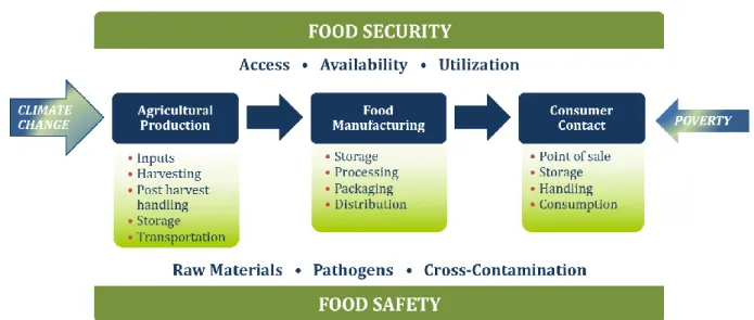
Nowadays, food security/safety has become a major concern (Figure 2) particularly since numerous contaminants are well known and detected in our environment such as dioxins, mycotoxins, heavy metals, pesticides, polycyclic aromatic hydrocarbons, drugs and hormones (Barreira et al. 2010). Among them, mycotoxins present in a large part of the food supply pose an important health problem. As a result of the increasing interest of thousands of researchers around the world, these toxins and their origins, toxicities, stabilities, biosynthetic pathways, control strategies, etc. are the subject of intensive studies. In this chapter, a general introduction on microscopic fungi, mycotoxins and their impact on human health is presented. More detailed information on the filamentous fungus Penicillium expansum and its secondary metabolites (e.g. the mycotoxin patulin), will be discussed in the second part of this introduction.
Figure 2 Risks impacting food safety and security throughout the business supply chain
A.1 Fungi: microscopic filamentous fungi
The general term fungi comprise organisms known as mushrooms, moulds and yeasts. Fungi are eukaryotic organisms that traditionally were classified in the plant kingdom. Due to their cell wall containing cellulose or glucans (true fungi) and chitin, also found in the animal kingdom (Méheust 2012), and the absence of chlorophyll (Berger and Guss 2005), these organisms were differentiated from plants, and nowadays constitute a separate kingdom, namely that of the “fungi”.
Fungi are one of the largest kingdoms of organisms on earth and play a key role in many ecosystems (Mueller and Schmit 2007). The kingdom is characterized by a large diversity estimated at more than 1.5 million species, although to date, only about 70,000 species have been identified (Paterson 2006). This kingdom includes macroscopic species (macromycetes) and
8
microscopic ones (micromycetes). The latter are not perceptible by the eye unless their development becomes pronounced (Tabuc 2007). They are ubiquitous organisms able to proliferate in the indoor and outdoor environment (Muro-Cacho et al. 2003) when physico-chemical conditions such as humidity, temperature, and the presence of organic and inorganic substrates allow it (Aydogdu and Gucer 2009). The fungal kingdom is composed of saprophytic fungi that decompose the ecosystem and play an essential role in carbon and nitrogen recycling, and of others affecting plants, animals and humans, causing plant diseases and mycoses (Lutzoni et al. 2004). We also distinguish fungi living symbiotically (e.g., mycorrhizal fungi, lichens, endophytes) (Gargas et al. 1995).
Fungi have a sexual and/or asexual reproduction. The spores, ensuring asexual multiplication, may have a role in their dispersion once produced, but may also play a role in the survival of the organism when environmental conditions become unfavourable (Madelin 1994). Fungi are classified into two broad categories: the unicellular yeast form and the mycelial multicellular form formed of hyphae (Redecker 2002). The mycelial form allows the fungus to have significant radial growth and to rapidly colonize a medium. In fact, hyphae that consist of heterocaryotic (Ascomycota and Basidiomycota) or coenocytic (Zygomycota and Glomeromycota) cells ensure the growth of mycelial fungi. Their extension is restricted to the apex. After division, the newly formed apical part can separate from the rest of the mycelium by a septum (septal mycelium in Ascomycota and Basidiomycota) or not (mycelium siphoned in Zygomycota) (Jennings and Lysek 1996). This mycelial form thus provides a maximum contact surface and allows search for nutrients in all three dimensions (Carlile and Watkinson 1994; Jennings and Lysek 1996).
A.2 Filamentous fungi and their secondary metabolites
Filamentous fungi are of importance in the human environment, in a beneficial or harmful way and have economic consequences. They are involved in different sectors such as the (agri-) food, pharmaceutical and cosmetic, as well as in the medical sector.
In the food industry, certain moulds are used to produce cheeses such as roquefort (Penicillium roqueforti) or camembert (Penicillium camembertii) (Ropars et al. 2012). They can also be used for the synthesis of organic acids such as citric acid or gluconic acid (Aspergillus niger), used as food additives (Karaffa et al. 2001). Some moulds are used for the synthesis/production of enzymes such as maltase and dextrinase converting maltose and starch into alcohol (Rhizopus oryzae). This process of alcoholic fermentation is of relevance for manufacturing rice alcohol in Asia (Lv et al. 2012). In the pharmaceutical industry, certain moulds are used for the synthesis of drugs, including antibiotics such as penicillin (Penicillium chrysogenum) or cephalosporins (Cephalosporium acremonium).
Filamentous fungi are therefore of industrial interest. Nevertheless, they represent a threat for the feed and food sector due to contamination of feed and foodstuffs. The proliferation of moulds, whether pathogenic or not, deteriorates food and feed (i.e. spoilage) and leads to unfavourable changes in dietary and organoleptic characteristics (appearance, texture, smell and flavour). This leads to heavy economic losses. Food commodities losses due to mycotoxin contamination represent above 25% of all food spoiled (Egmond et al. 2004).
In addition, the proliferation of moulds may lead to reduced food safety and increased health risks for the consumer. The production of toxic secondary metabolites such as mycotoxins (see the paragraph 1.3) represents a major risk for human and animal health. A given species of microscopic fungus can generate one or several mycotoxins, and the same mycotoxin can be produced by several mould species. They can be secreted both at pre- and postharvest stages.
A.3 Mycotoxins and their impact on human health
a. Historical overviewThe term "mycotoxin" comes from the combination of the Greek word "mycos" referring to mushroom, and the Latin word "toxicum" meaning poison (Jouany et al. 2009; Rai et al. 2012). The first outbreaks of mycotoxicosis, described in antiquity, were ergotism (Figure 3A) that were later named "St. Anthony's fire" (Figure 3B) because of the burning sensation felt by the victims (Bennett and Klich 2003; Gravesen 1979). This was at the origin of major epidemics that raged across the Old Continent during the Middle Ages, killing hundreds of thousands of people. The disease was caused by the consumption of rye flour produced from rye contaminated with fungi of the genus Claviceps (Figure 3C). It was only in the 19th century that scientists managed to isolate the alkaloids responsible for ergotism and to study their toxicological characteristics. Other mycotoxicoses have been described later, such as Trichothecene Toxic Food Aleucie (ATA), which occurred in Russia in the 1930s, or the Turkey "X" disease, which occurred in Great Britain in 1960. The latter caused an unusual veterinary crisis in England, in which nearly 100,000 turkeys were decimated with severe hepatic necrosis and biliary hyperplasia (Bennett and Klich 2003; Yiannikouris and Jouany 2002; Nesbitt et al. 1962). The cause of this disease has been attributed to aflatoxins, secondary metabolites produced by the fungus Aspergillus flavus that have proliferated on peanuts contained in the food of these birds (Medeiros et al. 2012; Nesbitt et al. 1962). After this event, a real "gold rush" of mycotoxins was born and lasted for about fifteen years (1960-1975), during which many scientists joined well-funded research programs (Bennett and Klich 2003).
10
Figure 3 Art as evidence of mycotoxin contamination. A: Paint exemplifying an ergotism outbreak; B: “Saint Anthony’s hallucinations” by Mathias Grünewald (effects of hallucinations associated to
ergotism); C: rye ear contaminated by Claviceps purpurea.
b. Definition and characteristics
Mycotoxins are low molecular weight secondary metabolites (<1,000 Dalton) produced by filamentous fungi (Marin et al. 2013). These metabolites are non-essential to the fungus life cycle, which means that they are not associated with fungal cell growth and development (Moss 1991). Nevertheless, once produced, they may confer some competitive advantages (Fox and Howlett 2008b).
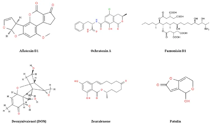
To date, 300 to 400 compounds have been recognized as mycotoxins with Aspergillus, Fusarium and Penicillium as the main producers, while many remain unknown (CAST 2003; Bryden 2016). Other genera such as Alternaria, Byssochlamys and Claviceps are known to produce mycotoxigenic compounds as well. The most commonly encountered mycotoxins are aflatoxins produced by Aspergillus species, ochratoxins produced by some species of Aspergillus and Penicillium, fumonisins, trichothecenes and zearalenone produced by Fusarium species, and patulin produced by several species of Aspergillus, Penicillium and Paecilomyces (Marin et al. 2013) (Figure 4). Patulin, the mycotoxin of interest in this thesis, is very toxic if ingested by humans and is present in fruits especially apples and pears (Moss 2008). Its toxicity and characteristics are described in
A
C
B
the third section of the introduction. Other mycotoxins have gained interest nowadays like for example citrinin, enniatins, etc. More studies should be done regarding them to be able to properly assess their toxicity and characteristics.
Figure 4 The chemical structures of major mycotoxins
Their enormous structural diversity, their countless biological effects and their different fungal producers make it hard to classify mycotoxins (Bennet and Klich 2003). The mycotoxins were classified based on their clinical effect, i.e., hepatotoxic, nephrotoxic, neurotoxic, immunotoxic and others, while cell biologists classify them as teratogens, mutagens, carcinogens and allergens. In order to prevent these risks, the International Agency for Research on Cancer (IARC) performed carcinogenic hazard assessment of some mycotoxins in humans and categorized them (e.g. patulin has an IARC of 3, aflatoxins (AFB1, AFB2, AFG1, AFG2 and AFM1) of 1, etc.). Their chemical structure (e.g., lactones), biosynthetic origin (e.g., amino acid-derived), illness they cause (e.g., aflatoxicosis) or fungal origin (e.g., Penicillium mycotoxins) could also be characteristics based on which mycotoxins could be classified (Bennet and Klich 2003). Based on the time point at which fungal invasion and mycotoxin formation take place, a distinction can be made between pre-harvest and post-harvest mycotoxins, referring to those produced by moulds growing on the field (e.g., Fusarium) and those produced by moulds at later stages such as harvest, transport, storage, processing, etc... However, this distinction is not always clear-out since post-harvest mycotoxins moulds can occur at pre-harvest stages as well.
12
c. Health effects
When mycotoxins are ingested, or sometimes inhaled, various toxic effects can be caused depending on the toxin (combination) and structures, the dose and duration of exposure and the person himself as well. They can induce toxicity in different organs and have various cellular and molecular mechanisms of action. Mycotoxins can cause acute or chronic toxicity. Even though chronic exposure to mycotoxins is considered as the main human and veterinary health burden (e.g. kidney toxicity, cancer induction, immune suppression), the best-known mycotoxin incidents were due to profound acute effects (e.g., human ergotism, Turkey X disease, stachybotryotoxicosis) (Bennet and Klich 2003; Cano et al. 2016).
The main problem we are facing is that several filamentous fungi capable of producing one or numerous mycotoxins could contaminate every raw material. In addition, most of the time, a food and feed item is composed of several raw materials. As a result, humans and animals are mostly not exposed to only one mycotoxin but more likely to a mixture of mycotoxins.Streit et al. (2013) showed that out of 17,316 food samples tested, 38% were contaminated with several mycotoxins simultaneously. Several studies indicated that interactions between mycotoxins present at the same time in a product might result in significantly higher toxicity than the individual toxicity of each mycotoxin (Smith et al. 1997; Speijers and Speijers 2004). In fact, as per Alassane-Kpembi et al. (2017), simultaneous exposure could result in synergistic(i.e., interaction resulting in greater effect than expected), additive (no interaction, i.e., as expected) or antagonistic (i.e., interaction leading to lesser effect than expected) effects. For example, a binary or ternary mixtures of type B trichothecenes (DON, NIV, and their acetylated derivatives) revealed synergistic cytotoxicity at low mycotoxin concentrations and additive or nearly additive effect at higher concentrations (Alassane-Kpembi et al. 2013). More studies on the interactions between mycotoxins are needed.
d. Regulations
Regulations exist and maximum levels of contamination by certain mycotoxins in food items have been set, but these legislations do not consider multi-contamination situations. In addition, of all the mycotoxins described, only thirty have proven toxic effects and less than a third of them are regulated. Maximum levels for aflatoxins (AFB1, AFB2, AFG1, AFG2 and AFM1), ochratoxin A (OTA), deoxynivalenol (DON), fumonisins (FB1, FB2), T-2 toxin, HT-2 toxin, zearalenone (ZEA) and
PAT are set by European commission (EC) Regulation No 1881/2006 and amendments. A limited number of mycotoxins (namely aflatoxins, deoxynivalenol, zearalenone, fumonisins and ochratoxin A) are regularly tested in the feed manufacturing process and are subject to regulations/guidance set by Commission Recommendation (EC) 576/2006 and Commission
Regulation (EU) No 165/2010. Mycotoxins are a very important problem of public health, quality and food safety and further research on this topic is still needed.
In the following sections (part B, C and D) of this introduction, we will be focusing on P. expansum and its secondary metabolites/mycotoxins with mean focus on PAT.
B. Secondary metabolism of Penicillium spp. and its regulation (review 1)
CHRISTELLE EL HAJJ ASSAF1,2, CHRYSTIAN DEL CARMEN ZETINA SERRANO1, ISABELLE P.
OSWALD1, SOPHIE LORBER1 AND OLIVIER PUEL1*
1Toxalim (Research Centre in Food Toxicology), Université de Toulouse, INRA, ENVT, INP-Purpan,
UPS, 31027 Toulouse, France
2Flanders Research Institute for Agricultural, Fisheries and Food (ILVO), Technology and Food
Science Unit, Brusselsesteenweg 370, 9090 Melle, Belgium
*Corresponding author Olivier PUEL
INRA UMR 1331 TOXALIM - INRA/INPT/UPS 180 chemin de Tournefeuille – BP 93173 F-31027 Toulouse cedex 3
Phone: +33 (0)582 06 63 36 Fax: +33 (0)561 28 52 44
16
Abstract
Penicillium, one of the most common fungi occurring in diverse range of habitats, has a worldwide distribution and a large economic impact on human health. Hundreds of the species belonging to this genus cause disastrous decays of food crops and are able to produce a varied range of secondary metabolites (SM), from which we can distinguish the harmful mycotoxins. Some Penicillium species are considered as important producers of patulin and ochratoxin A, two well-known mycotoxins. The production of these mycotoxins as well as other SM is controlled and regulated by different mechanisms. The aim of this review is to point out the different levels of regulation of SM in Penicillium sp. focusing on P. expansum. P. expansum, principal cause of spoilage of pome fruits throughout the past centuries, is ubiquitous and can infest a varied range of agricultural products during harvest, in storage, or even during processing. Besides, we discuss how the SM of the latter contribute to its virulence and pathogenicity and we end with an exposure of the different control treatments applied on P. expansum in order to inhibit its growth, decrease disease incidence and toxin accumulation.
Keywords: Penicillium spp, secondary metabolism, regulation, virulence, control of gene