HAL Id: hal-01833916
https://hal.archives-ouvertes.fr/hal-01833916
Submitted on 11 Jul 2018
HAL is a multi-disciplinary open access
archive for the deposit and dissemination of
sci-entific research documents, whether they are
pub-lished or not. The documents may come from
teaching and research institutions in France or
abroad, or from public or private research centers.
L’archive ouverte pluridisciplinaire HAL, est
destinée au dépôt et à la diffusion de documents
scientifiques de niveau recherche, publiés ou non,
émanant des établissements d’enseignement et de
recherche français ou étrangers, des laboratoires
publics ou privés.
of ion channels
Marwa Daghsni, Mohamad Rima, Ziad Fajloun, Michel Ronjat, Juan L.
Brusés, Ridha M’Rad, Michel de Waard
To cite this version:
Marwa Daghsni, Mohamad Rima, Ziad Fajloun, Michel Ronjat, Juan L. Brusés, et al.. Autism
throughout genetics: Perusal of the implication of ion channels. Brain and Behavior, Wiley Open
Access, 2018, Equipe IIb, pp.e00978. �10.1002/brb3.978�. �hal-01833916�
Brain and Behavior. 2018;e00978.
|
1 of 18 https://doi.org/10.1002/brb3.978wileyonlinelibrary.com/journal/brb3
1 | BACKGROUND
For several decades, autism was described as an infantile psychiatric disorder and was termed “childhood schizophrenia.” It was initially believed to be triggered by the psychopathological behavior of the
parents and in particular by the behavior of the mother toward her child (Sanua, 1983). In 1943, Leo Kanner characterized for the first time several cases of autism by describing the behavior of children between 2 and 8 years of age (Sanua, 1983). These children were characterized by deficits in social interactions and communication
Received: 19 November 2017
|
Revised: 1 March 2018|
Accepted: 18 March 2018 DOI: 10.1002/brb3.978R E V I E W
Autism throughout genetics: Perusal of the implication of ion
channels
Marwa Daghsni
1,2* | Mohamad Rima
3* | Ziad Fajloun
4| Michel Ronjat
1,5|
Juan L. Brusés
6| Ridha M’rad
2,7| Michel De Waard
1,5This is an open access article under the terms of the Creative Commons Attribution License, which permits use, distribution and reproduction in any medium, provided the original work is properly cited.
© 2018 The Authors. Brain and Behavior published by Wiley Periodicals, Inc.
*These authors contributed equally to this work.
1L’institut du Thorax, INSERM UMR1087/ CNRS UMR6291, Université de Nantes, Nantes, France 2Université de Tunis El Manar, Faculté de Médecine de Tunis, LR99ES10 Laboratoire de Génétique Humaine, 1007, Tunis, Tunisie
3Department of Neuroscience, Institute
of Biology Paris-Seine, CNRS UMR 8246, INSERM U1130, Sorbonne Universités, Paris, France 4Azm Center for Research in Biotechnology and Its Application, Lebanese University, Tripoli, Lebanon
5LabEx Ion Channels Science and
Therapeutics, Nice, France
6Department of Natural Sciences, Mercy College, Dobbs Ferry, NY, USA 7Service des Maladies Congénitales et Héréditaires, Hôpital Charles Nicolle, Tunis, Tunisie Correspondence Michel De Waard, Institut du Thorax, INSERM UMR1087, Nantes, France. Email: michel.dewaard@univ-nantes.fr Funding information M.D.W. is part of the laboratory of Excellence “Ion Channels, Science and Therapeutics” and is supported by a grant from the Agence Nationale de la Recherche (grant No. ANR- 11- LABX- 0015)
Abstract
Background: Autism spectrum disorder (ASD) comprises a group of neurodevelop-mental psychiatric disorders characterized by deficits in social interactions, interper-sonal communication, repetitive and stereotyped behaviors and may be associated with intellectual disabilities. The description of ASD as a synaptopathology highlights the importance of the synapse and the implication of ion channels in the etiology of these disorders.
Methods: A narrative and critical review of the relevant papers from 1982 to 2017
known by the authors was conducted.
Results: Genome-wide linkages, association studies, and genetic analyses of patients
with ASD have led to the identification of several candidate genes and mutations linked to ASD. Many of the candidate genes encode for proteins involved in neuronal development and regulation of synaptic function including ion channels and actors implicated in synapse formation. The involvement of ion channels in ASD is of great interest as they represent attractive therapeutic targets. In agreement with this view, recent findings have shown that drugs modulating ion channel function are effective for the treatment of certain types of patients with ASD.
Conclusion: This review describes the genetic aspects of ASD with a focus on genes
encoding ion channels and highlights the therapeutic implications of ion channels in the treatment of ASD.
K E Y W O R D S
autism, autism spectrum disorder, genetics, ion channels, synapse formation, synaptopathology, therapeutic targets
skills including eye contact avoidance, difficulties in understanding the emotions of others, hyper or hypo- reactivity, focused interests, and stereotyped repetitive behaviors. Because the clinical mani-festation of autism may differ significantly among patients, various pathologies were initially described including early infantile autism, childhood autism, Kanner’s autism, high functioning autism, atypi-cal autism, pervasive developmental disorder not otherwise speci-fied, childhood disintegrative disorder, and Asperger disorder. This group of disorders is currently encompassed within autism spec-trum disorder (ASD; American Psychiatric Association, 2013). The essential clinical manifestations of ASD are persistent in reciprocal social communication and social interaction and restrictive patterns of behavior, interests, or activities. These clinical features may be caused by a variety of genetic abnormalities and environmental fac-tors and may also temporarily overlap with other disorders including Rett syndrome (Park et al., 2016). ASD may be also associated with epilepsy (15%–47% of cases) and intellectual disability (8%–39% of cases; Ko, Kim, Kim, Song, & Cheon, 2016; La Malfa, Lassi, Bertelli, Salvini, & Placidi, 2004).
Genetic causes of ASD were first identified by epidemiologi-cal studies of human populations diagnosed with autism (Szatmari, Jones, Zwaigenbaum, & MacLean, 1998). Ozonoff et al. (2011) demonstrated that the prevalence of autism among siblings was 18 times higher than in the general population, suggesting the ex-istence of a familial heritability factor. Also, the imbalance in the sex ratio among ASD cases, with four to five boys affected for each affected girl, has led to the suggestion of a segregation linked to a sex chromosome and the implication of genes whose variations are expressed on a sex- linked recessive mode (Lai, Lombardo, Auyeung, Chakrabarti, & Baron- Cohen, 2015; Ozonoff et al., 2011). The sec- ond argument in favor of genetic causes of ASD is based on the ob-servation of a change in concordance rate between monozygotic and dizygotic twins, which was found to be 70%–90% in monozygotic twins compared with a lower rate of 0%–30% for dizygotic twins (Ronald & Hoekstra, 2014; Rosenberg et al., 2009). Thirdly, the ex-istence of chromosomal aberrations detected in patients with ASD also points toward genetic causes (Vorstman et al., 2006). Finally, genome-wide association studies (GWAS) have led to the identifi-cation of numerous ASD susceptibility genes that are located on various chromosomes, especially 2q, 5p, 7q, 15q, 17q and on chro-mosome X (Anney et al., 2010).
In addition, some patients with ASD were found to have vari- ations in syndromic Mendelian genes (e.g., FMR1 for fragile X syn-drome, TSC1 and TSC2 for tuberous sclerosis, and MECP2 for Rett syndrome; Liu & Takumi, 2014). The identification of variations in neuroligin genes (NLGN4X and NLGN3X) in patients with ASD sug-gested that proteins involved in synapse formation and synaptic transmission play an important role in the etiology of ASD (Jamain et al., 2003). Similarly, rare variations have been detected in genes coding for ion channels (e.g., CACNA1 and CACNB2), as well as pro-teins involved in synaptic structure, gene transcription, and chro-matin remodeling (e.g., NRXN1, CTTNBP2, CHD8, and SHANK3), indicating that altered synaptic plasticity and regulation of gene
expression may also be involved in the etiology of ASD (Cross- Disorder Group of the Psychiatric Genomics Consortium, 2013; De Rubeis et al., 2014; Durand et al., 2007).
This review examines the genetic basis of ASD and highlights the involvement of ion channel dysfunctions in the causes of this disorder.
2 | REVIEW
2.1 | Genetic aspects of ASD
During the last 30 years, a number of reviews have provided de-tailed description of the genetic architecture associated with ASD (Bourgeron, 2015; Devlin & Scherer, 2012; Li, Zou, & Brown, 2012; Liu & Takumi, 2014; Persico & Napolioni, 2013; Robert et al., 2017). This expansion of knowledge is due to the advances in molecular technologies, which allowed detecting chromosomal rearrange-ments, copy number variations (CNVs), and candidate genes in pa-tients with ASD.
2.1.1 | Chromosomal abnormalities and CNVs
in ASD
Chromosomal rearrangements have been identified in 5% of indi-viduals with ASD. These cytogenetic abnormalities are observed in chromosomes 5p15, 15q11–q13, 17p11, and 22q11.2 (Jacquemont et al., 2006; Sebat et al., 2007). Abnormalities affecting the 15q11– q13 region represent the most frequent variation associated with ASD accounting for close to 1% of all ASD cases (Badescu et al., 2016). Depending on the variation type and pattern of inheritance, this locus is associated with either Prader–Willi syndrome (PWS) or Angelman syndrome (AS) along with ASD (Badescu et al., 2016). This variation is based on whether the duplication affects the maternal or the paternal allele (Badescu et al., 2016). Besides structural rear-rangements, other abnormalities of chromosomal numbers or aneu-ploidies are detected in ASD including trisomy 21, Turner syndrome (45, X), and Klinefelter syndrome (47, XXY; Devlin & Scherer, 2012). Thanks to the comparative genomic hybridization (CGH) technique or SNPs array, CNVs were also found in multiple chromosomal re-gions at 1q21.1, 16p11.2, 17q12, and 22q11.2 (Jacquemont et al., 2006; Marshall et al., 2008; Matsunami et al., 2013; O’Roak et al., 2012; Pinto et al., 2010; Sebat et al., 2007). Further studies sup-ported the association with ASD of two recurrent de novo CNVs at 16p11.2 (duplication and deletion) and 7q11.23 (duplication; Levy et al., 2011; Sanders Stephan et al., 2011). The chromosomal dele-tion found at 7q11.23 has been linked to William’s syndrome, which includes intellectual disabilities, facial dysmorphic features, con-genital heart defect, and transient hypercalcemia. The intellectual disabilities suggest that this chromosomal region may also contain genes associated with social behaviors (Pinto et al., 2010). CNVs were found enriched in groups of genes implicated in cell signaling pathways that regulate neuronal development and cell prolifera-tion along with a group of genes associated with the GTPase/Ras
signaling pathway and neuronal plasticity (Pinto et al., 2010). CNVs studies demonstrated also that there is alteration in the fragile X mental retardation protein (FMRP) in patients with ASD. In fact, fragile X syndrome (X- Fra) is associated with 1%–2% of ASD cases. X- Fra syndrome is the second major cause of intellectual disability, which is caused by the expansion of CGG trinucleotide repeats in the FMR1 gene located on chromosome X and that encodes FMRP. This protein plays an essential role in synaptic plasticity by regulat-ing mRNA trafficking in the brain (Devlin & Scherer, 2012; Roberts, Tonnsen, McCary, Caravella, & Shinkareva, 2016).
These data emphasized the role of CNVs in ASD, and further in-vestigations in these regions have led to the identification of can-didate genes in particular with whole- exome and whole- genome sequencing studies.
2.1.2 | ASD candidate genes
Next- generation sequencing (NGS) techniques are very efficient tools for the identification of novel candidate genes associated with ASD. Most of the studies have been performed on sporadic cases of ASD. Using NGS, O’Rack et al. identified de novo variations in
FOXP1, GRIN2B, SCN1A, LAMC3, and rare inherited CNTNAP2
varia-tions (De Rubeis et al., 2014; O’Roak et al., 2011). Three genes were found in ASD probands with two de novo variations in each of these genes: BRCA2, FAT1, and KCNMA1 (Neale et al., 2012). These stud-ies also found significant enrichment of de novo variations in five ASD candidate genes including STXBP1, MEF2C, KIRREL3, RELN, and
TUBA1A (Neale et al., 2012). Likewise, a region of chromosome 7q
that includes the candidate genes RELN, FOXP2, WNT2, and CADPS2 has been implicated in ASD (Liu & Takumi, 2014). The extracellular glycoprotein RELN plays a key role in neuronal migration and cell interactions (Li et al., 2004). However, it appears that variations in
RELN are insufficient to cause ASD, suggesting that secondary ge-netic or epigenetic factors are behind these cases of ASD (Li et al., 2004). FOXP2 is a crucial gene for language development. Variations affecting this gene have been detected in individuals who lack the ability of acquiring communication skills. However, evidence sup-porting the involvement of FOXP2 in ASD remains scattered (Toma et al., 2013). The WNT2 gene belongs to the large WNT gene family, which is highly expressed during development of the central nerv-ous system and, therefore, it is not surprising that it could represent an ASD candidate gene (Kalkman, 2012; Li et al., 2004). Finally, the
CADPS2 gene encodes a calcium (Ca2+)- binding protein and
varia- tions in this gene have been linked to patients with ASD and intellec-tual disability (Bonora et al., 2014). Others genes encoding synaptic proteins linked to ASD were also identified by NGS: They include the glutamate receptors (GRIK2, GRIA3), the cell adhesion molecule CNTNAP2, and the scaffolding protein SHANK3. SHANK3 is in-volved in (i) synapse formation and maturation, (ii) the link between neurotransmitter receptors and ion channels, and (iii) the interac-tion with scaffolding proteins and gene regulatory proteins (e.g., protein of chromatin remodeling CHD8; Anney et al., 2010; Cotney et al., 2015; De Rubeis et al., 2014; O’Roak et al., 2011). NRXN1,
NLGN3/4X, and SHANK3 genes, which encode proteins involved in
neuronal cell adhesion and in the regulation of synaptic transmis-sion, are considered strong candidate loci for ASD (Weiss & Arking, 2009). Variations in those loci have also been detected in several patients with ASD (Anney et al., 2010; O’Roak et al., 2011; Table 1).
These approaches have also identified variants in genes encod-ing ion channels. Here, we describe these variations and highlight the role of ion channels in ASD.
2.2 | Ion channels and ASD
2.2.1 | Calcium signaling and voltage- gated Ca
2+channels in ASD
Ca2+ channels are present in many different cell types and they
me-diate Ca2+ influx in response to stimuli which can be a response to
(i) change in the membrane depolarization; known as voltage- gated channels or (ii) to a ligand- mediated activation (e.g., ryanodine re-ceptor (RyR), inositol triphosphate rere-ceptor (IP3R) in the reticulum).
In the brain, the elevation of intracellular Ca2+ concentration
acti-vates several signaling pathways that regulate important neuronal functions such as synaptogenesis, neuronal differentiation, and cell migration (Krey & Dolmetsch, 2007). Dysfunctions of these path-ways are responsible for abnormalities observed in patients with ASD, which include an increased cell density, changes in neuronal size, dendritic and axonal branching alterations, as well as in
neu-ronal connectivity (Krey & Dolmetsch, 2007). Voltage- gated Ca2+
channels are devised in two categories: high- voltage- activated chan-nels (HVA) and low- voltage- activated channels (LVA). HVA include L- type, the neuronal N- , P/Q- , and R- type. The low- voltage- activated
Ca2+ currents are represented by T- type channels. HVA are
com-posed by a principal transmembrane subunit α1 (Cav α) associated with a disulfide- linked α2δ (Cav α2δ) dimer, an intracellular β subunit (Cav β), and a transmembrane γ subunit (Cav γ), while LVA channels are composed only by α1 subunit. Both of HVA and LVA channels
control the passive flow of Ca2+ across membranes. Therefore,
al-teration in their components leads to defective channel function that translates themselves into a variety of neurological disorders including hemiplegic migraine, episodic and spinocerebellar ataxia, epilepsy, and ASD (Bidaud, Mezghrani, Swayne, Monteil, & Lory, 2006; Breitenkamp, Matthes, & Herzig, 2015; Heyes et al., 2015; Parellada et al., 2014; Stary et al., 2008; Zamponi, 2016).
The Timothy syndrome (TS) is a channelopathy described to be associated with ASD. TS is a multisystem disorder characterized by autistic features, cardiac abnormalities (QT prolongation), de-fective immune response, and syndactyly (Splawski et al., 2004).
Variations affecting the gene encoding the pore- forming α1
sub-unit of L- type voltage- gated Ca2+ channels are associated with TS.
Two genetic variations (G406R and G402S), affecting exon 8 of the
Cav1.2 channel α1 subunit gene (CACNA1C), have been associated
with TS. They impair the Cav1.2 inactivation and lead to prolonged
channel opening and consequent increase in Ca2+ flux (Barrett &
T A B LE 1 M aj or g en es im pl ic at ed in a ut is m s pe ct ru m d is or de r ( A SD ) G ene N ame Cy to gen et ic loc at ion Pr ot ei n func tio n A ss oc ia ted p ath olo gi es Re fer en ce s CH D8 C hr om odo m ai n hel ic ase DN A - bin din g p ro tein 8 /a ut is m s us ce pt i-bi lit y 18 (A U T1 8) 14 q1 1. 2 Tr an sc rip tio na l r ep re ss or n eg at iv el y re gu la te s W nt s ig na lin g p at hw ay b y bin din g t o b et ca te nin th er eb y in hibi tin g bin din g t o T C F4 A SD C ot ne y e t a l. ( 20 15 ), K ru m m , O ’R oa k, Sh en du re , a nd E ic hl er (2 01 4) , a nd O ’R oa k e t a l. (2 012 ) CN TN AP2 C ont ac as so ci ate d pr ote lik e 2/ au tis m s us ce pt ib ili ty 1 5 (A U T1 5) 7q 35–q 36 Pr ot ei n m em be r o f t he n eu re xi n su pe rf am ily i nv ol ve d i n n eu ra l a nd gl ia i nt er ac tio ns a nd c lu st er in g o f po ta ss iu m c ha nnel s i n neu ro ns Ep ile ps y, Pi tt –H op ki ns - li ke sy nd ro m e 1, A SD O ’R oa k e t a l. ( 20 11 ) a nd To m a e t a l. ( 20 13 ) C TT N BP2 C or ta ct in - bin din g p ro tein 2 7q 31 .31 M od ul at es th e m ob ili ty o f c or ta ct in in n eu ro ns . R eg ula te s s pin e m or ph og ene si s a nd s yn ap tic si gn al in g vi a PP 2A c om pl ex A SD C ros D is ord er G ro up o f th e P sy ch ia tr ic G en omic s C ons or tiu m (2 01 3) FM R1 Fr ag ile X m en ta l r et ar da tio n pr ot ei n Xq 27. 3 FM RP is a n RN A - b in di ng p ro te in in vo lv ed in R N A tr an sl at io n in neu ro ns Fr ag ile X s yn dr om e, A SD D ev lin a nd S ch er er ( 20 12 ) an d Ro be rt s et a l. (2 01 6) M EC P2 M et hy l- C pG - bin din g pr ot ei n 2 Xq 28 C hr om at as so ci ate d p ro te in th at re gu la te s g en e t ra ns cr ip tio n. I t i s re qu ire d f or t he m at ur at io n o f neu ro ns Re tt s yn dr om e, m en ta l re ta rd at io n X- lin ke d sy nd ro m ic 1 3, a ut is m su sc ep tib ili ty X - li nk ed 3 D ev lin a nd S ch er er ( 20 12 ) an d L iu a nd T ak um i (2 014 ) N LG N 3 N eu ro ligi n 3 Xq 13 .1 Li nk ed o nl y t o g lu ta m at er gi c po st sy na pt ic p ro tein s A sp er ger s yn dr ome s us cep ti-bili ty , a ut is m su sc ep tib ili ty X- lin ke d 1 Ja m ai n e t a l. ( 20 03 ) N LG N 4 N eu ro ligi n 4 X p2 2. 32 –p 22 .3 1 B in ds t o n eu re xi ns a nd l oc al ize d i n de nd rit ic s pin es M en ta l r et ar da tio n X- lin ke d, A sp er ge r s yn dr om e su sc ep tib ili ty X - li nk ed , a ut is m su sc ep tib ili ty X - li nk ed 2 Ja m ai n e t a l. ( 20 03 ) NR XN 1 N eu rex in 1 2p1 6. 3 N eu re xi ns , i nc lu di ng N R X N 1, a re c el l su rf ac e r ec ep to rs t ha t b in d ne ur ol ig in s t o f or m a C a 2+- dep en den t n eu re xi n/ ne ur ol ig in co m pl ex a t s yn ap se s i n t he c en tr al ne rv ou s s ys te m . T hi s c om pl ex i s re qu ire d f or neu ro tr an sm is si on a nd is i nv ol ve d i n t he f or m at io n o f sy na pt ic c on ne xi on Pi tt –H op ki ns - li ke s yn dr om e 2, sc hi zo ph re ni a, A SD A nn ey e t a l. (2 01 0) a nd G iri ra ja n e t a l. ( 20 13 ) PT EN Ph os ph at ase a nd te ns io n h om olo g 10 q2 3. 31 Tu m or s up pr es so r i nv ol ve d i n P I3 K si gn al in g p at hw ay a nd n eg at iv el y re gu la te s th e M A PK p at hw ay PT EN h am ar to m a t um or sy nd ro m e, m ac roc eph aly , aut is m M cB rid e et a l. (2 01 0) a nd O ’R oa k e t a l. ( 20 12 ) (Co nt in ue s)
G ene N ame Cy to gen et ic loc at ion Pr ot ei n func tio n A ss oc ia ted p ath olo gi es Re fer en ce s SH ANK 3 SH 3 an d m ul tip le a nk yr in re pe at do m ai ns 3 22 q1 3. 33 Sc af fo ld p ro te in a bu nd an t i n po st sy na pt ic e xc ita to ry s yna ps es w he re i t o rg an ize s r ec ep to r si gn al in g (e .g ., N M D A re ce pt or , m G lu R) A SD , P he la n– M cD er mi d sy nd ro m e, s chi zo ph re ni a D ur an d e t a l. ( 20 07 ) a nd Yi e t a l. (2 01 6) SY N G AP1 Sy na pt ic R as G TP as ac tiv at in g pr ot ei n 1 6p 21 .3 2 Ra s G TP as ac tiv at in g p ro te in t ha t i s la rg el y l oc al ize d i n d en dr iti c s pi ne s in ne oc or tic al p yr am id al neu ro ns . Su pp re ss es s ign al ing p at hwa ys lin ke d to N M D A re ce pt or (N M D A R) - m ed ia te d sy na pt ic pl as tic ity a nd A M PA re ce pt or (AM PAR ) M en ta l r et ar da tio n, A SD Pi nt o e t a l. ( 20 10 ) TS C1 Ha m ar tin 9q 34 .1 3 In te ra ct s w ith t ub er in t o f or m a co m pl ex tha t in hibi ts s ig na l tr an sd uc tio n t o t he d own st re am ef fe ct or s o f t he m amm al ia n t ar ge t ra pa m yc in pa th w ay (m TOR ). Im pli ca te d i n c ell p ro life ra tio n in hibi tio n Tu be ro us s cl eros is - 1 D ev lin a nd S ch er er ( 20 12 ) an d L iu a nd T ak um i (2 014 ) TS C2 Tu be rin 16p 13. 3 A ct s as a c ha pe ro ne fo r h am ar tin pr ot ei n Tu be ro us s cl eros is - 2 D ev lin a nd S ch er er ( 20 12 ) an d L iu a nd T ak um i (2 014 ) T A B LE 1 (Co nti nue d)
male patient affected with TS revealed a novel variation in CACNA1C gene (p.I1166T; Boczek et al., 2015). Electrophysiological analy-sis of HEK- 293 cells expressing this gene variant showed a shift in the peak channel activation and a reduced current density (Boczek et al., 2015; Table 2). L- type channels are predominantly expressed in the heart and brain. They are localized at dendrites and cell
bod-ies of mature neurons and regulate neuronal excitability and Ca2+-
dependent signaling cascades involving cAMP- binding protein (CREB) and myocyte enhancer factor 2 (MEF2; Krey & Dolmetsch, 2007; Simms & Zamponi, 2014). CACNA1C plays a key role in the development and functionality of the central nervous system by modulating gamma- aminobutyric acid (GABA) transmission and in-fluencing neuronal firing. In fact, mice with dysfunctional CACNA1C
show defects in N- methyl- d- aspartate (NMDA) receptor activity
leading to an NMDA- independent long- term potentiation in the CA1 region of the hippocampus that produces an acute decline in mem-ory. These observations indicate that CACNA1C may play a role in NMDA receptor- dependent signaling and in synaptic plasticity in the
hippocampus. In addition to ASD, SNPs in CACNA1C gene are linked to psychiatric disorders including schizophrenia and bipolar disorder (Li et al., 2015; Moosmang et al., 2005). In a large GWAS, two genes
encoding the α1 subunit of calcium channel (CACNA1C) and its
reg-ulatory β2 subunit (CACNB2) were strongly linked to psychiatric
dis-orders and ASD (Cross- Disorder Group of the Psychiatric Genomics Consortium, 2013).
In addition, three rare missense variations of CACNB2 (G167S, S197F, and F240L) were identified in families with ASD (Breitenkamp et al., 2014). Heterologous expression of these gene variants in HEK- 293 cells followed by electrophysiological analysis showed re-markable changes in channel kinetics characterized by an increased sensitivity of voltage- dependent inactivation for both G167S and S197F variants. Unlike these variations, the third variation F240L
showed a significant accelerated time- dependent inactivation
(Breitenkamp et al., 2014; Table 2). A deletion in chromosome region 12p13.33, that affects both the CACNA1C and the CACNA2D4 genes
coding for the α1 channel- forming subunit and the α2δ4 auxiliary
TA B L E 2 Impact of genetic variations associated with autism spectrum disorder (ASD) on ion channel’s function
Ion channels Genes Variations Type of variation Impact on ion channel References
Ca2+ channels CACNA1C p.I1166T Missense Shifts peak channel
activation and reduces current density
Boczek et al. (2015)
CACNA1D p.A749G; p.G407R Missense Changes kinetics of activation and inactivation
Pinggera et al. (2015)
CACNA1F p.I745T Missense Shifts channel inactivation ~30 mV and significantly slows the inactivation kinetics Hemara- Wahanui et al. (2005) CACNA1H p.R212C; p.R902W, p.R1871Q/p.A1874V; p.W962C Missense All these mutations reduce current density and voltage- dependent gating properties
Splawski et al. (2006)
CACNB2 p.G167S; p.S197F;p.F240L Missense G167S and S197F increase the sensitivity of voltage- dependent inactivation, and F240L shows an accelerated time- dependent inactivation Breitenkamp et al. (2014)
K+ channels KCNMA1 9q23/10q22 Translocation Reduces the activity of the
BKCa channel
Laumonnier et al. (2006)
KCNB1 p.I199F Missense Induces partial loss of function relative to biophysical defects of assembled homotetra-meric and heterotetra-meric channels
Calhoun et al. (2017)
KCNQ3 p.P574S Missense Reduces potassium current amplitude
Gilling et al. (2013)
Na+ channels SCN2A c.476+1G>A Splicing Produces a nonsense
mRNA and a truncated protein which alters the channel properties
subunit, respectively, was observed in patients with autistic manifes-tations (Smith et al., 2012). A chromosomal translocation of 2p:12p resulting in a deletion of both genes (CACNA1C and CACNA2D4) was detected in two ASD- affected individuals (Smith et al., 2012). Furthermore, a whole- exome sequencing study identified de novo
rare alleles in α1 subunit loci CACNA1D and CACNA1E (O’Roak et al.,
2012; Pinggera et al., 2015). The α1 subunit (Cav1.3) of L- type
chan-nels plays an important role in neuronal signaling and in brain func-tion including memory and behavior (Pinggera et al., 2015). De novo
variations in Cav1.3 subunit (CACNA1D) were identified in a cohort
of patients affected with autism along with intellectual disability (Pinggera et al., 2015). Using heterologous expression of the mutant proteins in tsA- 201 cells and whole- cell patch- clamp electrophysio-logical recordings revealed that these genetic variations affect the gating properties of the channel by changing the voltage- dependent kinetics of activation and inactivation (Pinggera et al., 2015; Table 2). A relevant study based on the analysis of signaling pathways impli-cated in ASD etiology of 1000 individuals with ASD from the Autism Genetic Resource Exchange (AGRE) identified SNPs in 146 genes. From 15 high- risk SNPs linked to ASD found in the study, two of
them are found in CACNA1A encoding CaV2.1 of Ca2+ channel and
in CACNA2D3 gene encoding for α2δ3 subunit of voltage- gated Ca2+
channel (Skafidas et al., 2014). A recent study in Chinese Han pop-ulation reported for the first time the association of two markers (rs7249246 and rs12609735) in CACNA1A gene with patients with ASD (Li et al., 2015). A variation in CACNA1F gene encoding for
CaV1.4 of Ca2+ channel was detected in a family with inherited night
blindness and ASD. This variation leads to the substitution of thre-onine by an isoleucine residue at codon 745 (p.I745T). Functional analysis of this variation performed in tsA- 201 cells demonstrated
that it affects channel kinetics causing the inactivation of the Ca2+
current (Hemara- Wahanui et al., 2005; Table 2).
T- type voltage- gated Ca2+ channels are known to play a key
role in the cerebral cortex and in the thalamus (Simms & Zamponi, 2014). Four heterozygous missense variations in CACNA1H gene,
encoding the Cav3.2 subunit of T- type channels, were found
as- sociated with decreased channel activity in six of 461 autistic pa-tients. This decrease could be a result of abnormal trafficking of the channel (Splawski et al., 2006; Table 2). Variations in the CACNA1H gene have also been associated with childhood absence epilepsy
(Splawski et al., 2006). Another α1 subunit of the T- type calcium
channel- encoding gene (CACNA1G) mapped at 17q11–q21 region was found to contain SNPs (rs12603122, rs757415, rs12603112, and rs198547) in patients with ASD (Lu, Dai, Martinez- Agosto, & Cantor, 2012). A statistical re- analysis of a GWAS data of patients with ASD from the AGRE revealed the association of the CACNA1I
gene encoding for CaV3.3 of Ca2+ channel in ASD (Hussman et al.,
2011). Yatsenko et al. studied 20 unrelated children affected with neurodevelopmental impairments, speech delay, and ASD using array- comparative genome hybridization and detected a duplica-tion in 9q34. This region contains the CACNA1B gene, and the 3′ region of EHMT1 gene implicated in Kleefstra syndrome, which is a genetic disorder characterized by intellectual disabilities, infantile
hypotonia, severe delay in expressive language, and facial dysmor-phism associated with other clinical signs. However, this duplication was described by the author as “benign” because it was also found in the control individual (Yatsenko et al., 2012; Table 3).
The importance of defective regulation of intracellular Ca2+ in
the pathophysiology of ASD is further supported by the association
between genes that encode plasma membrane Ca2+ pumps and ASD.
In fact, three studies from different human populations reported an association between the ATP2B2 gene coding for the plasma
mem-brane Ca2+ ATPase and ASD phenotypes (Yang et al., 2013). It should
be noted that ASD- associated genetic variations have been identi-fied in genes encoding Ca2+ channels and Ca2+ transport pumps, as
well as in genes encoding ion channels whose activities are under
Ca2+ modulation. To the best of our knowledge, until now, no
asso-ciation with other Ca2+ channels such as ligand- gated Ca2+ channels
(RyR, IP3R) has been found, hampering the exploration of novel cel-lular pathways.
2.2.2 | Potassium (K
+) channels in ASD
K+ channels are located in membranes of excitable and non-excitable
cells and they assure K efflux out of cells. According to their struc-ture and functions, K+ channels are segregated into four
catego-ries: the voltage- gated channels, inwardly rectifying (Kir), tandem pore domain (K2P), and the ligand- gated (Kligand) channels (Kuang, Purhonen, & Hebert, 2015). They all share a pore- forming α subunit
but different regulatory subunits are identified in each group. Ca2+-
activated potassium channels (BKCa) are ligand- gated K+ channels
that participate to several cell functions such as the regulation of hormone and neurotransmitter releases (Kuang et al., 2015). In fact,
BKCa are abundantly distributed throughout the brain and are mainly
localized at presynaptic terminals, where they partake in the adjust-ment of synaptic transmission and neuronal excitability (Kuang et al., 2015; Laumonnier et al., 2006). Laumonnier et al. (2006) observed a de novo balanced translocation of the 9q23/10q22 region that
houses the α1 subunit gene of BKCa channel (KCNMA1) in patients
with ASD. Electrophysiological experiments on lymphoblastoid cell lines derived from patients with ASD manifested a reduced activity of these channels. The authors also found a missense variation that alters a conserved domain of the channel in one patient with ASD
(Laumonnier et al., 2006; Table 2). Furthermore, a variation in the α1
subunit of BKCa channel (KCNMA1) has been implicated in
general-ized epilepsy and paroxysmal dyskinesia (Du et al., 2005). A novel
missense variation (c.595A.T) in KCNB1 gene that encodes KV2.1
voltage- gated potassium channel was detected in a patient with ASD associated with intellectual disability and epilepsy. This varia-tion causes the substituASD associated with intellectual disability and epilepsy. This varia-tion of isoleucine to phenylalanine at codon 199 (p.I199F) leading to significant depolarizing shifts in the voltage dependence of activation and inactivation of the channel (Calhoun, Vanoye, Kok, George, & Kearney, 2017; Table 2). The regulatory
β4 subunit gene of BKCa channel (KCNMB4) was classified as one
of the three predictive genes in ASD as it was strongly associated with SNPs- ASD- associated in a large meta- analysis study (Skafidas
et al., 2014; Table 3). On the other hand, a variation of KCNQ3 gene mapped to chromosome 8q24, encoding the voltage- gated
potas-sium channel Kv7.3, has been linked to epilepsy. This locus was found
disrupted as a consequence of a de novo chromosomal translocation in one patient with ASD. In addition, three patients with ASD shared a missense variation in KCNQ3. This variation could be described as a loss of function as identified by electrophysiological recordings in
Xenopus laevis oocytes (Gilling et al., 2013; Table 2).
These findings established a link between ASD and potassium channels and highlight their physiological importance in neuronal functions.
2.2.3 | Sodium (Na
+) Channels in ASD
Voltage- gated Na+ channels (Na
v) are essential for the initiation and
propagation of action potentials in neuronal cells, muscles, and heart
tissues. NaV channels are heteromeric complexes comprised of an
α subunit (pore- forming) associated with one or more β regulatory
subunits. We distinguish nine members of NaV channels (NaV1.1 to
NaV1.9) that differ by their structure but also by their ligand- specific
binding sites (toxins, drugs) which has led to their classification as critical drug targets (Bagal, Marron, Owen, Storer, & Swain, 2015).
Nav channels are primarily expressed in neurons and glial cells in
the central and peripheral nervous system. Variations affecting the
α subunit of Na+ channels and their accessory β subunit are known
to be responsible for Brugada syndrome, a cardiac disease (Weiss et al., 2003). In addition, several variations in SCN1A and SCN2A that
encode Nav1.1 and Nav1.2, respectively, are associated with
child-hood epilepsy and ASD (Weiss et al., 2003). Variations in SCN1A and SCN2A were shown to cause familial hemiplegic migraine and to be implicated in severe seizure syndrome, epilepsy, and Dravet syndrome (Craig, de Menezes, & Saneto, 2012; Weiss et al., 2003). It was shown that variations in SCN2A affect the calmodulin- binding
site of the channel and reduce its affinity for Ca2+. This site is crucial
for the binding between channel subunits and for connecting Na+
channels to Ca2+ signaling pathways (Weiss et al., 2003). Another
study using array- comparative genome hybridization identified a de novo deletion in a chromosome 2 region (2q24.2–q24.3) that con-tains SCN2A and SCN3A genes in a child with ASD (Celle, Cuoco, Porta, Gimelli, & Tassano, 2013). These findings were also validated by whole- exome sequencing study showing a significant associa-tion of SCN1A gene with the etiology of ASD (O’Roak et al., 2012; Sanders et al., 2012). Tavassoli et al. (2014) using whole- exome se-quencing found a de novo splice site variation in SCN2A gene in one patient with ASD. This variation (c.476+1G>A) that occurs at exon 4 of SCNA2A gene generates a truncated protein (Tavassoli et al.,
2014; Table 2). In addition, the α subunit 8 gene of a Na+ channel was
associated with ASD and identified by whole- genome sequencing in a family with ASD. In fact, a de novo heterozygous missense varia-tion was found in SCN8A gene (p.N1768D), which alters a conserved residue of the channel. The biophysical consequences of this
varia-tion are an increase in Na+ current and partial channel inactivation
(Veeramah et al., 2012; Table 2). In a large study of consanguineous
families with autism, a homozygous deletion of SCN7A gene was identified in one family, which is adjacent to SCN1A gene within the sodium channel gene cluster (SCN1A, SCN2A, SCN3A, and SCN9A) on chromosome 2 (Morrow et al., 2008; Table 3).
2.2.4 | Role of the ligand- gated ion channels, GABA,
glutamate, and cholinergic nicotinic receptors in ASD
Due to their crucial role in synaptic transmission, variations in GABA- A receptors are implicated in several severe neurological and neuropsychiatric disorders (Kang & Barnes, 2013). Patients with ASD have been reported to carry rearrangements abnormalities in chromosome 15q11–13 known as the imprinted region of Angelman/ Prader–Willi syndromes. This region houses a cluster of GABA re-ceptor genes that include GABRA5, GABRG3, and GABRB3, as well as CHRNA7 encoding the α7 subunit of the nicotinic acetylcholine receptor (nAChR; Hoppman- Chaney, Wain, Seger, Superneau, & Hodge, 2013). Moreover, polymorphisms in GABRA4 have also been associated with autism. Two SNPs located within 15q12 region were significantly linked to ASD, suggesting that this particular region ofGABRG3 gene is associated with an increased risk for ASD (Ben- Ari,
Khalilov, Kahle, & Cherubini, 2012; Kang & Barnes, 2013). ASD- associated polymorphisms have been identified in both the GRIK2 gene encoding the ionotropic glutamate receptor kainate 2 and the
GRM5 gene that encodes a metabotropic glutamate receptor. A re- cent study demonstrated a large spectrum of ASD phenotypes as-sociated with the 15q11–13 microdeletions. This region includes the CHRNA7 locus suggesting that CHRNA7 is a critical gene in ASD (Kuang et al., 2015; Laumonnier et al., 2006).
GABA- A receptors and Cl− concentration are important actors
for the excitation/inhibition balance in neurons during neurogenesis.
These two actors appear to be complementary in that the level of Cl−
concentration is crucial for GABAergic signaling. The regulation of
Cl− concentration in neurons is mediated by Cl− cotransporter (CCCs)
proteins anchored into the plasma membrane. Their roles are to
cou-ple the transport of Na+, K+, and Cl− and are named Na/K- Cl (NCC,
NKCC1, and NKCC2) transporters. There are four different K- CCCs (KCC1, KCC2, KCC3, and KCC4). In neurons, NKCC1 and KCC2 are
the predominant Cl− exchangers (Ben- Ari et al., 2012). A variation in
SLC12A2 gene encoding NKCC1 was reported to be linked to
schizo-phrenia. Functional experiments in Xenopus oocytes of this variation displayed an increased sensitivity to the NKCC blocker bumetanide. This evidence supports the hypothesis that NKCC1 activity is asso-ciated with schizophrenia and ASD because these two conditions share the same genetic background (Merner et al., 2016). In fact, it
was demonstrated that patients with ASD present an elevated intra-cellular Cl− concentration in neurons, suggesting that defective
ex-citability/inhibition balance could promote ASD due to an ineffective action of GABA leading to an abnormal chloride gradient (Ben- Ari et al., 2012). Bumetanide blocks NKCC1 and decreases intracellu-lar chloride concentration in neurons. A clinical study done on 60 children showed improvements in some ASD- related clinical mani-festations. Therefore, bumetanide is currently under investigation
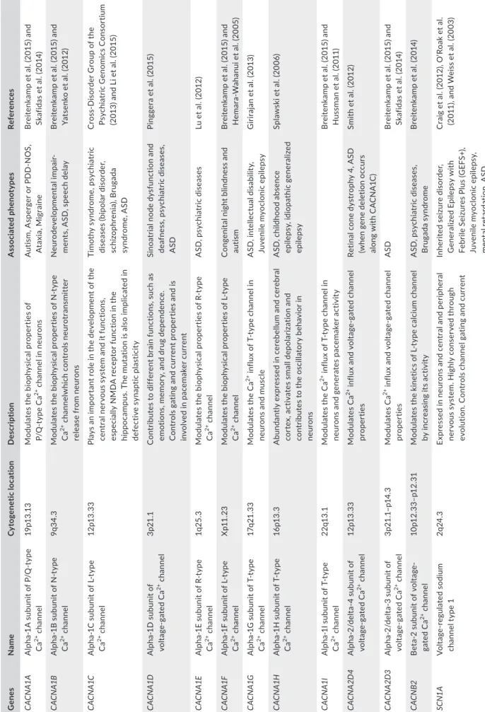
T A B LE 3 Io n ch an ne l g en es im pl ic at ed in a ut is m s pe ct ru m d is or de r ( A SD ) a nd re la te d pa th ol og ie s G ene s N ame Cy to gen et ic loc at ion D es cr ip tio n A ss oc ia ted ph en ot yp es Re fer en ce s CA CN A1 A A lp ha - 1 A s ub un it of P /Q - t yp e Ca 2+ c ha nne l 19 p13 .13 M od ul at es th e bi op hy si ca l p ro pe rt ie s of P/ Q - t yp e C a 2+ c ha nne l i n neu ro ns A ut is m , A sp er ge r o r P D D - N O S, A ta xi a, M ig ra in e B re ite nk am p e t a l. ( 20 15 ) a nd Sk af id as e t a l. ( 20 14 ) CA CN A1B A lp ha - 1 B su bu ni t o f N - t yp e Ca 2+ c ha nne l 9q 34 .3 M od ul at es th e bi op hy si ca l p ro pe rt ie s of N - t yp e Ca 2+ c ha nne lw hi ch c on tr ol s neu ro tr an sm itt er rel ea se fr om neu ro ns N eu ro de velo pm en ta l i mpa ir-m en ts , A SD , s pe ec h de la y B re ite nk am p e t a l. ( 20 15 ) a nd Ya ts en ko e t a l. ( 20 12 ) CA CN A1C A lp ha - 1 C s ub un it of L - t yp e Ca 2+ c ha nne l 12 p1 3. 33 Pl ay s a n i m po rt an t r ol e i n t he d ev el op m en t o f t he ce nt ra l n er vo us s ys te m a nd i t f un ct io ns , es pe ci al ly N M D A re ce pt or fu nc tio n in th e hi pp oc am pu s. T he m ut at io n i s a ls o i m pl ic at ed i n de fe ct iv e s yn ap tic pl as tic ity Tim ot hy s yn dr om e, ps ych ia tr ic di se as es (bip ola r di so rd er , sc hi zo ph re ni a) , B ru ga da sy nd ro m e, A SD C ros D is ord er G ro up o f t he Ps yc hi at ric G eno m ic s C on so rt iu m (2 01 3) a nd L i e t a l. ( 20 15 ) CA CN A1D A lp ha - 1 D s ub un it of vo lta ge - g ate d C a 2+ c ha nne l 3p 21 .1 C on tr ib ut es t o d iff er en t b ra in f un ct io ns , s uc h a s em ot io ns , mem or y, a nd d ru g dep en denc e. C on tr ol s g at in g a nd c ur re nt p ro pe rt ie s a nd i s in vo lv ed in p ac em ak er c ur ren t Si no at ria l no de d ys fu nc tio n a nd de af ne ss , ps ych ia tr ic di se as es , A SD Pi ng ge ra e t a l. ( 20 15 ) CA CN A1E A lp ha - 1 E su bu ni t o f R - t yp e Ca 2+ c ha nne l 1q 25 .3 M od ul at es th e bi op hy si ca l p ro pe rt ie s of R - t yp e Ca 2+ c ha nne l A SD , p sy ch ia tr ic d is ea se s Lu e t a l. ( 20 12 ) CA CN A1F A lp ha - 1 F su bu ni t o f L - t yp e Ca 2+ c ha nne l X p1 1. 23 M od ul at es th e bi op hy si ca l p ro pe rt ie s of L - t yp e Ca 2+ c ha nne l C on ge ni ta l n ig ht b lin dn es s a nd aut is m B re ite nk am p e t a l. ( 20 15 ) a nd H em ar a- W ah an ui e t a l. (2 00 5) CA CN A1G A lp ha - 1 G s ub un it of T - t yp e Ca 2+ c ha nne l 17q 21 .3 3 M od ul at es th e C a 2+ in flu x o f T - t yp e c ha nn el i n neu ro ns a nd m usc le A SD , i nt el le ct ua l d is ab ili ty , Ju ve nil e m yo cl on ic e pil ep sy G iri ra ja n e t a l. ( 20 13 ) CA CN A1H A lp ha - 1 H s ub un it of T - t yp e Ca 2+ c ha nne l 16p 13. 3 A bu nd an tly e xp re ss ed in c er eb el lu m a nd c er eb ra l co rt ex , ac tiv at es s m al l dep ol ar iz at io n a nd co nt rib ut es t o t he o sc ill at or y b eh av io r i n neu ro ns A SD , c hi ld ho od a bs en ce epi le ps y, idi op at hic g en er al iz ed epi le ps y Sp la w sk i e t a l. (2 00 6) CA CN A1I A lp ha - 1 I s ub un it of T - t yp e Ca 2+ c ha nne l 22 q1 3.1 M od ul at es th e C a 2+ in flu x o f T - t yp e c ha nn el i n ne ur on s a nd g en er at es p ac em ak er ac tiv ity B re ite nk am p e t a l. ( 20 15 ) a nd H us sm an e t a l. (2 01 1) CA CN A2D 4 A lp ha - 2 /d el ta - 4 s ub un it of vo lta ge - g ate d C a 2+ c ha nne l 12 p1 3. 33 M od ul at es C a 2+ in flu x a nd v olt ag ga te d cha nn el pr op er tie s Re tin al c on e dy st ro ph y 4, A SD (wh en g en e de le tio n o cc ur s al on g w ith C A C N A 1C ) Sm ith e t a l. ( 20 12 ) CA CN A2D 3 A lp ha - 2 /d el ta - 3 s ub un it of vo lta ge - g ate d C a 2+ c ha nne l 3p 21 .1– p1 4. 3 M od ul at es C a 2+ in flu x a nd v olt ag ga te d cha nn el pr op er tie s A SD B re ite nk am p e t a l. ( 20 15 ) a nd Sk af id as e t a l. ( 20 14 ) CA CN B2 B et 2 s ub un it o f v ol ta ge - gate d C a 2+ c ha nne l 10 p1 2. 33 –p1 2. 31 M od ul at es th e ki ne tic s of L - t yp e ca lc iu m c ha nn el by inc re as in g i ts ac tiv ity A SD , p sy ch ia tr ic d is ea se s, B rug ad a s yn dr om e B re ite nk am p et a l. ( 20 14 ) SC N1 A Vo lta reg ula te d so di um ch an nel ty pe 1 2q 24 .3 Ex pr es se d i n n eu ro ns a nd c en tr al a nd p er ip he ra l ne rv ou s sy st em . H ig hl y co ns er ve d th ro ug h ev ol ut io n. C on tr ol s c ha nnel g at in g a nd c ur re nt In he rit ed s ei zu re d is ord er , G en er al ize d E pi le ps y w ith Fe br ile S ei zu re s P lu s ( G EF S+ ), Ju ve ni le m yo cl on ic e pi le ps y, m en ta l r et ar da tio n, A SD C ra ig e t a l. ( 20 12 ), O ’R oa k e t a l. (2 01 1) , a nd W ei ss e t a l. ( 20 03 ) (Co nt in ue s)
G ene s N ame Cy to gen et ic loc at ion D es cr ip tio n A ss oc ia ted ph en ot yp es Re fer en ce s SC N2 A Vo lta reg ula te d so di um ch an nel ty pe 2 2q 24 .3 Ex pr es se d i n n eu ro ns a nd c en tr al a nd p er ip he ra l ne rv ou s s ys te m . C on tr ol s c ha nnel g at in g a nd cu rre nt Ea rly in fa nt ile ep ilep tic , en ce ph alo pa th y, b en ig n f am ili al in fa nt ile s ei zu re s, A SD C el le e t a l. ( 20 13 ) a nd W ei ss e t a l. (2 00 3) SC N3A Vo lta reg ula te d so di um ch an nel ty pe 3 2q 24 .3 Ex pr es se d i n n eu ro ns a nd c en tr al a nd p er ip he ra l ne rv ou s s ys te m . C on tr ol s b io ph ys ic al p ro pe rt ie s of the c ha nnel Ep ile ps y, A SD C el le e t a l. ( 20 13 ) a nd W ei ss e t a l. (2 00 3) SC N 7A Vo lta reg ula te d so di um ch an nel ty pe 7 2q 24 .3 N a +- s pe ci fic c ha nn el i n e xc ita bl e c el ls A SD (h om oz yg ou s de le tio n in aut is m ) M or ro w e t a l. (2 00 8) SC N 8A Vo lta reg ula te d so di um ch an nel ty pe 8 12 q13 .13 A lte rs th e re pe tit iv e fir in g pa tt er n of c er eb el la r Pu rk in je neu ro ns Cer eb el la r a ta xi a, ep ilep tic en ce ph alo pa th y e ar ly in fa nt ile , A SD W ei ss e t a l. ( 20 03 ) KC N M A1 C al ci um - a ct iva te d la rg e co nd uc ta nc e p ot as si um ch an ne l s ub fa m ily A 10 q2 2. 3 Sy na pt ic p ro tein reg ula to r o f n eu ro na l ex ci ta bili ty G en er al ize d e pi le ps y a nd pa ro xysmal d ysk in es ia (G EP D ), A SD La um on ni er e t a l. (2 00 6) KC NM B4 B K c ha nn el b et a s ub un it 4 12 q1 5 Reg ula to ry s ub un it o f B K cha nn el A SD Sk af id as e t a l. ( 20 14 ) KC N Q 3 Po ta ss iu m v olt ag ga te d ch an nel (M - c ha nnel ) 8q 24 .2 2 M od ul at es th e ki ne tic s of th e ch an ne l Ro la ndic e pi le ps y a nd idi op at hic gen er al iz ed ep ilep sy (IG E) in cl udin g b en ig n n eo na ta l co nv uls io ns , A SD G ill in g e t a l. ( 20 13 ) KC N Q 5 Po ta ss iu m v olt ag ga te d ch an nel (M - c ha nnel ) 6q1 3 Ex pr es se d i n b ra in a nd m us cl e a nd i m pl ic at ed i n sl ow ac tiv at io n o f t he c ha nn el. In ter ac ts w ith KC N Q 3 A SD G ill in g e t a l. ( 20 13 ) G RI K2 G lu ta m at e re ce pt or io no tr op ic ka in ate 2 6q 16 .3 G lu ta m at e r ec ep to rs a re the p re do m in an t ex ci ta to ry neu ro tr an sm itt er re ce pt or s i n t he ce nt ra l n er vo us s ys te m . C on ve rt s c he m ic al si gna l t o e le ct ric al im pul se M en ta l r et ar da tio n, A SD B en - A ri et a l. (2 01 2) , K an g an d B ar ne s ( 20 13 ), a nd L au m on ni er et a l. (2 00 6) G RI K3 G lu ta m at e re ce pt or io no tr op ic ka in ate 3 1p 34 .3 Pa ra lo g o f G RI K 2 Sc hi zo ph re ni a, A SD B en - A ri et a l. (2 01 2) , K an g an d B ar ne s, 2 01 3, a nd L au m on ni er et a l. (2 00 6) CH RN A7 A ce ty lc ho lin e re ce pt or , ne ur ona l n ic ot in ic , a lp ha - 7 su bu nit 15 q1 3. 3 Po st sy na pt ic G A B A er gi c in te rn eu ro n ac tiv ity . M ed ia te s fa st s ig na l t ra ns m is si on a t s yn ap se s Sc hi zo ph re ni a, A SD B en - A ri et a l. (2 01 2) , K an g an d B ar ne s ( 20 13 ), a nd L au m on ni er et a l. (2 00 6) G AB RG 3 G A B A - A g am m a su bu ni t o f G A B A re ce pt or fa m ily 15 q1 2 C on du ct s c hl or id e i on s u po n a ct iv at io n l ea di ng to h yp er po la riz at io n. C au se s i nh ib ito ry e ff ec t on neu ro tr an sm is si on Sc hi zo ph re ni a, A SD B en - A ri et a l. (2 01 2) , K an g an d B ar ne s, 2 01 3, a nd L au m on ni er et a l. (2 00 6) T A B LE 3 (Co nti nue d)
as a prospective drug by restoring the gradient and GABA inhibi-tion and, thereby, considered as a potential ASD- therapeutic agent (Lemonnier et al., 2012; Table 3).
2.3 | Ion channels and dysfunctional pathways
in ASD
Several genes encoding proteins involved in cellular pathways have been found enriched in ASD. These proteins are essentially implicated in synapse regulation (chromatin remolding, synaptic functions and protein synthesis and degradation; De Rubeis et al., 2014; Hormozdiari, Penn, Borenstein, & Eichler, 2015; Pinto et al., 2011; Ronemus, Iossifov, Levy, & Wigler, 2014; Uddin et al., 2014; Voineagu et al., 2011).
Synaptic regulatory proteins mainly concern: glutamatergic (e.g., GRIN2B) and GABAergic (e.g., GABRA3 and GABRB3) neurotrans-mission, neuronal connection (e.g., CNTNAP2) and ion permeabil-ity (e.g., CACNA1, CACNA2D3, and SCN1A), as well as proteins directly involved in synapse formation such as neurexins (NRXNs) and neuroligins (NLGNs). Among the scaffold proteins, there are proteins involved in the regulation of cell adhesion molecules and neurotransmitter receptors density in the synapse. This is the ex-ample of SHANK family proteins that assemble into large molecular platforms interacting with glutamate receptors, ion channels, actin cytoskeleton- associated proteins, and G protein- coupled signaling pathways (Grabrucker, Schmeisser, Schoen, & Boeckers, 2011). The SHANK proteins are associated with NMDA receptors via the gua-nylate kinase- associated protein (GKAP)/postsynaptic density- 95 (PSD- 95) complex and with metabotropic glutamate receptors type 1 (mGluR1) via the neuronal scaffolding protein Homer1. In addition, SHANK proteins can bind to several actin- regulatory molecules, such as cortactin (Durand et al., 2012). Mutations and CNVs (dele-tion and duplicasuch as cortactin (Durand et al., 2012). Mutations and CNVs (dele-tion) affecting SHANK genes have been associated with ASD. These variations resulted in actin accumulation in den-dritic spines, which alters the development and the morphology of dendrites (Durand et al., 2012).
Furthermore, neuronal dysfunctions are due to the modifica-tions in synthesis level of synaptic proteins caused by a defective mRNA regulation especially translation (Kelleher & Bear, 2008). This mechanism is controlled by several genes in particular mTOR and
FMR1. FMRP protein, encoded by FMR1 gene, binds to 400 different
mRNAs and represses their translation (Kelleher & Bear, 2008). The loss of FMRP protein results in fragile X syndrome that is present in 5% in patients with ASD. This protein acts downstream of the Ras- ERK signaling pathway via the complex FMRP–EIF4E–CYFIP1. This complex regulates the translation of more than 1,000 specific genes, many of which are ASD risk genes (De Rubeis et al., 2014). When CYFIP1 (cytoplasmic FMR1 interacting protein 1) binds to FMRP protein, the complex inhibits directly the translation of mRNA or in- directly by preventing the ribosomal translocation on mRNA. It is in-teresting to note that the expression of FMRP is under the control of
Ca2+/calmodulin- dependent protein kinase 4 (CAMKIV). However,
CYFIP1 and CAMKIV have been described as susceptibility genes in
ASD and together combined with an altered activity of FMPR en-hances the ASD risk (Waltes et al., 2014).
Another pathway implicating calcium signaling and ASD is the Mammalian target of rapamycin (mTOR) pathway also known as the mechanistic target of rapamycin kinase. MTOR gene is a tumor suppressor that regulates calcium signaling and mitochondrial func-tions. mTOR controls cells growth, proliferation, and differentiation, involved in synapse plasticity, and inhibits autophagy by preventing protein degradation. Interestingly, mTOR is upstream regulated by several mediators such as growth factors signals (e.g., insulin) or in neurons by the brain- derived neurotrophic factor (BDNF) through the phosphoinositide- 3- kinase (PI3K) activation the protein kinase B (Akt) and Ras to the extracellular signal- regulated kinase (Erk; Napoli et al., 2008; Schratt, Nigh, Chen, Hu, & Greenberg, 2004).
Both Erk and Akt act on the tuberous sclerosis complex (TSC1 and TSC2) by phosphorylating TSC2 inducing its dissociation of TSC1. TSC1 and TSC2 proteins act like GTPase proteins and down-regulate a small GTPase Rheb (Ras homolog enriched in brain) pro-tein via GAP propro-tein through a mechanism that remains unknown (Ma & Blenis, 2009).
Rheb is a direct activator of mTOR complex by activating its regulatory associated protein (raptor; Ma & Blenis, 2009). Once the mTOR complex is activated, it phosphorylates a series of protein such as the S6 kinase 1 (S6K1), the eukaryotic translation initiation factor 4E- binding protein 1 (eIF- 4BP1), and the carbamoyl- phosphate syn-thetase 2, aspartate transcarbamylase, and dihydroorotase (CAD). S6K1 and eIF- 4BP1 are essential for protein synthesis and poly-peptide translation in ribosomes and cell proliferation, while CAD is a key player in pyrimidine synthesis and so nucleotide synthesis (Ma & Blenis, 2009). In addition, the tuberous sclerosis complex can also be activated by AMPK, GSK3β, and p53 which leads the inhibi-tion of mTOR pathway. Furthermore, variainhibi-tions in TSC1 and TSC2 genes have been associated with ASD (Devlin & Scherer, 2012). Also, variations in mTOR pathway repressors, such as for the neurofibro-min 1 (NF1) gene NF1, cause neurofibromatosis type 1 syndrome as reported in 1% patients with ASD (Devlin & Scherer, 2012). The phosphatase and tension protein homolog (PTEN) is also known to downregulate mTOR pathway via both PI3K and AKT. Patients with ASD associated with cerebral malformation, like macrocephaly, have been found to carry variations in the PTEN gene in 7% of the cases (Devlin & Scherer, 2012; McBride et al., 2010; Figure 1).
Besides protein synthesis and translation that have been shown to be implicated in the process of ASD, the mechanism of protein degradation was also studied in ASD. Genetic studies indicate that ubiquitin–proteasome system is necessary for normal human cog-nitive function by regulating the synapse assembly and elimination (Mabb & Ehlers, 2010). The ubiquitin ligase enzyme Ube3A is a mem-ber of the E3 ubiquitin ligase family. The disruption of its activity leads to Angelman syndrome, while in turn the Angelman syndrome was described in ASD with CNVs and mutations in UBE3A gene (Greer et al., 2010). In Ube3A knockout mice, electrophysiological studies demonstrated an impaired long- term potentiation (LTP) in the hippocampus, which suggest that alteration of Ube3A results in
the loss of neuronal plasticity. In fact, Ube3A increases transcription through the myocyte enhancer factor 2 (MEF2) complex and regu-lates synapse function by ubiquitinating and degrading the synaptic protein Arc (activity- regulated cytoskeleton- associated protein). The role of Arc is to decrease long- term potentiation by promoting the internalization of AMPA receptors, which are the mediators of the excitatory neurotransmission in the central nervous system (Greer et al., 2010). On another hand, a decrease in AMPAR expression at synapses has been observed in patients with fragile X syndrome. This decrease is due to excessive mGluR5 signaling resulting in an increased Arc translation and consequently excessive AMPA recep-tors internalization (Dolen & Bear, 2008). In FMR1 knockout mice, injections of mGluR5 restore the AMPA receptors expression levels and prevent fragile X syndrome (Dolen et al., 2007).
Interestingly, it has been shown that an alteration of the
inhib-itory phosphorylation function of the Ca2+/calmodulin- dependent
protein kinase II (CamKII) is coupled to an increase in AMPA recep-tors expressed at the synapse (Rose, Jin, & Craig, 2009). In addition, mutations affecting this critical site of CamKII were shown to pre-vent the behavioral deficit in UBE3A gene- altered mice, suggesting that the Angelman syndrome is associated with a perturbation of CamKII functions (van Woerden et al., 2007; Figure 1).
Together, these studies emphasize the implication of ion chan-nels in the pathophysiology of ASD and strengthen the hypothesis that pharmacological manipulation of ion channels function is a po-tential therapeutic target in ASD.
2.4 | Ion channels and drug therapy in ASD
Ion channels have always been considered as powerful drug targets for the treatment of a wide range of pathologies owing to their cru-cial role as regulators of cell excitability (Kaczorowski, McManus, Priest, & Garcia, 2008).
In 1884, cocaine was discovered as the first anesthetic drug (Vandam, 1987). Several decades later, cocaine was described as a
Na+ channel blocker (Kyle & Ilyin, 2007; Vandam, 1987). This
ob-servation led the chemists to the production of novel analogs of cocaine, all classified under the term of “caine” and constituting a novel family of anesthetics (e.g., benzocaine, lidocaine; Casale, Symeonidou, & Bartolo, 2017; Tremont- Lukats, Megeff, & Backonja,
2000). Thereafter, drug- mediated modulation of Na+ channel
prop-erties was found to have other therapeutic functions such as anti-convulsants and antidepressants (e.g., carbamazepine) used in the treatment of neuropathic pain (Tremont- Lukats et al., 2000).
Valproic acid (VPA) is one of the most widely used anti- epileptic drugs for the treatment of tonico- clonic seizures that act by
modu-lating Na+ channel kinetics in neurons (Loscher, 2002). VPA is also
used for the treatment of bipolar disorder, anxiety, and migraine (Loscher, 2002). Studies showed that the exposure to VPA during pregnancy induces neurobehavioral abnormalities similar to autism traits in both rodents and humans (Bertelsen et al., 2016; Choi et al., 2016; Mony, Lee, Dreyfus, DiCicco- Bloom, & Lee, 2016). In fact, VPA treatment of postnatal rats was shown to affect DNA synthesis
and astrocyte proliferation and was associated with autistic behav-ior (Mony et al., 2016). A recent study showed that the phenotypic signs of ASD induced by VAP exposure in rats can be significantly improved or recovered by the administration of vitamin D in early stages of development (Du, Zhao, Duan, & Li, 2017). In addition,
it has been demonstrated that persistent Na+ current is
responsi-ble for hypoxia in neurons leading to neuronal damages (Faustino
& Donnelly, 2006). In fact, the persistence of Na+ currents leads to
the increased activity of Na+/Ca2+ exchangers in neurons, itself
re-sulting in an increase in Ca2+ cytoplasmic concentration (Faustino &
Donnelly, 2006). In order to correct this situation, it has been pro-posed that an increase in Na+ influx into cells prevents trauma in
the nervous system (Ates et al., 2007). Some Na+ channel blockers
(e.g., phenytoin, riluzole) showed neuroprotective activity in exper-imental spinal cord injury studies, in neurobehavioral studies and tissue recovery (Ates et al., 2007). It is important to mention that gabapentin, the first known drug in the treatment of neuropathic
pain, specifically binds to the α2δ1 subunit of N- type Ca2+ channels
and decreases the current (Zhu et al., 2017).
N- methyl- d- aspartate receptors (NMDAR) are well known to be
associated with psychiatric disorders (Lakhan, Caro, & Hadzimichalis, 2013). With no surprise, they were also linked to ASD risk (Lee, Choi, & Kim, 2015). Their activation follows the binding of glutamate once the D- serine or glycine co- agonists engage the specific allosteric site of the receptor (Kim et al., 2005). These two ligands were used in a clinical study on patients with severe schizophrenia as antipsy-chotic agents and were able to correct some negative clinical as-pects (Buchanan et al., 2007). In 1991, Haring et al. characterized an antibody, named B6B21, which showed a remarkable action in rat neurons by increasing long- term potentiation in CA1 pyramidal cells. This antibody has a high binding affinity for NMDA receptors. As a matter of fact, the authors concluded that B6B21 acts in a similar way to glycine on the receptor (Haring, Stanton, Scheideler, & Moskal, 1991). From B6B21, derived a family of small peptides called glyxines (Santini et al., 2014). One of these peptides, named GLYX- 13, was found to modulate NMDAR properties in a similar way to glycine. Treating ASD- affected rats with of GLYX- 13 resulted in promising improvements of autistic signs. Thereafter, authors sug-gested that this antibody might be a potential treatment for patients
affected by ASD (Santini et al., 2014). Moreover, d- cycloserine,
which is a partial NMDAR glycine agonist, is known to have effects on the behavioral deficits observed in autism and schizophrenia (Posey et al., 2004).
In a recent clinical trial carried out on 20 patients with autism, it has been shown that D- cycloserine treatment alleviated the stereo-typed behavior of these patients (Urbano et al., 2014). To go more into details, the administration of D- cycloserine during 8 weeks with different dosages showed to be effective on ASD manifestations in these patients without showing any side effects (Urbano et al., 2014). Additional studies will be required to determine the thera-peutic effect of this drug in ASD.
Concerning another therapeutic target, the implication of the acetylcholine receptor in ASD was demonstrated for the first time
by the analysis of postmortem adult brains from patients that suf-fered ASD (Martin- Ruiz et al., 2004). Analysis of mRNA levels by real- time PCR in different brain tissues (cerebral cortex and cere-bellum) showed a significant difference in the mRNA expression of several nicotinic acetylcholine receptor subunits (α3, α4, β2, and α7; Martin- Ruiz et al., 2004). Thus, it was suggested that the loss of nAChR functionality in the brain could be responsible for the ASD phenotype (Martin- Ruiz et al., 2004). Administration of
acetylcholine receptor activator donepezil to an ASD- affected boy proved beneficial for his cognitive skills after 6 weeks of treatment (Srivastava, Agarwal, & Pundhir, 2011). A randomized double- blind placebo- controlled trial using glutamine in autistic children showed significant improvements for some ASD clinical signs (Ghaleiha et al., 2014). The α7 nicotinic acetylcholine receptor en-coded by the CHRNA7 gene has also been associated with ASD (Deutsch, Urbano, Burket, Herndon, & Winebarger, 2011; Dineley, F I G U R E 1 Synaptic signaling pathways associated with autism spectrum disorder (ASD). Alterations in the mechanistic target of rapamycin complex (mTOR) are considered risk factors for ASD. mTOR is activated by Rheb- GTP. Upstream of Rheb is the tuberous sclerosis complex (TSC1–TSC2). TSC2 contains a GTPase- activating protein (GAP) domain that converts Rheb from GTP- bound form to its inactive GDP- bound form. Several upstream signaling pathways ranging from PI3K–AKT, Ras–ERK, LKB1–AMPK and Wnt–GSK3β pathways, positively or negatively regulate mTOR signaling. (AMPK, AMP- activated protein kinase; ERK, extracellular signal- regulated kinase; GSK3β, glycogen synthase kinase 3β; and PI3K, phosphoinositide 3- kinase). The mTOR pathway is also regulated by the brain- derived neurotrophic factor (BDNF) which binds to the tropomyosin- related kinase B (TRKB). BDNF plays a key role in the development and the plasticity of the central nervous system and it is considered a risk factor for ASD because increased levels of BDNF concentration have been observed in the serum and brain of patients with ASD. PI3K is also regulated by the synaptic protein SHANK, which is associated with metabotropic glutamate receptors type 1 (mGluR1) via the neuronal scaffolding protein HOMER1. The mTOR complex is a key modulator of protein synthesis by direct phosphorylation of 4E- binding proteins (4E- BPs) and activation of the ribosomal subunit S6 kinase (S6Ks), which in turn phosphorylate translation initiation factors. Thus, mTOR blocks the activation of cell autophagy and promotes cell proliferation, growth, and differentiation. The activity of the proteasome is also regulated by neuronal activity. The expression of UBE3A is increased through the transcription factor MEF2 and regulates the degradation of ARC protein, which promotes the internalization of AMPA- R and regulates
excitatory synapse development. Variations in the neuronal L- type Ca2+ channel α subunit CACNA1C have been associated with Timothy
syndrome and with ASD. In addition, Ca2+/calmodulin- dependent protein kinases are associated with components of the neuronal complex
including the fragile X mental retardation protein (FMRP) and its protein interaction CYFIP1, which also consider candidate genes in ASD. UBE3A: ubiquitin–protein ligase E3A; MEF2: myocyte- specific enhancer factor 2; ARC: activity- regulated cytoskeleton- associated protein; AMPR: AMPA receptors; CYFIP1: cytoplasmic FMRP- interacting protein 1 CACNA1C Ras PI3K Akt ErK TSC1/ TSC2 mTOR S6K1 eIF4E-BP1 mRNA translation and protein synthesis Cell proliferation PTEN Rheb GTP Rheb GDP Raptor Rapamycin Autophagy NF1 Tumor suppressors Proto-oncogenes AMPK GSK3β Ca2+ NMDAR AMPR CaMKII PSD95 SYNGAP1 mGluR1 SHANK PIKE ARC Proteasom e UBE3A BDNF TRKB HOMER1 CaMKIV