Rituximab therapy for hairy cell leukemia: a retrospective study
of 41 cases
Mathieu Leclerc&Felipe Suarez&Marie-Pierre Noël&Anne Vekhoff&Xavier Troussard&
Jean-François Claisse&Catherine Thieblemont&Frédéric Maloisel&Yves Beguin&
Jérôme Tamburini&Coralie Barbe&Alain Delmer
Received: 26 June 2014 / Accepted: 24 July 2014 / Published online: 7 September 2014 # Springer-Verlag Berlin Heidelberg 2014
Abstract The purine analogs (PAs) cladribine and pentostatin have transformed the prognosis of hairy cell leukemia (HCL). However, some patients still relapse after PAs, or fail to reach an optimal response, and new agents are needed to further improve treatment outcome. We retrospectively studied 41 HCL patients from 10 centers in France and Belgium, who received 49 treatment courses with the anti-CD20 monoclonal antibody rituximab. Most of the patients were treated at re-lapse (84 % of cases) and rituximab was combined to a PA in 41 % of cases. Overall, response rate is 90 % including 71 % complete hematologic responses (CHRs). Frontline treatment, combination therapy, and absolute neutrophil count were
associated with response in multivariate analysis. Three-year relapse-free and overall survivals are 68 and 90 %, respective-ly. When combined to a PA, rituximab yields a 100 % sponse rate, even beyond frontline therapy. In contrast, re-sponse rate is only 82 % (59 % CHR) when rituximab is used alone. In this latter setting, relapse rate is 56 % and median time to relapse is 17.5 months. All eight patients who were treated two times with the antibody responded again to re-treatment. We confirm the high efficacy of the combination rituximab + PA. However, when rituximab is used as mono-therapy, response rate is lower and the high relapse rate is a concern. Prospective clinical trials are needed to confirm the
M. Leclerc (*)
:
A. DelmerCentre Hospitalier Universitaire (CHU) de Reims, Hôpital Robert
Debré, Service d’Hématologie Clinique, Reims, France
e-mail: matleclerc@hotmail.fr A. Delmer
e-mail: adelmer@chu-reims.fr F. Suarez
Assistance Publique-Hôpitaux de Paris (AP-HP), Hôpital
Necker-Enfants Malades, Service d’Hématologie Adultes, Paris,
France
e-mail: felipe.suarez@nck.aphp.fr M.<P. Noël
Centre Hospitalier Régional Universitaire de Lille, Hôpital Claude Huriez, Service des Maladies du Sang, Lille, France
e-mail: marie-pierre.noel@chru-lille.fr A. Vekhoff
AP-HP, Hôpital Saint-Antoine, Service d’Hématologie,
Paris, France
e-mail: anne.vekhoff@sat.aphp.fr X. Troussard
CHU de Caen, Hôpital Femme-Enfant-Hématologie, Service
d’Hématologie Clinique, Caen, France
e-mail: troussard-x@chu-caen.fr
J.<F. Claisse
CHU d’Amiens, Hôpital Sud, Service d’Hématologie Clinique et de
Thérapie Cellulaire, Amiens, France e-mail: claisse.jean-francois@chu-amiens.fr C. Thieblemont
AP-HP, Hôpital Saint-Louis, Service d’Hématologie-Oncologie, Paris, France
e-mail: catherine.thieblemont@sls.aphp.fr F. Maloisel
Groupe Hospitalier Saint-Vincent, Clinique Sainte-Anne, Service
d’Oncologie et d’Hématologie, Strasbourg, France
e-mail: f.maloisel@solcrr.org Y. Beguin
CHU of Liège and University of Liège, Liège, Belgium e-mail: Yves.Beguin@chu.ulg.ac.be
J. Tamburini
AP-HP, Hôpital Cochin, Service d’Hématologie Clinique, Paris,
France
e-mail: jerome.tamburini@cch.aphp.fr C. Barbe
CHU de Reims, Hôpital Robert Debré, Service de Coordination à la Recherche Clinique, Reims, France
superiority of the combination rituximab + PA over PA alone, both as frontline therapy and at relapse.
Keywords Hairy cell leukemia . Rituximab . Immunotherapy . Chronic leukemias
Introduction
Hairy cell leukemia (HCL) is a rare and indolent lymphoid disorder, representing 2 % of all cases of lymphoid leukemias [1]. It is characterized by the presence of mature leukemic CD20-positive B cells showing both typical morphological features and phenotypic profile and accumulating in the bone marrow (BM), spleen, and liver. Hence, patients with HCL usually present with cytopenias, splenomegaly, and some-times hepatomegaly [2]. Treatment of HCL relies mainly on the purine analogs (PAs) cladribine and pentostatin, which have shown similar efficacy and constitute the gold standard of care either as frontline therapy or for relapsed patients [3–7]. However, despite the remarkable response rates obtain-ed with PA therapy, some patients will eventually relapse and the efficacy of these agents seems to decrease at each line of treatment [5]. The addition of new molecules to PAs may improve the response rates and prevent relapse.
Rituximab is a chimeric IgG1 kappa-type monoclonal antibody directed against the CD20 molecule. It was first used in relapsed patients with HCL more than 10 years ago [8], and several series of patients treated with rituximab as monotherapy were published in the following decade, reporting response rates ranging from 25 to 80 % [9–13]. These encouraging results have led to combine rituximab with PA, both in previously un-treated and in relapsed patients. As frontline therapy, combination with cladribine provided an impressive 100 % response rate in two separate cohorts that in-cluded a total of 63 patients [14, 15]. Moreover, mo-lecular responses were obtained in 70 % of these pa-tients. In the relapse setting, the efficacy of rituximab combined with either cladribine, pentostatin, or even fludarabine is also substantial with similar response and relapse rates, even though the number of reported patients is much lower [16, 17].
We designed a multicenter retrospective study of patients treated with rituximab for HCL, either frontline or at relapse and either in monotherapy or in combina-tion with other molecules. The objectives of the study were to assess the overall efficacy of rituximab in distinct clinical settings from a cohort of unselected patients representative of the population seen in the routine practice, and to identify prognostic factors for response, duration of response, and survival.
Methods
Patients
Patients were retrospectively recruited in 10 centers in France and Belgium from July 2002 to September 2012. They were eligible for inclusion in the study if they had a confirmed diagnosis of classical HCL and had received at least three subsequent injections of rituximab. No cases of HCL-variant were included. Diagnosis of HCL relied on identification of typical HCL cells in peripheral blood (PB) or BM with com-patible immunophenotyping (diagnostic score of 3/4 or 4/4 according to Matutes et al. [18]) and/or immunochemistry.
Rituximab therapy
Rituximab was administered intravenously at the dose of 375 mg/m2per injection in all patients but one who received two infusions of 1,000 mg at day 1 and day 15 as induction therapy and then a consolidation phase with the classic dos-age. Patients were allowed to receive several treatment se-quences with rituximab during the course of their disease.
Response assessment
Response to rituximab therapy was assessed during the first 6 months following the end of treatment, depending on avail-able data. Complete hematologic response (CHR) was defined as (i) the recovery of normal blood counts (absolute neutrophil count (ANC) ≥1.5×10⁹/L, hemoglobin level≥120 g/L for men and≥110 g/L for women, and platelet count ≥100×109/ L) and (ii) the absence of circulating HCL cells and clinical signs (mainly splenomegaly and hepatomegaly) of HCL; de-pending on the availability of a BM assessment after rituxi-mab therapy, CHR was further divided into three subgroups: stringent complete response (sCR) if BM evaluation showed no persistent leukemic cells, unconfirmed complete response (uCR) if no BM trephine biopsy or aspirate was performed after treatment, and CHR with persistent medullar infiltration by leukemic cells (iCHR). Partial response (PR) was defined as a≥50 % improvement for every CHR-defining criterion or normalization of at least one blood count (ANC, hemoglobin level, or platelet count), without circulating HCL cells. Pa-tients not meeting criteria for at least PR were classified as non-responders (NR).
Relapse
Relapse was defined either by reappearance of HCL cells in PB or BM or by a significant cytopenia (ANC≤1.5×10⁹/L, hemoglobin level ≤110 g/L, or platelet count ≤100×109/L) arising during follow-up, without other identifiable cause.
Time to relapse was defined as the time between the first rituximab infusion and relapse.
Minimal residual disease evaluation
Some patients included in this study had minimal residual disease (MRD) evaluation after treatment. For these patients, MRD was analyzed on a BM or PB sample using either immunophenotyping by flow cytometry or detection of an immunoglobulin heavy chain gene rearrangement by poly-merase chain reaction (PCR) [19,20].
Statistical analysis
Factors associated with treatment response were studied by univariate analysis (the Student, Wilcoxon, Chi-square, or Fisher’s exact tests, as appropriate) and by multivariate anal-ysis (binary logistic regression with ascending stepwise selec-tion and entry and exit thresholds set at 0.10). All variables with ap value <0.10 by univariate analysis were included in the multivariate analysis.
To assess potential predictive factors of relapse, univariate (log-rank tests) and multivariate (Cox stepwise regression with entry and removal limits set at 0.10) analyses were performed. All variables with ap value <0.10 by univariate analysis were included in the multivariate analysis.
Overall survival (OS) and relapse-free survival (RFS) curves were estimated with the use of the Kaplan-Meier method. For patients who received two separate treatment sequences with rituximab, only the first one was included in the statistical analyses of prognostic factors and survival.
Ap value of 0.05 was considered statistically significant. All analyses were performed with SAS® software, version 9.3 (SAS Inc, Cary, NC, USA).
Results
Patients’ characteristics
Forty-one patients were included in the study, for a total of 49 sequences of treatment with rituximab since eight of our patients were treated two times with this agent.
Patients’ characteristics at time of rituximab therapy are summarized in Table1. Most of them (76 %) were males, median age was 57 years, and one fifth was 70 or older. The median time elapsed between diagnosis and initiation of ri-tuximab therapy was more than 6 years (range 0–32 years).
Rituximab was used as part of frontline therapy in 16 % of cases (8 of 49 treatments performed). For the 41 remaining cases, the median number of previous therapeutic lines was 3 (range 1–8) with cladribine and pentostatin being the most
frequent previously used drugs (78 and 54 % of cases, respec-tively). All these patients had received at least one PA, and in 32 % of cases, they had received both cladribine and pentostatin. Other previous therapies included interferon in 20 cases (49 %), splenectomy in 7 cases (17 %), norethandrolone in 1 case (2 %), and, as mentioned above, rituximab in 8 cases (20 %).
For relapsed patients, overall response rate (ORR) to the therapeutic line immediately preceding rituximab (which in-cluded a PA in 76 % of cases) was 87 % (60 % CHR and 27 % PR).
Rituximab therapy
The median number of rituximab infusions per treatment course was 4 (range 3–12). Most of the patients received either four or eight infusions (65 and 15 % of cases, respectively). Rituximab was used as monotherapy in 55 % of cases and in combination with other agents in 45 % of cases. The most frequently associated molecule was cladribine (18 of 22 cases, 82 %), which was given either before (7 cases), concomitantly (8 cases), or after rituximab administration (3 cases). In the remaining cases, rituximab was combined with pentostatin (two patients), interferon (one patient), and steroids (one pa-tient). Rituximab was administered weekly in 88 % of cases (among whom 23 % subsequently received additional infu-sions at longer intervals). Median rituximab treatment dura-tion was 24 days (range 14–695).
Rituximab was given because of disease relapse or pro-gression (66 % of cases), contraindication to PA (12 %), failure of previous therapeutic line (10 %), detectable MRD after PA (10 %), and associated auto-immune hemolytic ane-mia (2 %).
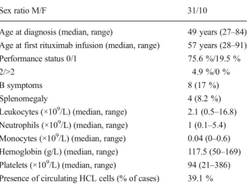
Table 1 Patients’ characteristics (n=41)
Sex ratio M/F 31/10
Age at diagnosis (median, range) 49 years (27–84)
Age at first rituximab infusion (median, range) 57 years (28–91)
Performance status 0/1 75.6 %/19.5 %
2/>2 4.9 %/0 %
B symptoms 8 (17 %)
Splenomegaly 4 (8.2 %)
Leukocytes (×109/L) (median, range) 2.1 (0.5–16.8)
Neutrophils (×109/L) (median, range) 1 (0.1–5.4)
Monocytes (×109/L) (median, range) 0.04 (0–0.6)
Hemoglobin (g/L) (median, range) 117.5 (50–169)
Platelets (×109/L) (median, range) 94 (21–386)
Presence of circulating HCL cells (% of cases) 39.1 %
Efficacy
Response assessment was available in 48 cases (Table 2). Whatever treatment modalities (monotherapy or combina-tion), ORR was 90 % (43 of 48 cases) and CHR was achieved in 34 cases (71 %), including 6 sCRs (13 %), 26 uCRs (54 %), and 2 iCHRs (4 %). PR was observed in nine cases (19 %) and treatment failure in five cases (10 %). Bone marrow evalua-tion was done in only 14 cases (29 %), mostly by BM aspirate (only three patients had a trephine biopsy), and was normal in 43 % of them.
All eight patients who received rituximab during frontline therapy achieved a CHR (three sCRs and five uCRs), even though it was combined with cladribine for five of them.
Regarding treatments given in relapsed patients, ORR was 88 % (including 65 % CHR and 23 % PR), with lower response rates when rituximab was used as monotherapy (24 cases, ORR 79 % and CHR 54 %) than when it was combined with cladribine or pentostatin (15 cases, ORR 100 %, CHR 86 %). Of note, all five treatment failures observed in this study were relapsed patients treated with rituximab alone.
Overall, when rituximab was used in combination with a PA (20 cases), ORR was 100 % (including 90 % CHR) versus 82 % (59 % CHR) when used as monotherapy (27 cases). No significant association was found between the number of administered rituximab infusions and the probability of re-sponse to treatment; particularly, ORR for patients treated with four infusions (90 %, including 71 % CHR) was compa-rable to that of patients receiving more than four infusions (93 %, including 73 % CHR).
Interestingly, all eight patients who had previously been treated with rituximab responded to re-treatment with this agent (6 uCRs, 1 iCHR, and 1 PR). Among them, five patients even had a better response at re-treatment, although for only one of them, rituximab was combined with a PA for this second treatment course.
Five parameters correlated with the likelihood of achieving a response to rituximab were identified by univariate analysis: first-line treatment, combination therapy, ANC and platelet count (with higher numbers increasing the probability of
achieving a response), and percentage of HCL cells in PB before treatment (with lower numbers increasing the proba-bility of achieving a response). Three independent factors persisted after multivariate analysis: first-line treatment (odds ratio (OR)=0.027, 95 % confidence interval (CI)=[0.001– 0.555],p=0.0192), combination therapy (OR=10.120, 95 % CI=[1.227–83.485], p=0.0316), and high ANC before treat-ment (OR=1.002, 95 % CI=[1.001–1.004], p=0.0060).
Nine patients had MRD evaluation after rituximab therapy, which was performed by immunophenotyping in six cases and by PCR in three cases. Two of them had undetectable disease. Significant diminution of MRD was observed in three additional cases, whereas three patients had stable MRD. The last patient had a very low positive MRD after rituximab, close to the detection threshold, but pre-treatment MRD was not available.
Relapses and duration of response
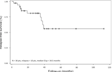
Among the 43 responses observed after rituximab therapy either alone or in combination, relapse or progression oc-curred in 15 cases (35 %), after a median duration of 19 months (range 2–39) since the first rituximab infusion (Table2). Three-year RFS was 68 % (Fig.1).
Regarding the eight patients who received rituximab during frontline therapy (median follow-up 30.5 months, range 4– 76), only 1 (13 %) relapsed at 38 months. On the other hand, when rituximab was used beyond frontline therapy (median follow-up 38 months, range 5–117), relapse rate was 40 % and median time to relapse was only 17.5 months (range 2–39). Concerning the eight patients who were re-treated with ritux-imab, five relapsed after this second treatment (63 %), at a median of 30 months.
When rituximab was given alone, the relapse rate was as high as 56 % compared to 14 % when used in combination with another drug (only 11 % when the other drug was a PA). Time to relapse was also shorter in case of monotherapy (median 17.5 vs 38 months).
The only independent prognostic factors for relapse/ progression identified by multivariate analysis were platelet
Table 2 Response to treatment, relapse rate, and duration of response
R rituximab, PA purine analog, ORR overall response rate, CHR complete hematologic response, mo months
Number ORR (%) CHR (%) Relapse
rate (%)
Duration of response (mo) (median, range)
Overall 48 90 71 35 26 (2–113) 1st line 8 100 100 13 30.5 (4–54) R alone 3 100 100 0 40 (12–54) R + PA 5 100 100 20 29 (4–38) Relapse 40 88 65 40 23 (2–113) R alone 24 79 54 63 23 (7–113) R + PA 14 100 86 7 24.5 (10–79) R + other 2 100 50 50 21 (2–40)
infusion requirement before rituximab therapy (hazard ratio (HR)=17.783, 95 % CI=[1.112–284.486], p=0.0419) and hemoglobin level after treatment (with lower numbers increas-ing the probability of relapse) (HR=0.943, 95 % CI=[0.892– 0.998],p=0.0406).
Overall survival
After a median follow-up of 36 months (range 4–117), 6 of 41 patients had died (15 %). Death occurred after a median of 37 months (range 9–76) since first rituximab infusion. Three-year OS for the whole cohort was 90 % (Fig.2). All of six patients either did not respond to treatment or relapsed, except one who died in remission.
Discussion
Whereas the largest series published to date concerns the use of rituximab in combination with PA for frontline therapy of HCL [14, 15], this situation represents only 5 of the 49 treatments reported in our study, with an excellent treatment outcome (100 % CHR). Most of our patients were treated at relapse, after multiple courses of therapy and a median time of more than 6 years since HCL diagnosis. This can explain the low incidence of splenomegaly, B symptoms, and severe cytopenias at time of initiation of rituximab treatment. In this relapse setting, the response rate we report (88 % ORR in-cluding 65 % CHR) is satisfactory and equivalent to the one observed at the previous line of therapy, even though combi-nation to PA seems to provide a real benefit in this patient
Fig. 1 Relapse-free survival.Pts
patients,f/up follow-up
Fig. 2 Overall survival.Pts
population. There is scarce data available concerning the use of frontline rituximab as monotherapy. Only four cases have been reported to date and two of these patients have responded to treatment [10,21]. Here, we report three additional cases of patients not eligible for frontline PA who received rituximab alone. They all met uCR criteria and none of them had relapsed after 12, 40, and 54 months of follow-up. Thus, this treatment approach is feasible and might represent a good alternative to interferon for patients who have contraindication to PA, even though more data are needed to confirm our results.
Another interesting issue that has not been assessed in the literature is whether or not patients who have already received rituximab for HCL may benefit from re-treatment with this agent. In fact, only one case has been reported so far [22]. Eight of the 41 patients of our study were treated two times with rituximab during the course of their disease. The re-sponse rate at re-treatment is excellent (100 % ORR including 88 % CHR), and most surprisingly, five of these eight patients had a better response to re-treatment as compared with first treatment with rituximab, and none of them had a worst response to re-treatment. Although the number of cases is low, these results seem to support the possibility of multiple rituximab therapy during the course of HCL, even in case of non-optimal response to first treatment.
Despite the good response rate reported in our study, re-lapse, which occurred in one third of our patients, remains a concern, mostly when rituximab is used alone and beyond frontline therapy. In this latter situation, the relapse rate we observe (63 %) is higher than the ones previously reported in the literature (ranging from 0 to 50 %) [9–13]. The longest follow-up in our cohort (median 45 months for these patients) may explain this difference. However, even in case of combi-nation with PA, relapse rate in our study (11 % after a median follow-up of 24.5 months) is still higher than published data, although to a lesser extent [15,16].
This study is the largest cohort of patients treated with rituximab for HCL reported to date. The large inclusion criteria we used have allowed us to study this molecule in a wide range of clinical situations, some of which have barely been explored in the literature. Most of all, our patients are not selected patients for prospective clinical trials but likely represent a good sample of the population met in routine clinical practice. On the other hand, one may argue that a limitation of this study is the wide heterogeneity of our cohort. Additionally, our data do not allow us to draw firm conclusions regarding the role of rituximab for MRD eradication. In fact, only very few patients had an MRD evaluation after treatment. More-over, results are difficult to interpret because of the het-erogeneity of the techniques and samples used for each patient. The low rate of MRD negativity we observe after rituximab (two of nine patients) may also be due to the
fact that MRD evaluation was often performed quite early after treatment, whereas previously published data have shown that molecular responses increase over time and are still quite low at 2 and even 6 months [19,14].
One of the limitations of this work may also concern the choice of response criteria, which are slightly different from the ones published in the guidelines from the British Committee for Standards in Haematology [6]. In these guidelines, response assessment and distinction between PR and CR rely mainly on BM evaluation. However, according to the recently published recommendations of the French Society of Hematology for the management of HCL patients [23], BM trephine biopsy evaluation after treatment is not mandatory in France and is barely real-ized outside prospective clinical trials. Indeed, in our retrospective study, less than a third of patients had a BM evaluation after rituximab therapy (and only three had a BM trephine biopsy), making response assessment incongruous in 70 % of cases regarding the British guide-lines. Therefore, one should keep in mind that the response rate we report may have been overestimated by the criteria we used and the fact that most patients did not have BM evaluation. On the other hand, as it has already been shown that the optimal response may not occur earlier than 2 or 3 months after treatment, response rate may also have been underestimated in a few cases in which response assessment was done too early because of insufficient follow-up or missing data. We believe, however, that the criteria we chose are well adapted to routine clinical practice as they are based on the concept of CHR, which is easy to assess. Another limitation of this study is the relatively short median follow-up (36 months) compared to other published series of patients suffering from this chronic disease.
In conclusion, regarding the results of this and previous studies, and before further data is available concerning the efficacy and toxicity of BRAF inhibitors, we think rituximab therapy for HCL should be considered either frontline, i.e., for patients not eligible for treatment with a PA or not achieving CR or negative MRD after PA, or at relapse, in combination with a PA whenever possible given the high relapse rate and short response duration when used as monotherapy. Random-ized prospective studies are warranted to confirm the superi-ority of the combination rituximab + PA over PA alone.
Conflict of interest The authors declare that they have no conflict of
interest.
Authors’ contribution ML and AD designed the study; ML, FS,
MPN, AV, XT, JFC, CT, FM, YB, JT, and AD provided the data; ML and CB performed the statistical analysis; ML and AD wrote the paper; all authors read and approved the final manuscript and agreed to submission.
References
1. Foucar K, Falini B, Catovsky D, Stein H (2008) Hairy cell leukaemia, vol 1. WHO classification of tumours of haematopoietic and lym-phoid tissues. IARC Press, Lyon
2. Bouroncle BA (1979) Leukemic reticuloendotheliosis (hairy cell leukemia). Blood 53(3):412–436
3. Spiers AS, Moore D, Cassileth PA, Harrington DP, Cummings FJ, Neiman RS, Bennett JM, O’Connell MJ (1987) Remissions in hairy-cell leukemia with pentostatin (2′-deoxycoformycin). N Engl J Med 316(14):825–830
4. Piro LD, Carrera CJ, Carson DA, Beutler E (1990) Lasting remissions in hairy-cell leukemia induced by a single infusion of 2-chlorodeoxyadenosine. N Engl J Med 322(16):1117–1121 5. Else M, Dearden CE, Matutes E, Garcia-Talavera J, Rohatiner AZ, Johnson SA, O’Connor NT, Haynes A, Osuji N, Forconi F, Lauria F, Catovsky D (2009) Long-term follow-up of 233 patients with hairy cell leukaemia, treated initially with pentostatin or cladribine, at a median of 16 years from diagnosis. Br J Haematol 145(6):733–740 6. Jones G, Parry-Jones N, Wilkins B, Else M, Catovsky D (2012)
Revised guidelines for the diagnosis and management of hairy cell leukaemia and hairy cell leukaemia variant*. Br J Haematol 156(2): 186–195
7. Flinn IW, Kopecky KJ, Foucar MK, Head D, Bennett JM, Hutchison R, Corbett W, Cassileth P, Habermann T, Golomb H, Rai K, Eisenhauer E, Appelbaum F, Cheson B, Grever MR (2000) Long-term follow-up of remission duration, mortality, and second malig-nancies in hairy cell leukemia patients treated with pentostatin. Blood
96(9):2981–2986
8. Hagberg H (1999) Chimeric monoclonal anti-CD20 antibody (ritux-imab)—an effective treatment for a patient with relapsing hairy cell
leukaemia. Med Oncol 16(3):221–222
9. Lauria F, Lenoci M, Annino L, Raspadori D, Marotta G, Bocchia M, Forconi F, Gentili S, La Manda M, Marconcini S, Tozzi M, Baldini L, Zinzani PL, Foa R (2001) Efficacy of CD20 monoclonal anti-bodies (Mabthera) in patients with progressed hairy cell leukemia.
Haematologica 86(10):1046–1050
10. Hagberg H, Lundholm L (2001) Rituximab, a chimaeric anti-CD20 monoclonal antibody, in the treatment of hairy cell leukaemia. Br J
Haematol 115(3):609–611
11. Nieva J, Bethel K, Saven A (2003) Phase 2 study of rituximab in the treatment of cladribine-failed patients with hairy cell leukemia. Blood
102(3):810–813
12. Thomas DA, O’Brien S, Bueso-Ramos C, Faderl S, Keating MJ,
Giles FJ, Cortes J, Kantarjian HM (2003) Rituximab in relapsed or
refractory hairy cell leukemia. Blood 102(12):3906–3911
13. Zenhausern R, Simcock M, Gratwohl A, Hess U, Bargetzi M, Tobler A (2008) Rituximab in patients with hairy cell leukemia relapsing after treatment with 2-chlorodeoxyadenosine (SAKK 31/98).
Haematologica 93(9):1426–1428
14. Cervetti G, Galimberti S, Andreazzoli F, Fazzi R, Cecconi N, Caracciolo F, Petrini M (2008) Rituximab as treatment for minimal residual disease in hairy cell leukaemia: extended follow-up. Br J
Haematol 143(2):296–298
15. Ravandi F, O’Brien S, Jorgensen J, Pierce S, Faderl S, Ferrajoli A,
Koller C, Challagundla P, York S, Brandt M, Luthra R, Burger J, Thomas D, Keating M, Kantarjian H (2011) Phase 2 study of cladribine followed by rituximab in patients with hairy cell leukemia.
Blood 118(14):3818–3823
16. Else M, Dearden CE, Matutes E, Forconi F, Lauria F, Ahmad H, Kelly S, Liyanage A, Ratnayake V, Shankari J, Whalley I, Catovsky D (2011) Rituximab with pentostatin or cladribine: an effective combination treatment for hairy cell leukemia after disease recurrence. Leuk Lymphoma 52(Suppl
2):75–78
17. Gerrie AS, Zypchen LN, Connors JM (2012) Fludarabine and ritux-imab for relapsed or refractory hairy cell leukemia. Blood 119(9):
1988–1991
18. Matutes E, Morilla R, Owusu-Ankomah K, Houliham A, Meeus P, Catovsky D (1994) The immunophenotype of hairy cell leukemia (HCL). Proposal for a scoring system to distinguish HCL from B-cell disorders with hairy or villous lymphocytes. Leuk Lymphoma 14(Suppl 1):57–61
19. Cervetti G, Galimberti S, Andreazzoli F, Fazzi R, Cecconi N, Caracciolo F, Petrini M (2004) Rituximab as treatment for minimal residual disease in hairy cell leukaemia. Eur J Haematol 73(6):412– 417
20. Matutes E, Meeus P, McLennan K, Catovsky D (1997) The signifi-cance of minimal residual disease in hairy cell leukaemia treated with deoxycoformycin: a long-term follow-up study. Br J Haematol 98(2): 375–383
21. Malfuson JV, Doghmi K, Fagot T, Souleau B, De Revel T, Nedellec G (2006) Efficacy of rituximab in hairy cell leukemia. Presse Med 35(5 Pt 1):801–802
22. Buckstein R, Patel H, Chesney A, Reis M, Imrie K (2006) Multiply relapsing hairy cell leukemia responsive to repeated
courses of rituximab: a case report. Hematology 11(4):267–
270
23. Cornet E, Delmer A, Feugier P, Garnache-Ottou F, Ghez D, Leblond V, Levy V, Maloisel F, Re D, Zini J, Troussard X (2014) Recommendations of the SFH (French Society of Haematology) for the diagnosis, treatment and follow-up of hairy cell leukemia. Ann Hematol In press