The RPAP3-Cterminal domain identi
fies R2TP-like
quaternary chaperones
Chloé Maurizy
1,2
, Marc Quinternet
3
, Yoann Abel
1,2
, Céline Verheggen
1,2
, Paulo E. Santo
4,5
, Maxime Bourguet
6
,
Ana C.F. Paiva
4,5
, Benoît Bragantini
7
, Marie-Eve Chagot
7
, Marie-Cécile Robert
1,2
, Claire Abeza
1,2
, Philippe Fabre
7
,
Philippe Fort
8
, Franck Vandermoere
9
, Pedro M.F. Sousa
4,5
, Jean-Christophe Rain
10
, Bruno Charpentier
7
,
Sarah Cianférani
6
, Tiago M. Bandeiras
4,5
, Bérengère Pradet-Balade
8
, Xavier Manival
7
& Edouard Bertrand
1,2
R2TP is an HSP90 co-chaperone that assembles important macro-molecular machineries. It
is composed of an RPAP3-PIH1D1 heterodimer, which binds the two essential AAA+ATPases
RUVBL1/RUVBL2. Here, we resolve the structure of the conserved C-terminal domain of
RPAP3, and we show that it directly binds RUVBL1/RUVBL2 hexamers. The human genome
encodes two other proteins bearing RPAP3-C-terminal-like domains and three containing
PIH-like domains. Systematic interaction analyses show that one RPAP3-like protein, SPAG1,
binds PIH1D2 and RUVBL1/2 to form an R2TP-like complex termed R2SP. This co-chaperone
is enriched in testis and among 68 of the potential clients identi
fied, some are expressed in
testis and others are ubiquitous. One substrate is liprin-
α2, which organizes large signaling
complexes. Remarkably, R2SP is required for liprin-
α2 expression and for the assembly of
liprin-
α2 complexes, indicating that R2SP functions in quaternary protein folding. Effects are
stronger at 32 °C, suggesting that R2SP could help compensating the lower temperate of
testis.
DOI: 10.1038/s41467-018-04431-1
OPEN
1IGMM, CNRS, Université de Montpellier, Montpellier 34293, France.2Equipe labélisée Ligue Nationale Contre le Cancer, 34293 Montpellier, France. 3CNRS, INSERM, IBSLOR, Université de Lorraine, Nancy F-54000, France.4iBET, Instituto de Biologia Experimental e Tecnológica, Apartado 12, Oeiras 2781-901, Portugal.5Instituto de Tecnologia Química e Biológica António Xavier, Universidade Nova de Lisboa, Av. da República, Oeiras 2780-157, Portugal. 6Laboratoire de Spectrométrie de Masse BioOrganique, Université de Strasbourg, CNRS, IPHC UMR 7178, Strasbourg 67000, France.7CNRS, IMoPA, Université de Lorraine, Nancy F-54000, France.8CRBM, University of Montpellier, CNRS, 1919 Route de Mende, Montpellier 34090, France.9IGF, CNRS, Université de Montpellier, Montpellier 34090, France.10Hybrigenics Services, Paris 75014, France. These authors contributed equally: Chloé Maurizy, Marc Quinternet. Correspondence and requests for materials should be addressed to X.M. (email:xavier.manival@univ-lorraine.fr)
or to E.B. (email:edouard.bertrand@igmm.cnrs.fr)
123456789
T
he R2TP complex was discovered in S. cerevisiae as an
HSP90 co-factor
1. R2TP is an unusual co-chaperone
because it appears specialized in quaternary protein
fold-ing, and in particular in the assembly of key cellular machineries
important for cell growth
2. Important R2TP clients are the small
nucleolar ribonucleoprotein particles (snoRNPs), which are
required for ribosomal RNAs maturation
3. More recently, other
substrates were described, including the U4 and U5 spliceosomal
snRNAs
4–6, the nuclear RNA polymerases
7–9, and the family of
PI3K-like kinases (PIKKs), which comprises mTOR, DNA-PK,
ATR, ATM, SMG1 and TRRAP
10,11. Given the role of these
cli-ents in cell growth and proliferation, it was hypothesized that
R2TP mediates some of the tumorigenic effects elicited by
HSP90
12. Newly identified clients involved in DNA damage
response corroborate this possibility
13.
In humans, R2TP is composed of a core that associates with
prefoldins and additional factors to form the R2TP/Prefoldin-like
complex, recently renamed PAQosome
2,7–9. The R2TP core is
composed of four subunits (Fig.
1
a): PIH1D1, RPAP3, and the
two related AAA+ATPases RUVBL1 and RUVBL2 (also called
Pontin and Reptin). RPAP3 directly binds HSP90 and forms a
stable heterodimer with PIH1D1
14,15. These components are
believed to function as adapter and regulatory factors, while
RUVBL1 and RUVBL2 are catalytic components that likely
possess a chaperone activity
16. The analysis of snoRNP biogenesis
suggests that R2TP functions through a stepwise process, where
HSP90 stabilizes clients before their assembly, followed by the
independent recruitment of several snoRNP components by
R2TP, and ending in the loading of RUVBL1/2 on the client
complex
17. The molecular role of RUVBL1 and RUVBL2 still
remains elusive. Structural studies showed that they form
hex-americ rings typical for this class of enzymes
18. In addition, they
contain an insertion called domain II, which protrudes from the
ATPase ring and forms a
flexible structure whose conformation
depends on the presence and nature of bound nucleotides
19,20.
RUVBL1/2 make ATP-dependent contacts with some cofactors
and client proteins
21,22, suggesting that ATP loading and
hydrolysis may act as a switch to control the binding and release
of clients. Accordingly, it has been proposed that RUVBL1/
2 stabilize interactions between subunits of target complexes
17, or,
alternatively, that they stimulate the dissociation of specific
assembly factors
23. Recent structural analyses showed that
RUVBL1/2 cycle between hexameric and dodecameric forms,
with client binding promoting dimerization of the hexamer, and
ATP hydrolysis dissociation of the dodecamer
16. This provides a
glimpse of how these enzymes may chaperone their clients.
Structural studies revealed that PIH1D1 has two domains. Its
N-terminal region harbors the conserved PIH domain that
pos-sesses a phospho-peptide binding pocket responsible for
recog-nizing some substrates
14,15. Its C-terminal region folds as a CS
domain and mediates hetero-dimerization with RPAP3
14,15,24.
RPAP3 has a C-terminal domain of unknown function
(RPAP3-Cter), and a middle region composed of six tetratricopeptide
repeats (TPR) arranged in two consecutive domains (Fig.
1
b).
Structural studies showed that they bind HSP90 through
five
conserved residues forming a carboxylate clamp, which catches
the last C-terminal residues of the chaperone (-MEEVD
15,25). A
recent cryo-EM structure of the yeast R2TP revealed that a single
copy of the Tap1p:Pih1p dimer binds an hetero-hexamer of
Rvb1/2 (the yeast homologs of RPAP3, PIH1D1 and RUVBL1/2,
respectively
26,27). This interaction involves the DII domain of the
ATPases and the linker separating the PIH and CS domains of
Pih1p
26–28.
In this study, we elucidate the 3D structure of the RPAP3
C-terminal domain and we show that it binds directly the RUVBL1/
2 ATPases. A similar domain is also present in a human protein
called SPAG1, and we demonstrate that it forms an R2TP-like
complex that functions in quaternary protein folding.
Results
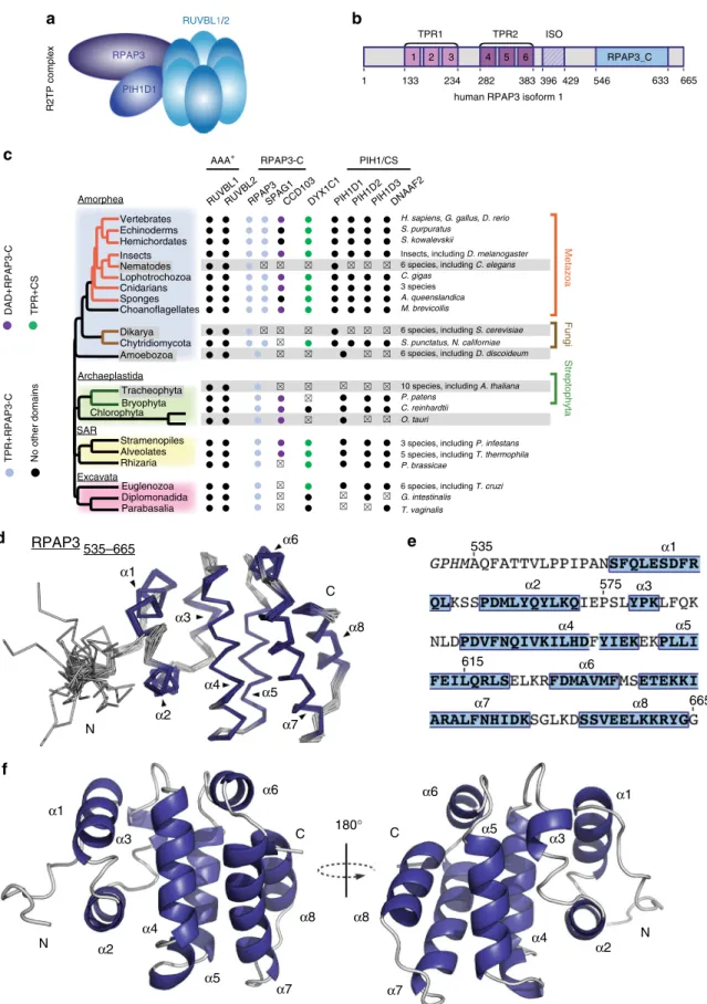
The RPAP3 C-ter domain co-evolved with PIH-domain
pro-teins. Although extensive structural studies have been performed
with R2TP proteins
2, the structure and function of RPAP3-Cter is
unknown. This domain is absent in the S. cerevisiae homolog of
RPAP3, and we thus performed an evolutionary analysis (Fig.
1
c).
Besides RPAP3, the human genome encodes two proteins
har-boring a RPAP3-Cter domain: SPAG1, which contains three TPR
domains, and CCDC103, which has a N-terminal dynein
attachment domain. Proteins with both an RPAP3-Cter and a
TPR domain occur in all eukaryotic lineages (Fig.
1
c; blue dots in
the RPAP3_C column). A single such protein was likely present
at the root of Eukaryotes, and a duplication event having
occurred between Amoebozoa and Opisthokonta (Fungi and
Metazoa) generated RPAP3 and SPAG1. The human genome
encodes four proteins with PIH domains: PIH1D1-3 and
DNAAF2 (also known as Kintoun). DNAAF2 associates with
DYX1C1
29, a CS- and TPR- containing protein that we included
in our analysis. Most eukaryotic lineages encode three
PIH-proteins and one DYXC1 ortholog. These PIH-proteins are thus of
ancient origin, as previously noted
30. Interestingly, the
duplica-tion of the RPAP3 ancestor is mimicked by a similar
con-temporary duplication of the ancestor of PIH1D1, which
generated PIH1D1 and PIH1D2. Note that neither RUVBL1/2
nor other HSP90 co-chaperone were duplicated (Fig.
1
c).
Alto-gether, this analysis indicates that the ancestral form of RPAP3
had both TPR and RPAP3-Cter domains, suggesting an
impor-tant function for this domain. This protein further co-evolved
with the ancestor of PIH1D1, pointing to a link with R2TP.
The NMR structure of RPAP3-Cter reveals a helical 3D fold.
To get more information on RPAP3-Cter, we performed
struc-tural studies. We expressed fragments 535–665 and 547–665 of
human RPAP3. Only the largest domain, i.e., RPAP3
535-665, was
soluble after expression and purification from E. coli
(Supple-mentary Fig.
1
A, B). Using NMR, the intensity and distribution of
peaks in the
1H-
15N HSQC spectrum revealed a properly folded
domain (Supplementary Fig.
1
C). We obtained a well-resolved set
of 20 water-refined structures (Table
1
), revealing that this
domain is composed of 8
α-helices that pack together to form a
globular object (Fig.
1
d–f). Interestingly, the residues 541–548
form a loop that protects several hydrophobic amino-acids from
solvent, including Leu542, Pro543, Ile545, and Pro546
(Supple-mentary Fig.
1
D). This loop contributes to the solubility of
RPAP3-Cter since its truncation in RPAP3
547-665leads to protein
aggregation. We submitted our structure to the DALI server to
search for potential structural homologs. No strong candidate
could be identified using a top DALI Z-score below 8, with a
mean RMSD and identity percentage of 3.6 Å and 9.7%,
respec-tively. We concluded that RPAP3-Cter adopts a 3D fold, linked to
an uncharacterized biological function.
RPAP3-Cter associates with RUVBL1/2 and some R2TP
cli-ents. To get insights into the function of RPAP3-Cter, we
char-acterized its partners by performing a proteomic analysis in
human cells. We fused this domain to GFP and stably expressed it
in HeLa cells using site-specific Flp-In integration. Following
differential labeling of GFP-RPAP3-Cter and control cells with
isotopic amino-acids (SILAC), whole cell extracts were
immuno-precipitated (IP) with anti-GFP antibodies and pellets were
sub-jected to quantitative mass-spectrometry analysis (Fig.
2
a). About
20 proteins associated with RPAP3-Cter with high abundance
R2TP complex
RPAP3
PIH1D1
RUVBL1/2
human RPAP3 isoform 1
TPR1 TPR2 ISO 1 1 133 234 282
RPAP3
N N N 535–665 665 575 615 535 C C 180° C α1 α1 α1 α1 α3 α3 α3 α3 α6 α6 α6 α6 α8 α8 α8 α8 α5 α5 α5 α5 α4 α4 α4 α4 α2 α2 α2 α2 α7 α7 α7 α7 383 396 429 546 633 665 2 3 4 5H. sapiens, G. gallus, D. rerio S. purpuratus
S. kowalevskii
Insects, including D. melanogaster 6 species, including C. elegans C. gigas
3 species A. queenslandica M. brevicollis
6 species, including S. cerevisiae S. punctatus, N. californiae 6 species, including D. discoideum
10 species, including A. thaliana P. patens
C. reinhardtii O. tauri
3 species, including P. infestans 5 species, including T. thermophila P. brassicae
6 species, including T. cruzi G. intestinalis T. vaginalis Amorphea Vertebrates Echinoderms Hemichordates Insects Nematodes Lophotrochozoa Cnidarians Sponges Choanoflagellates Chytridiomycota Archaeplastida Tracheophyta Bryophyta SAR Stramenopiles Alveolates Rhizaria Excavata Euglenozoa Diplomonadida Parabasalia Dikarya Chlorophyta Amoebozoa Streptophyta Fungi Metazoa
RPAP3SPAG1CCD103DYX1C1PIH1D1PIH1D2PIH1D3DNAAF2
RPAP3-C PIH1/CS RUVBL1RUVBL2 AAA+ TPR+RPAP3-C DAD+RPAP3-C No other domains TPR+CS 6 RPAP3_C
a
b
c
d
e
f
Fig. 1 Solution structure of the C-terminal domain of human RPAP3 a Schematic representation of the human R2TP complex. b Domain architecture of RPAP3 (numbering corresponds to amino-acids of isoform 1).c Conservation of RPAP3 and PIH repertoires across Eukaryotes. Members in which TPR (Pfam: 13414) or Dynein attachment (Pfam: 15867) domains are associated to the canonical RPAP3_C are colored as indicated at the left. Clades or species that have lostflagella or in which cilia are not motile are in gray background. Members that were not found are indicated by x. d Backbone view of the superposition of the best 20 NMR structures of human RPAP3-Cter, withhelices indicated in violet. e Sequence of RPAP3-Cter, with corresponding α-helices.f Backbone orthogonal views (180 °C) of RPAP3-Cter structure in ribbon representation with correspondingα-helices
(intensity) and specificity (SILAC ratio). The top hits were
RUVBL1/2, while the others belonged to several classes: (i)
known R2TP clients (SHQ1 and NOP58 for snoRNPs, PRPF8,
and other U5 proteins for snRNAs, POLR2A for RNA
poly-merase II); (ii) known R2TP cofactors (ZNHIT2) and/or
RUVBL1/2 partners (C12ORF45, C2ORF44, DPCD)
6,17,21; (iii)
PAQosome subunit (PFDN2)
2; and (iv) chaperones (HSP70
isoforms and its regulator BAG2). Overall, these data suggest that
RPAP3-Cter may play an important role in interacting with
RUVBL1/2 and some R2TP clients.
RPAP3-Cter directly binds RUVBL1/2. We then performed
pairwise yeast two-hybrid assays (Y2H) between RPAP3-Cter and
its putative partners (Supplementary Fig.
2
A). We detected a
positive signal with RUVBL2, but not with the other tested
pro-teins. To determine whether this interaction is direct, we used
recombinant proteins for in vitro binding assays. RUVBL1/2 were
co-expressed in E. coli and they purified predominantly as a
dodecamer (see below). We then assessed the binding to
RPAP3-Cter using NMR spectroscopy. We took advantage of the
13C-labeling to monitor the effect of RUVBL1/2 on the methyl groups
of RPAP3-Cter. Indeed, methyl groups are able to provide strong
signals even at low protein concentrations and/or basic pHs
31.
The NMR signal of
13C-labeled RPAP3-Cter decreased
dramati-cally in presence of RUVBL1/2 hetero-multimers (Fig.
2
b), while
only minor effects were seen with RUVBL1 or RUVBL2 alone
(Supplementary Fig.
2
B). Next, we used SPR and observed robust
interactions between RPAP3-Cter and RUVBL1/2
hetero-multimers (Fig.
2
c), with a K
Dof 4.2 nM in a 1:1 interaction
model calculation (Table
2
). These data demonstrated that
RPAP3-Cter binds heteromeric forms of RUVBL1/2.
RUVBL1/2 cycle between hexamers and
hetero-dodecamers
20, and we thus addressed the stoichiometry of the
interaction. First, we co-expressed RPAP3-Cter with RUVBL1/2
in E. coli and analyzed the complex by gel-filtration (Fig.
2
d, e).
The eluting volumes of RUVBL1/2 alone and
RUVBL1/2:RPAP3-Cter corresponded to molecular masses of dodecameric and
hexameric forms of RUVBL1/2, respectively. Next, we performed
mass spectrometry analysis under non-denaturing conditions
(native MS), of either RUVBL1/2 alone, or RUVBL1/2
co-purified with recombinant RPAP3-Cter (Fig.
2
f). In the free state,
RUVBL1/2 formed hetero-hexameric and hetero-dodecameric
complexes, but hexameric forms became largely dominant upon
RPAP3-Cter binding. Note also that one hexamer can bind
multiple Cter domains. These data indicate that
RPAP3-Cter binds predominantly RUVBL1/2 hetero-hexamers.
Next, we analyzed the binding of RPAP3-Cter to RUVBL
mutants altered in their ATPase cycle. For this, we turned to
LUMIER interaction experiments. In this quantitative co-IP
assay, the prey and bait proteins are respectively fused to Renilla
luciferase (RL) and FLAG-tagged Firefly luciferase
(3xFLAG-FFL). Following transient expression in human HEK293 cells, the
bait is IP’ed with anti-FLAG antibodies, or without antibodies as
control. The RL and FFL luciferase activities are then measured in
the input and pellet, and the co-IP efficiency is expressed as the
percent of prey co-immuno-precipitated, relative to that of the
bait, providing a direct measurement of binding efficiencies
(Supplementary Fig.
2
D). In these experiments, we used canonical
mutations of AAA
+ ATPases
32, expected to prevent nucleotide
binding (K to M mutant in the Walker A domain), nucleotide
hydrolysis (E to Q in the Walker B-domain), or coupling between
adjacent monomers (R to E in the Arg
finger). The effect of
several of these mutations was previously validated using the
Chaetomium
thermophilum
orthologs
of
RUVBL1
and
RUVBL2
20. Interestingly, while the K-M and R-E mutations
had little effects on the association of either RUVBL1 or RUVBL2
with RPAP3-Cter, the E-Q mutations diminished binding
(Supplementary Fig.
2
D). Taken together, these data indicated
that RPAP3-Cter made a direct, high-affinity interaction with
hexameric RUVBL1/2. This interaction did not require nucleotide
and was weakened in mutants deficient in ATP hydrolysis.
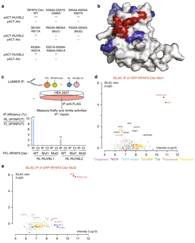
RPAP3-Cter binds R2TP clients through RUVBL1/2. Our
proteomic analysis of RPAP3-Cter showed that it binds not only
RUVBL1/2, but also a range of R2TP clients. To test whether
these latter interactions depend on RUVBL1/2, we generated a
series of RPAP3-Cter mutants. Structure-guided analysis selected
solvent-exposed residues potentially involved in RUVBL1/2
interaction (Supplementary Fig.
3
A, B). Alanine mutants were
screened by Y2H assays, and two of them indeed lost interaction
with RUVBL2 (Fig.
3
a): R623A-M626A (Mut1) and
F630A-S632A (Mut2). Both mutants localized similar to wild-type
RPAP3-Cter (Supplementary Fig.
3
C), suggesting no major
alteration. The mutated residues are conserved across evolution
(Supplementary Fig.
3
D). They were located next to each other in
the 3D structure of RPAP3-Cter (Fig.
3
b), suggesting that they
may constitute a conserved binding site for RUVBL1/2. LUMIER
interaction assays confirmed the Y2H data, as both RPAP3-Cter
Mut1 and Mut2 lost interaction with RUVBL1 and RUVBL2
(Fig.
3
c). Next, we performed SILAC quantitative proteomic
analyses of the RPAP3-Cter mutants. Remarkably, both mutants
lost all of the RPAP3-Cter partners identified previously (Fig.
3
d,
e), with the exception of SHQ1 that remained weakly bound to
RPAP3-Cter Mut1. Taken together, these data suggest that
RUVBL1/2 are required for the association of RPAP3-Cter with
its other partners.
Table 1 NMR and re
finement statistics for top-20
RPAP3
535-665structures
RPAP3535-665
NMR distance and dihedral constraints Distance constraints Total NOE 3707 Intra-residue 855 Inter-residue 2852 Sequential (|i – j| = 1) 849 Medium-range (|i – j| ≤ 4) 1033 Long-range (|i – j| ≥ 5) 970 Total dihedral angle restraints 230
ϕ 110
ψ 120
Structure statistics after AMBER refinement Violations
Distance constraints (Å) 0.10 ± 0.03 Dihedral angle constraints (°) 6.70 ± 1.94 Max. dihedral angle violation (°) 9.99 Max. distance constraint violation (Å) 0.14 Violation occurrences
Distances constraints ( > 0.2 Å) 0 Dihedral angle constraints ( > 5°) 1.30 ± 1.26 R.m.s. deviations from idealized geometry
Bond lengths (Å) 0.013 ± 0.00
Bond angles (°) 1.85 ± 0.05
R.m.s. deviation to best structurea(Å)
All heavy atoms 1.33 ± 0.29
All backbone atoms 0.98 ± 0.38
Heavy atoms in secondary structures 0.94 ± 0.07 Backbone atoms in secondary structures 0.37 ± 0.05
Identification of RPAP3-like/PIH-like complexes. As described
above, the human genome encodes two other proteins bearing
RPAP3-Cter-like domains, SPAG1 and CCDC103, and three
others containing PIH domains, PIH1D2, PIH1D3 and DNAAF2
(Fig.
1
c, Fig.
4
a). This suggested that these proteins could form
R2TP-like complexes, by associating to each other and with
RUVBL1/2 through their RPAP3-Cter-like domains. To test this
possibility, we
first performed systematic pairwise LUMIER co-IP
assays between RPAP3-like and PIH-like proteins (Fig.
4
b). In
these tests, we also added DYX1C1 because this protein associates
with DNAAF2
29and bears some structural features of RPAP3
and PIH1D1 (e.g., TPR and CS domains), although it lacks PIH
and RPAP3-Cter domains. We also included the known splicing
isoforms of DYX1C1 and RPAP3, including the RPAP3 isoform 2
that does not interact with PIH1D1
33. Finally, we added HSP70,
HSP90, and STIP1, a factor promoting the transfer of clients from
HSP70 to HSP90
34. These proteins were fused at their N-termini
to Renilla luciferase or to 3xFLAG-Firefly luciferase. The plasmids
were transiently transfected in HEK293 cells, and LUMIER IPs
were performed (Fig.
4
b). As above, the interaction strength was
Binding and dissociation of RUVBL1/2 on immobilized RPAP3-Cter 5 10 11 12 Intensity (Log10) C2orf44 RUVBL1 RPAP3-Cter RUVBL2 DPCD SHQ1 C12orf45 SNRNP200 EFTUD2 NOP58 BAG2 PRPF8 ZNHIT2 HSPA8 HSPA7 HSPA1A PFDN2 HSPA9 HSPA5 SILAC ratio (Log2) 0 2 4 6 8 POLR2A
Chaperone R2TP R2TP-related SnoRNP U5 Polymerase Ribosome
GFP SILAC IP OF GFP-RPAP3-Cter RPAP3-Cter RPAP3-Cter + RUVBL1/2 2 1 0 NMR signal intensity
Superose 6 elution profile
Volume (ml) RUVBL1/RUVBL2
RUVBL1/RUVBL2/RPAP3-Cter
Absorbance (mAU) Absorbance (mAU)
7.5 6.5 5.5 4.5
Response units (RU)
3.5 2.5 1.5 0.5 –0.5 –1.5 16 14 12 10 8 6 4 2 0 0
RUVBL1/2 hexamer region 313113 ± 26 Da 313 113 ± 26 Da RUVBL1/2 hexamer 329 145 ± 26 Da 1:1 RUVBL1/2 hexamer : RPAP3-Cter 344 740 ± 20 Da 1:2 RUVBL1/2 hexamer : RPAP3-Cter 8000 8250 8500 8750 9000 9250 9500 8000 8250 8500 8750 9000 9250 9500 m/z m/z m/z MS spectra 38+ 37+ 36+ 39+ 38+ 37+ 36+ 38+ 37+ 36+ 39+ 38+ 37+ 39+
RUVBL1/2 hexamer region
RUVBL1/2 hexamer
RUVBL1/2 + RPAP3-Cter
4000 5000 6000 7000 8000 9000 10,000 11,000 12,000 13,000 14,000 15,000 16,000 17,000 18,000
m/z MS spectra
RUVBL1/2
RUVBL1/2 dodecamer region 626 305 ± 8 Da
RUVBL1/2 dodecamer region
10 20 30 40 50 60 70 80 90 100 110 400 kDa 250 150 100 75 50 37 25 20 15 10 1 2 VE = 74.6 VE = 80.9 350 300 250 200 150 100 50 0 –100 100 300 500 700 900 75.2 nM 37.6 nM 18.8 nM 9.4 nM 4.7 nM 2.4 nM 1.2 nM 0.6 nM 0.3 nM 0.15 nM Time (s) 6 7 8 9 1 H (ppm)
a
b
c
d
e
f
expressed as the fraction of co-IP’ed prey protein normalized to
that of the bait. Negative controls included four unrelated
pro-teins, which were tested against all the RPAP3-like and PIH-like
proteins (Supplementary Fig.
4
A). This allowed us to rigorously
identify specific interactions (see Methods). These experiments
revealed that all the TPR-containing proteins interacted with
HSP70, HSP90 and STIP1, although the co-IP efficiency was low
(Fig.
4
b and S
4
B). This could be due to a low affinity or to the fact
that HSPs have many partners that potentially compete with baits
in extracts. Most interestingly, we uncovered three strong
asso-ciations between RPAP3-like and PIH-like proteins (30–40% of
co-IP efficiencies): RPAP3-iso-1 with PIH1D1, DYX1C1-iso-a
with DNAAF2, and SPAG1 with PIH1D2. Two other strong
interactions were found, between DYX1C1-iso-c and PIH1D3,
and between DNAAF2 and SPAG1, although the IP efficiencies
were 5–10 times less than for the previous couples (4–8% of co-IP
efficiencies). Finally, a number of weak but significant
interac-tions were also found (Fig.
4
b and S
4
C), suggesting that
addi-tional PIH1D1-like/RPAP3-like pairs may also form, albeit at low
efficiencies.
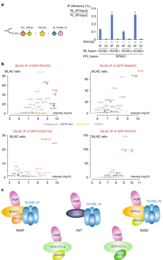
PIH1D2 facilitates the binding of SPAG1 to RUVBL1/2. We
then tested whether SPAG1 and CCDC103 would bind RUVBL1/
2, also using LUMIER assays (Fig.
4
c). SPAG1, RPAP3-iso-1 and
iso-2 interacted with both RUVBL proteins, while CCDC103 did
not. Surprisingly however, the isolated C-terminal domain of
SPAG1 failed to interact with RUVBL1 or RUVBL2. The
C-terminal domains of RPAP3, SPAG1, and CCDC103 have an
overall identity of only 25%, and the sequence differences may
modulate their affinity for RUVBL1/2, from a high affinity
binding for RPAP3-Cter, to a low or no binding for SPAG1-Cter
and CCDC103-Cter. Indeed, the amino-acids required for the
binding of RPAP3-Cter to RUVBL1/2 are not strictly conserved
in SPAG1 and CCDC103 (Supplementary Fig.
3
B). Since the
full-length SPAG1 protein bound better to RUVBL1/2 than the
iso-lated SPAG1-Cter domain, we further tested whether its PIH1D2
partner would enhance binding. We repeated the LUMIER assay
in presence of co-expressed, untagged PIH1D2 (Fig.
5
a). Indeed,
PIH1D2 increased the binding of SPAG1 to RUVBL1 and
RUVBL2 by nearly 3-fold, achieving a binding efficiency similar
to that of full-length RPAP3. Taken together, these data indicate
that SPAG1 binds RUVBL1/2 and that the binding determinants
likely include several regions within SPAG1 and PIH1D2.
This result prompted us to test whether the PIH-like proteins
would interact with RUVBL1/2. LUMIER assays indicated that
significant interactions could be detected (Supplementary
Fig.
4
D), but at a low level (5–20 fold weaker than with RPAP3).
Human cells contain several complexes related to R2TP. The
LUMIER assays indicate that SPAG1 can interact with PIH1D2
and RUVBL1/2, possibly forming a complex similar to R2TP. To
obtain direct evidence that this complex exists, and to determine
whether additional R2TP-like complexes may form, we turned to
quantitative SILAC proteomics. We fused GFP to PIH1D2,
PIH1D3, DNAAF2, and CCDC103, stably expressed the fusions
in HeLa cells and used them as baits in proteomic experiments
(Fig.
5
b and Supplementary Data
1
). We did not
find any partner
for GFP-CCDC103. For GFP-PIH1D3, we found a strong
asso-ciation with LRIF1, a nuclear factor enriched in testis. Only
minute amounts of RUVBL1/2 were detected, in agreement with
the weak interaction observed in the LUMIER assays.
DYX1C1-iso-c was also not detected, possibly because of its low abundance
in HeLa cells (Gtex dataset
35). For GFP-DNAAF2, we found a
strong association with DYX1C1-iso-a, and again a weak binding
to RUVBL1 and RUVBL2 that agreed with the LUMIER assays
(Fig.
4
b and S
4
C). In the GFP-PIH1D2 IP, the three most
pro-minent proteins were SPAG1, RUVBL1 and RUVBL2, all found
with both a high specificity and abundance (Fig.
5
b). HSP70 and
its regulator BAG2 were also significantly enriched. This
indi-cated the existence of an R2TP-like complex containing PIH1D2,
SPAG1, RUVBL1/2, which associated with HSP70 chaperones.
The other GFP-PIH1D2 partners found in this experiment were
involved in a variety of processes ranging from metabolism to
RNA processing, and may represent clients of this chaperone
system. No prefoldin nor prefoldin-like proteins were detected,
Fig. 2 RPAP3-Cter interacts with RUVBL1/2 hexamers. a SILAC proteomic analysis of RPAP3-Cter. The graph depicts the proteins identified in anti-GFP immuno-precipitates of HeLa cells expressing GFP-RPAP3-Cter. Each dot is a protein and the color code is indicated below the graph.X-axis: protein abundance (Log10 of signal intensity); y-axis: enrichment over a control IP (Log2 of SILAC ratio).b NMR interaction analysis of RPAP3-Cter with recombinant RUVBL1/2 complex. The graph depicts 1D NMR METHYL-SOFAST-HMQC spectra in the methyl region of13C-labeled RPAP3-Cter alone (top lane) or mixed with recombinant RUVBL1/2 complex (bottom lane). Intensity of the NMR signal (arbitrary units,Y-axis) is plotted against the1H chemical shift (in ppm,X-axis). c SPR binding assays of RPAP3-Cter with RUVBL1/2. The graph depicts the response upon injecting the RUVBL1/2 complex (t = 0 s), or upon washing (t= 300 s), on immobilized RPAP3-Cter. X-axis: time (s); Y-axis: response (arbitrary units). These data have been obtained with the same batch of RUVBL1/2 complex as in the control experiment (Fig. S2E, F).d Chromatographic analysis of the RUVBL complexes. The graph depicts the chromatograms of purified RUVBL1–RUVBL2 (dashed gray, left Y axis) or RUVBL1-RUVBL2-RPAP3-Cter (black line, right Y axis), on a Superose 6 16/70 XK. X-axis: elution volume; Y-axis: absorbance. e Electrophoresis of the purified RUVBL1–RUVBL2–RPAP3–Cter complex. The gel shows the peak fraction of the complex eluted from the column (black line in d), with a purity estimated to ~95 %. Lane 1: Precision Plus Protein Unstained Standards (Biorad); Lane 2: denatured RUVBL1–RUVBL2–RPAP3–Cter complex. Black and white arrows: RUVBL1 and RUVBL2 (52 and 53 kDa, respectively); gray arrow: RPAP3-Cter (15 kDa).f Native mass spectrometry analysis of recombinant RUVBL complexes. The upper mass spectrum presents the purified RUVBL1/2 complex. The bottom mass spectrum presents the same complex after addition of RPAP3-Cter. Y-axis: signal intensity; X-axis: m/z. Insets: zoom over the 8000–9000 m/z region. Schematics depict the complex observed. Blue: RUVBL proteins; red: RPAP3-Cter
Table 2 RUVBL1/2:RPAP3
–Cter affinity and kinetic interaction parameters determined by SPR
His_RUVBL1_Flag_RUVBL2 interaction with immobilized RPAP3-CterKD(M) kd(s−1) ka(M−1s−1) na
4.18×10⁻⁹± 1.96×10⁻⁹ 1.12×10⁻³± 2.58×10⁻⁴ 2.97×10⁵± 6.35×10⁴ 3
but we noted that an interaction of SPAG1 with WDR92 has been
previously described
21.
Collectively, the LUMIER and proteomic data thus defines
three types of complexes related to R2TP (Fig.
5
c). A
first
complex is composed of RPAP3-iso-2 in association with
RUVBL1/2. This complex is identical to the canonical R2TP
except that it lacks PIH1D1, and was thus named R2T. A second
complex comprises SPAG1, PIH1D2 and RUVBL1/2. This
complex shares an organization similar to R2TP and was named
R2SP. Another related complex could form with SPAG1,
HEK 293T RPAP3-Cter HEK 293T FLAG FFL IP anti-FLAG Measure firefly and renilla activities
IP / inputs + RL RUVBL1/2 8 6 4 2 0 RL-RUVBL1 RL-RUVBL2 IP WT IP efficiency (%): RL (IP/INPUT) FL (IP/INPUT) LUMIER IP: WT K584A-D587A-RPAP3 Cter-D589A E604A-K605A-K607A *** ** ** – – – Q616A-R617A R623A-M626A (Mut1) F630A-S632A (Mut2) ** – – – – – K636A-K637A E657A-E658A-K660A-K661A pACT-RUVBL2 pACT-Alix pACT-RUVBL2 pACT-Alix pACT-RUVBL2 pACT-Alix ** ** – – 5 10 11 12 MVP ACYP2 IFI16 USP9X DDX24 PRMT1 SERPINB1 0 2 4 6 Intensity (Log10) SILAC ratio (Log2) RPAP3-Cter SHQ1 RPAP2 FFL-RPAP3-Cter :
SILAC IP of GFP-RPAP3-Cter Mut1
Chaperone R2TP R2TP-related SnoRNP U5 Polymerase Ribosome
GFP RUVBL1 RPAP3_Cter CSRP2 IAH1 CAPNS1 FUS HSP70 GFP SILAC IP of GFP-RPAP3-Cter Mut2
Intensity (Log10) RUVBL2 HSPA7 0 2 4 6 SILAC ratio (Log2) ** ** ** 10 α8 α3 α6 α4 α1 N R623 M626 F630 S632 Ct IP Ct IP Ct IP Ct IP Ct IP Ct
Mut1 Mut2 WT Mut1 Mut2 6 7 8 9
5 6 7 8 9 10 11 12
a
b
c
d
e
Fig. 3 The RPAP3 C-terminal domain interacts with R2TP clients via RUVBL1/2 multimers. a Yeast two-hybrid analysis of interactions between RUVBL2 and RPAP3-Cter mutants. Alix is a negative control. ***: strong interaction; **: medium; *: weak;−: no interaction. b Molecular surface representation of RPAP3-Cter structure by specifying the location of the mutants that lost interaction with RUVBL1/2.c LUMIER assay showing the in vivo interaction between RPAP3-Cter and RUVBL1/2 mutant proteins. Top panel: schematic representation of the assay. Bottom panel: graph plotting the IP efficiency of the indicated proteins. The values are the IP efficiencies of the co-precipitation of the RL fusion proteins (IP/Input), normalized by the IP/Input values obtained with the anti-FLAG IP of the 3xFLAG-FFL fusion protein. Error bars: standard deviation. Stars: values significantly greater than six-times the mean value obtained in the control IPs without anti-FLAG antibody (Ct). **p-value < 0.001 (Z-test). d,e SILAC proteomic analysis of the partners of RPAP3-Cter-Mut1 and RPAP3-Cter-Mut2, respectively. Legend as in Fig.2a
DNAAF2 and RUVBL1/2 (i.e., an R2SD complex). However, this
complex remains hypothetical since it was detected in LUMIER
but not in proteomic experiments. Finally, we observed two
heterodimers containing a PIH-like protein associated with a
DYX1C1
isoform:
DNAAF2/DYX1C1-iso-a
and
PIH1D3/
DYX1C1-iso-c. These interactions were previously reported
29,36,
but the isoform specificity of DYX1C1 was not known.
The occurrence and composition of these complexes is
corroborated by our evolutionary analyses (Fig.
1
c). The parallel
duplication leading to RPAP3 and SPAG1 on one side, and PIH1D1
and PIH1D2 on the other, is consistent with their respective
incorporation into R2TP and R2SP. Similarly, some species contain
an RPAP3 ortholog but lack a PIH1D1/PIH1D2 gene, thus
mirroring the existence of R2T in human cells. The frequent
co-occurrence of DYX1C1 with either DNAAF2 or PIH1D3 is also
consistent with the association observed for the human proteins.
R2TP-like components are enriched in testis. To gain insights
into the function of these R2TP-related complexes,
fluorescent
microscopy was performed with the stable cells expressing the
GFP-tagged proteins. These proteins localized to different cellular
areas, suggesting specialized functions (Supplementary Fig. 5).
Interestingly, DYX1C1-iso-c was nuclear and concentrated in
punctate structures, as did its partner PIH1D3. It is also worth
noting that RPAP3-iso-2 was mainly nuclear while RPAP3-iso-1
was mainly cytoplasmic. Thus, alternative splicing determines not
only the partners but also the localization of these proteins.
3xFlag-FFL-X
RL-Y
IP efficiency : RL (IP/input) / FFL (IP/input) (%)
DYX1C1-iso-a DYX1C1-iso-b DYX1C1-iso-c RPAP3-iso-1 RPAP3-iso-2 SPAG1 CCDC103
HSP70 0.4 0.3 1.2 3.0 1.8 6.7 1.1 HSP90 0.1 0.0 0.3 0.2 0.1 0.7 0.3 STIP1 1.0 0.3 1.9 1.9 1.3 4.8 1.7 PIH1D1 0.0 0.0 0.2 41.0 0.7 0.3 0.1 PIH1D2 0.1 0.0 0.3 0.2 0.2 31.3 0.1 PIH1D3 0.8 0.1 4.2 0.3 0.2 0.8 0.2 DNAAF2 42.1 0.3 0.1 0.3 0.8 7.7 0.1 RPAP3-Like FLAG FFL + RUVBL1/2 RL HEK 293T IP anti-Flag Measure firefly and renilla activities
IP / inputs IP SPAG1-Cter SPAG1 IP efficiency (%): RL (IP/input) FL (IP/input)
RPAP3-Iso1 RPAP3-Iso2 DYX1C1-c CCDC103 CCDC103-Cter
0 0.5 1 1.5 2 RL-RUVBL1 RL-RUVBL2 ** ** ** ** ** ** RPAP3 CCDC103 PIH1D1 PIH1D2 PIH1D3 DNAAF2 CC CC CS domain CS domain CS domain CS domain PIH domain PIH domain PIH domain PIH 1 1 1 1 1 10 182 254 349 837 111 208 214 6 8 145 225 315 159 199 286 290 32 96 189 208 242 TPR1 TPR2 TPR1 TPR1 TPR1 TPR1 TPR1 TPR2 TPR3 TPR2 DYX1C1 SPAG1 ISO RPAP3_C RPAP3_C RPAP3_C RPAP3_C 6 5 4 3 2 1 6 5 4 3 2 1 3 2 1 2 1 1 iso 1 iso 2 iso a iso b iso c CS domain CS domain CS domain 1 1 133 133 1 3 1 3 87 87 290 290 1 1 209 309 445 554 623 724 802 891 926 3 87 290 399 357 376 381 323 420 234 234 282 282 383 383 512 599 631 396 429 546 633 665 6 5 4 7 8 9 3 2 1
a
b
c
Ct IP Ct IP Ct IP Ct IP Ct IP Ct IP CtFig. 4 RPAP3-like and PIH1-like proteins interact with each other. a Architecture of the human proteins containing a RPAP3-Cterminal domain (RPAP3-C), or a PIH domain (PIH). Coiled-coil (CC), CHORD-containing proteins and SGT1 domain (CS) and TPR domains (TPR) are also indicated. Different splicing isoforms of RPAP3 and DYX1C1 are shown, with their variable domains in hatched violet (RPAP3), or yellow (DYX1C1).b Summary of pairwise LUMIER interaction assays between the indicated proteins. The values are the efficiencies of the co-precipitation of the RL fusion proteins (IP/Input), expressed in percent of the efficiencies obtained with the 3xFLAG-FFL fusions. p-values are shown in Supplementary Fig.4C.c LUMIER interaction assays between RL-RUVBL1/2 and RPAP3-like proteins tagged with 3xFLAG-FL. Legend as in Fig.3c. Stars: values significantly greater than six-times the mean value obtained in the control IP (Ct). **p-value < 0.001 (Z-test)
Next, we examined existing data to determine in which tissues
these factors are expressed (Gtex dataset
35; Supplementary
Fig.
6
A). The expression patterns of RUVBL1, RPAP3, and
PIH1D1 looked similar to each other, with a rather ubiquitous
expression. DNAAF2 and SPAG1 were also broadly expressed,
but with a moderate enrichment in testis. Interestingly, PIH1D2,
PIH1D3, and RUVBL2 were highly expressed in testis, suggesting
an important role in this organ.
SPAG1 FLAG FFL + RUVBL1/2 RL RUVBL1 RUVBL1 IP Ct IP Ct IP Ct IP Ct SPAG1 – – + + – – + + PIH1D2:
RL fusion: RUVBL2 RUVBL2
FFL fusion: + PIH1D2 0.3 0.4 0.2 0.1 0 RL (IP/input) FL (IP/input) HSP SPAG1 R2SP RUVBL1/2 PIH1D2 RPAP3-2 R2T HSP DYX1C1-a HSP RUVBL1/2 R2SD DYX1C1-c PIH1D3 HSP SPAG1 RUVBL1/2 HSP DNAAF2 DNAAF2 ACTB MYH9 FLNC GFP HSP70 SPAG1 PIH1D2 GAPDH FLNA RUVBL2 EEF1A2 MAGE2 ENO1 PKM SILAC ratio SILAC IP of GFP-PIH1D2 Intensity (log10) 0 20 40 60 80 ZBTB1 RUVBL1 BAG2
Chaperone R2TP-like Ribosome Others
SILAC IP of GFP-DNAAF2 Intensity (log10) SILAC ratio DNAAF2 RTF1 PPM1G EIF2AK1 DYX1C1-a RAD18 CFDP1UBE3DPHGDH C11orf58 TCOF1 CDK11A GFP RUVBL1 RUVBL2 0 20 40 60 6 7 8 9 10 6 7 8 9 10 5 HAX1 HSP70 NUP93 SILAC IP of GFP-CCDC103 Intensity (log10) SILAC ratio 0 20 30 10 CCDC103 HSC70 HSP70 HSP90 DNAJA1 DNAJB6 RUVBL2 VBP1 BAG6KPNA2 SP2 SILAC IP of GFP-PIH1D3 Intensity (log10) SILAC ratio 0 50 100 150 GFP PIH1D3 RUVBL1 RUVBL2 LRIF1 GFP IP efficiency (%): 6 7 8 9 10 5 5 6 7 8 9 10 11
a
b
c
Fig. 5 Identification of R2TP-like complexes. a LUMIER interaction assays between SPAG1 and RUVBL1/2. Legend as in Fig.3c excepted that an untagged PIH1D2 was co-expressed (lanes+), or SMD1 as control (lanes−). b SILAC proteomic analysis of the partners of PIH1D2, DNAAF2, GFP-CCDC103, and GFP-PIH1D3, performed in HeLa cells. Legend as in Fig.2a. The color code is indicated between the graphs.c Models of possible R2TP-related complexes
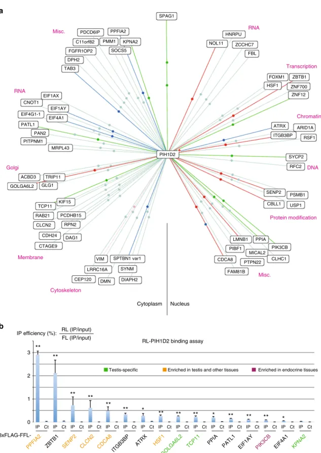
PIH1D2 has both ubiquitous and testis-enriched partners. To
characterize the function of R2SP, we
first searched for partners
by performing yeast two-hybrid screens using PIH1D2 as bait.
Given its expression pattern, we screened two human libraries,
from lung cancer cell lines and from testis (Fig.
6
a). The screens
revealed a total of >60 potential partners (46 from the lung and 32
from testis; Fig.
6
a and Supplementary Data
2
). Nine proteins
were found in both libraries, including SPAG1, indicating the
high quality of the screens. These PIH1D2 partners are involved
in a variety of functions, ranging from DNA metabolism,
tran-scription and RNA processing, and up to cytoskeletal
organiza-tion, membrane-related processes and trafficking.
Next, we selected 16 proteins to test LUMIER assays and could
validate most of them (Fig.
6
b). Taken together, these data
demonstrated that PIH1D2 has a range of partners involved in a
variety of processes. Some partners were enriched in testis and
others were ubiquitous (Gtex dataset
35, Fig.
6
b).
R2SP facilitates quaternary protein folding. Next, we tested
whether R2SP has a chaperone activity toward its partners. Since
PIH1D2 is poorly expressed in HeLa cells, in contrast to the other
components of R2SP (Supplementary Fig.
6
B), we generated
HeLa cells stably expressing GFP-PIH1D2, and thus having a
fully assembled R2SP complex (see Fig.
5
b). To measure its
chaperone activity, we fused to Firefly luciferase the PIH1D2
partners previously identified, and we transiently transfected
them in HeLa-PIH1D2 and parental HeLa cells. Remarkably,
five
fusions were significantly more expressed in presence of
GFP-PIH1D2, reaching a threefold increase in one case (Fig.
7
a). These
were PPFIA2 (liprin-α2), ZBTB1, TCP11, PATL1 and PIK3CB. In
contrast, the expression of a broad series of control proteins,
including unrelated factors (Alix, FFL) or known R2TP-substrates
(EFTUD2, PRPF31, NOP58), was identical in both cell lines
(Fig.
7
a). Next, we repeated the experiments at 32 °C, the optimal
temperature of testis where PIH1D2 is highly expressed. The
effects of R2SP on proteins levels were generally more important
at this temperature. Altogether, this suggested that R2SP
enhanced expression of some of its partners and had a stronger
effect at the testis temperature.
Since R2TP is involved in the assembly of its target complexes
2,
we hypothesized that R2SP could do the same. To test this, we
focused on liprin-α2 (PPFIA2). Indeed, this is the strongest
binder of PIH1D2 and it is also the most sensitive to the presence
of PIH1D2. In addition, liprins are important scaffolding
molecules that bring together a diverse set of factors in order to
control cell adhesion, cell migration, and organization of the
active synaptic zone
37. Liprins possess a long coiled-coil domain
at their N-terminus, followed by a linker and three Sterile Alpha
Motif domains (SAM). The coiled-coil domain of liprin-α2 can
dimerize or heteromerize with liprin-α1 and α3
38. This domain
also binds several proteins, including RIMS1, a protein involved
in the docking of exocytic vesicles
37,39. The SAM domain of
liprin-α2 interacts with the kinase CASK, as well as with the
tyrosine phosphatases LAR (PTPRF), PTPRD and PTPRS
38. In
addition, it can simultaneously interact with liprin-β to organize
higher order molecular assemblies
39.
We
first determined whether R2SP could interact with the
partners of liprin-α2. We fused liprin-α1, liprin-β2 (PPFIBP2),
CASK and RIMS1 to Renilla Luciferase and to
flagged Firefly
Luciferase, and performed LUMIER assays with R2SP subunits
(Fig.
7
b). Indeed, CASK, liprin-β2 and RIMS1 interacted with
SPAG1. Next, we tested whether R2SP would facilitate the
association of liprin-α2 with its partners. FLAG-FFL-liprin-α2
was transfected in HeLa and HeLa-PIH1D2 cells, and LUMIER
IPs were performed with RL-tagged liprin-α2 partners. Of note, a
larger amount of the FLAG-tagged liprin-α2 plasmid was
transfected in HeLa cells to compensate for its lower level of
expression in this cell line, such that a similar ratio of bait over
preys was obtained in HeLa and HeLa-PIH1D2 cells. The
efficiency of co-precipitation of liprin-α1 by liprin-α2 was similar
in HeLa and HeLa-PIH1D2 cells. In contrast, liprin-β2, CASK,
RIMS1 and PTPRS were all co-precipitated more efficiently by
liprin-α2 in HeLa-PIH1D2 cells (2.3–3.9 fold; Fig.
7
c). These data
demonstrate that the presence of PIH1D2, and thus of a full R2SP
complex, promotes the association of liprin-α2 with several of its
targets. This indicates that R2SP is involved in quaternary protein
folding.
Discussion
R2TP is a conserved HSP90 co-chaperone that is involved in the
assembly of key cellular complexes
2. S. cerevisiae and human
R2TP share a similar organization but striking differences
dis-tinguish their RPAP3/Tah1p subunit. Human RPAP3 contains
two central TPR domains that bind HSP70 and HSP90, and we
show here that its C-terminal domain adopts a helix-bundle fold
and bind RUVBL1/2 hexamers. In contrast, S. cerevisae Tah1p is
six times smaller than human RPAP3 and consists of a single
short TPR domain that functions as an adapter between Hsp90
and Pih1p
15,25. In particular, Tah1p lacks the RPAP3-Cter
homology domain and consequently, the Rvb ATPases are
mainly recruited by Pih1p in yeast
26,28. This structural difference
likely translates into different functions. In S. cerevisiae, TAH1
knock-out displays much milder phenotypes than PIH1
40, while
Drosophila and mouse RPAP3 are essential genes (
41and
unpublished observations).
The PIH domain of PIH1D1 recruits some client proteins via a
phosphopeptide-binding pocket that binds DSpDD/E motifs
14,15.
We show here that RPAP3-Cter binds a large number of R2TP
clients and is thus also involved in client recruitment.
Interest-ingly, binding of these clients is lost in RPAP3 mutants that no
longer bind RUVBL1/2. RPAP3-Cter could make cooperative
interactions with RUVBL1/2 to bind the clients, or it may only
bind the ATPases, which would in turn recruit the clients. An
interesting possibility would be that RPAP3-Cter maintains the
ATPases in a conformation suitable for client binding, in a
manner analogous to CDC37 for HSP90
42. In agreement with this
idea, RPAP3-Cter has different affinities for RUVBL mutants
locked at different stages of their ATPase cycle.
It was recently proposed that the RUVBLs cycle between single
and double ring structures and that this may give them chaperone
activity
16. Interestingly, dimerization of the hexameric rings
involves their DII domains, and a recent cryo-EM structure of the
S. cerevisiae R2TP complex revealed that the Tah1p:Pih1p
het-erodimer also associates with this domain. The position of Tah1p:
Pih1p thus appears ideal to regulate the formation of double ring
structures. Given the very different interaction of human
RPAP3-PIH1D1 with RUVBL1/2, it will be interesting to determine
whether a similar structural arrangement is conserved in human
R2TP.
SPAG1 has an organization similar to RPAP3, with three TPR
domains preceding the RPAP3-Cter-like domain. We show here
that SPAG1 forms an R2TP-like complex with RUVBL1/2 and
PIH1D2, which we termed R2SP. Our two-hybrid screen
indi-cates that a short region downstream the third TPR of SPAG1 is
involved in PIH1D2 binding (Supplementary Data
2
).
Interest-ingly, the difference between the two isoforms of RPAP3 occurs at
a similar location, and this region also determines binding to
PIH1D1 (ref.
33and Fig.
4
b). This is reminiscent of the binding of
Tah1p to Pih1p, where a short C-terminal region of Tah1p,
located immediately downstream the TPR, interacts with the CS
Cytoplasm Nucleus RNA Transcription Chromatin DNA Protein modification Misc. Misc. RNA Golgi Membrane Cytoskeleton PPFIA2 SPAG1 KPNA2 SOCS5 PDCD6IP C11orf82 FGFR1OP2 DPH2 TAB3 PMM1 HNRPU NOL11 ZCCHC7 FBL ZNF12 ATRX ARID1A RSF1 ITGB3BP SYCP2 RFC2 ZNF700 ZBTB1 FOXM1 HSF1 SENP2 PIH1D2 CBLL1 USP1 PSMB1 CLHC1 PIK3CB MICAL2 PPIA PIBF1 PTPN22 FAM81B CDCA8 VIM SPTBN1 var1 KIF15 PCDHB15 RPN2 DAG1 CDH24 CTAGE9 CLCN2 RAB21 TCP11 GLG1 TRIP11 ACBD3 GOLGA6L2 SYNM DIAPH2 DMN LRRC16A CEP120 LMNB1 MRPL43 PITPNM1 PAN2 PATL1 EIF4A1 EIF1AY EIF1AX CNOT1 EIF4G1-1 IP Ct IP Ct IP Ct IP Ct IP Ct IP Ct IP Ct IP Ct IP Ct IP Ct IP Ct IP Ct IP Ct IP Ct IP Ct IP Ct 0 1 2 3 IP efficiency (%): RL (IP/input) FL (IP/input)
RL-PIH1D2 binding assay
3xFLAG-FFL-Testis-specific Enriched in testis and other tissues Enriched in endocrine tissues
**
**
**
**
**
*
*
**
*
**
**
**
**
**
**
PPFIA2 ZBTB1 SENP2 CLCN2 CDCA8
ITGB3BP
ATRX HSF1
GOLGA6L2
TCP11 PPIA PATL1 EIF1AY PIK3CB EIF4A1 KPNA2
a
b
Fig. 6 Identification of PIH1D2 partners. a Results of yeast two-hybrid screens using human PIH1D2 as bait and performed with human libraries from lung carcinoma cell lines and testis. The color of the lines indicate the strength of the Y2H interaction (PBS score). a red; b dark blue; c green; d light blue. Lines with two dots indicate that the prey was found in the two libraries.b Validation of the hits found in the yeast two-hybrid screens by LUMIER co-IP assays. The graph depicts the results of LUMIER co-IP assays performed with the indicated proteins. Error bars: standard deviation. Stars: values significantly greater than six-times the mean value obtained in the control IPs without anti-FLAG antibody (Ct). *p-value < 0.05; **p-value < 0.001 (Z-test)
domain of Pih1p
15,25. Taken together, these data suggest a model
in which PIH-like and RPAP3-like proteins interact via an
interface composed of a CS-domain on one side and a short
region downstream the TPRs on the other. This type of
interac-tion may also extend to DYX1C1 since its three isoforms, which
differ in TPR domains and the downstream
flanking region,
interact with different PIH partners.
Our interaction assays indicate that cells may contain other
related complexes. First, an R2T complex that contains
RPAP3-iso-2 and RUVBL1/2 but lacks a PIH1-like component. This
complex may be specialized in nuclear functions. Second, an
R2SD complex composed of SPAG1, DNAAF2 and RUVBL1/2. It
was not detected in our proteomic analyses and may thus form
only in specific cell types. Finally, DYX1C1 isoforms associate
with DNAAF2 and PIH1D3 but apparently without binding the
RUVBLs. We do not exclude the possibility that additional
pro-teins may be present, as the prefoldins in the case of R2TP.
Overall, this study highlights the variety of R2TP-like complexes.
0 0.2 0.4 0.6 0.8 1 1.2 *
Relative firefly luciferase activity
in GFP-PIH1D2-expressing versus parental HeLa cells
1 0 3 5 7 9 37 °C 32 °C * * * * * * * * * * * IP efficiency (%): RL (IP/input)
FL (IP/input) 3xFLAG-FFL-PPFIA2 (liprin-α2) binding assay
RL
fusion : CASK PPFIA1
(liprin-α1) PPFIBP2 (liprin-β2) PTPRS RIMS1 HeLa HeLa-PIH1D2 20 16 12 8 4 0 2.5 2.0 1.5 1.0 0.5 0 SPAG1 RuvBL1/2 PIH1D2 HSP Coiled-coil SAM domains Liprin β dimer CASK PTPRS RIMS1
Testis-specific Enriched in testis and other tissues Enriched in endocrine tissues
Controls PIH1D2 partners
FFL fusions :
CASK PPFIBP2 RIMS RL
fusion : CASK PPFIBP2 RIMS
3xFLAG-FFL-SPAG1 3xFLAG-FFL-PIH1D2 IP Ct IP Ct IP Ct IP Ct IP Ct IP Ct IP Ct IP Ct PPFIA1 PPFIA1 IP efficiency (%): RL (IP/input) FL (IP/input) ** ** ** ** ** ** ** ** ** ** ** ** ** ** ** FFL ALIX
EFTUD2PRPF31 NOP58PPFIA2ZBTB1SENP2 CLCN2 CDCA8ITGB3BP ATRX HSF1
GOLGA6L2
TCP11 PPIAPATL1EIF1AYPIK3CBEIF4A1KPNA2
IP Ct IP Ct IP Ct IP Ct IP Ct IP Ct IP Ct IP Ct IP Ct IP Ct Liprin α2 partners Liprin α2 dimer
a
b
c
d
With the exception of PIH1D2, all the R2TP-related proteins
analyzed here have been previously linked to the formation of
cilia and to the assembly of axonemal dyneins
29,36,43. Our and
previous evolutionary analysis showed a loss of these proteins
specifically in species lacking cilia (Fig.
1
c and
30). However,
several lines of evidence suggest that they have additional
func-tions: (i) R2SP interacts and is involved in the biogenesis of
proteins unrelated to cilia function; (ii) the strongest proteomic
partner of DNAAF2 is RTF1, which is a nuclear protein involved
in transcription; (iii) our CCDC103 two-hybrid screen performed
with a testis library revealed only a few proteins related to cilia
function (see Supplementary Data
2
); (iv) GFP-tagged PIH1D3
and DYX1C1-iso-c are predominantly nuclear, with an
accu-mulation in uncharacterized nuclear dots. In the future, it will be
interesting to characterize the various functions of these
R2TP-related proteins and to determine the balance of direct and
indirect effects in cilia formation.
R2SP enrichment in testis could be due to two reasons: (i) it
has testis-specific clients; (ii) it helps ubiquitous proteins to adapt
the particular environment of testis. Our data suggest that both
possibilities occur, since some putative clients of R2SP are
enri-ched in testis, while others are ubiquitous. Indeed, some clients
exhibited a stronger dependency on R2SP at 32 °C, the
tem-perature of testis. This suggests that proteins selected to function
at 37 °C may require additional help to function at 32 °C.
In the case of liprin-α2, we show that R2SP is required for its
expression and association with its partners. Interestingly, the
R2SP subunit SPAG1 binds several liprin-α2 partners while
PIH1D2 binds liprin-α2 itself. This suggests a model in which the
proteins to be assembled are brought together by R2SP, thus
giving them the possibility to interact with each other and to
access the chaperoning activity of the RUVBL1/2 ATPase
(Fig.
7
d). The independent recruitment of multiple subunits of
client complexes may be a general mechanism of action for this
class of chaperones.
Liprin-α2 is a conserved scaffolding protein expressed in the
brain and to a lesser extends in testis (Gtex Portal,
35). It plays an
important role in neurons, where it participates to the
organi-zation of the synaptic active zone and in the coordinated
exo-cytosis of pre-synaptic vesicles
44. Interestingly, the acrosomal
reaction also requires the simultaneous exocytosis of a large
number of vesicles, and this process is also dependent on
liprins
45. A parallel has been thus drawn between the synapse and
the acrosome, leading to the term acrosomal synapse
46. Since
PIH1D2 is also expressed in the brain, it will be interesting to
determine whether R2SP participates to synaptic transmission,
through its action on liprins.
Methods
Cell culture. HeLa Flp-In cells were a gift of S. Emiliani (Institut Cochin, Paris)47.
HEK293 cells were from the ATCC collection. HeLa Flp-In and HEK293 cells were grown in Dulbecco’s modified eagle medium (DMEM) containing 10% fetal bovine serum (FBS), glutamin (2.9 mg/ml), and penicillin/streptomycin (10 U/ml), at 37 ° C, 5% CO2. For SILAC, a 3×-FLAG-GFP tag was fused at the N-terminus of the
indicated proteins and the fusions were stably expressed in HeLa H9 cells by Flp-In
recombination, using the CMV promoter to drive expression48. Clones were selected in hygromycin (150μg/ml), picked individually and characterized by Western blots andfluorescence microscopy.
Plasmids and cloning. DNA cloning was performed using standard techniques and with the Gateway™ system (InVitrogen). For pairwise two-hybrid tests, plas-mids were based on pACTII and pAS2ΔΔ48. For the LUMIER assays, the baits and
preys were expressed in HEK293 cells from the CMV promoter for the 3xFLAG-FFL fusions, or from the mouse L30 promoter for the RL fusions. The cDNAs were all of human origin except for RUVBL1 and RUVBL2, which were from mouse. Plasmids for in vitro expression in Escherichia coli are described below. Detailed maps and sequences are available upon request. RUVBL1, RUVBL2 are cloned in the pETDuet vector (Novagen) by manufacturer (GenScript) between NcoI and HindIII, and NdeI and XhoI, respectively. RPAP3-Cter PCR-amplified fragment were cloned between the NdeI and BamH1 sites in custom pET-Based vector (pnEA-3CH,49) (Supplementary Table1).
Purification of the human RUVBL1-RUVBL2. RUVBL1 carries an N-terminal 6×His-tag followed by thrombin cleavage site, while RUVBL2 has an N-terminal FLAG-tag followed by a TEV cleavage site. The RUVBL1-RUVBL2 complex was expressed in Escherichia coli (DE3) (Novagen, 71400), with 100 µM IPTG over-night at 18 °C. The complex was immobilized in a 5 ml HistrapTMHP (GE
Healthcare) and eluted with 300 mM imidazole. Anti-Flag M2 Affinity Gel (Sigma) was used as a second affinity step. FLAG_FH8 tag was cleaved by incubating 18 h at 4 °C with 1% (w/w) HRV-3C protease (Thermo Fisher Scientific). Two size exclusion steps separated oligomeric species, FLAG_FH8 and protease. Superdex S200 and Superose 6 column (GE Healthcare) equilibrated in 20 mM Tris–HCl pH 8.0, 150 mM NaCl, 5% glycerol, 2 mM MgCl2and 0.5 mM TCEP resulted in a
stable dodecameric complex eluting as a single peak from Superose 6 (Supple-mentary Fig.2C).
Purification of the human RUVBL1-RUVBL2-RPAP3 complex. RUVBL1 and RUVBL2 were co-expressed E. coli (DE3; Novagen, 71400) containing the pRARE2 plasmid, during 24 h at 30 °C in EnPresso® B animal-free Media (BioSilta), by adding 100 µM of IPTG, in a New Brunswick™ (Innova®) 44R Shaker at 225 rpm. RUVBL1 was described previously50, while RUVBL2 carried a C-terminal FLAG_FH8 Tag51preceded by a Human Rhino 3C cleavage site (HRV-3C). RPAP3535-665did not any tag.
The RUVBL1-RUVBL2-RPAP3535-665(R1R2R3) complex was purified, as
described50, but in the presence of ADP and with replacement of the FlagTrap by a Hydrophobic interaction column (HIC), followed by the Superose 6 column. Peak fractions collected from the HisTrap were incubated with 5 mM CaCl2during 1 h
and loaded onto an HiPrepTMOctyl FF 16/10 column (GE Healthcare) equilibrated in Buffer C (20 mM Tris–HCl pH 8.0, 200 mM NaCl, 5 % glycerol, 2 mM MgCl2, 5
mM CaCl2, 0.5 mM TCEP, 300 µM ADP). Bound proteins were eluted using Buffer
D (Buffer C without CaCl2, supplemented with 5 mM EDTA). To remove the
FLAG_FH8 tag the collected samples were incubated 18 h at 4 °C with 1% (w/w) HRV-3C protease (Thermo Fisher Scientific). To separate oligomeric species, we used a Superose 6 column equilibrated in 20 mM Tris–HCl pH 8.0, 150 mM NaCl, 5% glycerol, 2 mM MgCl2, 0.5 mM TCEP and 400 µM ADP. Elution resulted in a
single peak containing a stable R1/R2 hexameric complex bound to RPAP3-Cter (Fig.2f). The peak fractions were pooled and concentrated to 12.5 mg/ml using a 10 kDa Cut-off Amicon Ultra centrifugalfilter (Millipore). All purification steps were carried out at room temperature and were monitored by NuPage Bis-Tris gels (Invitrogen, NP0302).
Purification of RPAP3535-665and NMR sample preparation. Recombinant13C/ 15N-labeled RPAP3
535-665domain with a cleavable 6xHis-tag were overexpressed in
E. coli (DE3) pRARE2 (Novagen) overnight at 20 °C in a minimal M9 medium complemented with15N-NH
4Cl and13C-d6-Glucose and purified on TALON
beads (Clonteth) in 25 mM HEPES, pH 7.5, 300 mM NaCl and eluted from the resin by cleavage with the HRV-3C protease (GE Healthcare). Afinal size exclusion chromatography (S75, GE Healthcare) performed in 10 mM NaPi, pH 6.4, 150 mM NaCl, and 0.5 mM TCEP provided15N/13C-labeled samples at a concentration of 1 mM after concentration with Amicon Ultra-15 centrifugalfilter unit (Millipore). Fig. 7 The R2SP complex promotes the stabilization of its clients and the assembly of liprin-α2 complexes. a R2SP enhances expression of some of its clients. The graph depicts the relative expression levels of the indicated FFL-fusion proteins in HeLa cells expressing PIH1D2, vs. parental HeLa cells not expressing it. Dark blue: experiment performed at 37 °C; light blue: experiment performed at 32 °C. Values are normalized by the mean of controls (left); error bars: standard deviation. **p-value < 0.02 with a t-test involving all the control samples (n > = 3). b Binding of PPFIA2-related proteins to SPAG1 and PIH1D2. The graph depicts LUMIER interaction assays between the indicated proteins. Error bars: standard deviation. Stars: values significantly greater than six-times the mean value obtained in the control IPs without anti-FLAG antibody (Ct). *p-value < 0.05; **p-value < 0.001 (Z-test). c R2SP promotes association of liprin-α2 (PPFIA2) with its partners. The graph depicts LUMIER interaction assays between PPFIA2 and its partners, in HeLa cells expressing or not PIH1D2. Legend as in Fig.3c, with single black stars indicating ap-value < 0.05, and double black stars a p-value < 0.001 (Z-test comparing values of the FLAG IPs with six-times the mean value obtained in the control IP). Orange stars: comparison of HeLa and HeLa-PIH1D2 cells. Error bars: standard deviation. *p-values < 0.05; **p-values < 0.005 (T-test). d Assembly of liprin-α2 complexes by R2SP