T. Desaive*, B. Lambermont*, P. Kolh*, G.M. Shaw**, J.G. Chase*** *Cardiovascular Research Center, University of Liege, Liege, Belgium **Christchurch Hospital Intensive Care Unit, Christchurch, New Zealand
*** Department of mechanical engineering, University of Canterbury, Christchurch, New Zealand
Abstract: Critically ill patients are highly variable in their response to care and treatment. This variability and the search for improved outcomes have led to a significant rise in the use of protocolised care to reduce variability in care. However, protocolised care does not address the variability in outcome due to inter- and intra- patient variability. This lack of specificity defines the opportunity for patient-specific approaches to diagnosis, care and patient-management that are complementary to, and fit within, protocolised approaches.
Computational models of human physiology offer the potential, with clinical data, to create patient-specific models that capture a patient’s physiological status. Such models can provide new insights into patient condition by turning a series of sometimes confusing clinical data into a clear physiological picture. More directly, they can track patient-specific condition and thus provide new means of diagnosis and opportunities for optimising therapy.
This article presents the concept of model-based therapeutics and the use of computational models in cardiovascular critical care in specific. This concept is illustrated by means of examples in monitoring disease states and defining new clinically relevant metrics.
Keywords: Cardiovascular system, Decision support and control of biomedical systems
1. INTRODUCTION
Critically ill patients exhibit a high variability in response to care and treatment. In particular, variability in outcome arises from variability in care and variability in the patient-specific response to care (Chase et al., 2011). The greater the variability, the more difficult the patient’s management and the more likely a lesser outcome becomes. Hence, the recent rise in importance of protocolised care to minimise the iatrogenic component due to variability in care. Recent studies (Wendon, 2010, Kavanagh and Meyer, 2005) have shown that protocols are potentially most applicable to groups with well-known clinical pathways and limited co-morbidities, where a “one size fits all” approach can be effective. Those outside this group may thus receive lesser care and outcomes compared to the greater number receiving benefit.
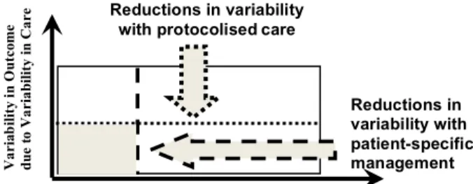
In a recent review paper (Chase et al., 2011), we summarized this problem with a plot (Fig. 1). This figure emphasizes the role of variability in care that protocolised care can reduce, and the role of a different, potentially less reducible, component due to inter- and intra-patient variability in response to treatment. The larger the area, the more difficult the patient can be to manage.
♦ This work was supported by the FNRS (Belgium), the Univ
of Canterbury, the French Community of Belgium (Actions de Recherches Concertées – Académie Wallonie-Europe).
Thus, protocolised care reduces only the non-patient portion of this diagram. Equally, those whose clinical pathway is “straightforward” and can benefit most from protocolised care, are likely to have limited inter- and intra-patient variability in response to treatment. Hence, the smallest, least variable case is one in which intra-patient response is either reduced or managed in a patient-specific fashion, thus separating the final area into several smaller ones.
Figure 1 : Variability in outcome of the critically ill patient defined by variability in response to therapy and variability in care. Shaded area defines the target zone for patient-specific care (Chase et al., 2011).
The aim of this paper is to show that model-based methods can provide patient-specific care that is therefore robust to
Reductions in variability with protocolisedcare
Reductions in variability with patient-specific management
Variability in Outcome due to Intra- and Inter- Patient Variability in Response to Therapy
Va ri ab ili ty in O ut co m e due t o V ar ia bi lit y in C ar e
these intra- and inter- patient variabilities. We examine and review a new, emerging therapeutic approach that provides for individualised care that accounts for intra- and inter- patient variability within an overall protocolised and evidence-based framework. We will focus on cardiovascular monitoring and diagnosis in Intensive Care Unit (ICU) patients, but the overall approach is readily generalizable to other areas of intensive care medicine.
Cardiovascular monitoring is essential in intensive care, enabling hemodynamic disturbances to be recognized, and thus leading to specific treatments. However, it is unclear whether more invasive monitoring actually improves patient outcome. Many different sensors and therapeutic instruments are available, but a major problem is to take this diverse set of clinical data and transform it into a clear and simple physiological context. Moreover, complex interactions between these variables and the body’s natural reflex response can hide the underlying hemodynamic instability. The randomised controlled trial (RCT) is often regarded as the most reliable instrument on which to base treatment selection. Nevertheless, a problem with deriving protocols from large RCT’s is that they are often developed based on empirical evidence and in a “one size fits all” context. These protocols are therefore not well suited to the highly dynamic and variable needs of ICU patients (Vincent, 2010). For example, in terms of patient outcome, there is little evidence to guide the choice of inotropic drugs, although there is a lot of knowledge on the hemodynamic effects of the drugs. Thus, when an intensive care clinician is looking at an individual patient, the reality is that they must consider many combinations of different disease scenarios based on frequently conflicting data on a patient’s condition. In addition, there are no specific guidelines for treatment even once a diagnosis has been made. Therefore, successful diagnosis and treatment often relies on experience and intuition of clinical staff, increasing the likelihood for clinical errors.
Hence, the major features that are lacking in current cardiovascular system (CVS) management are the means to identify and track meaningful physiological parameters of individual patients, and the flexibility to change therapy dynamically in response to patient specific dynamics. It is these two conclusions that define the overall problem, and demonstrate the need for a unified and more consistent approach to managing hemodynamics in the critically ill. This problem illustrates the potential for developing a robust cardiovascular system model that describes the essential dynamics of the circulation as a whole, and can thus improve monitoring, diagnostics and prediction. Most importantly, the model must be able to adapt to an individual patient and be identifiable within a reasonable time period (<5 minutes) using standard ICU measurements. To create such a model, needs an understanding of the major physiological processes involved in the hemodynamic responses to both disease states and therapy, while the validation requires “in-silico” testing, animal trials and finally human trials.
We will review hereafter the unique work done jointly by the Cardiovascular Research Center at the University of Liege
(Belgium) and the Mechanical Engineering department at the University of Canterbury (New Zealand).
2. METHODS
2.1 Cardiovascular system model
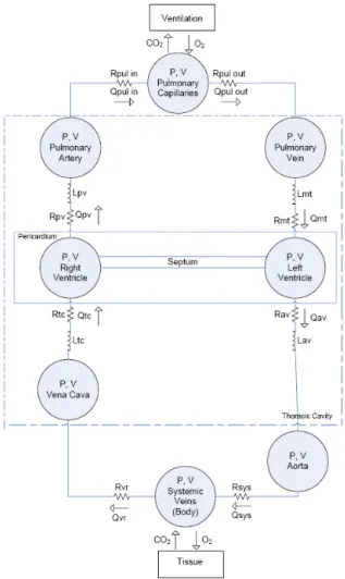
Approaches to modelling the human circulation can be grouped into either Finite Element (FE) or Pressure Volume (PV) approaches. The advantage of FE techniques is that they offer accurate results, but require immensely detailed inputs such as muscle fibre orientations, structures and mechanical properties (Kerckhoffs et al., 2007, Legrice et al., 1997). Limitations on the availability of detailed clinical patient specific data mean that FE methods are not well suited as rapid diagnostic tools. In contrast, PV methods divide the circulation into a series of elastic chambers separated by resistances, and inductors simulating inertial effects where required. Each elastic chamber models a section such as the ventricles, or the atria, each with their own pressure-volume relationship. Only a minimal number of parameters are thus needed, allowing these models to be solved in very reasonable times suitable for immediate clinical feedback.
Figure 2: Minimal model of the CVS (Starfinger et al., 2008d, Starfinger et al., 2008c).
We developed such a minimal CVS model, which is a lumped parameter model of 6-8 elastic chambers. The original model has been extended (Chase et al., 2010, Smith et al., 2004, Smith et al., 2006, Starfinger et al., 2008b, Starfinger et al., 2008d, Starfinger et al., 2008c, Starfinger et al., 2007) and an overview of the 8 chamber version used in this research is given in Figure 2. Each chamber is characterised by the flow in and out of the chamber, the pressure up- and downstream, the resistances of the heart valves, and inertia of the blood. The model also accounts for ventricular interaction by means of the septum displacement. This CVS model uses the classical concept of time varying elastance proposed by Suga et al. (Suga et al., 1973) to simulate the cardiac muscle activation. More specifically, the upper and lower limits of the elastance are defined by the systolic pressure–volume relationship (ESPVR) and end-diastolic pressure–volume relationship (EDPVR) (Burkhoff et al., 1988). By combining the ESPVR and EDPVR an equation relating the ventricular pressure (P) to the ventricular volume (V):
𝑃 = 𝑒 𝑡 𝐸!"#$% 𝑉 − 𝑉! + 1 − 𝑒 𝑡 𝑃!(𝑒! !!!! − 1) (1)
where P0, V0 and λ are respectively, the pressure gradient and
the volume at zero pressure and the curvature, while Eesrvf is
the RV end-systolic elastance and e(t) is the activation function (Figure 3) that accounts for ventricular activation (Smith et al., 2004, Smith et al., 2006).
Figure 3. Plot of the activation function e(t).
2.2 Parameter identification method
Once we have defined a CVS mathematical model, we need to develop a parameter identification method in order to obtain a patient-specific model. A lot of effort has been done to obtain efficient, fast and reliable identification methods. The original parameter identification method used in this research has already been shown to rapidly and accurately identify virtually the entire parameter set in the presence of significant measurement noise (Desaive et al., 2008, Hann et al., 2006, Starfinger et al., 2008a, Starfinger et al., 2008d, Starfinger et al., 2008c) and limited measurements (Hann et al., 2009, Hann et al., 2010).
Recent improvements of the identification method (Revie et al., 2011a, Revie et al., 2011b) have led to a more clinically
applicable method. The new method only requires a minimal set of discrete measurements, such as stroke volume and mean aortic pressure, that are easily obtainable in the ICU. Whereas the previous method required measurements of continuous waveforms, including the highly invasive (or expensive) measures of the time varying ventricular volume traces, which require a lot of computation processing. The measurements used by the new approach are far fewer and far less intensive to obtain and utilize.
It is important to note that in this model-based approach, it is not intended to perfectly match the pressure and volume waveform shapes, but only the minimum (diastolic) and maximum (systolic) values. Hence, it seeks to capture the primary, clinically relevant dynamics for decision support at the bedside, rather than a potentially more perfect physiological picture.
3. RESULTS AND DISCUSSION
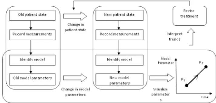
The model-based approach summarized in Figure 4 (Revie et al., 2011a) has been successfully tested on experimental animal models of pulmonary embolism (Starfinger et al., 2007), PEEP titrations and volume therapy decision support (Starfinger et al., 2008d, Starfinger et al., 2008c), and septic shock experiments (Desaive et al., 2008, Starfinger et al., 2008b).
Figure 4. Flow chart of the model-based approach (Revie et al., 2011a).
Hereafter, we will briefly review some of the latest results obtained by our groups in cardiovascular monitoring and diagnosis.
3.1 Monitoring of acute pulmonary embolism
In a recent study by Revie et al. (Revie et al., 2011a), the subject-specific computer models accurately captured the
increase in pulmonary resistance (Rpul), the main
cardiovascular consequence of acute pulmonary embolism (Figure 5), in all five pigs trials, which related well (R2 =
0.81) with the experimentally derived pulmonary vascular
resistance. Figure 5 also shows that Rpul systemically
overestimates the experimentally derived pulmonary vascular
resistance (PVR)(Ghuysen et al., 2008), but importantly, the
t240 after pulmonary embolization.
Figure 5: Identified subject-specific pulmonary resistance during an acute pulmonary embolism pig trial.
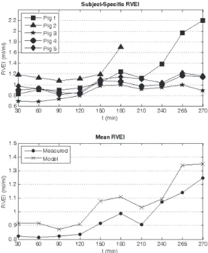
Figure 6. Identified right ventricular expansion index (RVEI) during acute pulmonary embolism.
A direct cause of the increase of PVR during pulmonary embolism is pulmonary hypertension and the right ventricle compensates this pressure increase by dilation, as it is a compliant chamber. This results in a leftward shift in the intraventricular septum, which can be measured by the right
ventricle expansion index (RVEI), namely the ratio of the right and left end diastolic volumes. Averaged across all five pigs, the identified RVEI compared well with the true RVEI
with a correlation coefficient of R2 = 0.92 (Revie et al.,
2011a).
Figure 6 shows that the modelled RVEI increases in all the trials, with sharp increases noticed for pigs 1 and 2 near the end of their trials, indicating that they are in a near-death state. Whereas only minor increases (< 25%) are noticed for pigs 3, 4, and 5, which survive the trials. Hence, the modelled RVEI provides a good indication of how well the pigs are coping with acute pulmonary embolism.
3.2 Defining ventriculo-arterial coupling metric
Blood flow creates a continuous interaction between the cardiac ventricle and the arterial tree. This interaction, known as ventriculo-arterial coupling, is the main determinant of stroke volume and ejection pressure because it relates effort or force capacity to output resistance or afterload. Theoretically, acute assessment of the ventriculo-arterial coupling should lead to optimal hemodynamic therapy (Lambermont and D'Orio, 2006).
In a recent study, Desaive et al (Desaive et al., 2012) have shown that model-based approach offers potentially a new reliable procedure to assess the right ventricle contractility, as well as the right ventricular-arterial coupling. They also
defined a new model-based metric (𝐸!"#$%/𝑅!"#$%) that, with
adequate calibration, can effectively monitor coupling in clinical real-time and thus be used to guide therapy.
Figure 7: Mean experimental ( 𝐸!"!"#/𝐸!!"#) and identified
(𝐸!"#$%/𝑅!"#$%) right ventricular-arterial coupling as a
function of time for all pigs.
Figure 7 presents the RV-vascular coupling using 𝐸!"#$%/
𝑅!"#$% during an endotoxic shock experiment as a further
marker of the impact of sepsis. It shows a good correlation between the identified coupling metric and the experimental
coupling. The overall trends over time, beginning with a sharp fall to a value approaching 1.0 and uncoupling, followed by a recovery due to hemofiltration treatment.
4. CONCLUSIONS
The Cardiovascular Research Center at the University of Liege and the Department of Mechanical Engineering at the University of Canterbury have developed a unique collaboration on model-based therapeutics. This collaboration has led to the development of clinically validated models of physiological systems for diagnosis and decision support. In cardiovascular critical care, they designed minimal models allowing for rapid and efficient identification using clinical measurements easily obtained in the ICU. Validation has been performed using animal data, showing the applicability of the model-based patient-specific approach to diagnose and track disease states but also to design new clinically relevant metrics that could be followed in real-time.
REFERENCES
Burkhoff, D., Alexander, J., Jr. & Schipke, J. 1988. Assessment of Windkessel as a model of aortic input impedance. Am J Physiol, 255, H742-53.
Chase, J. G., Le Compte, A. J., Preiser, J. C., Shaw, G. M., Penning, S. & Desaive, T. 2011. Physiological modeling, tight glycemic control, and the ICU clinician: what are models and how can they affect practice? Ann Intensive Care, 1, 11.
Chase, J. G., Starfinger, C., Hann, C. E., Revie, J. A., Stevenson, D., Shaw, G. M. & Desaive, T. 2010. Model-based prediction of the patient-specific response to adrenaline. The Open Medical
Informatics Journal, 4, 149-163.
Desaive, T., Lambermont, B., Ghuysen, A., Kolh, P., Dauby, P. C., Starfinger, C., Hann, C. E., Chase, J. G. & Shaw, G. M. 2008. Cardiovascular Modelling and Identification in Septic Shock - Experimental validation. Proceedings of the 17th IFAC World
Congress July 6-11, 2008, Seoul, Korea.
Desaive, T., Lambermont, B., Janssen, N., Ghuysen, A., Kolh, P., Morimont, P., Dauby, P. C., Starfinger, C., Shaw, G. M. & Chase, J. G. 2012. Assessment of ventricular contractility and ventricular-arterial coupling with a model-based sensor. Comput
Methods Programs Biomed.
Ghuysen, A., Lambermont, B., Kolh, P., Tchana-Sato, V., Magis, D., Gerard, P., Mommens, V., Janssen, N., Desaive, T. & D'orio, V. 2008. Alteration of right ventricular-pulmonary vascular coupling in a porcine model of progressive pressure overloading.
Shock, 29, 197-204.
Hann, C. E., Chase, J. G., Desaive, T., Froissart, C. B., Revie, J., Stevenson, D., Lambermont, B., Ghuysen, A., Kolh, P. & Shaw, G. M. Robust parameter identication for model-based cardiac diagnosis in critical care. 7th IFAC Symposium on Modelling
and Control in Biomedical Systems, 2009 Aalborg, Denmark.
Hann, C. E., Chase, J. G., Desaive, T., Froissart, C. B., Revie, J., Stevenson, D., Lambermont, B., Ghuysen, A., Kolh, P. & Shaw, G. M. 2010. Unique parameter identification for cardiac diagnosis in critical care using minimal data sets. Comput Methods Programs
Biomed.
Hann, C. E., Chase, J. G. & Shaw, G. M. 2006. Integral-based identification of patient specific parameters for a minimal cardiac model. Comput Methods
Programs Biomed, 81, 181--192.
Kavanagh, B. P. & Meyer, L. J. 2005. Normalizing physiological variables in acute illness: five reasons for caution. Intensive Care Med, 31, 1161-7.
Kerckhoffs, R. C., Neal, M. L., Gu, Q., Bassingthwaighte, J. B., Omens, J. H. & Mcculloch, A. D. 2007. Coupling of a 3D finite element model of cardiac ventricular mechanics to lumped systems models of the systemic and pulmonic circulation. Ann Biomed
Eng, 35, 1-18.
Lambermont, B. & D'orio, V. 2006. The role of right
ventricular-pulmonary arterial coupling to
differentiate between effects of inotropic agents in experimental right heart failure. Crit Care Med, 34, 2864-5.
Legrice, I. J., Hunter, P. J. & Smaill, B. H. 1997. Laminar structure of the heart: a mathematical model. Am J
Physiol, 272, H2466-76.
Revie, J. A., Stevenson, D. J., Chase, J. G., Hann, C. E., Lambermont, B. C., Ghuysen, A., Kolh, P., Morimont, P., Shaw, G. M. & Desaive, T. 2011a. Clinical detection and monitoring of acute pulmonary embolism: proof of concept of a computer-based method. Ann Intensive Care, 1, 33. Revie, J. A., Stevenson, D. J., Chase, J. G., Hann, C. E.,
Lambermont, B. C., Ghuysen, A., Kolh, P., Shaw, G. M., Heldmann, S. & Desaive, T. 2011b. Validation of subject-specific cardiovascular system models from porcine measurements. Comput
Methods Programs Biomed.
Smith, B. W., Chase, J. G., Nokes, R. I., Shaw, G. M. & Wake, G. 2004. Minimal haemodynamic system model including ventricular interaction and valve dynamics. Medical Engineering \& Physics, 26, 131-139.
Smith, B. W., Chase, J. G., Shaw, G. M. & Nokes, R. I. 2006. Simulating transient ventricular interaction using a minimal cardiovascular system model. Physiol
Meas, 27, 165--179.
Starfinger, C., Chase, J. G., Hann, C. E. & Shaw, G. M. 2008a. Simulating the patient-specific adrenaline dose response using a cardiovascular system model.
Cardiovascular Engineering.
Starfinger, C., Chase, J. G., Hann, C. E., Shaw, G. M., Lambermont, B., Ghuysen, A., Kolh, P., Dauby, P. C. & Desaive, T. 2008b. Model-based identification and diagnosis of a porcine model of induced endotoxic shock with hemofiltration. Math Biosci, 216, 132--139.
Starfinger, C., Chase, J. G., Hann, C. E., Shaw, G. M., Lambert, P., Smith, B. W., Sloth, E., Larsson, A., Andreassen, S. & Rees, S. 2008c. Model-based identification of PEEP titrations during different volemic levels. Comput Methods Programs Biomed, 91, 135--144.
Starfinger, C., Chase, J. G., Hann, C. E., Shaw, G. M., Lambert, P., Smith, B. W., Sloth, E., Larsson, A., Andreassen, S. & Rees, S. 2008d. Prediction of hemodynamic changes towards PEEP titrations at different volemic levels using a minimal cardiovascular model. Comput Methods Programs
Biomed, 91, 128--134.
Starfinger, C., Hann, C. E., Chase, J. G., Desaive, T., Ghuysen, A. & Shaw, G. M. 2007. Model-based cardiac diagnosis of pulmonary embolism. Comput
Methods Programs Biomed, 87, 46--60.
Suga, H., Sagawa, K. & Shoukas, A. A. 1973. Load independence of the instantaneous pressure-volume ratio of the canine left ventricle and effects of epinephrine and heart rate on the ratio. Circ Res, 32, 314-22.
Vincent, J. L. 2010. We should abandon randomized controlled trials in the intensive care unit. Crit Care
Med, 38, S534-8.
Wendon, J. 2010. Critical care "normality": individualized versus protocolized care. Crit Care Med, 38, S590-9.