Journal of Applied Biosciences 133: 13584 - 13591
ISSN 1997-5902
Evaluation of decreased haematocrit and
haemoglobin levels in Plasmodium falciparum
infected individuals from South-western Nigeria.
Mary Aigbiremo Oboh, Khadim Diongue, Mamadou Alpha Diallo,Aida S. Badiane,Daouda Ndiaye.
Parasitology and Mycology Laboratory, Le Dantec University Hospital, 30 Avenue Pasteur, BP 16477, Dakar, Senegal.
* To whom correspondence should be addressed to- Mary Aigbiremo Oboh aigbi4god@gmail.com
Original submitted in on 9th October 2018. Published online at www.m.elewa.org on 31st January 2019 https://dx.doi.org/10.4314/jab.v133i1.10
ABSTRACT
Objective: Plasmodium parasite is responsible for the breakdown of red blood cells, resulting into life threatening situation. Thus, an observational study of the parasitaemic impact of P. falciparum on some haematological parameters in comparison to non-infected individuals was carried out in two endemic state of Nigeria (Edo and Lagos).
Methodology and Results: Blood samples collected from individuals (from September 2016-March, 2017) aged 2 years and above, were subjected to rapid diagnostic test (RDT) and microscopy assay to determine the presence of P. falciparum. Further, auto-haematology analyser and/or microcentrifuge where available were employed to acquire information on the haematocrit and haemoglobin levels. Of the 2376 collected samples, three hundred (12.6%) were positive by RDT, out of which Plasmodium falciparum was detected microscopically in 137. The mean haematocrit (PCV) level (37.36±0.37) of the negative samples was significantly higher (p<0.001) than the positive samples (29.6± 0.6). Same relationship was observed when the mean haemoglobin of negative samples (12.08±0.12) was compared with those of positive samples (9.9± 0.2). Those with high parasite density had significantly (p<0.001) low haematocrit (PCV) as well as haemoglobin (p<0.001).
Conclusion and application of findings: The findings from this study reveals serious impact of high P. falciparum burden on haemoglobin and haematocrit in infected individuals, the need to intensify efforts in delivering malaria control interventions especially to priority need areas such as in Edo State cannot be overemphasized. Thus, concerted efforts by all stakeholders in such areas is highly needed if malaria infection will ultimately be eliminated from the country.
Keywords: malaria burden, Plasmodium falciparum, vulnerable, parasite density, haemoglobin, haematocrit, anaemia
INTRODUCTION
Nigeria and other sub-Saharan African countries carry the heaviest malaria burden. In, 2015 alone, an estimated 212 million global malaria cases were recorded, a great proportion (90%) of which
occurred in Africa. Similarly, of the 429 000 deaths observed globally during the same time period, 92% were from the African region (WHO 2016). The most vulnerable or at risk individual such as
the pregnant women (Oscar and Aguzie, 2017), children under five years (Hemingway et al., 2016) and other immune-compromised individual suffer most of the consequence of this disease especially in high transmission area (WHO and UNICEF, 2005). These consequences are usually in the form of grave pathological and biochemical outcomes impacted on infected persons. The density of malaria parasite is an important determining factor of the pathological and biochemical outcome (Ayatse and Ekanem, 1994; Achidi et al., 1996). Plasmodium parasite has been said to be responsible for the breakdown of red blood cells through cascades of processes such as destruction of red blood cells (RBC) by the reticulo-endothelial system in the spleen, phagocytosis or rupturing of infected cells, removal of uninfected cells as a result of antibody sensitization (inflammatory responses) (Kurtzhals et al., 1999), all of which lead to anaemia. Anemia result when the body lacks the required quantity of the oxygen-carrying cells (haemoglobin) which are synthesized in the bone marrow under the influence of erythropoietin produced by the kidney. In order, to assess the anaemic condition of a patient at any given time, the full blood count is carried out to evaluate the quantity, morphology and size of various blood cells (Philips and Plasvol, 1992; Ajibola et al., 2012). Anaemia is measured
primarily by determining the level of haemoglobin, however other haematological parameters such as packed cell volume (PCV) also known as haematocrit, as well as mean corpuscular haemoglobin concentration (MCHC) are also used to determine the anaemic stage. The physiological pathology of anaemia is multifaceted and complicated resulting into majority of the morbidity and mortality recorded in this infection especially vulnerable groups who have reduced immunity. Difficulty in appropriately diagnosing malaria as a result of low parasite density or where the clinical features of malaria are confused with that of other infections could result to death sometimes attributable to anaemia, hypoglycaemia (<40mg/dl), metabolic acidosis (BCO
32-<15mmol/l), hyper lactataemia (lactate > 5mmol/l), renal impairment (serum creatinine >265µmol/l) amongst others (Philips and Plasvol, 1992; WHO 2012). A major target of the global technical strategy (GTS) hinges on the elimination of malaria from 35 countries where malaria is transmitted and more importantly reducing malaria mortality by 90% (WHO, 2016). To this end, this study was designed to assess the impact of parasite burden on two haematological factors (anaemia and haematocrit), which could in combination with other pathological outcomes results to death in severe cases.
MATERIALS AND METHODS
Study areas: Samples were collected from four local government areas (LGAs) in Lagos (Eti-Osa, Ibeju-lekki, Kosofe and Ikorodu) and two in Edo (Oredo and Ikpoba-Okha).Generally, Lagos State has a double rainfall pattern with an annual rainfall of 1400mm-1800mm with a short break called “August break’. There are two climatic conditions-the dry season (lasting from November to March) and the wet season (from April to October) with a temperature range of 30-38°C (Ayeni, 2016). Malaria transmission normally occurs throughout the year with its peak transmission occurring during the raining season (Odugbemi et al., 2016). In Edo, the rainy season begins in March/April and ends in October/November, thus providing a favourable condition for mosquito breeding (NPC, 2010; Ekhaese and Amole, 2014). Ikpoba-Okha has a land mass of 862km2 with 371, 106 inhabitants and shares a
western boundary with Oredo LGA. Three of these study LGAs are urban areas (Eti-Osa, Kosofe and Oredo) while the rest are sub-urban (Ibeju-Lekki, Ikorodu and Ikpoba-Okha).
Inclusion and exclusion criteria: Individuals from ≥2 years of age visiting the various hospitals who have assented/consented to partake in the study were all included. Additionally, patients with clinical symptoms of malaria detected by a febrile condition of ≥37.5°C were also participants while pregnant women and those with other complicated infections were excluded.
Sampling: a total of 2376 patients were recruited in September 2016 to March 2017 from all study sites after detailed briefing of the purpose of the study. Using RDT kits, they were all screened, those found positive by this initial diagnostic test were further screened by microscopy, while 137 (use as negative control) of
those found negative by both diagnostic methods, were
randomly selected from the different LGAs to compare their haematological values with the microscopically positive samples as shown in Figure 1.
Figure 1: Algorithm of sample processing employed Sample collection and Sample Preparation: Venous blood was collected from each patient into EDTA containers for RDT analysis as well as smear preparation. Each container corresponds to a patient’s unique identity. Following the manufacturer’s instruction, Care Start® P.f (Access Bio Inc, 65 Chyde Road Suite A, Somerset NJ 08873 USA, Batch number M014L04-M014M10) was used to carry out a rapid diagnosis, and those samples found positive by this preliminary test were processed further microscopically. Preparation of thin and thick blood films followed same protocol employed in our previous article (Olusegun-Joseph et al., 2016). Samples detected as microscopically positive were blindly checked by a trained microscopist at the Nigerian Institute of Medical Research, Lagos. The packed cell Volume (PCV in %), also known as haematocrit level as well as haemoglobin were measured for all microscopically
positive samples with the auto-analyzer (Mindray Suzhou Coming Chengye Medical Technology Co. Ltd, China) and/or micro-centrifuge (for haematocrit determination only) where available. Utilizing the method described by Strumia et al (1954), heparinized capillary tube was three-quarter filled and sealed with plasticene, thereafter it was spun for 15minutes at 10 000rpm and the values gotten directly from micro-haematocrit chart.
Definitions of parameters: Haematocrit (PCV %); Female: ≤30 is low, ≥31≤36 Normal and ≥37 High, while for Male: <32 is Low, ≥33≤40 is Normal and ≥41 is High.
Haemoglobin, Hb (g/dl): Female: Hb>12 normal, mild anaemia 12≤Hb>6.5, Severe anaemia Hb<6.5, while for male Hb>13 normal, mild anaemia 13≤Hb>7, Severe anaemia Hb≤7 (WHO, 2011).
Total patients screened =2376
Subjects from Lagos State=1947
Subjects from Edo State=429 Positive by RDT=157 Negative by RDT=1790 Negative by RDT=286 Positive by RDT=143 Negative by Microscopy =82 Positive by Microscopy =75 Positive by Microscopy =62 Negative by Microscopy =81
Intensity of infection (parasite/µl): ≤1,000 is Low parasitaemia, ≥1,001 ≤ 10,000 is Moderate and > 10,000 is high (WHO, 2010).
Data analysis: Data were entered in SPSS version 21.0 and Pearson’s correlation was used to determine the level of association between variables at a significant level of 0.5, the association of the intensity of microscopically positive samples were tested with chi-square to see if it has any association with other variables such as Parked cell volume (PCV (%)), Haemoglobin (g/dl), Intensity of infection (parasite/µl), LGAs, Age group and Sex. In same manner, the
association between packed cell volume or haematocrit levels and the other variables were tested using chi-square. Sample paired t-test was used to determine the association between the haemoglobin and haematocrit of positive and negative samples.
Ethical consideration: This study was approved by the Institutional Review Board (IRB/16/347) Nigerian Institute of Medical Research, The Lagos State Health Service Commission and the Edo State Hospital Management Board. All participants were duly briefed on the purpose of the study and those who gave consent and/or assent were recruited into the study. RESULTS
Distribution of samples: In all, 2376 patient samples comprising of 1058 males and 1318 females were examined, first by RDT and then by microscopy. Three hundred (300) samples were positive by RDT, out of
which Plasmodium falciparum was detected microscopically in 137 and Plasmodium ovale (excluded from further analysis) in one (1) sample (Table 1).
Table 1: Samples breakdown by RDT and microscopy
Sex Examined by RDT Positive by RDT
(%) Positive by Microscopy (%)
Males 1058 132 (12) 68(6.4)
Females 1318 168 (12) 69(5.2)
Total 2376 300 (12.6) 137 (5.8)
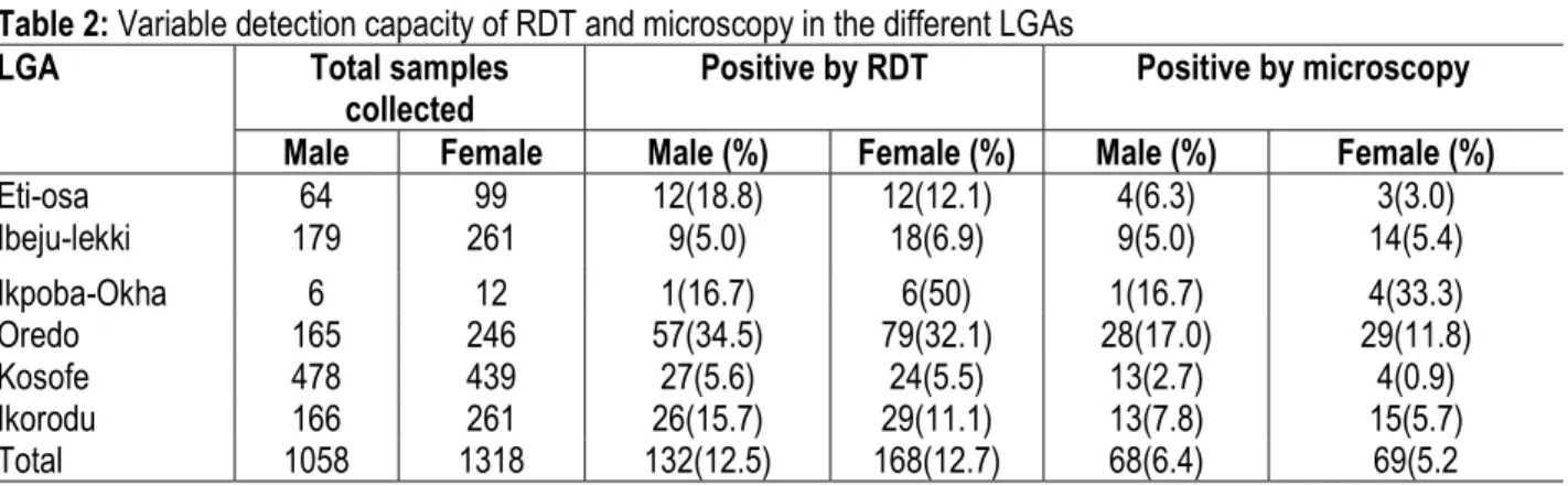
Note: Plasmodium ovale positive sample was excluded from further analysis. Prevalence of malaria by RDT and microscopy in
different LGAs: On the whole, Ikpoba-Okha has the highest prevalence both by RDT 7(38.9%) and
microscopy 5(27.8%), this is closely followed by Oredo with 136 (33.1%) and 57 (13.9%) samples positive by RDT and microscopy respectively. (Table 2).
Table 2: Variable detection capacity of RDT and microscopy in the different LGAs
LGA Total samples
collected Positive by RDT Positive by microscopy
Male Female Male (%) Female (%) Male (%) Female (%)
Eti-osa 64 99 12(18.8) 12(12.1) 4(6.3) 3(3.0) Ibeju-lekki 179 261 9(5.0) 18(6.9) 9(5.0) 14(5.4) Ikpoba-Okha 6 12 1(16.7) 6(50) 1(16.7) 4(33.3) Oredo 165 246 57(34.5) 79(32.1) 28(17.0) 29(11.8) Kosofe 478 439 27(5.6) 24(5.5) 13(2.7) 4(0.9) Ikorodu 166 261 26(15.7) 29(11.1) 13(7.8) 15(5.7) Total 1058 1318 132(12.5) 168(12.7) 68(6.4) 69(5.2
Correlation of age, haemoglobin and parasite density in subjects positive and negative for malaria parasite: There was no statistical difference (p=0.777) between the age of positive and negative subjects. On the contrary, the mean haematocrit level
(37.36±0.37) of the negative sample is significantly higher (p<0.001) than that of the positive sample (29.6± 0.6). Same relationship was observed when the mean haemoglobin of negative samples (12.08±0.12) was compared with the positive (9.9± 0.2). (Table 3).
Table 3: Age, haemoglobin and parasite density in microscopically positive and negative samples
Variables Positive samples Negative samples t-test
Minimum Maximum Mean (S.E) Minimum Maximum Mean (S.E)
Age (Years) 2 85 20.9±1.4 3 85 21.48±1.3 0.777
PCV (%) 6 55 29.6± 0.6 23 42 37.36±0.37 0.000
Haemoglobin (g/dl) 2 18 9.9± 0.2 7 14 12.08±0.12 0.000
Parasite density
(Parasite/µl of blood) 73 905600 23739.1± 7592.9 0 0 0
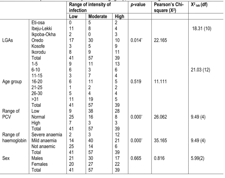
Relationship of parasite density on some variables: As expected, those with high parasite density (see above definition) has significantly (P<0.001) low haematocrit (PCV). Very similarly, high parasite counts
also impact significantly (P<0.000*) on the
haemoglobin. However, there was no observable impact of parasite counts on the different study locations, age and sex (Table 4).
Table 4: Relationship between the intensity of infection age group, PCV and haemoglobin Range of intensity of
infection p-value Pearson’s Chi-square (X2)
X2 tab (df) Low Moderate High
LGAs Eti-osa 0 5 2 0.014* 22.165 18.31 (10) Ibeju-Lekki 11 8 4 Ikpoba-Okha 2 0 3 Oredo 17 30 10 Kosofe 3 5 9 Ikorodu 8 9 11 Total 41 57 39 Age group 1-5 9 11 13 0.519 11.111 21.03 (12) 6-10 6 3 6 11-15 3 7 4 16-20 6 11 5 21-25 1 2 2 26-30 5 4 4 >31 11 19 5 Total 41 57 39 Range of PCV Low 9 38 28 0.000* 26.062 9.49 (4) Normal 25 16 8 High 7 3 3 Total 41 57 39 Range of
haemoglobin Severe anaemia 2 3 12 0.000*
35.165 9.49 (4) Mild anaemia 14 40 21 Not anaemic 25 14 6 Total 41 57 39 Sex Males 21 30 17 0.665 0.816 5.99(2) Females 20 27 22 Total 41 57 39 DISCUSSION
The burden and severity of malaria is usually borne by children under the age of five, pregnant women, the elderly and those whose immune system has been compromised as a result infection (Hemingway et al.,
2016; Oscar and Aguzie, 2017). The effect is usually grievous in sub-Saharan Africa where the eco-climatic condition of the environment favours the breeding of the non-vertebrate host, enhancing the seasonal and/or
continuous transmission of the disease coupled with the simultaneous inaccessibility to quality health care. In this study, samples collected from six different study sites with varying endemic status showed marked difference in prevalence by RDT as well as microscopy. It is noteworthy that one sample which was positive by RDT was microscopically positive for Plasmodium ovale as against Plasmodium falciparum given the fact that the RDT used is a species specific one (P.f HRP2). This could be explained by the fact that either that was a mixed infection in which case the former species was not picked in the analysis, or it was a cross reaction which will be verified in the follow-up study using PCR. Although, the microscopy results shows a lower prevalence than the RDT, it cannot be said that the RDT over diagnosed as it is well established that histidine rich protein-2 circulates in the blood long after (up to 2 weeks) parasites must have been cleared by antimalarial therapy (Wongsrichanalai et al., 2007). Probing further into the practices of the study population (which is outside the scope of this present study) would have revealed if drugs were used prior to hospital visitation and as such substantiate the claim of drug reaction on the parasite. Other studies showing same trend of lower prevalence by microscopy as against RDT where both were used include that carried out in Lagos, Nigeria (Olusegun-Joseph et al., 2016) and also in Tanzanian, though among children (Sumari et al., 2017). Such results however need validation by PCR in order to ascertain the true diagnostic status of the individuals. Two of the six study sites (Oredo and Ikpoba-Okha) stand out in their prevalence both by microscopy as well as by RDT. They have relatively higher prevalence than all the study sites and this could be attributed to several factors amongst which are the eco-climatic condition of the study sites which are both located in the South-Southern part of the country where rainfall is moderately high. These factors support the
breeding of the non-vertebrate host, thus enhancing transmission for a prolonged period during the year. This could also be due to the fact that, the level of intervention targeting both the vector as well as human hosts is relatively minimal compared to the other study state where various interventions ranging from indoor residual spraying, intermittent preventive treatment, free net distribution are ongoing (Odugbemi et al . , 2016). The mean haemoglobin and haematocrit level of the uninfected group is significantly higher than those detected as positive by microscopy. Although there are various factors implicated in haemoglobin reduction (Olutola and Mokuolu,2012), it is however not by random in this study that presence of parasites in the blood of infected cases have cause a reduction in these two haematological parameters especially in study subjects with high and very high intensity of infection as shown by the Chi-square result. Similar findings to this study is seen in the works of Ojurongbe et al (2014) though among HIV infected individuals and Oseghale et al (2012) in Osogbo and Edo State Nigeria respectively. The intensity of infection was significantly higher in Oredo than in all other study sites, this again could be attributed to the eco-climatic condition and minimal intervention programmes. Additionally, those in age groups ≥31, 1-5 and 16-20 years of age represent the highest microscopically positive samples in decreasing order and in this group was high and very high intensity of infection recorded more than in other groups. The implication of this is that over time with prolong exposure to infection and development of immunity against the parasites, there will be a reduction in the manifestation of symptoms in these set of individuals (asymptomatic carriers). Thus, the need to seek medical care would not be there and ultimately, they will serve as parasite reservoir, enhancing transmission in that area and even beyond when there is migration.
CONCLUSION
Government should therefore intensify efforts in delivering malaria control interventions such as artemisinin combination therapy and long lasting insecticide treated nets to priority need areas such as in
Edo State if pre-elimination and ultimately elimination of this disease is a paramount goal to be achieved in the health sector of any country.
COMPETING INTEREST
ACKNOWLEDGEMENT.
The author is grateful to the participants for their willingness to participate and the laboratory staff of all the hospitals for assisting in sample collection. MAO
designed the work, collected data, performed the experiment; MAO, KD and MAD analysed data, all authors contributed to the manuscript.
REFERENCE
Achidi, E. A., Salimonu, L. S., Asuzu, M. C., Berzins, K. and Walker, O (1996). Studies on Plasmodium falciparum parasitaemia and development of anaemia in Nigerian infants during their first year of life. Am J Trop Med Hyg. 55(2): 138-143.
Ajibola, M., Oloruntoba, A. C. and Yetusoko, N. D (2012). Evaluation of PCV and Hemoglobin Variations among Malaria Positive and Malaria Negative Patients at the ECWA Community Health Centre Bukuru, Jos. Nig J Pharmacy 2(6): 65-69.
Ayatse, J. O. and Ekanem, E. E (1994). Plasmodium falciparum malaria: its effect on some haematological parameters in normal and sickle cell Nigerian children. Trop Med Parasitol. 45: 219-222.
Ayeni, A. O (2016). “Increasing population, Urbanization and climatic factors in Lagos State, Nigeria: The nexus and implication on water demand and supply”, Journal of Global Initiatives: Pol Pedag Perspect. 11(2):69-87 Ekhaese, E. N. and Amole, B (2014). Benin domestic
architecture “a tabula rasa” for transition: From pre-independence to contemporary architecture. Int J Soc Sci Entrep. 1(9):1-23 Hemingway, J., Shretta, R., Wells, T. N. C., Bell, D.,
Djimdé, A. A., Achee, N. and Qi, G (2016). Tools and Strategies for Malaria Control and Elimination: What Do We Need to Achieve a Grand Convergence in Malaria? PLoS Biol. 14, e1002380.
Kurtzhals, J. A., Addae, M. M., Akanmori, B. D. and Danyo, S (1999). Anaemia caused by asymptomatic Plasmodium falciparum infection in semi-immune African school children. Trans R Soc Trop Med Hyg. 93 (6): 623-627.
Odugbemi, B. A., Wright, K.O., Onajole, A. T., Kuyinu, Y. A., Goodman, O. O., Odugbemi, T. O and Odusanya, O. O (2016). A malariometric survey of under‑fives residing in indoor residual spraying‑implementing and non‑implementing communities of Lagos, Nigeria. Mal J. 15:458-461
Olusegun-Joseph, T. S., Oboh, M. A. and Uduak, M. U (2016). A survey of malaria prevalence and antimalarial preventive measures amongst students of University of Lagos, Nigeria- Afr J Clin Experl Microbiol. 17(4):267-273.
Ojurongbe, O., Oyeniran, O. A., Alli, O. A. T., Taiwo, S. S., Ojurongbe, T. A. Olowe, A. O.,
Opaleye, O. O. and Adeyeba, O.A (2014). Prevalence of Plasmodium falciparum Parasitaemia and Its Correlation with Haematological Parameters among HIV-Positive Individuals in
Nigeria. J Trop Med.
doi.org/10.1155/2014/161284.
Olutola, A. and Mokuolu, O (2012). Severe Malaria Anaemia in Children In: Anemia. Donald Silverberg (Ed.) ISBN: 978-953-51-0138-3, InTech, 31pp.
Oscar, I. and Aguzie, N (2017). Pregnancy-associated Malaria, Challenges and Prospects in sub-Sahara Africa. Clin. Mother Child Heal. 15: e1000282.
Oseghale, F. O., Okogun, G. R. A., Akhile, A.and Omolumen, L. E. S (2012). Relationship between malaria parasitaemia and packed cell volume among primary school pupils in Ekpoma. Int J Basic Applied Innovat Res. 1(4): 111 - 115
Phillips, R. E. and Pasvol, G (1992). Anaemia of Plasmodium falciparum malaria. Baillierers Clin Haematol. 2:315-30.
Strumia, M. M., Sample, A. B. and Hart, E. D (1954). An improved microhaematrocrit method. Am J Clin Path. 24(9):1016-1024
Sumari, D., Mwingira, F., Selemani, M., Mugasa, J., Mugittu, K. and Gwakisa, P (2017). Malaria prevalence in asymptomatic and symptomatic children in Kiwangwa, Bagamoyo district, Tanzania. Mal J. 16:222-229
World Health Organization (2016). Eliminating Malaria. WHO/HTM/GMP/2016.3. 28pp Accessed 14th
March 2016.
World Health Organization (2015). Global Technical Strategy for Malaria 2016–2030. Geneva:
World Health Organization (WHO); (http://www.who.int/malaria/areas/global_techn ical_strategy/en, accessed 16 March 2017). World Health Organization (2011). Haemoglobin
concentrations for the diagnosis of anaemia and assessment of severity. Vitamin and Mineral Nutrition Information System. WHO/NMH/NHD/MNM/11.1. Geneva: World
Health Organization.
http://www.who.int/vmnis/indicators/haemoglo bin.pdf. Accessed 23 June 2017.
World Health Organization (2010). Basic Malaria Microscopy. Part 1; Learner’s guide, 2nd
Edition, 88pp
World Health Organization (2005). Protecting vulnerable groups in malaria-endemic areas in Africa through accelerated deployment of insecticide-treated nets.
Wongsrichanalai, C., Barcu, M. J., Muth, S., Sutamihardja, A. and Wernsdorfer, W. H (2007). A review of malaria diagnostic tools: Microscopy and rapid diagnostic test (RDT). Am J Trop Med Hyg. 77:119-127.