HAL Id: inserm-02333383
https://www.hal.inserm.fr/inserm-02333383
Submitted on 25 Oct 2019HAL is a multi-disciplinary open access
archive for the deposit and dissemination of sci-entific research documents, whether they are pub-lished or not. The documents may come from teaching and research institutions in France or abroad, or from public or private research centers.
L’archive ouverte pluridisciplinaire HAL, est destinée au dépôt et à la diffusion de documents scientifiques de niveau recherche, publiés ou non, émanant des établissements d’enseignement et de recherche français ou étrangers, des laboratoires publics ou privés.
Sex Differences in Spontaneous Degranulation Activity
of Intrahepatic Natural Killer Cells during Chronic
Hepatitis B: Association with Estradiol Levels
Zuzana Macek Jilkova, Thomas Decaens, Alice Marlu, Hélène Marche,
Evelyne Jouvin-Marche, Patrice Marche
To cite this version:
Zuzana Macek Jilkova, Thomas Decaens, Alice Marlu, Hélène Marche, Evelyne Jouvin-Marche, et al.. Sex Differences in Spontaneous Degranulation Activity of Intrahepatic Natural Killer Cells dur-ing Chronic Hepatitis B: Association with Estradiol Levels. Mediators of Inflammation, Hindawi Publishing Corporation, 2017, 2017, pp.1-5. �10.1155/2017/3214917�. �inserm-02333383�
Research Article
Sex Differences in Spontaneous Degranulation Activity of
Intrahepatic Natural Killer Cells during Chronic Hepatitis B:
Association with Estradiol Levels
Zuzana Macek Jilkova,
1,2Thomas Decaens,
1,2,3Alice Marlu,
3Hélène Marche,
1,2Evelyne Jouvin-Marche,
1,2and Patrice N. Marche
1,21Université Grenoble-Alpes, IAB, 38000 Grenoble, France
2INSERM U1209, 38000 Grenoble, France
3Département d’Hépato-Gastro-Entérologie, CHU-Grenoble Alpes, 30700 La Tronche, France
Correspondence should be addressed to Patrice N. Marche; patrice.marche@inserm.fr
Received 17 October 2016; Revised 25 February 2017; Accepted 2 March 2017; Published 2 April 2017 Academic Editor: Alex Kleinjan
Copyright © 2017 Zuzana Macek Jilkova et al. This is an open access article distributed under the Creative Commons Attribution License, which permits unrestricted use, distribution, and reproduction in any medium, provided the original work is properly cited.
Major sex differences are observed in the prevalence, intensity, and severity of hepatitis B virus (HBV) infection. Here, we
investigated degranulation activity of circulating and intrahepatic natural killer (NK) cells from HBV and HCV chronically
infected patients before any treatment (n = 125). The frequency of CD107+NK cells in the female liver was significantly higher
compared to that in males during chronic HBV infection (p = 0 002) and correlated with the plasma levels of estradiol
(correlation coefficient r = 0 634; p < 0 0001). Our results clearly show sex differences in degranulation activity of intrahepatic
NK cells of HBV-infected patients. This probably contributes to the ability of females to better deal with HBV disease.
1. Introduction
The liver is an immune-privileged organ in which antigen-rich blood is pressed through a network of microscopic vessels called sinusoids where blood is scanned by intrahepa-tic (IH) immune cells. IH lymphocyte population is selectively enriched in natural killer (NK) cells, which play critical roles in controlling both viral hepatitis infections and liver tumorigenesis.
Major sex differences in hepatitis B virus (HBV) infection and the male susceptibility for hepatitis-related hepatocellular carcinoma (HCC) have been described. However, distinct mechanisms have remained enigmatic. In fact, the prevalence, intensity, and severity of HBV disease itself are consistently higher in men than in women [1–3]. The higher incidence of HBV in men for sure contributes to sex differences in occurrence of HCC, but even among HBsAg-positive indi-viduals, liver cancer mortality is two times higher in males
compared to females [1]. Sex-specific differences in exposure to risk factors, such as alcohol consumption or drug use in male population, do not fully explain the greater severity of HBV disease and the higher occurrence of HCC in males compared to females. For instance, same sex differences are also observed during animal experiments. Understanding the mechanisms that enable females to better deal with HBV disease and to reduce their risk of developing HCC needs to be elucidated.
It is known that females often exhibit greater humoral and cell-mediated immune responses to infection than do males [1, 4, 5]. Similarly, numerous in vitro and in vivo experiments have demonstrated that sex hormones directly or indirectly affect and modify the actions of immune cells [6]. The female and male livers show considerable sexual dimorphism, and when taking into account that sex hormones are notably metabolised in the liver, the effects of sex hormones on IH immune cell actions are expectable. Hindawi
Mediators of Inflammation
Volume 2017, Article ID 3214917, 5 pages https://doi.org/10.1155/2017/3214917
Mediators of Inflammation, Volume 2017 (2017), Article ID 3214917. https://doi.org/10.1155/2017/3214917
Therefore, the objective of this study was to investigate degranulation activity of peripheral and IH-NK cells during chronic hepatitis B infection with a focus on sex differences.
2. Methods
2.1. Patients. One hundred twenty-five patients included in this study were prospectively selected prior to any treatment (Department of Gastroenterology and Hepatology, Grenoble University Hospital). HBV-infected patients (n = 43, 63% men) were positive for anti-HBV antibody (tested by ELISA 3, Ortho Diagnostic Systems, USA), positive for HBV DNA in serum as measured by qPCR (Amplicor HCV, Roche Diagnostic Systems), and negative for human immunodefi-ciency virus (HIV) and, HCV infections and did not have any autoimmune hepatitis (AIH) antibody. HCV-infected patients (n = 82, 52% men) were positive for anti-HCV antibody (tested by ELISA 3, Ortho Diagnostic Systems, USA), positive for HCV RNA in serum as measured by RT-qPCR (Amplicor HCV, Roche Diagnostic Systems), and negative for HIV and HBV infections and did not have AIH antibody. Alcohol consumption of patients was lower than 30 g/day in men and 20 g/day in women. The main characteristics of all patients are described in Table S1 in Supplementary Material available online at https://doi.org/ 10.1155/2017/3214917.
Liver biopsies were divided into two parts: one part for histological examination and the other part for immunolog-ical analyses. Histologimmunolog-ical examination was assessed by expe-rienced liver pathologists. Paired blood and liver samples were obtained in 16 HBV-infected patients (Supplementary Figure1C).
The study was performed in accordance with the Decla-ration of Helsinki and French legislation and received approval of the Grenoble University Hospital ethical com-mittee (03/APTF/1). All study participants provided written informed consent.
2.2. Flow Cytometric Analysis. Immediately after the liver biopsy procedure, cells were recovered by mechanical disrup-tion and stained for flow cytometric analysis as described previously [7]. Similarly, peripheral blood cells were immu-nostained. Live/dead cells were discriminated by a Zombie UV™ Fixable Viability Kit. The following antibodies were used for surface staining: CD45 (APC/Cy7, Clone HI30, Bio-Legend), CD3 (PerCP/Cy5.5, Clone UCHT1, BioBio-Legend), and CD56 (APC, Clone HCD56, BioLegend). Surface stain-ing of CD107a (Pacific Blue, LAMP-1, Clone H4A3, 0.25μg/sample) was used to study degranulation activity [8–13]. Data were acquired on BD-LSRII flow cytometer (BD Biosciences), collected with BD FACSDiva 6.3.1 soft-ware, and analyzed using FCS Express V3 and V6 software.
2.3. K562 Target Cell Activation. IH immune cells were incubated for 3 h at 37°C with or without K562 target cells (cell : target = 1 : 1) in the presence of monensin (Sigma). Degranulation activity was monitored by detection of cell surface CD107a.
2.4. NK Cell Lines. Cell lines were cultured in RPMI 1640 medium supplemented with 1% of antibiotics (Pen Strep, Life Technologies), recombinant IL2, and 1 mM sodium pyruvate and with 10% fetal bovine serum for KHYG1 and NK92 cell lines and with 10% heat-inactivated human serum, type AB for an NKL cell line. All sex hormones: estrone (E9750),
β-estradiol (E8875), testosterone (T1500), and
4,5α-dihydro-testosterone (A8380), were purchased from Sigma. Hormones were added to the culture media in a concentration of 10 nM for 24 h before the stimulatory experiment by ±K562 target cells.
2.5. Measurements of Hormone Levels. Levels of estradiol and testosterone were measured in serum by commercially available ELISA kits (Abcam, UK), according to manufac-turer’s protocols.
2.6. Statistical Methods. Analyses were performed using the statistical software GraphPad Prism 6 (GraphPad Software, CA, USA). Gaussian distribution was tested by the D’Agostino-Pearson omnibus normality test. The t-test was used in the case of normal distribution of data and the nonpara-metric Mann-Whitney test in the case of nonnormality. The Wilcoxon matched-pairs signed-rank test was used to test differences between K562 nonstimulated and K562 stimulated cells. Pearson’s correlation coefficients were used for the linear relationship between two variables.
3. Results
Immune cells from blood and from fresh liver biopsies of infected patients (Supplementary Table1) were analyzed by flow cytometry. Among CD45high+population, NK cells were
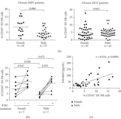
identified (Supplementary Figure 1A). To investigate the NK cell cytolytic properties, we determined the CD107a expres-sion, which is considered a marker of degranulation, that is, release of lytic granules toward the target cells [12, 13]. Mean percentage of CD107a+IH-NK cells was significantly higher in liver biopsies from HBV-infected females (n = 16) com-pared to HBV-infected males (n = 27), with a frequency of CD107a+ IH-NK 8.4± 1.2% in females compared to 4.5± 0.6% in males (Figure 1(a)). In HCV-infected patients, the frequency of CD107+ IH-NK cells was similar between females (4.7± 0.6%, n = 39) and males (5.1 ± 0.4%, n = 43).
When the cohorts were compared regardless of gender, we observed no difference in the frequency of CD107+ intra-hepatic NK cells in HBV patients compared to HCV patients. However, the frequency of CD107+intrahepatic NK cells was significantly higher in HBV-infected women than in HCV-infected women (p = 0 0096) or men (p = 0 0206) and corre-sponded to degranulation activity that can be observed in patients with AIH (Supplementary Table2).
To exclude the possibility that observed sex differences in the functional degranulation activity of NK cells were caused by differences in severity of HBV disease, 16 HBV-infected males matched for age and severity of liver injury (ALT levels, METAVIR activity grade, and fibrosis stage) were compared with 16 females (Supplementary Table 3). Similarly as in the whole cohort, the percentage of
CD107a+IH-NK cells was 2.2-fold higher in females than in males (p = 0 0031) (Supplementary Figure 1B). On the other hand, the frequency of CD107+NK cells in blood sam-ples was very low, both among females (0.64± 0.16%) and among males (0.63± 0.14%) (Supplementary Figure 1C). A similar low frequency of spontaneously degranulating NK cells was found previously in blood of HCV-infected patients, showing that the degranulation process of NK cells occurs mainly in the liver [13]. Interestingly, our preliminary results showed that even the percentage of IFNγ+ IH-NK cells is 2.6-fold higher in HBV-infected females than in males (p = 0 0044) (Supplementary Figure 1D), while no sex difference in IFNγ+ IH-NK cells is observed in the
cohort HCV-infected patients. All together, these results indicate that sex differences in degranulation activity of NK cells are specific for the HBV-infected liver.
To analyze whether the stimulated degranulation capac-ity of IH-NK cells differs between sexes, liver biopsies of HBV-infected females (n = 7) and males (n = 7) were divided in two parts and immune cells of one part were incubated for 3 hours with K562 target cells. In accordance with the results
described above, the mean frequency of CD107+IH-NK cells without any stimulation was significantly higher in females compared to males (p = 0 033). This difference was statisti-cally significant specifistatisti-cally in CD107+
CD56DimIH-NK cell population (p = 0 046) (Figure 1(b), Supplementary Table 4). Interestingly, upon stimulation, we observed a 3.2-fold increase in the mean frequency of CD107+ CD56Dim IH-NK cells of males (p = 0 031) but only a 1.6-fold increase in that of females (p = 0 047) (Figure 1(b)). Thus, after in vitro K562 stimulation, the frequencies of CD107+CD56Dim IH-NK cells did not differ between the sexes (p = 0 872) under-lining that the degranulation capacity (in vitro-stimulated degranulation) of IH-NK cells is equal in males and females. In summary, these results suggest that NK cells in the liver of HBV-infected women are specifically activated.
To investigate if the activity of NK cells is modified by sex hormones, we analyzed serum from chronically HBV-infected patients by ELISA. A strong correlation was observed between the spontaneous degranulation activity of IH-NK cells and levels of estradiol (Figure 1(c),p < 0 0001) while no correlation was observed in testosterone levels
Chronic HBV patients % CD107 + IH -NK cells 0.006 % CD107 + IH-NK cells Male
Female Female Male
Chronic HCV patients 0.602 0 5 10 15 20 0 5 10 15 20 n = 27 n =16 n = 39 n = 43 (a) K562 stimulation ‒ + ‒ + 0.047 0.031 0.046 0.872 0 5 10 15 20 25 Female n = 7 Male n = 7 % CD107 + IH-NK cells 10 15 20 0 50 100 150 200 250 Female Male r = 0.634, p<0.0001 Es tradio l (pg/mL) 0 5 % CD107+ IH-NK cells (b) (c)
Figure 1: (a) Degranulation activity of IH-NK cells of chronic HBV- and HCV-infected patients directly after the recovery of liver biopsies.
Gaussian distribution was tested by the D’Agostino-Pearson omnibus normality test. The Mann-Whitney test was used to test the HBV
cohort and the t-test to test the HCV cohort. (b) Frequencies of degranulating IH-NK CD56Dimcells of chronically HBV-infected patients
stimulated in vitro by±3 h of K562 target cells. The Mann-Whitney test was used to compare between females and males, while the
Wilcoxon matched-pairs signed-rank test was used to compare before and after K562 stimulation. (c) Correlation of estradiol serum levels
with spontaneous degranulation capacity of intrahepatic NK cells of chronically HBV-infected patients. To test significance, Pearson’s
correlation coefficients were used. Each symbol represented a patient and mean values are indicated by lines.
3 Mediators of Inflammation
(Supplementary Figure1E). Interestingly, no other correla-tions were observed between the spontaneous degranulation activity of IH-NK cells and age, ALT levels, METAVIR activ-ity grade, fibrosis stage, or viral load in HBV-infected patients (Supplementary Table1).
To clarify if NK cell functions are dependent on sex hor-mones, we used three different well-established human NK cell lines: KHYG1 (originally from female), NK92, and NKL (originally from male), and stimulated them by sex hor-mones. Even though NK cells express sex hormone receptors [14–16], no direct effect of estradiol or other sex hormones on degranulation activity of human NK cell lines was observed (Supplementary Figure1F). However, the fact that NK cell lines are of peripheral blood origin and HBV infection was missing in this scenario makes drawing conclu-sions about the possible indirect action of estrogens on NK cells difficult.
Taken together, our results show that during HBV infec-tion, the degranulation activity of IH-NK cells is associated with estradiol levels.
4. Discussion
Our results clearly show sex differences in the spontaneous degranulation activity of IH-NK cells of HBV-infected patients in correlation with levels of circulating estradiol.
Previously, in vitro assessment of degranulation activity after K562 stimulation of NK cells from the blood of healthy subjects showed higher stimulated activity in men compared to women [17], depending on menstrual cycle [18], but spon-taneous activity of NK cells was not determined. In our study, we did not observe sex differences in the spontaneous activity of circulating NK cells of HBV-infected patients, but in vitro stimulation of IH-NK cells by K562 target cells showed a 3.2-fold increase in degranulation in men compared to only a 1.6-fold increase in women. In fact, as IH-NK cells in the female liver were already activated, further in vitro activation was not as effective as in males. Therefore, it is important to distinguish spontaneous and in vitro-stimulated degranula-tion activity of NK cells when interpreting the results.
Higher immune responses in females not only can result in faster clearance of infections but also contribute to increased susceptibility to autoimmune diseases [1, 4, 5]. The direct role of overactivated NK cells in the liver damages occurring during the course of autoimmune hepatitis has been described [19]. However, in our cohort of HBV-infected women, we did not observe higher liver damages even though the frequency of activated IH-NK cells was sig-nificantly increased compared to that of men. This is proba-bly due to the fact that the degranulation activity of IH-NK cells is not increased constantly in HBV-infected females but correlates with estradiol levels, which rise and fall during the menstrual cycle with a peak of estradiol level during only 2-3 days during the late follicular phase. The association with levels of circulating estradiol also explains high heterogeneity in frequency of CD107+ IH-NK cells in HBV-infected females (Figure 1(a)).
The protective effects of estrogen are thought to enable women to clear the HCV infection and thus progress slower
to the disease than in men [20] and NK cells contribute to this difference, since NK p46 expression on NK cells is higher in HCV-infected females compared to males [21]. In HBV infection, higher spontaneous degranulation activity of NK cells in females was never reported probably due to the fact that research is mainly focused on circulating NK cells which differ radically from NK cells in the liver where these immune cells have unique phenotypic features and func-tional properties [19]. Moreover, during chronic HBV infec-tion, a specific cross talk is established between different immune cells in the infected liver. It has been shown that for instance, impaired interactions between plasmacytoid dendritic cells (pDCs) and NK cells reduce immune control of HBV and lead to chronic infection [22]. Interestingly, pDCs are known to be strongly positively regulated by estro-gens [23]. Therefore, one plausible explanation is that estradiol-activated pDC may increase degranulation activity of NK cells in HBV-infected women. However, the exact mechanism on how estrogens stimulate degranulation activ-ity of NK cells in the liver needs to be further investigated.
5. Conclusions
In this study, we provide evidence that the frequency of CD107+IH-NK cells in the female liver is higher compared to that in males during chronic HBV infection and correlates with the estradiol levels. This phenomenon can contribute to sex-related differences in intensity and severity of HBV disease.
Abbreviations
AIH: Autoimmune hepatitis HBV: Hepatitis B virus HCC: Hepatocellular carcinoma HCV: Hepatitis C virus IH: Intrahepatic NK: Natural killer.
Conflicts of Interest
All authors have no conflict of interest.
Acknowledgments
The authors would like to thank the patients enrolled in this study for their participation. The authors are grateful to Caroline Aspord for scientific discussion and Emilie Fugier for the technical assistance. This work was supported by grants from the Institut National de la Santé et de la Recherche Médicale (INSERM) and the Agence Nationale de Recherches sur le Sida et les hépatites virales (ANRS). Zuzana Macek Jilkova was supported by 2014-1 ANRS fellowship.
References
[1] S. L. Klein,“Sex influences immune responses to viruses, and
efficacy of prophylaxis and treatments for viral diseases,”
BioEssays: News and Reviews in Molecular, Cellular and
Devel-opmental Biology, vol. 34, no. 12, pp. 1050–1059, 2012.
[2] S. H. Wang, P. J. Chen, and S. H. Yeh,“Gender disparity in chronic hepatitis B: mechanisms of sex hormones,” Journal
of Gastroenterology and Hepatology, vol. 30, no. 8, pp. 1237–
1245, 2015.
[3] M. Montella, G. D'Arena, A. Crispo et al., “Role of sex
hor-mones in the development and progression of hepatitis B virus-associated hepatocellular carcinoma,” International Journal of Endocrinology, vol. 2015, Article ID 854530, p. 9, 2015.
[4] C. Giefing-Kroll, P. Berger, G. Lepperdinger, and B.
Grubeck-Loebenstein, “How sex and age affect immune responses,
susceptibility to infections, and response to vaccination,”
Aging Cell, vol. 14, no. 3, pp. 309–321, 2015.
[5] S. L. Klein, I. Marriott, and E. N. Fish,“Sex-based differences in
immune function and responses to vaccination,” Transactions
of the Royal Society of Tropical Medicine and Hygiene, vol. 109,
no. 1, pp. 9–15, 2015.
[6] L. M. Pennell, C. L. Galligan, and E. N. Fish, “Sex affects
immunity,” Journal of Autoimmunity, vol. 38, no. 2-3,
pp. J282–J291, 2012.
[7] Z. Macek Jilkova, S. Afzal, H. Marche et al.,“Progression of
fibrosis in patients with chronic viral hepatitis is associated
with IL-17(+) neutrophils,” Liver International: Official
Journal of the International Association for the Study of the Liver, vol. 36, no. 8, pp. 1116–1124, 2016.
[8] M. R. Betts, J. M. Brenchley, D. A. Price et al.,“Sensitive and
viable identification of antigen-specific CD8+ T cells by a flow
cytometric assay for degranulation,” Journal of Immunological
Methods, vol. 281, no. 1-2, pp. 65–78, 2003.
[9] B. Kramer, C. Korner, M. Kebschull et al., “Natural killer
p46High expression defines a natural killer cell subset that is potentially involved in control of hepatitis C virus replication
and modulation of liver fibrosis,” Hepatology (Baltimore,
Md), vol. 56, no. 4, pp. 1201–1213, 2012.
[10] S. Varchetta, D. Mele, S. Mantovani et al.,“Impaired
intrahe-patic natural killer cell cytotoxic function in chronic hepatitis C virus infection,” Hepatology (Baltimore, Md), vol. 56, no. 3,
pp. 841–849, 2012.
[11] G. Ahlenstiel, B. Edlich, L. J. Hogdal et al.,“Early changes in
natural killer cell function indicate virologic response to
inter-feron therapy for hepatitis C,” Gastroenterology, vol. 141, no. 4,
pp. 1231–1239, 2011, 9 e1-2.
[12] E. Jouvin-Marche, Z. Macek Jilkova, M. A. Thelu et al., “Lymphocytes degranulation in liver in hepatitis C virus car-riers is associated with IFNL4 polymorphisms and ALT
levels,” The Journal of Infectious Diseases, vol. 209, no. 12,
pp. 1907–1915, 2014.
[13] E. Fugier, H. Marche, M. A. Thelu et al.,“Functions of liver
natural killer cells are dependent on the severity of liver
inflammation and fibrosis in chronic hepatitis C,” PloS One,
vol. 9, no. 4, article e95614, 2014.
[14] M. Pierdominici, A. Maselli, T. Colasanti et al., “Estrogen
receptor profiles in human peripheral blood lymphocytes,”
Immunology Letters, vol. 132, no. 1-2, pp. 79–85, 2010.
[15] E. M. Curran, L. J. Berghaus, N. J. Vernetti, A. J. Saporita, D. B.
Lubahn, and D. M. Estes,“Natural killer cells express estrogen
receptor-alpha and estrogen receptor-beta and can respond to estrogen via a non-estrogen receptor-alpha-mediated
path-way,” Cellular Immunology, vol. 214, no. 1, pp. 12–20, 2001.
[16] S. Laffont, N. Rouquie, P. Azar et al., “X-chromosome
complement and estrogen receptor signaling independently
contribute to the enhanced TLR7-mediated IFN-alpha pro-duction of plasmacytoid dendritic cells from women,” Jour-nal of Immunology (Baltimore, Md: 1950), vol. 193, no. 11,
pp. 5444–5452, 2014.
[17] G. Yovel, K. Shakhar, and S. Ben-Eliyahu,“The effects of sex,
menstrual cycle, and oral contraceptives on the number and
activity of natural killer cells,” Gynecologic Oncology, vol. 81,
no. 2, pp. 254–262, 2001.
[18] S. S. Souza, F. A. Castro, H. C. Mendonca et al.,“Influence of
menstrual cycle on NK activity,” Journal of Reproductive
Immunology, vol. 50, no. 2, pp. 151–159, 2001.
[19] Z. Tian, Y. Chen, and B. Gao, “Natural killer cells in liver
disease,” Hepatology (Baltimore, Md), vol. 57, no. 4,
pp. 1654–1662, 2013.
[20] R. Baden, J. K. Rockstroh, and M. Buti, “Natural history
and management of hepatitis C: does sex play a role?”
The Journal of Infectious Diseases, vol. 209, Supplement 3, pp. S81–S85, 2014.
[21] L. Golden-Mason, A. E. Stone, K. M. Bambha, L. Cheng, and
H. R. Rosen,“Race- and gender-related variation in natural
killer p46 expression associated with differential
anti-hepatitis C virus immunity,” Hepatology (Baltimore, Md),
vol. 56, no. 4, pp. 1214–1222, 2012.
[22] J. Martinet, T. Dufeu-Duchesne, J. Bruder Costa et al., “Altered functions of plasmacytoid dendritic cells and reduced cytolytic activity of natural killer cells in patients
with chronic HBV infection,” Gastroenterology, vol. 143, no. 6,
pp. 1586–96.e8, 2012.
[23] C. Seillet, S. Laffont, F. Tremollieres et al., “The TLR-mediated
response of plasmacytoid dendritic cells is positively regulated by estradiol in vivo through cell-intrinsic estrogen receptor
alpha signaling,” Blood, vol. 119, no. 2, pp. 454–464, 2012.
5 Mediators of Inflammation
1
Supplementary Information
Sex differences in spontaneous degranulation activity of intrahepatic natural killer cells
during chronic hepatitis B; association with estradiol levels
Zuzana Macek Jilkova
1, 2, Thomas Decaens
1, 2, 3, Alice Marlu
3, Hélène Marche
1, 2, Evelyne
Jouvin-Marche
1,2and Patrice N Marche
1, 21
Université Grenoble-Alpes, IAB, F38000 Grenoble, France;
2INSERM U1209, F-38000
Grenoble, France;
3CHU-Grenoble Alpes, Département d’Hépato-Gastro-Entérologie, F30700
La Tronche.
3
Supplemetary Figure 1:
Gating strategy and degranulation of NK cells
A) Gating strategy to investigate degranulation activity of NK cells. Dead cells were excluded
and immune cells were identified according their FSC and SSC parameters (left panel). Cells
were further gated based on their CD45
high+expression and NK cells (CD56
+CD3
−), NKT
cells (CD56
+CD3
+) and T cells (CD56
−CD3
+) were selected. To study degranulation activity,
surface expression of CD107 was analysed. B) Degranulation of NK cells in liver of selected
chronic HBV-infected patient cohort. C) Degranulation of NK cells from blood of chronic
HBV-infected patients. D) Frequency of IFN gamma
+NK cells in the liver of chronic
HBV-infected patients. In this cohort, the liver cell suspension was stained by surface antibodies:
CD45, CD3, CD56, then fixed, permeabilized, and stained for IFN
BV421, Clone 4S.B3)
E) Correlation of testosterone serum levels with spontaneous degranulation capacity of
intrahepatic NK cells of chronic HBV-infected patients. F) Frequencies of CD107+ human
NK cell lines cultured in media -/+ sex hormones for 24h and stimulated by -/+ 3h of K562
target cells. Human NK cell lines: KHYG1 (originally from female (F)), NK92 and NKL
(originally from male (M)). Cell line experiments were performed in duplicates and repeated
at least three times. Gaussian distribution was tested by D'Agostino & Pearson omnibus
normality test and non-parametric Mann-Whitney test was used to test HBV cohort.
4
Chronic HBV infection
Chronic HCV infection
Females
Males
p-value
Females
Males
p-value
N
16
27
39
43
Age (yr)
*44.5 ± 2.9
40 ±2.6
0.279
53.8 ±1.8
50.0 ±1.4
0.116
ALT (IU/L)
*39.0 ± 4.2 58.4 ±10.9
0.217
69.7 ±10.5 107 ±13.8
0.033
AST (IU/L)
*36.8 ± 4.1
39.9 ±6.0
0.921
48 ±6.2
68 ±11.1
0.117
Viral load (IU/mL)
#7574
7435
0.188
646000
869850
0.185
Metavir activity
A0
4
6
0
0
A1
9
18
21
14
A2
3
2
14
21
A3
0
1
4
8
Metavir fibrosis
F0
1
5
0
0
F0/1
3
1
0
0
F1
11
15
22
19
F2
1
2
13
15
F3
0
3
3
5
F4
0
1
1
4
Supplementary Table 1
Demographic and clinical parameters of cohorts.
Demographic and clinical parameters of chronically HBV and HCV-infected patients.
*Data
are Mean ± SE,
#5
Frequency of CD107
+intrahepatic NK cells
Etiology (n=F/M)
Females
Males
p-value
Chronic HBV (16/27)
8.4 ± 1.2
4.5 ± 0.6
0.0061
Chronic HCV (39/43)
4.7 ± 0.6
5.1 ± 0.4
n.s.
NASH (8/11)
4.6 ± 0.5
3.5 ± 0.3
n.s
AIH (15/2)
8.2 ± 0.9
9.2± 0.8
n.s
Supplementary Table 2
Degranulation of intrahepatic natural killer cells during liver diseases.
Spontaneous degranulation activity of intrahepatic natural killer cells during chronic hepatitis
B (HBV), chronic hepatitis C (HCV), Nonalcoholic steatohepatitis (NASH) and Autoimmune
hepatitis (AIH). Data are Mean ± SE.
6
Supplementary Table 3
Demographic and clinical parameters of HBV cohort.
Demographic and clinical parameters of chronically HBV-infected patients (n=16). Data are
Mean ± SE.
Females
Males
n = 16
n = 16
p-value
Age (yr)
44.5 ± 2.9
43.7 ± 2.4
0.82
ALT (IU/L)
39.0 ± 4.2
40.2 ± 3.9
0.83
AST (IU/L)
36.8 ± 4.1
30.8 ± 3.4
0.13
Metavir activity
A0
4
5
A1
9
10
A2
3
1
Metavir fibrosis
F0
1
3
F0/1
3
1
F1
11
10
F2
1
2
7
Non-stimulated
Stimulated
F
M
p-value
F
M
p-value
CD 107
+NK cells
7.8
[2.8-17.1]
4.5
[1.8-6.9]
0.033
12.6
[9.7-20.5]
14.8
[4.9-30.74]
0.965
CD107
+CD56
BrightNK cells
6.7
[0-38.9]
3.5
[0-11.1]
0.104
9.1
[0-44.4]
8.8
[7.7-40.0]
0.592
CD107
+CD56
DimNK cells
8.0
[2.7-18.2]
4.7
[1.9-6.5]
0.046
12.2
[9.6-22.7]
13.3
[15.1-4.6]
0.872
Supplementary Table 4
Degranulation of intrahepatic CD56 Bright versus Dim natural killer cells.
Frequency of intrahepatic CD107a
+NK cells of chronically HBV-infected females (F, n=7)
and males (M, n=7). Without (Non-stimulated) or with (Stimulation) K562 target cells. Data
are Median [Min-Max].
Submit your manuscripts at
https://www.hindawi.com
Stem Cells
International
Hindawi Publishing Corporation
http://www.hindawi.com Volume 2014
Hindawi Publishing Corporation
http://www.hindawi.com Volume 2014
MEDIATORS
INFLAMMATIONofHindawi Publishing Corporation
http://www.hindawi.com Volume 2014
Behavioural
Neurology
Endocrinology
International Journal ofHindawi Publishing Corporation
http://www.hindawi.com Volume 2014 Hindawi Publishing Corporation
http://www.hindawi.com Volume 2014
Disease Markers
Hindawi Publishing Corporation
http://www.hindawi.com Volume 2014
BioMed
Research International
Oncology
Journal ofHindawi Publishing Corporation
http://www.hindawi.com Volume 2014
Hindawi Publishing Corporation
http://www.hindawi.com Volume 2014
Oxidative Medicine and Cellular Longevity
Hindawi Publishing Corporation
http://www.hindawi.com Volume 2014
PPAR Research
The Scientific
World Journal
Hindawi Publishing Corporation
http://www.hindawi.com Volume 2014
Immunology Research
Hindawi Publishing Corporation
http://www.hindawi.com Volume 2014
Journal of
Obesity
Journal ofHindawi Publishing Corporation
http://www.hindawi.com Volume 2014
Hindawi Publishing Corporation
http://www.hindawi.com Volume 2014
Computational and Mathematical Methods in Medicine
Ophthalmology
Journal ofHindawi Publishing Corporation
http://www.hindawi.com Volume 2014
Diabetes Research
Journal ofHindawi Publishing Corporation
http://www.hindawi.com Volume 2014
Hindawi Publishing Corporation
http://www.hindawi.com Volume 2014 Research and Treatment
AIDS
Hindawi Publishing Corporation
http://www.hindawi.com Volume 2014
Gastroenterology Research and Practice
Hindawi Publishing Corporation
http://www.hindawi.com Volume 2014