HAL Id: hal-02921467
https://hal.archives-ouvertes.fr/hal-02921467
Submitted on 23 Nov 2020HAL is a multi-disciplinary open access archive for the deposit and dissemination of sci-entific research documents, whether they are pub-lished or not. The documents may come from teaching and research institutions in France or abroad, or from public or private research centers.
L’archive ouverte pluridisciplinaire HAL, est destinée au dépôt et à la diffusion de documents scientifiques de niveau recherche, publiés ou non, émanant des établissements d’enseignement et de recherche français ou étrangers, des laboratoires publics ou privés.
Structure, function and assembly of the long, flexible
tail of siphophages
Romain Linares, Charles-Adrien Arnaud, Séraphine Degroux, Guy Schoehn,
Cécile Breyton
To cite this version:
Romain Linares, Charles-Adrien Arnaud, Séraphine Degroux, Guy Schoehn, Cécile Breyton. Struc-ture, function and assembly of the long, flexible tail of siphophages. Current Opinion in Virology, Elsevier, 2020, 45, pp.34-42. �10.1016/j.coviro.2020.06.010�. �hal-02921467�
Structure, function and assembly of the long,
flexible tail of siphophages
Romain Linares
1,3, Charles-Adrien Arnaud
2,3, Séraphine Degroux
1, Guy Schoehn
1and
Cécile Breyton
11Univ. Grenoble Alpes, CNRS, CEA, Institute for Structural Biology, F-38000 Grenoble, France 2Hockmeyer Hall of Structural Biology, Purdue University, West Lafayette, IN 47907, USA
Corresponding author: Cécile Breyton ([email protected])
3These authors contributed equally to this work.
Abstract
Bacteriophages, viruses that infect bacteria, are the most abundant biological entities on Earth. Siphophages, accounting for ~60% of known phages, bear a long, flexible tail that allows host recognition and safe delivery of the DNA from the capsid to the cytoplasm of the infected cell. Independently from their host (Gram positive or Gram negative) and the nature of their receptor at its surface (polysaccharide or protein), the core tail architecture of all caudophages and of phage-derived systems share the same structural organisation and are thought to be homologous. Here, we review the recent advances in the structure, function and assembly of the core tail architecture of siphophages.
Introduction
The vast majority of known bacterial viruses are tailed bacteriophages. They consist of a capsid containing a densely packed double-stranded DNA and a tail. Depending on the morphology of their tail, phages are classified as Siphoviridae (long flexible tail), Myoviridae (long contractile tail) and Podoviridiae (short tail). The assembly pathway of the capsid and of the long tail are independent: DNA-full capsids and assembled tails connect to form the complete virion, which is liberated with cell lysis. Because its interaction with the cell surface triggers host infection, the phage tail is an extremely interesting study subject in terms of its assembly pathway, structure, host recognition and cell wall perforation mechanisms. Indeed, the tail serves to recognise the host and safely deliver the genome into the bacterial cytoplasm. Thus, at its distal extremity, the tail tip complex is equipped with Receptor Binding Proteins (RBPs), which are present in one to several copies (up to e.g. 54 in siphophage p2 and 72 in myophage CBA120). In siphophages, optional side tail fibres may also be present. The core of the tip complex is formed by a ring of the hexameric Distal Tail Protein (DTP) and a trimeric ring of the Baseplate Hub Protein (BHP). RBPs are attached either via the DTP or at the extremity of a central fibre that is attached to the BHP (Fig. 1A). On the proximal side of the tip complex, the tail continues as a long tube, formed by the oligomerisation of the Tail Tube Protein (TTP) around the Tape Measure Protein (TMP). The tube ends with the Terminator Protein and in some cases the Tail Completion Protein (Fig. 1A). In myophages, the tail tube is enveloped by the sheath. Whereas the primary sequences of structural tail proteins of sipho- and myo-phages have diverged and share very low sequence similarity, the relative position of genes coding for tail structural proteins in phage genomes is remarkably conserved (Fig. 1B), pointing to a common ancestor. This hypothesis is further consolidated by the striking structural similarity within phage tail proteins [1]. An extensive review that summarises many decades of research and that covers a broad range of topics concerning the non-contractile tails of siphophages was published in 2012 [2]. The present short review is an update, highlighting the work that has been published since then, focusing on the structure, function and assembly of the non-contractile, flexible phage tails. The proximal extremity of siphophage tails
As mentioned above, full capsids and assembled tails come together to form the complete phage particle. It was shown, for SPP1, that on the tail side, this “glue” function is performed by the terminator protein [3]. The terminator protein is a highly conserved protein within long-tailed
phages [2,4,5](Linares et al., in preparation) that terminates the tail tube, making a final hexameric ring after the last TTP hexamer [6]. The same fold, reminiscent of the TTP fold, is conserved in myophages to terminate not only the tail tube, but also the sheath [7].
The tail completion protein, product of another highly conserved gene, is believed to be present at the proximal end of the siphotail, possibly to help correct positioning of the DNA after capsid-tail joining. No further information is available other than its detection in T5 virions and purified tails [4], but it was not identified in the structure of the head-to-tail interface of SPP1 [6]. For an interesting head-to-tail architecture discussion, see [8,9].
The Tail Tube Protein (TTP)
The flexible tail tube is formed by the stacking of hexameric rings of the TTP (also called Major Tail Protein). Its structure consists of a sandwich of 2 antiparallel β sheets, an α helix on one side and a long loop [10], and upon polymerization, the fold becomes more structured [11–13]. This fold is shared by the terminator protein, the DTP and the BHP of siphophages (Fig. 2), but also of the homologous proteins of myophages and related contractile injection systems [14]. Interestingly in T5, the TTP results in gene duplication and fusion, and the tail tube results in the stacking of trimeric rings of the TTP (Fig. 2,3A)[12].
Far from being just a passive architecture linking the capsid to the tip complex, the tail tube has multiple functions. TTP are commonly observed to self-assemble in vitro as seen with SPP1-gp17 [15], T5-pb6 [12] and vB_EcoS_NBD2-gp39 [16]. In vivo however, tail assembly is an extremely regulated process, and “wild” self-assembly does not seem to occur. Whether assembly of the tail tube is driven by self-assembly properties or not remains to be determined. The tail tube contains the TMP in a metastable state (see below) and facilitates transfer of DNA, with its negatively charged lumen surface, as seen for T5 and 80a (Fig. 3B). Interestingly, it does not seem to be the case for l [13], maybe because of its viral entry requiring larger amount of the host energy-driven machinery. This will need however to be confirmed with higher resolution data.
The tail tube of Siphoviridae had also been suggested to carry out a role of signal transmission leading to the opening of the capsid and DNA release upon receptor binding, through TTP conformational changes [17]. However this hypothesis was based on negative stain, low resolution data. Since then, higher-resolution structures of T5 [12] andλ[13] tubes have been solved pre- and post-interaction with the host receptor, revealing no conformational changes of the TTP. Thus, it is now hypothesised that DNA release is triggered by TMP release (see §TMP). Capsid opening would occur when tail and capsid assemble or upon host binding. Note that these two hypotheses are not fully exclusive and may furthermore be extended to Myoviridae. Although the fold conservation of core building blocks of the tail advocates for a common mechanism, different phages might have adopted different strategies. A deeper knowledge of the head-to-tail interfaces will yield answers on this missing step of the viral assembly and entry.
In siphophages, the TTP is free to carry ‘decoration’ domains. Ig-like domains of the Big2, I-set and FN3 families are commonly found as C-terminal extensions to Caudovirales structural proteins, and especially TTPs [18]. These domains are thought to enhance infectivity through weak interactions with the cell wall. In tail tubes, they are arranged in different fashions along the tube: T5 TTP has three domains per ring, sticking out tangentially to the TTP ring and perpendicular to the tube axis, whereas the six domains per ring are arranged parallel to the tube axis in λ (Fig. 3A); lactophage 1358 has three consecutive domains on each TTP, but p2 and 80a have none (Fig. 1B,3A). In SPP1, the TTP gene yields two proteins through a programmed translational frameshifting, with or without a C-terminal Ig-like domain, in a 3:1 ratio (Fig. 1B) [15,19].
In T5, the first tube ring after the DTP is formed by a minor tail protein, p140, which has a similar fold as the TTP but lacks the decoration domain. It is instead covered by the ‘collar’, a dodecameric ring to which are attached the side fibres, making p140 a bona fide member of the tip complex (Fig. 1B, 2)(Linares et al., in preparation). This is reminiscent of myophages, which also bear a modified TTP ring within their baseplate [14].
Decoration domains could also be involved in the mechanical qualities of the tube: interactions between the Ig-domain and the T5 tube were reported [20] and in SPP1, the absence of the Ig-like domain affects tail flexibility [19]. In phage 80α, which lacks the TTP decoration domain, the TTP C-terminus makes additional inter-ring interactions, which could indicate that the lack of Ig-like domains would need to be compensated (Fig. 3A)[21]. A C-terminal extension is also observed in p2 TTP, which also lacks a decoration domain.
Unlike those of contractile systems, tubes of Siphoviridae are not required to be rigid to perforate the cell wall. Flexibility of siphophages tubes was attributed to a difference in N-terminus and other loops, making more extensive inter-ring interactions in rigid tube assemblies [12,13]. Modulation of tail flexibility and its impact on particles infectivity could prove an interesting field of study in the near future.
The Tape Measure Protein (TMP) and the Tail Associated Chaperones (TACs)
As its name suggests, the TMP determines the length of the tail tube, both in Siphoviridae and in Myoviridae, as elegantly demonstrated on l [22], and more recently on TP901-1 [23]. These latter authors further dissect the requirement of the different domains of the protein. No structure of TMP is yet available, but it is proposed to be located, in a stoichiometry of ~6 and as a helix bundle or coiled-coil, in the lumen on the tail tube, with its N-terminus at the proximal end of the tail, and its C-terminus at the distal end. In the C-terminal sequence, hydrophobic segments and enzymatic domains involved in peptidoglycan hydrolysis can be identified (e.g. [24]). In several phages, a fragment of the C-terminus is cleaved (e.g. [4, 25]), but whether this is a general trait has not been systematically investigated. In a very recent siphophage tip structure, the last 21 amino acids of the TMP of phage 80a were solved, as a trimer [21]. In our own structure of phage T5 tip, we also see three copies of the 35 C-terminal residues of the TMP. These are the C-terminus of the full-length protein, showing that the small proteolysed C-terminal fragment is retained in the tail tube. Mass spectrometry analysis shows that this fragment includes the peptidoglycan hydrolase domain of the protein (Linares et al., in preparation), suggesting that this domain needs to be independent from the rest of the protein during cell wall perforation. Interestingly, both in 80a and T5, only the last C-terminal residues are resolved, because of specific interactions with the BHP. Densities belonging to the rest of the TMP are present but is uninterpretable in terms of residues. This could be due to the low/a-specific/no interaction of the TMP with the inner surface of the tail tube. During tail assembly, the long TMP is stabilised by chaperones. In the majority of phages, two proteins, named from l G and GT, are synthesised, via a programmed translational -1 frameshift. Efficacy of frameshift determines the G/GT ratio, which has been shown to be crucial for correct tail assembly, as well as the covalent link between G and T in the GT protein [26]. Structural analyses suggest that G coats the TMP as a spiral, preventing TMP aggregation, with occasional insertions of GT in the G spiral. Indeed, despite sequence and oligomerisation mode divergence, the spiral quaternary structure, which would stabilise the TMP without the need for sequence recognition, seems to have been conserved [27,28]. In l, TTP polymerisation around the TMP was shown to be initiated by the GT–TTP interaction, which would recruit and convert the TTP into an assembly competent conformation, acting as an oligomerisation nucleus [28]. It was proposed that the TMP is folded in the tail tube in a metastable conformation, which would relax, after host binding, into a conformation of lesser energy inducing expulsion from the tail after host binding [12] (reminiscent of the metastable sheath fold of Myoviridae and of the unfolding/refolding of core proteins of
Podoviridae). Expulsion of the TMP from the tube would trigger DNA release from the capsid.
Proteolysis of the C-terminus could induce a transition from a stable chaperone-bound state, to a metastable TTP-bound state. Supporting this hypothesis is the fact that in l, proteolysis occurs only after tail assembly is complete [25], and the observation that purified tails are not stable: the first sign of aging in T5 tails is the release of the TMP from the proximal end of the tail. Due to space limitation, we will not discuss in details the fate of the TMP after expulsion. In a nutshell: the TMP is proposed to form the channel that could span the membrane(s) [23,24], but spanning of the inner membrane would require interaction with a host protein [29]. Furthermore, in HK97 and TP-J34, the TMP is the target of the Superinfection Exclusion protein, protecting lysogens from over-infection [29,30]. The Tail Tip Complex Located at the distal end of the tail, the tip complex is the host-recognition apparatus of the phage. Although very diverse in size and shape, from large baseplate-like structures to very pared-down ones, the tip comprise two structurally conserved proteins, the BHP (or Tal) and the DTP (or Dit) that are also the core of myophages and bacterial phage-related injection systems [14]. The tip complex serves as a hub for RBPs binding, either directly or indirectly (via e.g. TP901-1-BppU [31] or Tuc2009-BppA [32]) for polysaccharide binding RBPs, or at the extremity of the central fibre.
Over the past decade, several siphophage tip structures, or parts thereof, have become available, often by combination of EM and X-ray crystallography, and more recently with high-resolution cryo-EM, both for Gram-positive (SPP1 [33], TP901-1 [34], p2, [35], Tuc2009 [32] and 80α [21]) and Gram-negative infecting phage T5 [Linares et al., in preparation]. Tail assembly has been extensively studied for phage l. Recent studies on phages p2 and TP901-1 confirm the central role of DTP/BHP oligomerisation and assembly, together with the TMP/TAC complex, which seems to be the initiator complex. Assembly of the initiator complex is followed by polymerisation of the TTP to form a tube, and the attachment of peripheral proteins to the baseplate [36–38].
The Distal Tail Protein (DTP)
The DTP is located between the first TTP ring and the BHP trimer and assembles as a hexameric ring continuing the tail tube (Fig. 1A,2,4A). In a remarkable paper from 2010, two back to back hexamers have been suggested in p2, from fitting the baseplate crystal structure in a cryo-EM map of the virion at 22 Å resolution [35]. In view of the extremely concerved core of the baseplate, this seems however unlikely. Previously thought to be specific to Gram-positive infecting siphophages, it appears now clearly that DTPs are a widespread building block also shared by Gram-negative infecting siphophages [39] and myophages [14]. DTPs exhibit a TTP fold, even though sequence is very poorly conserved, to which decoration domains can be grafted (Fig. 4A)[21,33,34,39], but this is not always the case [39,40]. Gram-positive siphophages often exhibit a galectin domain, inserted at the C-terminal extremity of the TTP domain [41], whereas an oligosaccharide/oligonucleotide-binding fold domain is found in Gram-negative phage T5, inserted in a loop of the TTP domain [39](Fig. 4A). “Evolved DTPs" have also been reported to exhibit domain insertions [42], able to act as bona fide RBPs [43,44]. All these decoration domains share oligosaccharide binding properties and contribute, as TTP Ig-like domains, to unspecific cell wall binding. In “evolved DTPs” however, these domains are directed against the same polysaccharides as the RBPs, suggesting co-evolution to adapt to the same host-specific cell wall associated polysaccharide type [42].
The Baseplate Hub Protein (BHP)
The BHP assembles as a trimer below the DTP ring (Fig. 1A, 2,4B). Although being very variable in length, sipho- and myophage BHPs share a four “Hub Domain” (HD) common scaffold. HDI and III appear to be structurally very conserved and consist each of a TTP fold, continuing the tail tube with a pseudo 6-fold symmetry. It thus acts as an adaptor between the upper DTP hexamer and the optional trimeric central fibre (Fig. 2). In siphophages, HDII and IV form the distal part of the BHP, which closes the tail tube (Fig. 2,4B). This is different from myophages and phage-related injection systems, in which the tail tube is closed by the central membrane piercing needle (gp5-like) [14, 45]. BHP can also form partially (e.g. T5 [Linares et al., in preparation]) or probably completely (l and SPP1 [33,46]) the central fibre, which can partly be composed of fibronectin domains (Fig. 1B,2,4B)[Linares et al., in preparation]. In l, the BHP is formed by several proteins: gpL is related to HDI and gpJ to HDIII-IV. GpK and gpI, although located between these two proteins in the morphogenetic block, appear unrelated to BHP HDII, but gpI could serve the function, considering its gene position in the genome.
Other decorative/accessory domains can also be found in BHP: C-terminal domains with peptidoglycan hydrolase enzymatic activities are reported, suggesting a role in cell wall degradation and which first gave them the name “Tail Associate Lysozyme” (Tal) [1,36,40,47] (Fig. 1B). In Tuc2009 and TP901-1, these domains can be removed by proteolysis during morphogenesis, leading to two phage populations: one, without the hydrolase domain, with enhanced host-binding capacities, while the other one, bearing the hydrolase domain, is able to infect cell in stationary phase, for optimized infection [48]. Other decorative domains, with putative carbohydrate-binding activities, have also been reported [43].
Following interaction with the receptor, the HDII-IV domains widen, opening the tail tube. This was suggested by the different conformations of SPPI and 1358 baseplate and tip low resolution structures [33,49] and directly confirmed, following receptor interaction, on T5 tip [Linares et al., in preparation]. The opening of the BHP would destabilise interactions with the TMP, liberating it and committing the phage to infection.
The Receptor Binding Proteins (RBPs) and Side Fibre Proteins
The size limit of the review does not permit us to detail this aspect. RBPs represent the most divergent proteins in phage tails, and different strategies and protein have been developed to adapt to the very different cell surface of hosts. Very good reviews, concerning mostly Gram-positive recognition by phages, have been recently published [49–53].
Concluding remarks
With the wealth of phage protein structures and more recently, with the “resolution revolution” of cryo-EM, of entire phages, and the always more powerful in silico prediction tools, it has become clear that phages share much more than previously thought. Indeed, despite great sequence diversity, we can but acknowledge great structure conservation. A common building block, the TTP fold, is decorated with different accessory domains to serve different functions and interact with different partners, to finally form the tail. This original module is not only common to all the proteins of the siphotail tube (Fig. 2), but is also shared by the myotail proteins and all the bacterial phage-derived injection system tube protein. It appears the same fold is also shared by podophage knob proteins [54], and maybe even by non-structural proteins [55]!
However, much is still unclear. The chain of events and conformational changes, starting with RBP interaction with its bacterial receptor and resulting in cell wall perforation and DNA ejection, are still not understood; the formation and structure of the channel spanning the bacterial envelope, the interaction with host factors, are also of particular interest, and start to become accessible, e.g. by cryo-electron tomography [56,57], promising fascinating years to come in the study of phage structural biology.
Acknowledgments
We thank Paulo Tavares and Rudi Lurz for the image of SPP1 for the graphical abstract. This work acknowledges funding from the Grenoble Instruct-ERIC center (ISBG; UMS 3518 CNRS-CEA-UGA-EMBL) within the Grenoble Partnership for Structural Biology (PSB), supported by FRISBI (ANR-10-INBS-05-02) and GRAL, financed within the University Grenoble Alpes graduate school (Ecoles Universitaires de Recherche) CBH-EUR-GS (ANR-17-EURE-0003), and from the grant ANR-16-CE11-0027-01. IBS acknowledges integration into the Interdisciplinary Research Institute of Grenoble (IRIG, CEA).
Figures
Figure 1: A. Schematic representation of siphophages. Conserved tail protein are indicated. The position of the tail completion protein is not known. The pink arrows indicate chaperones. TTP and BTP proteins do not always show decoration domains. When the receptor is a polysaccharide, RBPs are present in multiple copies and bound directly or indirectly to the DTP, and the central fibre protein can be absent (left). When the phage targets a protein as main receptor, the RBP is located at the end of the central fibre, as an individual protein or a domain of the central fibre protein (right). The side fibres are not always present. The colour code is the same as that of panel B. Terminator protein, TTP, DTP, BHP HDI-I and HDIII share the same fold, represented by the apple-like shape. The capsid and the tail are not on the same scale. B. General arrangement of siphophage tail genes. The region encoding tail proteins is shown for E. coli siphophages λ and T5, B. subtilis siphophage SPP1, L. lactis siphophages TP901-1 and p2, S. aureus siphophage 80α, as well as for E. coli myophage Mu. Genes encoding proteins of homologous function/structure are shown in the same colour. Abbreviation are that of panel A. Similarities were established by structural data and through HHpred searches. No frameshift consensus sequences were found in neither T5 nor 80a TAC gene (but an overlap with the following ORF is possible in both cases); one is clearly identified in p2 TAC, but there is no possible overlap with the following ORF.
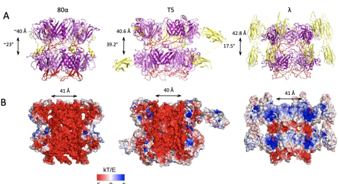
Figure 2: Structure of the tail tip of T5, highlighting the common features of the TTP fold in the different tail proteins. The side helix is coloured blue, the beta sandwich, pale yellow and the large loop, magenta (from Linares et al., in preparation). The terminator protein, reminiscent of the TTP, is that of l (3FZ2). Figure 3: A. Side views of known tube assembly of siphophages (TTP). Helical rise and twist are indicated. 80α tube structure was solved as part of the tip, thus helical parameters were not refined through the reconstruction process; the two rings are not as fully reconstructed and have slightly different conformations. The large loop is represented in red while C-terminal structures are in yellow. B. Electrostatic charge distribution of the lumen of siphophages tubes. 80α and T5 are vastly negative; this was proposed to smooth DNA transfer through the tube during viral entry. In contrast, l has negatively charged inter-ring loops while most of the inner-tube surface is not. Charge distribution in l tube was proposed to be involved in the assembly/pH sensitive disassembly of the tube of l [13].
Figure 4: Structural homologies of DTPs and BHPs. A. Top views of the DTP hexameric ring of phages T4 (5IV5), T5 (4JMQ), SPP1 (2X8K), 80α (6V8I), p2 (2WZP) and TP901-1 (4V96). The TTP fold is coloured red and decoration domains are coloured blue. B. Side views of the monomeric (top) and top views (bottom) of the trimeric BHP of phages Mu (1WRU), p2 (2WZP), 80a (6V8I) and T5 (to be published). Hub Domains I-IV are coloured blue, green, yellow and orange, respectively, on one monomer. In T5, the extension of HDII is coloured cyan, and in 80a and T5, C-terminal extensions are coloured red. In 80a-BHP the C-terminal domain, predicted to fold as a lipase domain, is not resolved and in T5, the second fibronectin domain is poorly resolved.