HAL Id: hal-02296759
https://hal-amu.archives-ouvertes.fr/hal-02296759
Submitted on 1 Dec 2020
HAL is a multi-disciplinary open access
archive for the deposit and dissemination of
sci-entific research documents, whether they are
pub-lished or not. The documents may come from
teaching and research institutions in France or
abroad, or from public or private research centers.
L’archive ouverte pluridisciplinaire HAL, est
destinée au dépôt et à la diffusion de documents
scientifiques de niveau recherche, publiés ou non,
émanant des établissements d’enseignement et de
recherche français ou étrangers, des laboratoires
publics ou privés.
Sponge digestive system diversity and evolution: filter
feeding to carnivory
Nelly Godefroy, Emilie Le Goff, Camille Martinand-Mari, Khalid Belkhir,
Jean Vacelet, Stephen Baghdiguian
To cite this version:
Nelly Godefroy, Emilie Le Goff, Camille Martinand-Mari, Khalid Belkhir, Jean Vacelet, et al.. Sponge
digestive system diversity and evolution: filter feeding to carnivory. Cell and Tissue Research, Springer
Verlag, 2019, 377 (3), pp.341-351. �10.1007/s00441-019-03032-8�. �hal-02296759�
Sponge digestive system diversity and evolution: filter feeding to carnivory
Nelly Godefroy1
• Emilie Le Goff1• Camille Martinand-Mari1• Khalid Belkhir 1• Jean Vacelet2•
Stephen Baghdiguian 1
Abstract
Sponges are an ancient basal life form, so understanding their evolution is key to understanding all metazoan evolution. Sponges have very unusual feeding mechanisms, with an intricate network of progressively optimized filtration units: from the simple choanocyte lining of a central cavity, or spongocoel, to more complex chambers and canals. Furthermore, in a single evolutionary event, a group of sponges transitioned to carnivory. This major evolutionary transition involved replacing the filter-feeding apparatus with mobile phagocytic cells that migrate collectively towards the trapped prey. Here, we focus on the diversity and evolution of sponge nutrition systems and the amazing adaptation to carnivory.
Keywords Sponge . Porifera . Evolution . Choanocyte . Filtration . Carnivory
Introduction
Sponges, or Porifera, are traditionally considered as one of the key groups for understanding basal metazoan evolution. The Porifera are a rich taxon of 8000 to 9000 described species, currently divided into four monophyletic classes: Demospongiae, Calcarea, Homoscleromorpha and, the most dif- ferentiated form, the Hexactinellida. Despite the prevalent view
of sponges as Bsimple^ animals, they have very sophisticated
feeding mechanisms involving the whole body and including a network of water filtration units, known as the aquiferous sys- tem. Studying the aquiferous system morphology of sponge classes, subclasses, orders and suborders will show us a very clear and interesting story of the successive modifications. Thus, the system has various degrees of complexity from the simplest
asconoid organization to the most complex leuconoid and sole-
noid filtration units (Fig. 1a). The great anatomical diversity of
the five sponge organization plans raises a number of questions concerning which are the most ancestral and which are the most efficient in terms of filtration. But whatever the evolutionary history of the classic filtering sponge aquiferous system, these different architectures, which aimed at optimizing food intake by increasing the exchange surfaces of the filtration system with the medium, never translated into a radical change in the general organization of the body plan, unlike that which occurred in carnivorous sponges (see below). In contrast to the diversity of Calcarea that possess the five types of body plan, in Hexactinellida, only leuconoids and syconoids are observed. All Homoscleromorpha are leuconoids, with a possible exception of Oscarella filipoi where a sylleibid system has
been described in Perez and Ruiz (2018) and all the extant
Demospongiae are leuconoids (Fig. 1c). It is likely that the
evo-* Jean Vacelet [email protected] * Stephen Baghdiguian
1
ISEM, CNRS, EPHE, IRD, Univ Montpellier, Montpellier, France
2 IMBE, CNRS, IRD, Station Marine d’Endoume, Aix Marseille Univ,
Univ Avignon, 13007 Marseille, France
lution in the class Calcarea (sponges characterized by calcareous spicules) occurred by the complexification of the asconoid type into syconoid, sylleibid, leuconoid, or solenoid organization
(Cavalcanti and Klautau 2011). The same scenario can be con-
sidered for the other three sponge classes with a secondary loss of asconoid, syconoid, sylleibid and/or solenoid forms. However, to date, there are no fossil remains or other evidence available to strengthen this hypothesis.
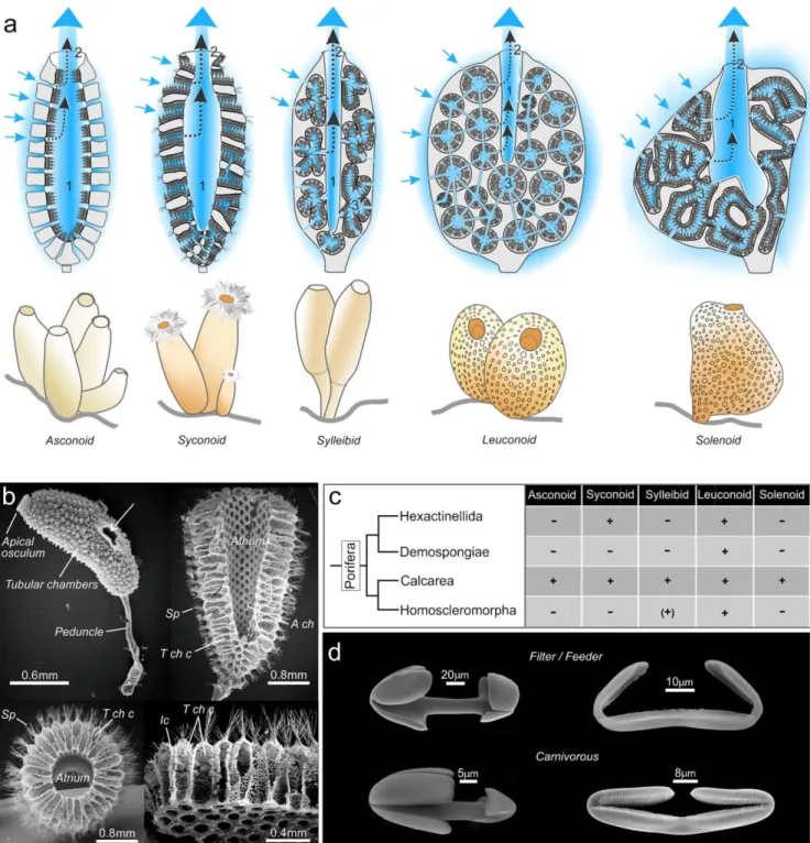
Fig. 1 Sponge nutrition system anatomy. a Schematic representation of the five sponge morphotypes. Arrows indicate the direction of the seawater flow into the spongocoel or atrium (1) and out through the oscule exhalant pore (2). In the more complex types, the water initially passes through a choanocyte chamber (3). Note numbers are deliberately faint to preserve the realism of the graphics. BGraphic design: ©Laurence Meslin - CNRS 2018.^ b Scanning electron microscope view of Sycon
sycandra. The upper left shows a whole specimen. The other micrographs
show different sections obtained by cryo-fracture after double fixation in glutaraldehyde and osmium followed by ultra-fast congelation in liquid nitrogen and metallization. Micrographs illustrate the morphological or- ganization of the syconoid system; sp, spicules; A ch, atrium chamber; T ch c, tubular choanocyte chamber; ic, inhalant canals; in left upper panel,
the arrow shows an additional exhalant orifice. c Phylogeny of the four classes of sponges in relation to the presence + or absence − of the five sponge morphotypes, (+) means that sylleibid organization was found only in one species in Homoscleromorpha. d Scanning electron micros- copy of representative spicules of filter/feeder sponges (upper panels) and carnivorous sponges (lower panels). Left upper panel: Palmate anisochela from Mycale (Oxymycale klausjanusorum, Van Soest, 2018), Mycalidae (filtering sponges) (courtesy of Rob Van Soest). Left lower panel: Palmate anisochela from an undescribed Asbestopluma, Cladorhizidae (carnivo- rous). Right upper panel: Placochela from Guitarra solorzanoi Cristobo, 1998, Guitarridae (filtering sponges) (courtesy of Javier Cristobo). Right lower panel: Placochela from Euchelipluma pristina Topsent, 1909, Cladorhizidae (carnivorous)
Anatomical generalities
The simplest of the five extant sponge plans, the asconoid form, is found only in the smallest calcareous sponges such as
Ascaltis, Clathrina and Leucosolenia. The asconoids are char-
acterized by inhalant pores (or ostia) directly connected to the large central spongocoel cavity, which is lined with a monolayer of flagellated choanocytes. In syconoids, the choanocytes line a multitude of small radial channels rather than directly lining the
spongocoel (Fig. 1b). This organization plan was an evolution-
ary advance because it increases the contact surface area be- tween the cells of the animal and the surrounding water, allowing greater absorption of food and increased body size. The syconoid body plan is found among Calcarea, for instance, in the genera Grantessa, Sycon and Sycettusa. In the third form, the sylleibids, the choanocyte chambers are radially arranged around invaginations of the atrial cavity. The sylleibid body plan is found in Calcarea Leucilla, Leucandra and Jenkina genera. In the fourth form, the leuconoids, which is the only form present in all four classes of sponges, the choanocytes are collected in elongated or spherical choanocyte chambers. These chambers are not linked to each other, but rather they communicate with the spongocoel and ostia through channels. This organization has the effect of slowing down the inward flow, which facilitates the filtration of the organic matter. This form is predominant in large calcareous sponges and essential- ly all of the d e mo s po nge s , he xac t i ne l li ds and homoscleromorphs are leuconoid. In the fifth and final filtering sponge body plan, solenoids, the filtration sys- tem is formed by anastomosing tubes lined with choano- cytes on the inner side and the atrium lacks choanoderm. The solenoid body plan was only recently described, in 2011, in calcareous sponges of the genus Leucascus
(Cavalcanti and Klautau 2011) (Fig. 1c). Evolution of
the choanocyte lining and the choanocyte chamber thus represented an efficiency drive that improved the nutri- tion process at the cellular and tissue levels, respectively. This efficiency drive, both in volume filtration, particle of organic matter retention and nutrient transfer, is most pronounced in the Hexactinellida, which are specially adapted to the deep sea. In these sponges, the leuconoid flagellar chambers, composed of a thin layer of anucleate ch oa nom ere s ( o r b ra nc hed c hoa no cyt es), a re complemented by a single, multinucleate syncytium ram- ifying throughout the sponge. This trabecular reticulum fulfills diverse functions, such as food transport in the whole animal and coordination of the filtering activity
in the absence of a nervous system (Leys et al. 2007).
Another efficiency adaptation that was developed to a greater or lesser degree in different types of sponge body plans is the formation of the mesohyl space that is made of a gelatinous extracellular matrix between the pinacoderm and choanoderm. The number of symbiotic
bacteria increases with the density of this structure to
maximize digestion (see the BInvolvement of the micro-
biota in the carnivorous diet^ section).
Nutrition/digestion in filtering sponges
In filtering sponges, nutritive particles are mostly trapped by flanges at the apical pole of choanocytes. Food particles are intracellularly digested by lysosomal acid phosphatases, as well as by alpha glucosidases, beta hexosaminidases and ca-thepsin D (Hahn-Keser and Stockem 1998). Cysteine protein-
ases like cathepsin L were biochemically implicated in the digestive process of Geodia cydonium also (Krasko et al.
1997). After intravacuolar digestion, the nutritive vacuoles
are exocytosed from choanocytes and delivered to pinacocytes and archeocytes, which pass them on to another cell type via the mesohyl space. Proof of this came when Ephydatia
fluviatilis were fed latex beads and the beads appeared in
choanocytes after 10 min of incubation, in exopinacocytes after 20 min, in mesohyl and endopinacocytes after 40 min and finally in some archeocytes after up to 12 h (Willenz
1980). Fluid pinocytosis is a type of endocytosis allowing
the capture of macromolecules and solutes in small vesicles that are redirected and incorporated into lysosomes. This kind of endocytosis has been observed for beta glycogen and iron saccharate transport in the leuconoid Demospongiae Suberites
massa (Diaz 1979). Many questions relating to the precise
kinetics and transfer routes of filtered food still need to be studied. At this stage, however, it is reasonable to propose the following scenario. Some of the choanocytes that have captured nutritive micro-particles transfer food to all the other cells via archeocytes, while a distinct subpopulation of the choanocytes transdifferentiate into additional archeocytes with regenerative potential. Thus, the diet would maintain the energetic needs of the sponge while ensuring its growth by forming new choanocyte chambers and all that without changing the global morphotype of the animal throughout its life cycle. Note that growth is limited as much by the compe- tition from other species in the ecosystem as by the nature and abundance of the nutriments in the medium.
Nutrition and digestion in carnivorous
species: data from
Lycopodina hypogea
The discovery of L. hypogea (Demospongiae), initially called
Asbestopluma hypogea and its carnivorous behavior by
Vacelet and Boury-Esnault in 1995 completely changed our
vision of sponge aquiferous system evolution. L. hypogea is not unique in this respect and indeed over a hundred carnivo- rous species have been i dentified exclusively in Demospongiae and most of them from deep waters.
L. hypogea and the related species Lycopodina occidentalis
are currently the only carnivorous sponges to be accessible to experimentation in the laboratory but only L. hypogea can be
harvested by scuba divers (Vacelet and Duport 2004; Riesgo
et al. 2007; Martinand-Mari et al. 2012). The study of
Lycopodina revealed that these carnivorous sponges were
not even sponges in the classical sense as they no longer meet
the phylum definition of sponges of Bergquist (1978) as Ba
sedentary, filter-feeding metazoan, which utilizes a single lay-er of flagellated cells (choanocytes) to pump a unidirectional water current through its body.^ These species have devel- oped a design that is unique among metazoans that enables
them to feed on macro-prey by phagocytosis (Fig. 2a–c). The
phagocytic cells act independently Bin their own interest^
completely without relying on the organism’s macro scale digestive cavity or specialized glands (Vacelet and Duport
2004). The independent acquisition of a carnivorous diet in
various flowering plants is also a spectacular example of ad- aptation and convergent evolution. However for plants, live- prey is only a supplementary food that is accommodated with- out a complete change in the nutrition system. For example, all the leaves of the insectivorous pitcher plant Cephalotus
follicularis are photosynthetic but only some leaves are car-
nivorous (Fukushima et al. 2017). Conversely to the situation
in plants, the reorganization of the nutrition/digestion system in carnivorous sponges was accompanied by major morpho- functional modifications that led to a strictly carnivorous diet
(Vacelet and Duport 2004; Martinand-Mari et al. 2012). In
L. hypogea, fasting animals are characterized by the presence
of many highly elongated filaments that play a decisive role in the capture of prey. After capture of prey, which are usually small crustaceans under 8 mm long and throughout the diges- tive process, these filaments regress rapidly before reforming, just as quickly, during the next fasting period. The average duration of an experimentally established cycle is approxi- mately 9 days. These morphogenetic transformations are di- rectly linked to an extremely dynamic cell turnover that in- volves coordination between programmed cell death (apopto- sis, autophagy), proliferation and cell migration (Martinand-
Mari et al. 2012). L. hypogea, like many other carnivorous
sponges, is attached to its substrate by a peduncle. At the base of the peduncle, there is a niche of stem cells that contributes to the overall cellular renewal of the body. When prey is ingested, there is a massive proliferation of these cells as shown by BrdU/EdU incorporation (Martinand-Mari et al.
2012). Sponge stem cells proliferate and then differentiate into
digestive cells that migrate along the central needle-like spic- ules of the peduncle to the prey at around 0.2 mm per hour (see
movie in supplementary data of Martinand-Mari et al. 2012) at
least in L. hypogea or other small species such as
L. occidentalis. Indeed, it is difficult to assume that such a
mechanism can occur in carnivorous sponges sometimes sur- passing the meter of length. The ensuing digestion ensures the
energy requirements of the animal including for its reproduc- tion. Although it has not yet been demonstrated, we can hy- pothesize that some of these stem cells (probably archeocytes that are described as totipotent cells in most studied sponges)
(Funayama 2013) could then transdifferentiate to allow the
regeneration of filaments, while others among them could
act as a pool of Bmemory cells,^ returning to the peduncle
base in wait for new prey. Finally, the hard skeletal parts of the prey, such as the chitinous exoskeleton of the crustaceans,
are possibly digested by endogenous chitinase (Table 1), prob-
ably in synergie with chitinase of bacterial origin (see Fig. 2e
and Vacelet and Duport 2004; Dupont et al. 2013, 2014), or
partly rejected for the hardest parts. Overall then, we see above that the complexity of regulation of cell dynamics and turn- over in L. hypogea rivals that of bilateral metazoans
(Martinand-Mari et al. 2012).
Spicule diversity in filtering and carnivorous
sponges
As explained above, the strategy for nutrition and digestion is completely different in filter feeding and carnivorous species. As well as developing mobile phagocytic cells (and losing the ability to filter feed), carnivorous sponges also needed to de- velop further secondary adaptations to efficiently capture large prey. The carnivores achieved this with an erect body shape that incorporates long filaments. In some genera, e.g.,
Chondrocladia, the filaments have specialized spheres at their
extremities that play the role of a float. In all the carnivores, the filaments have a regular lining of small siliceous hook-like spicules, generally under 100 μm long called microscleres (e.g., anisochelae, placochelae) that make a Velcro-like lining on their surface. These Velcro-lined filaments are thus perfect- ly adapted to trap the setae, or thin appendages, of diverse invertebrates, most often small crustaceans. The shape of these spicules reveals a close phylogenetic relationship be- tween the carnivorous sponges and some filter-feeding
demosponges of the order Poecilosclerida (Fig. 1d). The rela-
tionship is confirmed by molecular data and certainly thus not the result of evolutionary convergence (Fig. 43 of Hestetun
Fig. 2 Phagocytic activity analyzed by transmission electron microscopy during L. hypogea nutrition. a Archeocyte (ar) before nutrition. b Archeocyte/digestive cell (ardc) surrounding a crustacean cell (cc) 40 h after prey capture. At this stage, numerous phagosomes (ph) illustrate the intense phagocytic activity. c Detail of crustacean skeletal muscle (cskm) being digested by archeocyte/digestive cells (ardc). d Bacteriocyte (bc) in close contact with a sponge cell (sc) containing intravacuolar bacteria (ivb) at the beginning of the phagocytic process. e Numerous extracellular bacteria in the vicinity of a crustacean cuticle fragment (ccut). n, nucleus; ib, intracellular bacteria; psdp, pseudopod. Transmission electron micros- copy was performed as previously described (Vacelet and Duport 2004)
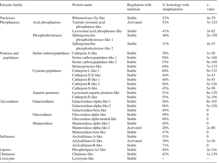
Table 1 Survey of hydrolases found in a differential transcriptomic analysis of unfed or fed L. hypogea. The percentage of homology with homologs in the carnivorous/filter-feeding barrier reef sponge
Amphimedon queenslandica that has been fully sequenced (Srivastava
et al. 2010) is mentioned with the e-values of the alignments
Enzyme family Protein name Regulation with nutrition
% homology with Amphimedon
e-
value Nucleases Ribonuclease Oy-like Stable 22% 4e-29 Phosphatases Acid phosphatases Tartrate-resistant acid Activated 52% 7e-124
phosphatase-like
Lysosomal acid phosphatase-like Stable 41% 3e-82 Phosphodiesterases Sphingomyelin Stable 46% 2e-158
phosphodiesterase-like 1
Sphingomyelin Stable 31% 4e-53 phosphodiesterase-like 2
Protease and peptidases
Serine-carboxypeptidases Cathepsin A-like Stable 30% 1e-50 Serine carboxypeptidase-like 1 Stable 56% 3e-160 Serine carboxypeptidase-like 2 Stable 53% 9e-169 Sérine protease-like Stable 44% 7e-133 Cysteine-peptidases Cathepsin L-like 1 Stable 60% 9e-132 Cathepsin X/Z-like Stable 44% 3e-43 Cathepsin B-like 1 Stable 46% 5e-93 Cathepsin B-like 2 Stable 56% 5e-126 Cathepsin O-like Stable 45% 5e-99 Aspartic-proteases Lysosomal aspartic protease-like Stable 51% 8e-129
Cathepsin D-like Stable 55% 3e-156 Glycosidases Galactosidases Galactosidase alpha-like 1 Stable 56% 8e-165 Galactosidase alpha-like 2 Stable 56% 5e-150 Galactosidase beta-like Stable 49% 0 Glucosidases Glucosidase alpha-like Stable 49% 0 Glucosidase alpha neutral-like Stable 68% 0 Mannosidase Mannosidase alpha-like 1 Stable 49% 0
Mannosidase alpha-like 2 Activated 28% 2e-80 Mannosidase beta-like Stable 47% 0 Sulfatases Arylsulfatase A-like Stable 51% 0
Arylsulfatase G-like Activated 39% 1e-56 Arylsulfatase B-like Stable 71% 0 Lipases Phospholipase A2-like Stable 44% 2e-116 Chitinases Chitinase-like Stable 42% 1e-139
Lysozyme Lysozyme-like Stable – –
Experimentally, total RNAwas isolated with RNeasy kit (QIAGEN) for eight individuals (two individuals for each condition: unfed, 15 h, 40 h and 216 h after nutrition). Libraries were constructed with a TruSeq stranded mRNA sample prep kit (Illumina, ref. RS-122-2101) according to the manufacturer instructions. Briefly, poly-A eucaryotic RNAs were purified using oligo-d(T) magnetic beads from 500 ng of total RNA (to effectively eliminate bacterial RNA). The poly-A+ RNAs were fragmented and reverse-transcribed using random hexamers, Super Script II (Life Technologies, ref. 18064-014) and actinomycin D. During the second-strand generation step, dUTP substituted dTTP. This prevents the second strand being used as a template in the final PCR amplification. Double-stranded cDNAs were adenylated at their 3′ ends before ligation was performed using Illumina’s indexed adapters. Ligated cDNAs were amplified following 15 cycles of PCR and PCR products were purified using AMPure XP Beads (Beckman Coulter Genomics, ref. A63881). Libraries were verified using a Bioanalyzer on a DNA1000 chip (Agilent) and quantified with a KAPA Library quantification kit (Roche, ref. KK4824). Eight libraries were pooled in equimolar amounts and sequencing was performed on a HiSeq2000 using the paired end 2 × 100-nt protocol on 1 lane. This sequencing produced between 17 and 24 million passed filter clusters per library (http://www.mgx.cnrs.fr/). Artemia sequences (i.e., contaminants acquired from prey ingestion) were removed and clean reads were de novo assembled into contigs using the Trinity transcriptome assembly software by the platform MBB from Montpellier (https://mbb.univ-montp2.fr/). Sequences from L. hypogea were extracted from these databases by TBLASTN alignments with protein sequences from other model organisms on the MBB platform. The L. hypogea isolated sequences were scanned with interproscan 5 (http://www.ebi.ac.uk/Tools/pfa/iprscan5/) to verify the presence of functional or structural domains specific for each protein. In parallel, each selected sequence was used to conduct TBLASTN searches on NCBI (all databases) to rename the sequence. The best assembly contigs were used as a reference for aligning reads from the 8 different L. hypogea individuals to estimate the expression levels (Bowtie, RSEM). From these normalized expression levels, appropriate statistical methods (Edger, Imb) were applied to identify genes whose expression levels vary significantly at least by a factor of 4 between conditions
et al. 2017). It is not known what the microsclere spicules do
in the filter feeding as they seem randomly dispersed in the sponge tissue. The filter-feeder microscleres are not involved
in providing skeletal structure, as this function is provided by larger spicules, from 150 μm to some millimeters or more, called megascleres, in both filtering and carnivorous species.
It is remarkable that in carnivorous sponges, the same spicules appear to have acquired a fundamental function in prey cap- ture by simply changing their arrangement. The morphologi- cal similarity of cheloid microscleres of non-carnivorous and
carnivorous sponges is illustrated in Fig. 1(d). The anisochelae
and the placochelae of the massive filter feeders Mycale and
Guitarra are randomly dispersed, while in carnivorous
sponges such as Asbestopluma and Euchelipluma, the anisochelae and the placochelae form a regular lining at the filament surfaces. An intermediate stage between a filter- feeding sponge, with an aquiferous system and micro-phagic digestion and a carnivorous sponge, without the aquiferous system, is represented by the genus Chondrocladia, classified in the family Cladorhizidae along with the other carnivorous sponges. In Chondrocladia, the choanocyte chambers persist but they no longer ensure the role of filtration/digestion but rather function to inflate the spheres at the apical pole of the sponges where the prey catching spicules are arranged. There was therefore a functional inversion of the choanocyte cham- bers in these intermediate species. Note the main function of the choanocytes in these intermediate architectures is structur- al, like the role of the spicules in filter species. The spicules then have evolved from a mainly architectural role in filter/ feeder species to forming structures exclusively involved in prey capture in the carnivorous species. Finally, in most strict- ly carnivorous sponges, the choanocyte chambers have completely disappeared, leading to a total reorganization of the body plan and digestion system.
Functional consequences of the carnivorous
diet
Filtering and carnivorous sponges developed different strate- gies for nutrition because the food for the latter species is much larger, up to 8 mm, whereas filtered particles are in the 1 nm to 2 μm range. These different strategies do not imply differences in phagocyte size (around 10 μm). Another major difference is that nutritive particles are delivered to the cells via the inhalant channels in the filtering species, whereas in the carnivores, which do not have any of these structures, it is the phagocytic cells that migrate towards the food after capture of a prey
(Martinand-Mari et al. 2012). Finally, in filtering species, there
is no major morphological change associated with nutrition, as it is practically continuous, although there can be occasional breaks in filtration with orifice closures during reorganization of the aquiferous system. In L. hypogea on the other hand, where food intake is discontinuous and dependent on the cap- ture of a prey, which can arrive months apart, there are dramatic morphologic rearrangements during a digestion cycle during which the sponge changes form and returns to the initial state
(Fig. 3a–d). Our lab is currently conducting an analysis of the
differential expression of the molecular actors that control this
unique form of shape homeostasis in carnivorous sponges. Extending the results obtained in Lycopodina to the deep- water carnivorous sponges suggests this cyclical mode of nu- trition could be an adaptation to the oligotrophic environments of the abyssal conditions. This hypothesis is consistent with the fact that indeed hexactinellids, which have the most efficient filtration/nutrition system of any sponges, have difficulty reaching a depth of 6770 m, whereas carnivorous species are found below 8000 m, including one at 8840 m. The tendency towards carnivorous behavior and macro-prey in deep-sea spe- cies has also been described in other invertebrate groups. Thus, large copepods were found in the digestive tract of abyssal sea squirts (e.g., in Octanemidae, Ascidiidae and in the Sorberacea
class) (Monniot and Monniot 1975, 1991). These tunicates
likely switch between filtering and carnivorousness and thus their evolution was less drastic and they only have a small anatomical modification of the branchial sac without any major reorganization of the overall body plan.
Conversely, carnivorous sponges are concrete proof that a radical functional transition is possible between a system of nutrition/digestion by filtration and a one of feeding by capture of prey, with total loss of the filtration system. This transition was accompanied by a profound change in the overall organi- zational plan and especially of the digestion system. In
L. hypogea, this morpho-functional reorganization is directly
associated with the acquisition of an axial polarity of the whole organism (polarity being defined as a structure that is different at one pole with respect to the opposite one) that, equally remarkably, resembles the apico-basal polarity at the cellular
level of the epithelial digestive system in mammals (Fig. 3e, f).
Digestion of large prey necessitates a panel of digestive enzymes and we found there is a remarkable diversity of en- zyme equipment in L. hypogea. All the main families of lyso- somal enzymes are present in the L. hypogea transcriptome, i.e., the nucleases, phosphatases, proteases, peptidases, glyco-
sidases, sulfatases and lipases (Table 1). Most of these en-
zymes are expressed at a constant level throughout the diges- tion cycle. However, some, including tartrate-resistant acid phosphatase type 5-like, mannosidase alpha-2-like and arylsulfatase G-like, are induced by a nutritional stimulus
(Table 1). These preliminary data, however, allow us to ini-
tially say that the mechanisms involved in intracellular diges- tion in L. hypogea are at least partly subject to physiological regulation and also that these mechanisms are as complex biochemically as those of higher vertebrates. Finally, the pres-
ence of lysozyme and of a chitinase (Table 1) suggests there is
a preliminary extracellular digestion in carnivorous sponges to degrade the prey, which are almost exclusively small crusta- ceans. Various chitinases and other bacterial enzymes must also contribute to this degradation, as suggested by Vacelet
and Duport (2004). Taken together, these considerations sug-
gest that the nutrition/digestion strategy of the whole organism in carnivorous sponges is phenomenologically similar to that
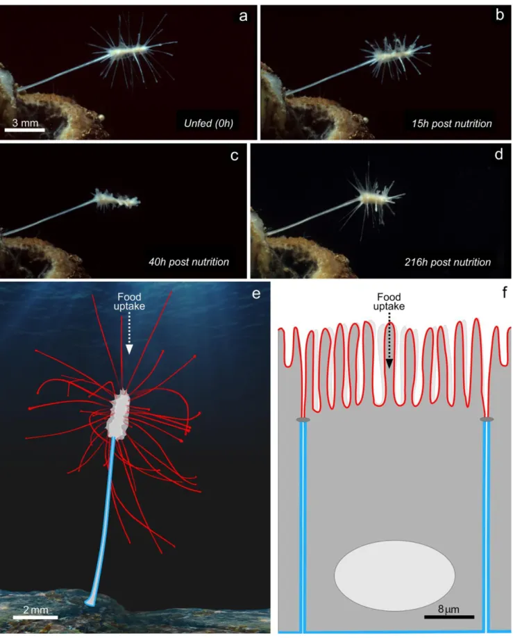
Fig. 3 Nutrition/digestion features associated with carnivorous diet. a–d Morphological changes of Lycopodina hypogea during a digestion cycle at the indicated times after introduction of prey. The long filaments of the apical region regress completely 40 h post-nutrition (c) to reform at the end of the digestive cycle (d). e and f Functional analogy between
L. hypogea and mammalian digestive cell. Schematic representation of
apico-basal polarity of carnivorous sponge (e) and a digestive epithelial cell (f). The red lines represent the apical pole and the blue line the basolateral domain. BGraphic design: ©Laurence Meslin - CNRS 2018^
of the cells of the digestive tube of most bilaterians. From bacteria to higher vertebrates, the regulation of the composi- tion of the proteins at the membrane and especially their asym- metric distribution, has played a determining role in the morpho-functional diversification of prokaryotic and eukary- otic cells. For example, in the bacterium Caulobacter
crescentus, the polarized distribution of the flagellum anchor
protein allows differentiation of an individual mother cell into an immobile pedunculated cell plus another flagellated mobile
cell (Gober et al. 1991). In these bacteria, it is the asymmetric
membrane-protein distribution that allows genuine social be- havior in the colony and that optimizes the use of nutrient resources. In the absorptive intestinal cells of vertebrates (known as enterocytes), an asymmetry of the biochemical composition of apical and basolateral membrane domains leads to a morphological differentiation of the apical domain into a specialized brush border. Here, polarity is at the origin of a functional differentiation of the two membrane domains and an increase of the exchange surface of the apical mem- brane domain with the lumen of the digestive tract. By analo- gy, polarity is also found in the difference in cell composition and global shape between the basal and apical regions of
L. hypogea. This difference results in a functional differentia-
tion of the whole organism and the establishment of many long filaments in the apical region. These filaments increase the interaction surface of the prey capture system with the medium, thus increasing and optimizing nutrition by vectorial transport of nutrients. This efficient directional transport is
illustrated in a cell or organism, respectively, in Fig. 3 (e, f).
It is likely that it is this efficient nutritive system that has allowed carnivorous sponges to colonize the extreme depths of the world’s oceans.
Involvement of microbiota in the carnivorous
diet
Marine filtering sponges harbor a complex and diverse com- munity of microbes and the carnivorous L. hypogea is no exception as around 800 operational taxonomic units of bac- terial and archaeal phyla have been documented in this model
carnivore (Dupont et al. 2013, 2014). These bacteria likely
help with the digestion of prey as they include strains with chitinolytic activity. A recent study shows that similar micro- bial communities are present in other carnivorous sponges,
Chondrocladia grandis and Cladorhiza oxeata and that there
are bacterial communities that are specific to either sponge but also that certain bacterial taxa are enriched in distinct anatom- ical regions. For example, bacteria of the genera Colwellia and
Reichenbachiella are found mainly in the capture spheres (api-
cal p o s i t i o n ) whi l e the b a c t e r i a of the ge n e r a
Robiginitomaculum, Maritimimonas, Granulosicoccus, Mycobacterium, Anderseniella and Nitrospira are
concentrated in the peduncle (basal position) (Verhoeven
and Dufour 2017). Digestion therefore likely relies on both
bacterial and lysosomal enzymes. In L. hypogea, the prey is literally engulfed in a few hours by archeocytes that have differentiated into digestive cells and by bacteriocytes that
have numerous cytoplasmic bacteria (Fig. 2b–d). The exact
species of bacteria inside and outside the bacteriocytes (Fig.
2d, e) have not yet been identified, but transmission electron microscopy data strongly suggest their involvement in the
extracellular and intracellular digestion process (Fig. 2).
Thus, the bacteria cooperate with the digestive cells and con- tribute to the total degradation of the prey. The entire autolytic/ phagocytic process lasts between 8 and 10 days for larger prey in the 8-mm range. Interestingly, the bacterial communities associated with carnivorous sponges are not very different from those of filtering sponges. There are a few examples of bacterial host specificity. For example, methanotrophic bacte- ria are only found in the carnivorous Cladorhiza
methanophila (Hestetun et al. 2016). Thus, the acquisition of
a strict carnivorous diet was not accompanied by a significant related modification of the diversity of associated bacterial communities; the bacteria in filtering sponges are equally im- portant for the efficient digestion of prey in carnivorous sponges. There is equivalence for host lysosomal enzymes also, which have a comparable diversity and primary structure
in carnivorous and filter/feeder sponges (Table 1). In summa-
ry, most of the biochemical and microbiological actors already present in classical models in sponge biology also contribute to the digestion processes of carnivorous sponges. However, in some cases (e.g., the secreted enzyme lysosyme that is present in L. hypogea transcriptome but not in the genome
of the filter-feeding sponge A. queenslandica, see Table 1),
we can suppose that subtle biochemical innovations have oc- curred during the filter feeder/carnivorous diet transition.
Conclusion
Several plant lineages have adapted to include active predation into their modus operandi but none have abandoned their ances- tral strategy of photosynthesis. By contrast, in sponges, the major evolutionary transition to a new axial polarity of the digestion system that led to the acquisition of carnivory enabled some sponges to relinquish passive filter feeding. Indeed, in organisms that have a digestive cavity or an aquiferous system as in filtering sponges, it is the food that is brought to the digestive system while it is the opposite in carnivorous sponges. This inversion of morpho-functional polarity was accomplished by a remark- able organizational plasticity in the basic sponge body plan. Identifying the molecular and cellular actors involved in this plasticity could help us better understand the evolutionary history and beginnings of early metazoans. One theory, still debated, is that sponges and choanoflagellates that both capture nutritive
particles with an apical sieve might have evolved directly from a unicellular common ancestor. More generally, the question now is how the sponges have evolved from this common ancestor to multicellular filtering forms. Similarly, the transition from the sponge phylum to other metazoans is complex and still uncertain. One side-branch of this evolution was the transition of sponges to the carnivorous diet, which evolved just one time. The sponges and cnidarian lineages diverged 600 million years ago, whereas the split between filtering and carnivorous sponges was more recent and occurred only in the Poecilosclerida order of the Demospongiae. Since it is now clearly established that Porifera
are the most basal metazoans (Simion et al. 2017; Pett et al.
2019), we can therefore postulate that three or four sponge phyla
derived from the Urmetazoa and kept the filter-feeder organiza- tion and exclusively intracellular phagocytosis, while the other Metazoa lost the canal system and choanocyte chamber. However, it was clearly a major evolutionary task to go from a sponge to a Cnidaria or other metazoans. Indeed, as stated by
Cavalier-Smith: Bit is selectively immensely harder for organisms
that feed by swallowing others or bits of them (a purely eukary-otic propensity) to switch from intracellular phagocytosis, as in amoebae or ciliates, to eating with a multicellular mouth and gut, whose cells have novel functions and structures absent in their unicellular ancestors. Animal feeding is effective only if novel cell types cooperate at a higher organizational level^ (Cavalier-
Smith 2017). The complete loss of filter-feeding apparatus in
sponges and its replacement by another type of organization leading to carnivory is a rare example of such a major transition in evolution. Therefore, further cellular and molecular studies of carnivorous sponges could improve our understanding of the evolutionary innovations that have led to the formation of a stomodeum, a mouth and a coelenteron in cnidarians and to the development of a coelom with lengthening of the stomodeum and formation of the digestive tract in all other metazoans.
Acknowledgments We thank Laurence Meslin (CNRS, BPlateforme Communication Scientifique Visuelle – ISEM^), for the drawing and elaborating the figures. For the transcriptomic data, we benefited from the Montpellier Bioinformatics Biodiversity platform supported by the LabEx CeMEB, an ANR BInvestissements d’avenir^ program (ANR- 10-LABX-04-01).
Compliance with ethical statements
Conflict of interest The authors declare that they have no conflict of interest.
Ethical approval All applicable international, national and/or institu- tional guidelines for the care and use of animals were followed.
References
Bergquist PR (1978) Sponges. Hutchinson, London & University of California Press, Berkeley & Los Angeles, pp 1–268
Cavalcanti FF, Klautau M (2011) Solenoid: a new aquiferous system to Porifera. Zoomorphology 130:255–260. https://doi.org/10.1007/ s00435-011-0139-7
Cavalier-Smith T (2017) Origin of animal multicellularity: precursors, causes, consequences—the choanoflagellate/sponge transition, neurogenesis and the Cambrian explosion. Philos Trans R Soc B 372:20150476. https://doi.org/10.1098/rstb.2015.0476
Diaz J (1979) Variations, différenciations et fonctions des catégories cellulaires de la démosponge d’eaux saumâtres, Suberites massa Nardo, au cours du cycle biologique annuel et dans des conditions expérimentales. Docteur d’Etat Mention Sciences, Université des sciences et techniques du Languedoc
Dupont S, Corre E, Li Yet al (2013) First insights into the microbiome of a carnivorous sponge. FEMS Microbiol Ecol 86:520–531. https:// doi.org/10.1111/1574-6941.12178
Dupont S, Carre-Mlouka A, Domart-Coulon I et al (2014) Exploring cultivable Bacteria from the prokaryotic community associated with the carnivorous sponge Asbestopluma hypogea. FEMS Microbiol Ecol 88:160–174. https://doi.org/10.1111/1574-6941.12279 Fukushima K, Fang X, Alvarez-Ponce D et al (2017) Genome of the
pitcher plant Cephalotus reveals genetic changes associated with carnivory. Nat Ecol Evolut 1:0059. https://doi.org/10.1038/s41559- 016-0059
Funayama N (2013) The stem cell system in demosponges: suggested involvement of two types of cells: archeocytes (active stem cells) and choanocytes (food-entrapping flagellated cells). Dev Genes Evol 223:23–38. https://doi.org/10.1007/s00427-012-0417-5 Gober JW, Alley MR, Shapiro L (1991) Positional information during
Caulobacter cell differentiation. Curr Opin Genet Dev 1:324–329 Hahn-Keser B, Stockem W (1998) Intracellular pathways and degrada-
tion of endosomal contents in basal epithelial cells of freshwater sponges (Porifera, Spongillidae). Zoomorphology 117:223–236. https://doi.org/10.1007/s004350050047
Hestetun JT, Dahle H, Jørgensen SL et al (2016) The microbiome and occurrence of methanotrophy in carnivorous sponges. Front Microbiol 7:1781. https://doi.org/10.3389/fmicb.2016.01781 Hestetun JT, Tompkins-Macdonald G, Rapp HT (2017) A review of
carnivorous sponges (Porifera: Cladorhizidae) from the boreal North Atlantic and Arctic. Zool J Linnean Soc 181:1–69. https:// doi.org/10.1093/zoolinnean/zlw022
Krasko A, Gamulin V, Seack J et al (1997) Cathepsin, a major protease of the marine sponge Geodia cydonium: purification of the enzyme and molecular cloning of cDNA. Mol Mar Biol Biotechnol 6:296–307 Leys SP, Mackie GO, Reiswig HM (2007) The biology of glass sponges.
Adv Mar Biol 52:1–145. https://doi.org/10.1016/S0065-2881(06) 52001-2
Martinand-Mari C, Vacelet J, Nickel M et al (2012) Cell death and re- newal during prey capture and digestion in the carnivorous sponge Asbestopluma hypogea (Porifera: Poecilosclerida). J Exp Biol 215: 3937–3943. https://doi.org/10.1242/jeb.072371
Monniot C, Monniot F (1975) Abyssal tunicates: an ecological paradox. Ann Inst océanogr 51(1):99–129
Monniot C, Monniot F (1991) Découverte d’une nouvelle lignée évolutive chez les ascidies de grande profondeur : une Ascidiidae carnivore. C R Acad Sci Paris III:383–388
Perez T, Ruiz C (2018) Description of the first Caribbean Oscarellidae (Porifera: Homoscleromorpha). Zootaxa 4369:501–514. https://doi. org/10.11646/zootaxa.4369.4.3
Pett W, Adamski M, Adamska M et al (2019) The role of homology and orthology in the phylogenomic analysis of metazoan gene content. Mol Biol Evol. https://doi.org/10.1093/molbev/msz013
Riesgo A, Taylor C, Leys SP (2007) Reproduction in a carnivorous sponge: the significance of the absence of an aquiferous system to the sponge body plan. Evol Dev 9:618–631. https://doi.org/10.1111/ j.1525-142X.2007.00200.x
Simion P, Philippe H, Baurain D et al (2017) A large and consistent phylogenomic dataset supports sponges as the sister group to all other animals. Curr Biol 27:958–967. https://doi.org/10.1016/j.cub. 2017.02.031
Srivastava M, Simakov O, Chapman J et al (2010) The Amphimedon queenslandica genome and the evolution of animal complexity. Nature 466:720–726. https://doi.org/10.1038/nature09201 Vacelet J, Boury-Esnault N (1995) Carnivorous sponges. Nature
373(6512):333–335
Vacelet J, Duport E (2004) Prey capture and digestion in the carnivorous sponge Asbestopluma hypogea (Porifera: Demospongiae). Zoomorphology 123:179–190. https://doi.org/10.1007/s00435-004-0100-0
Verhoeven JTP, Dufour SC (2017) Microbiomes of the Arctic carnivo- rous sponges Chondrocladia grandis and Cladorhiza oxeata suggest
a specific, but differential involvement of bacterial associates. Arctic Sci 4:186–204. https://doi.org/10.1139/as-2017-0015
Willenz P (1980) Kinetic and morphological aspect of particle ingestion by the freshwater sponge Ephydatia fluviatilis L. In: Smith DC, Tiffon Y (eds) Nutrition in the lower Metazoa. Pergamon Press, Oxford & New York