HAL Id: hal-03036109
https://hal.archives-ouvertes.fr/hal-03036109
Submitted on 2 Dec 2020HAL is a multi-disciplinary open access archive for the deposit and dissemination of sci-entific research documents, whether they are pub-lished or not. The documents may come from teaching and research institutions in France or abroad, or from public or private research centers.
L’archive ouverte pluridisciplinaire HAL, est destinée au dépôt et à la diffusion de documents scientifiques de niveau recherche, publiés ou non, émanant des établissements d’enseignement et de recherche français ou étrangers, des laboratoires publics ou privés.
Quantification and imaging of HER2 protein using
nanocrystals conjugated with single-domain antibodies
S Glukhov, M Berestovoy, P Chames, D Baty, I Nabiev, A Sukhanova
To cite this version:
S Glukhov, M Berestovoy, P Chames, D Baty, I Nabiev, et al.. Quantification and imaging of HER2 protein using nanocrystals conjugated with single-domain antibodies. Journal of Physics: Conference Series, IOP Publishing, 2017, International Symposium Physics, Engineering and Technologies for Bio-Medicine, 784, pp.012016. �10.1088/1742-6596/784/1/012016�. �hal-03036109�
PAPER • OPEN ACCESS
Quantification and imaging of HER2 protein using
nanocrystals conjugated with single-domain
antibodies
To cite this article: S Glukhov et al 2017 J. Phys.: Conf. Ser. 784 012016
View the article online for updates and enhancements.
Related content
Segmentation of HER2 protein
overexpression in immunohistochemically stained breast cancer images using Support Vector Machines
Raquel Pezoa, Luis Salinas, Claudio Torres et al.
-HER2 specific delivery of methotrexate by dendrimer conjugated anti-HER2 mAb
Rameshwer Shukla, Thommey P Thomas, Ankur M Desai et al.
-Molecular dynamic simulation of Trastuzumab F(ab’)2 structure in corporation with HER2 as a theranostic agent of breast cancer
S Hermanto, M Yusuf, A Mutalib et al.
-Recent citations
Effects of different quantities of antibody conjugated with magnetic nanoparticles on cell separation efficiency
Amir Hossein Haghighi et al
Quantification and imaging of HER2 protein using
nanocrystals conjugated with single-domain antibodies
S Glukhov1, M Berestovoy1, P Chames2, D Baty2, I Nabiev1,3 and A Sukhanova1,3 1
Laboratory of Nano-Bioengineering, National Research Nuclear University MEPhI (Moscow Engineering Physics Institute), Moscow, Russian Federation
2
INSERM U1068 and CNRS UMR7258, Cancer Research Center, Marseille, France, Institut Paoli-Calmettes and Aix-Marseille, Marseille, France
3
Laboratory of Research in Nanoscience, LRN - EA4682, University of Reims Champagne Ardenne, Reims, France
E-mail: [email protected]
Abstract. This study dealt with quantification and imaging of human epidermal growth factor
receptor 2 (HER2), an important prognostic marker for cancer diagnosis and treatment, using specific quantum-dot-based conjugates. Fluorescent inorganic nanocrystals or quantum dots (QDs) are extremely highly resistant to photobleaching and have a high emission quantum yield and a continuous range of emission spectra, from the ultraviolet to the infrared regions. Ultrasmall nanoprobes consisting of highly affine anti-HER2 single-domain antibodies (sdAbs or "nanobodies") conjugated with QDs in a strictly oriented manner have been designed. QDs with a fluorescence peak maxima at wavelengths of 562 nm, 569 nm, 570 nm or in the near-infrared region were used. Here, we present our results of ISA quantification of HER2 protein, in situ imaging of HER2 protein on the surface of HER2-positive SK-BR-3 cells in immunohistochemical experiments, and counting of stained with anti-HER2 conjugates HER2-positive SK-BR-3 cells in their mixture with unstained cells of the same culture in flow cytometry experiments. The data demonstrate that the anti-HER2 QD–sdAb conjugates obtained are highly specific and sensitive and could be used in numerous applications for advanced integrated diagnosis.
1. Introduction
Several types of fluorescent labels are used in biological and medical applications: heterocyclic organic dyes, fluorescent proteins, and fluorescent inorganic nanocrystals or quantum dots (QDs). The main advantages of QDs over other fluorescent dyes are an extremely high resistance to photobleaching, bright photoluminescence, and the possibility of obtaining a continuous range of emission colors from ultraviolet to infrared [1]. These characteristics make it possible to use QDs for imaging both fixed and live biological samples [2], as well as for obtaining contrast images of samples of different thicknesses in multiphoton applications [3, 4, 5]. Antibodies carrying fluorescent labels have become widely used for imaging specific protein markers, cells, and tissues in biology and medicine.
Detection of specific protein cancer markers in clinical samples is important for theranostics, a promising approach at the interface of therapeutics and diagnostics [6, 7]. Human epidermal growth factor receptor 2 (HER2) has been found to be an extremely important marker for detection of International Symposium Physics, Engineering and Technologies for Bio-Medicine IOP Publishing IOP Conf. Series: Journal of Physics: Conf. Series 784 (2017) 012016 doi:10.1088/1742-6596/784/1/012016
International Conference on Recent Trends in Physics 2016 (ICRTP2016) IOP Publishing Journal of Physics: Conference Series 755 (2016) 011001 doi:10.1088/1742-6596/755/1/011001
Content from this work may be used under the terms of theCreative Commons Attribution 3.0 licence. Any further distribution of this work must maintain attribution to the author(s) and the title of the work, journal citation and DOI.
malignant tumors, assessment of the stage of their development, and medical prognosis [8]. The development of antitumor therapy where HER2 serves as a target is underway [9, 10].
Immunoglobulins of cartilaginous fishes belonging to the nurse shark antigen receptor (IgNAR) family [11, 12] and similar to G-type immunoglobulins of camelids [13] stand apart from other immunoglobulins serving as antibodies in mammals. The antigen binding surface of these antibodies consists of a single globular domain, in contrast to two domains in typical mammalian immunoglobulins. An isolated variable domain of a camelid similar to G-type immunoglobulin capable of antigen binding is termed a single-domain antibody (sdAb). The size of sdAbs (weighing as little as 13 kDa) makes them the smallest sensors or capture molecules for specific nanoprobes currently used in biomedicine [14]. The technique for obtaining recombinant sdAbs allows their primary amino acid sequence to be additionally tagged.
In this study, we estimated the performance of nanoprobes containing photoluminescent nanocrystals with different spectral characteristics and highly affine sdAbs against HER2 in detecting this cancer marker.
2. Materials and methods
2.1. Cell cultures
The SK-BR-3 and MDA-MB-231 cell cultures were kindly provided by Dr. Baryshnikova (Laboratory of Experimental Diagnosis and Biotherapy of Tumors, Blokhin Cancer Research Center, Moscow, Russia). The cultures were grown on cover slips or chamber slides to 80% confluence in RPMI 1640 supplemented with 10% bovine serum at 37°C in an atmosphere of 5% CO2.
2.2. Antibodies
Single-domain antibodies against HER2 containing a C-terminal Cys–(His)6 peptide [5, 15, 16] were kindly provided by Dr. Baty (CNRS UMR 7258, Inserm U1068, Centre de Recherche en Cancérologie de Marseille).
2.3. Quantum dots
CdSe/ZnS core/shell QDs with emission peaks maxima at 562 nm, 569 nm, and 570 nm (QDs562, QDs569, and QDs570, respectively) dissolvable in an organic solvent were kindly provided by Dr. P. Samokhvalov (National Research Nuclear University MEPhI, Moscow, Russia). The PbS/CdS/ZnS core/core-periphery/shell QDs with an emission peak maximum in the near infrared region (1120 nm, QDsNIR) were kindly provided by Dr. Sungjee Kim (Department of Chemistry Pohang University of Science & Technology Pohang, South Korea).
2.4. Transfer of quantum dots to the aqueous phase
The procedure is described in detail elsewhere [14]. In brief, 10 mg of QDs was dissolved in 1 ml of chloroform, an equal volume of methanol was added, and the mixture was stirred and centrifuged at 14,000 rpm. The supernatant was removed, and the pellet was dissolved in methanol, after which 200 μl of a 10 mg/ml solution of DL-cysteine in methanol was added dropwise while stirring to a final volume of 1 ml. The QDs were settled by centrifugation at 14,000 rpm and washed with methanol three times, centrifuging the mixture at the same speed every time. The pellet was vacuum-dried and dissolved in water while adding 1 M NaOH dropwise and stirring in a vortex. The resultant QD solution was sonicated and filtered through a filter with a mesh size of 0.22 μm. The size variation of the dissolved nanoparticles was estimated by dynamic light scattering using a ZetaSizer ZS device (Malvern). A 150 mg/ml solution of the complex polymer HS–(CH2)m–EGn–OH (Prochimia) was added to the aqueous suspension of 10 mg of QDs prepared for water-solubilization, and the mixture was incubated at 4°C for at least 12 h. Unbound molecules were removed by centrifugation on Amicon Ultracel-10K filters. Milli-Q water filtered through a filter with a mesh size of 0.22 μm was used for washing. The final purification of the QDs was performed in PD MiniTrap Sephadex G-25
columns (GE Heathcare). The size variation of the nanoparticles was estimated by dynamic light scattering using a ZetaSizer ZS device (Malvern).
2.5. Bioconjugation
The procedure is described in detail elsewhere [14]. In brief, a solution of the N-(p-maleimidophenyl) isocyanate crosslinker was added to 50 μl of a 2 mg/ml aqueous solution of QDs coated with modified polyethylene glycol buffered with sodium borate (pH 8.5) to a 50-fold molar excess of the crosslinker over the QDs. The reaction mixture was incubated at room temperature in the dark for 30 min while gently stirring. The reaction of QD activation was terminated by gel filtration of the preparation through Sephadex G-25 columns equilibrated with 50 mM phosphate buffer saline (PBS).
Disulfide bonds in the antibody preparation were reduced by incubation in a tris(2-carboxyethyl)phosphine solution at a molar ratio of 1:10 for 30 min; the preparation was purified in PD MiniTrap Sephadex G-25 columns. The sdAbs treated with tris(2-carboxyethyl)phosphine were mixed with the maleimid-activated QDs at a molar ratio of 10:1. The reaction was carried out at room temperature in the dark for 2 h while gently stirring. The preparation of the conjugates was purified in Superdex 200 columns equilibrated with 50 mM PBS.
2.6. Agarose gel electrophoresis
Electrophoresis of the QDs and QD–sdAb conjugates was performed in 0.5% agarose gel at a potential gradient of 1 V/cm in tris-acetic buffer saline for 30–60 min. Gel documentation was performed using a ChemiDoc MP system (Bio-Rad).
2.7. Immunosorbent assay
The adsorption of HER2 protein (10 mg/ml) was carried out in 50 mM carbonate/bicarbonate buffer saline (pH 9.4) in an immunological plate at 4°C overnight. The plate was washed with PBS three times, with tween 20 added to a volume concentration of 0.1% (PBST). The plate was blocked with a 1% casein solution in PBS (Thermofisher) at room temperature for 1.5 h on a rotator and washed with PBST three times. A 0.3 mg/ml solution of QD–sdAb conjugates was used to prepare a series of successive dilutions with PBS to ratios of 1:100, 1:1000, and 1:10,000. The conjugates diluted 1:100 in the absence of the antigen were used as a negative control. The incubation was carried out at room temperature in the dark for 1 h. After the end of the immune reaction, the plate was washed four times with PBST. The results were read at excitation and emission wavelengths of 500 and 562 nm.
2.8. Immunohistochemistry
The protocol [17] with our modifications was used. The cells were washed from the medium with D-PBS and fixed with a 4% formaldehyde solution in 1 PBS at 4°C for 10 min. Nonspecific binding sites were blocked by incubating the slides in a 1% bovine serum albumin (BSA) solution in PBS at room temperature for 40 min. Then, they were incubated in the presence of QD569–sdAb solution diluted 1:50, 1:100, or 1:300 in the blocking solution of BSA in PBS for 1 h. After the immune reaction, the slides were washed with D-PBS–0.1% Tween 20 three times. Total DNA was stained with Hoechst solution (10 μg/ml) at 4°C overnight. Finally the slides were washed twice with D-PBS and once with PBS. The preparations were mounted in a 50% glycerol solution in PBS and stored at 4°C overnight before imaging.
2.9. Microscopy
Fluorescent microscopy was carried out using a Zeiss Axioscope microscope with an A-Plan 40/0.65 lens upon fluorescence excitation with a mercury lamp. The fluorescence detection channels were 370–430 nm for Hoechst and 520–555 or 593–610 nm for QDs569.
International Symposium Physics, Engineering and Technologies for Bio-Medicine IOP Publishing IOP Conf. Series: Journal of Physics: Conf. Series 784 (2017) 012016 doi:10.1088/1742-6596/784/1/012016
2.10. Flow cytometry
The attached SK-BR-3 and MDA-MB-231 cell cultures were grown to 80% confluence, detached from the culture vessels with 0.05% EDTA in PBS without Ca2+ or Mg2+ cations, and washed twice with an FACS buffer saline (D-PBS containing Ca2+/Mg2+ and 0.5% BSA). 105 cells in 50 μl of the FACS buffer saline were incubated in the presence of monoclonal anti-HER2 antibodies conjugated with Alexa Fluor 488 in the dark at 4°C for 1 h and then washed with the FACS buffer saline. After that, stained cells were counted using a flow cytometer.
The possibility of counting HER2-expressing cells with the use of QD570–sdAb conjugates was estimated as follows. The SK-BR-3 cells grown to 80% confluence were detached with 0.05% EDTA in PBS without Ca2+ or Mg2+ cations and washed twice with an FACS buffer saline (D-PBS containing Ca2+/Mg2+ and 0.5% BSA). 105 cells in 50 μl of the FACS buffer saline were incubated in the presence of QD570–sdAb conjugates at a concentration of 30 μg/ml in the dark at 4°C for 1.5 h and then washed with the FACS buffer saline. After that, conjugate-stained cells and unstained cells from the same culture were mixed in different proportions. The stained cells were counted using a flow cytometer, and the proportions of the experimentally detected stained cells and the introduced into the analyzed mixture stained cells were compared.
3. Results and discussion
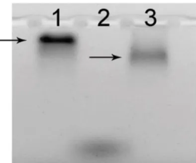
The conjugates of sdAbs and QDs were obtained as described in the "Materials and Methods". The design of the sdAbs in combination with the selected cross-linker and surface functional groups of the QDs made it possible to conjugate all the sdAbs to the QDs in the same predetermined orientation ensuring the maximum accessibility of the antigen-binding sites of the antibodies for the antigen molecules [14]. In order to preliminarily estimate the results of conjugation between QDs and sdAbs, we compared the electrophoretic mobilities of unconjugated QDs and QD–sdAb conjugates (Figure 1).
Figure 1. The electropherogram of QDs and QD–sdAb conjugates in agarose gel. Lanes: 1, QDs with an emission peak in the near-infrared region (QDsNIR); 2, bromophenol blue; 3, conjugates of QDsNIR with anti-HER2 sdAbs
The changes in mobility indicated successful conjugation of QDs and sdAbs. The potential applications of the conjugates were estimated in experiments with immune-sorbent assay (ISA), immunohistochemistry (IHC), and flow cytometry.
In order to estimate the possibility of using the conjugates in ISA, recombinant HER2 was adsorbed to wells of an immunological plate at a concentration of 10 mg/ml, after which, solutions of conjugates of the QDs with emission maximum at 562 nm and anti-HER2 sdAbs (QD562–sdAbs) with different concentrations were added to the wells. The results of the analysis (Figure 2) have shown that the HER2 antigen can be detected using QD562–sdAb conjugates at a concentration of 3 μg/ml.
Figure 2. Analysis of the effect of the QD562–sdAb conjugate concentration on the intensity of the fluorescence signal of the conjugates specifically binding HER2. The ordinate shows the fluorescence signal intensity; the abscissa shows successive dilutions of the original 0.3 mg/ml solution of QD562–sdAbs. In the negative control, the antigen concentration was zero and the conjugate concentration was 3 μg/ml.
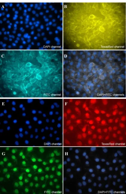
The possibility of using conjugates of anti-HER2 sdAbs and QDs with an emission peak at 569 nm (QDs569) in IHC was employed in experiments with cultured SK-BR-3 cells, which are known to overexpress HER2 protein located on the cell membrane surface [18]. Cultures of MDA-MB-231 cells, which do not show high level of HER2 expression [18], served as a negative control. The HER2 expression in the cell cultures was confirmed by flow cytometry (data not shown) shortly before the IHC procedure. The fluorescent images demonstrated the staining of the cell membrane of the positive cells (Figures 3, B–D) and only background staining of the cytoplasm and nucleus in HER2-negative cells (Figures 3, F–H). Thus, the experiments showed highly specific detection of the HER2 surface antigen by means of IHC using QD569–sdAb conjugates.
International Symposium Physics, Engineering and Technologies for Bio-Medicine IOP Publishing IOP Conf. Series: Journal of Physics: Conf. Series 784 (2017) 012016 doi:10.1088/1742-6596/784/1/012016
Figure 3. Microscopic images of cultures of (A–D) SK-BR-3 and (E–H) MDA-MB-231 immunostained with QD569 conjugated with anti-HER2 sdAbs. The fluorescent detection channels were: 370–430 nm for Hoechst (DAPI channel, panels A, D, E, H), 520–555 nm for QDs569 (FITC channel, panels С, D, G, H), and 593–610 nm (TexasRed channel, panels B, F). Optical magnitude, 40.
The possibility of using conjugates of anti-HER2 sdAbs and QDs with an emission peak at 570 nm (QDs570) to count of stained with anti-HER2 conjugates HER2-expressing cells in a mixture with other cells was demonstrated in flow cytometry experiments. A portion of SK-BR-3 cells was incubated with anti-HER2 conjugates as described in the "Materials and Methods" and mixed with unstained cells. Subsequent estimation of the percentage of correctly recognized cells stained with QD570–sdAb conjugates has shown that the technique allows the detection of HER2-positive cells even if their proportion in the population is as small as 0.5% (Figure 4).
Figure 4. Results of counting the cells stained with anti-HER2 sdAbs conjugated with QDs570. The black numbers above the plots show the percentages of SK-BR-3 cells stained with the conjugates that were included into the analyzed mixtures; the pink numbers at the upper right corners of the plots show the percentages of stained SK-BR-3 cells detected in the analyzed mixtures by means of flow cytometry.
4. Conclusions
The results of our study allow us to propose a method for fabrication of QD-based nanoprobes with advanced functional characteristics, including an increased sensitivity. The ultraminiature nanoprobes consisting of anti-HER2 sdAbs conjugated with QDs in a highly oriented manner developed in our studies have numerous implications for advanced integrated diagnostic procedures.
Acknowledgments
We a grateful to Dr. Pavel S. Samokhvalov (National Research Nuclear University MEPhI, Moscow, Russia) for kindly providing us with quantum dots fluorescing in the visible spectral region, Dr. Sungjee Kim (Department of Chemistry Pohang University of Science & Technology Pohang, South Korea) for kindly providing us with quantum dots fluorescing in the near-infrared region, Dr. Tina Van den Broeck, Line De Kimpe, and Dr. Frans Nauwelaers (BD Biosciences, Erembodegem, Belgium) for the assistance with flow cytometry experiments, and Dr. Baryshnikova (Laboratory of Experimental Diagnosis and Biotherapy of Tumors, Blokhin Cancer Research Center, Moscow, Russia) for the assistance with immunohistochemical experiments.
This study was supported by the Federal Target Program for Research and Developments of the Ministry of Education and Science of the Russian Federation (grant no. 14.584.21.0012, contract no. RFMEFI58415X0012).
References
[1] Sukhanova A, Devy J, Venteo L, Kaplan H, Artemyev M, Oleinikov V, Klinov D, Pluot M, Cohen J H M and Nabiev I 2004 Anal. Biochem. 324 60
[2] Bilan R, Fleury F, Nabiev I and Sukhanova A 2015 Bioconjugate Chem. 26 609 [3] Larson D R 2003 Science. 300 1434
[4] Deerinck T J 2008 Toxicol. Pathol. 36 112
[5] Hafian H, Sukhanova A, Turini M, Chames P, Baty D, Pluot M, Cohen J H M, Nabiev I and Millot J-M 2014 Nanomed. Nanotechnol. 10 1701
[6] Wang S, Zhang C, Wang G, Cheng B, Wang Y, Chen F, Chen Y, Feng M and Xiong B 2016 Theranostics 6 1877
[7] Kelkar S S and Reineke T M 2011 Bioconjugate Chem. 22 1879
[8] Harris L, Fritsche H, Mennel R, Norton L, Ravdin P, Taube S, Somerfield M R, Hayes D Fand Bast R C 2007 J. Clin. Oncol. 25 5287
[9] Khalil H S, Langdon S P, Goltsov A, Soininen T, Harrison D J, Bown J, Deeni Y Y 2016 Oncotarget, doi: 10.18632/oncotarget.12425
[10] Chotprakaikiat W, Allen A, Bui-Minh D, Harden E, Jobsri J, Cavallo F, Gleba Y, Stevenson F K, Ottensmeier C, Klimyuk V, Savelyeva N 2016 OncoImmunology 5 e1166323
International Symposium Physics, Engineering and Technologies for Bio-Medicine IOP Publishing IOP Conf. Series: Journal of Physics: Conf. Series 784 (2017) 012016 doi:10.1088/1742-6596/784/1/012016
[11] Roux K H, Greenberg A S, Greene L, Strelets L, Avila D, McKinney E C, Flajnik M F 1998 Proc. Natl. Acad. Sci. U.S.A. 95 11804
[12] Greenberg A S, Avila D, Hughes M, Hughes A, McKinney E C and Flajnik M F 1995 Nature 374 168
[13] Conrath K, Wernery U, Muyldermans S and Nguyen V Dev. Comp. Immunol. 27 87
[14] Sukhanova A, Even-Desrumeaux K, Chames P, Baty D, Artemyev M, Oleinikov V and Nabiev I 2012 Nat. Protoc. Exch. doi:10.1038/protex.2012.042
[15] Rakovich T Y, Mahfoud O K, Mohamed B M, Prina-Mello A, Crosbie-Staunton K, Van Den Broeck T, De Kimpe L, Sukhanova A, Baty D, Rakovich A, Maier S A, Alves F, Nauwelaers F, Nabiev I, Chames P and Volkov Y 2014 ACS Nano 8 5682
[16] Even-Desrumeaux K, Fourquet P, Secq V, Baty D and Chames P 2012 Mol. Biosyst. 8 2385 [17] Wu X, Liu H, Liu J, Haley K N, Treadway J A, Larson J P, Ge N, Peale F and Bruchez M P
2003 Nat Biotechnol. 21 41
[18] Subik K, Lee J F, Baxter L, Strzepek T, Costello D, Crowley P, Xing L, Hung M C, Bonfiglio T, Hicks D G and Tang P 2010 Breast Cancer (Auckl) 20 35