HAL Id: hal-02555574
https://hal.archives-ouvertes.fr/hal-02555574
Submitted on 15 Sep 2020
HAL is a multi-disciplinary open access
archive for the deposit and dissemination of
sci-entific research documents, whether they are
pub-lished or not. The documents may come from
teaching and research institutions in France or
abroad, or from public or private research centers.
L’archive ouverte pluridisciplinaire HAL, est
destinée au dépôt et à la diffusion de documents
scientifiques de niveau recherche, publiés ou non,
émanant des établissements d’enseignement et de
recherche français ou étrangers, des laboratoires
publics ou privés.
chelate: a novel structural entry for Mn2+-based
imaging agents
Daouda Ndiaye, Maryame Sy, Agnès Pallier, Sandra Même, Isidro de Silva, S.
Lacerda, Aline Nonat, Loic Charbonniere, Éva Tóth
To cite this version:
Daouda Ndiaye, Maryame Sy, Agnès Pallier, Sandra Même, Isidro de Silva, et al.. Unprecedented
kinetic inertness for a Mn2+-bispidine chelate: a novel structural entry for Mn2+-based imaging
agents. Angewandte Chemie International Edition, Wiley-VCH Verlag, 2020, 59 (29), pp.11958-11963.
�10.1002/anie.202003685�. �hal-02555574�
1
novel structural entry for Mn
2+
-based imaging agents
Daouda Ndiaye,
[a]Maryame Sy,
[b]Agnès Pallier,
[a]Sandra Même,
[a]Isidro de Silva,
[c]Sara Lacerda,
[a]Aline M. Nonat,
[b]Loïc J. Charbonnière*
[b]and Éva Tóth*
[a][a] D. Ndiaye, A. Pallier, Dr. S. Même, Dr. S. Lacerda, Dr. É. Tóth
Centre de Biophyisique Moléculaire, CNRS UPR 4301, Université d’Orléans rue Charles Sadron, 45071 Orléans, France
E-mail: [email protected] [b] M. Sy, Dr. A. M. Nonat, Dr. L. J. Charbonnière
Equipe de Synthèse Pour l’Analyse
Université de Strasbourg, CNRS, IPHC UMR 7178 F-67000 Strasbourg, France
E-mail : [email protected] [c] I. da Silva
CEMHTI, CNRS UPR3079, Université d’Orléans F-45071 Orléans 2, France
Supporting information for this article is given via a link at the end of the document.((Please delete this text if not appropriate))
Abstract: The search for more biocompatible alternatives to Gd3+
-based MRI agents, and the interest in 52Mn for PET imaging call for
ligands that form inert Mn2+ chelates. Given the labile nature of Mn2+, high inertness is challenging to achieve. The strongly preorganized
structure of the 2,4-pyridyl-disubstituted bispidol ligand L1 endows its
Mn2+ complex with exceptional kinetic inertness. Indeed, MnL1 did
not show any dissociation for 140 days in the presence of 50 eq. of
Zn2+ (37°C, pH 6), while recently reported potential MRI agents
MnPyC3A and MnPC2A-EA have dissociation half-lives of 0.285 h and 54.4 h under similar conditions. In addition, the relaxivity of
MnL1 (4.28 mM-1s-1 at 25°C, 20 MHz) is remarkable for a
monohydrated, small Mn2+ chelate. In vivo MRI experiments in mice
and determination of the tissue Mn content evidence rapid renal clearance of MnL1. Additionally, L1 could be radiolabeled with
52
Mn and the complex revealed good stability in biological media.
Introduction
The high diagnostic value of contrast agents makes them indispensable for many Magnetic Resonance Imaging protocols. For 35 years now, Gd-complexes have been used in millions of human examinations and considered among the safest
diagnostic drugs. However, the recent emergence of
nephrogenic systemic fibrosis and its causal link to Gd-exposure, as well as the evidence on brain and bone accumulation of Gd
have alerted the medical community.[1] In response to these
safety concerns, some of the linear Gd3+ chelates have been
recently withdrawn and the use of others has been restricted. In
this context, the replacement of Gd3+ with more biocompatible,
safer paramagnetic metal ions has become a major objective. Being an essential metal ion, as well as a good relaxation agent due to its five unpaired electrons (in the high spin state), slow
electron spin relaxation and fast water exchange, Mn2+ is an
obvious alternative.[2] Indeed, recent years have witnessed
increasing interest in Mn2+ complexes as potential MRI agents.[3]
In addition, manganese has a positron-emitting radioisotope,
52Mn, with interesting decay properties for Positron Emission
Tomography (PET) (t1/2 = 5.6 d, max. β+-energy: 575 keV).
Given its low β+ decay intensity (29.6%), PET resolution as good
as 1.2 mm could be achieved which is particularly important in small animal imaging.[4] The long t1/2 makes 52Mn particularly
adapted to image slow biological processes. Altogether, Mn2+ is
the unique metal ion to offer detection capabilities in both MRI
and PET.[5]
Thermodynamically stable and kinetically inert complexation of Mn2+ is indispensable to avoid in vivo release of the free metal ion, which could potentially cause toxicity during MRI where large contrast agent quantities are needed, or lead to off-target signals in PET examinations. In addition, for good MRI efficiency, the complex should contain at least one inner sphere water
molecule. However, the lower charge (with respect to Gd3+) and
the lack of ligand-field stabilization energy for the high-spin d5 electron configuration are not favorable to achieve high thermodynamic stability.
The highly labile nature of Mn2+ sets an even more difficult
challenge to meet. Generally, ligand rigidity and preorganization
promote kinetic inertness, and indeed, the most promising Mn2+
complexes are based on rigidified linear or macrocyclic chelators (Scheme 1). For instance, the trans-1,2-cyclohexylene backbone and the pyridine function ensure rigidity and are essential structural elements to provide slow dissociation of the MnPyC3A
chelate (Scheme 1), a potential Mn2+-based agent which has
successfully passed to in vivo experiments.[3a, 6] Among
macrocyclic chelates, the non-hydrated MnDOTA was reported
to have high inertness,[7] while the monohydrated [Mn(cisDO2A)]
was found already ~20-fold more labile (dissociation half-lives,
t1/2, estimated to 1061 h and 48 h, respectively, at pH 7.4).[8]
Recently, derivatives of the pyridine-containing pyclen
macrocycle were found to form stable and very inert Mn2+
chelates.[3c] With t1/2 = 8×10³ h estimated for pH 7.4, the
Mn(PC2A-EA) complex bearing an ethyleneamine pendant arm and proposed as a pH-sensitive agent, has the highest resistance to dissociation ever reported for a monohydrated
Mn2+ complex. Nevertheless, this dissociation half-life remains a
million times below the value for GdDOTA (t1/2 =2.7 × 10⁹ h, pH
2
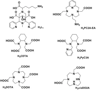
Scheme 1. Ligands discussed.
In the aim of substantially improving the kinetic inertness of Mn2+
chelates while ensuring good relaxation properties and water
solubility, we are exploring bispidine
(3,7-diazabicyclo[3.3.1]nonane) derivatives. Bispidine-type ligands
represent a versatile platform in coordination chemistry.[9]
Pendant arms with varying coordinating functions can be attached in different positions to the bicyclic amine core in order to tune the coordination properties for metal ions of various sizes, coordination numbers or coordination geometries. Depending on the substituents, the ligand can adopt different conformations
and configurations.[10] Among those, the chair-chair conformer is
best suited for stable metal coordination and thanks to its exceptionally preorganized structure, bispidine derivatives tend to form very inert complexes. Bispidine complexes have been investigated with a large variety of metal ions and for different
applications. Concerning Mn2+, the very scarce reports involve
pyridine derivative bispidine ligands and are restricted to studies
in the solid state or in non-aqueous solvents.[11]
We hypothesized that the 2,4-pyridyl-disubstituted bispidol
derivative L1 bearing one methylene carboxylic acid (Scheme 1)
could be a good chelator for Mn2+. First, the very rigid bispidine
scaffold can endow MnL1 with high kinetic inertness. The ligand
is expected to coordinate in a five-dentate manner, involving two pyridine and two bispidine nitrogens as well as the methylene carboxylate. This leaves one coordination site for a hydration water, important for MRI application. The non-coordinating pending carboxylates might additionally create a second sphere
contribution and increase proton relaxivity. Finally, L1 can be
readily functionalized for biological targeting via the lysine terminal amine. This scaffold has been previously investigated for the complexation of 64/67Cu2+[12] and 68Ga3+.[13] A three
dimensional representation of the putative structure of the MnL1
complex is shown in Scheme 2.
We evidence here that despite a moderate thermodynamic
stability, MnL1 has a resistance to acid catalyzed dissociation
and transmetalation which is unprecedented for a Mn2+ chelate.
In addition, the relaxation efficiency of the complex was
assessed by a combined 17O NMR and NMRD study. The
complex was injected into mice and its biodistribution and
pharmacokinetics were monitored by MRI. Finally, the 52Mn
radiolabelling of L1 was explored and the stability of the
radiocomplex was investigated.
Scheme 2. Three dimensional representation of the putative structure of MnL1.
Results and Discussion
L1 was synthesized in 4 steps as previously reported.[12]
Thermodynamic stability and kinetic inertness of MnL1
Ligand protonation constants were obtained by pH potentiometry and are reported in Table 1. By analogy to other bispidine ligands,[14] we attributed the first protonation constant to one of the tertiary amine groups, while the other amine of the bispidine skeleton remains likely unprotonated in the chair-chair
conformation.[14b] Further protonation steps supposedly occur on
the lysine amine (logKH2 = 10.31, typical of the lysine amine
protonation constant)[15] and then on the three carboxylates,
while the pyridine nitrogens are not protonated above pH 2.
Thermodynamic stability constants of MnL1 and ZnL1 were
assessed by pH potentiometry (Table 1). In the case of Mn2+,
slow complex formation prevented direct titration; therefore,
batch samples were prepared (Fig. S1). MnL1 is three orders of
magnitude less stable than ZnL1, in accordance with the Irving–
Williams series. Figure 1 depicts the species distribution for
MnL1 as a function of pH, supported by the pH-dependent
relaxivities. Protonated complexes form with both Zn2+ and Mn2+;
the first protonation constants are practically identical to logKH2 of the ligand and correspond to the protonation of the non-coordinating lysine amine. The similarity between the second
protonation constant of the ligand and logKMnHL or logKZnHL
further supports the attribution of the ligand protonation steps. At
physiological pH, this species protonated on the lysine amine
is the only complex present.
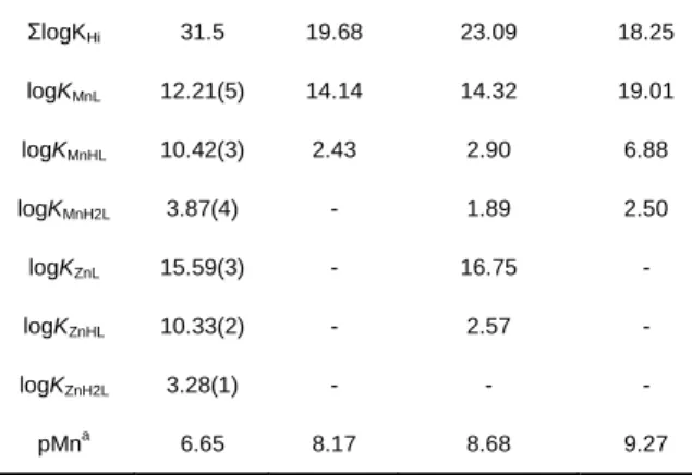
Table 1. Ligand protonation constants, stability constants of ML complexes
and pMn values. Values in parenthesis correspond to one standard deviation.
L1 PyC3A [3a] trans-CDTA[16] PC2A-EA[6] logKH1 11.44(1) 10.16 9.54 11.34 logKH2 10.31(2) 6.39 5.97 8.93 logKH3 4.71(5) 3.13 3.60 6.91 logKH4 2.76(5) - 2.52 1.97 logKH5 2.22(4) - 1.46 -
3
ΣlogKHi 31.5 19.68 23.09 18.25 logKMnL 12.21(5) 14.14 14.32 19.01 logKMnHL 10.42(3) 2.43 2.90 6.88 logKMnH2L 3.87(4) - 1.89 2.50 logKZnL 15.59(3) - 16.75 - logKZnHL 10.33(2) - 2.57 - logKZnH2L 3.28(1) - - - pMna 6.65 8.17 8.68 9.27 apMn calculated for cMn = clig = 10 -5
M; pH 7.4
Figure 1. Species distribution curves calculated for MnL1 (1 mM) and
pH-dependent relaxivities (▲) measured at 25°C, 60 MHz.
The stability of MnL1 is relatively modest, as shown by the pMn
value of 6.65 calculated for 10-5 M Mn2+ and L
1 concentration
and pH 7.4 (Table 1). Nevertheless, once the chelate is formed, it has an exceptional resistance to dissociation. Kinetic inertness of metal complexes is typically assessed by transmetalation experiments with endogenously available metal ions, such as
Zn2+, which is present at ~10 M concentration in the blood and
represents the most abundant potential competitor of Mn2+.
Different transmetalation protocols have been reported for Mn2+
complexes, some allowing a simple comparison of the kinetic inertness under given conditions, others giving also access to
the underlying dissociation mechanisms. We followed
transmetalation in the presence of 10 or 50 equivalents of Zn2+,
at pH 6 (50 mM MES buffer), 37°C, in a similar way to that
proposed by Caravan et al.[3a] At either of the Zn2+
concentrations, MnL1 did not show any relaxivity change for the
140 days of observation (Fig. 2). Under similar conditions (37°C,
pH 6.0, 25 Zn2+ equivalents), dissociation half-lives, t
1/2, of 0.285
h and 54.4 h have been reported respectively for MnPyC3A[3a]
and MnPC2A-EA[3c], two reference compounds considered as
potential MRI probes with good kinetic inertness. Analogous experiments have been carried out at more acidic pH values as
well (pH 3.1-6.0, 50 equivalents Zn2+, 37 °C; Fig. 2). Under these
pseudo-first order conditions, the dissociation rate constant, kobs,
has been determined, and the estimated transmetalation half-lives vary between 22.7 h at pH 3.13 and 130 days at pH 5.07.
The inertness of MnL1 was also assessed in highly acidic
solutions where the dissociation is much faster (0.01-1.0 M HCl,
25°C; Fig. 3). The dissociation rate constants, kobs, show a
second order dependence on the proton concentration and could be fitted to Eq. 1.
𝑘𝑜𝑏𝑠= 𝑘0 + 𝑘1[𝐻+] + 𝑘2[𝐻+]2 (Eq. 1)
The fit resulted in k1 = (1.6±0.1)×10-3 s-1 M-1 and k2 = (5.0±0.1)×10-4 s-1 M-2, while k0 was fixed to 0, otherwise the fit
yielded a negative value with a large error (-3.5±2)×10-5 s-1. We
should note that the kobs values determined above in the Zn2+
transmetalation study in the pH 3.1-5.1 interval also show a
second order dependence on [H+] (Fig. S2), with k
1 =
(2.2±0.2)×10-3 s-1 M-1, in the same order of magnitude as the one
above. This rate constant, characterizing the proton assisted dissociation pathway, is two orders of magnitude lower than that
reported for MnPC2A-EA which is the most inert hydrated Mn2+
chelate reported so far.[3c] At this point, it is difficult to extrapolate the dissociation half-life of the complex for physiological conditions, and in particular, more data are needed to describe the role of metal-assisted dissociation processes in the overall dissociation. These studies are in progress, but they are long due to the extremely low dissociation rates. Nevertheless, we can conclude that the rigid and preorganized bispidine skeleton
provides very high kinetic inertness, unprecedented for a Mn2+
chelate.
Figure 2. Time-dependent variation of the longitudinal proton relaxation rates
in a 1 mM MnL1 solution at 37°C and 60 MHz, 0.1 M KCl, in the presence of
50 equivalents of Zn2+. Empty symbols at pH 6.00 correspond to 10 equivalents of Zn2+
. The curves represent the fit of the experimental data to yield the observed rate constants, kobs.
Mn2+ MnL MnH2L MnHL 4 4.5 5 5.5 6 6.5 0 20 40 60 80 100 2 4 6 8 10 r1 (m M -1.s -1) F ra c ti o n s (% ) pH 8 9 10 11 12 13 14 15 0 50 100 150 1 /T1 /s -1 t / days pH 3.13 3.75 4.16 5.07 6.00
4
Figure 3. Pseudo-first order rate constants determined for the dissociation of
MnL1 in acidic solutions. The curve corresponds to the fit of the experimental
data to Eq. (1) as explained in the text.
Relaxation properties of MnL1
Proton relaxivities have been recorded for MnL1 as a function of
the magnetic field, at 25 and 37°C, in water (pH 7) and in human serum (Figs. 4a and S3). The NMRD data were complemented
by variable temperature 17O relaxation rate measurements (Fig.
4b) which give access to the hydration number and the water exchange parameters. The relaxivity is remarkably high for a Mn2+ complex (r1 = 4.28 mM-1s-1 and 3.37 mM-1s-1 at 25°C and 37°C, respectively, 20 MHz, pH 7 in water). In comparison, relaxivities of 3.3 and 2.5 mM-1s-1 (20 MHz, 25 and 37 °C) and
3.52 mM-1s-1 (20 MHz, 25°C) were reported for the
monohydrated complexes MnPCy3A and MnPC2A-EA. One
hydration water molecule is confirmed for MnL1 from the
temperature-dependent 17O transverse relaxation rates using the
method of Gale et al. (q = 0.85; Fig. S4).[17]
The temperature-dependent transverse 17O relaxation rates and
the NMRD profiles have been analysed in the frame of the
Solomon-Bloembergen-Morgan theory to determine the
parameters describing water exchange and rotational dynamics. The fit yielded kex298 = (5.1±0.7)×107 s-1 and Hǂ = 10.6 kJ/mol for the rate and the activation enthalpy of the water exchange. The water exchange rate is similar to that reported for MnPyC3A (kex298 = 5.4×10⁷ s-1)[3a] while it is about ten times lower than
kex298 on MnEDTA (kex298 = 47×107 s-1).[18] The bispidine MnL1 chelate is structurally very different from other previously studied
Mn2+ complexes, and the reason for this relatively slow water
exchange is difficult to identify. Nevertheless, this exchange rate remains high enough not to limit proton relaxivity. The rotational correlation time, R298 = (100±2) ps, is higher than the values
reported for small molecular weight Mn2+ complexes (e.g. 56 ps
for MnEDTA).[19] This might reflect that R is overestimated due to second sphere contribution to relaxivity from the non-coordinating carboxylates. Therefore, we also performed the analysis of the NMRD data by including an additional contribution from a second sphere relaxation mechanism to the overall relaxivity. To describe this second sphere mechanism,
we assumed two 2nd sphere water molecules (with an estimated
Mn-H proton distance of 3.2 Å, 50 ps residence time and an activation enthalpy of 10 kJ/mol, based on previous simulations
on Gd3+ complexes; see Fig. S5 and ESI for all details).[20] This
resulted in a rotational correlation time, R298 = (72±4) ps which
seems more reasonable on the basis of the molecular weight of the complex. This is obviously not a firm proof of a second sphere relaxivity contribution; nevertheless, it is evident that the relaxivities measured tend to be higher than the typical values
reported for monohydrated Mn2+ chelates.
The relaxivities remain identical in the presence of up to 50 equivalents of citrate, carbonate and phosphate (Fig. S6). They
increase to 6.46 and 5.12 mM-1s-1 in human serum at 25 and
37 °C, respectively (20 MHz, Fig. 4a), a tendency similar to that observed for instance for MnPyC3A and attributed to low affinity binding to serum proteins.
Figure 4. a) Proton relaxivities of MnL1 measured in water at 25°C (♦) and
37 °C (▲) and in human serum at 37 °C (∆), and b) reduced transverse 17
O relaxation rates (9.4 T). The lines represent the simultaneous fit of the experimental points as explained in the text.
52
Mn radiolabeling of L1
The need for long-lived -emitting isotopes and the emergence
of a combined PET-MRI technology have recently promoted an
increasing interest in the production and complexation of 52Mn.
We have produced and purified 52Mn at the cyclotron of the
Orléans CNRS campus (see ESI for details). 52Mn was
recovered as 52MnCl
2 solution after purification through 2 anion
exchange resin AG© 1-X8 columns. The radiolabelling of L1 was
tested under different conditions of pH, temperature and 52Mn:L1
ratio. The radiolabelling reactions were followed by radioTLC at 5, 15, 30 and 60 min. All reactions were performed in presence of ascorbic acid to prevent any oxidation reaction. Optimized reaction conditions were obtained for pH 7, 70 °C, 1h, achieving
99% labelling yield. 52MnL
1 is highly hydrophilic with a logP = -1.96±0.06, which falls within the range reported for other Mn complexes, e.g. MnDPDP or Mn(EDTA-BTA) with logP values of
-3.07 and -1.84, respectively.[21] The in vitro stability of the
radiocomplex was assessed in different media at pH 7.4 (water, PBS and saline (0.9% NaCl)) and in the presence of 0.6 mM
0 2 4 6 8 10 0.01 0.1 1 10 100 re la x iv it y / m M -1s -1 1H Larmor freuency / MHz 16.6 16.7 16.8 16.9 17 2.8 3.3 3.8 ln (1 /T 2 r) 1000/T / 1/K
a)
b)
0 0.0008 0.0016 0.0024 0 0.2 0.4 0.6 0.8 1 1.2 kobs / s -1 [H+] / M5
HSA (Fig. 5), revealing its stability in all media up to 18h, and a slightly lower to moderate stability at 24h.
Figure 5. In vitro stability of 52MnL1 in different media at pH 7.4 (water, PBS
and saline (0.9% NaCl)) and in the presence of 0.6 mM HSA, at different time points: 0, 1, 18 and 24 h (darker to lighter colour, respectively).
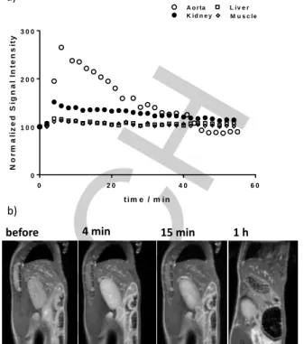
MRI biodistribution and pharmacokinetics of MnL1
To validate the potential in vivo use of MnL1, we have evaluated
the biodistribution in wild type mice by MRI at 7 T. The signal intensity of the main organs (kidney, liver, muscle, lung) and the aorta was followed during 1 h post intravenous injection (0.06 mmol/kg dose; Fig. S7). Figure 6 depicts normalized signal intensity for the analyzed organs. The highest signal intensity
was reached around 4 min after injection. MnL1 shows renal
clearance and the uptake in the liver is limited. The estimated blood half-life is 21 min, typical of small molecules and similar to
previously reported contrast agents.[3a, 22] This distribution profile
was further confirmed by ex vivo ICP-OES measurements performed at the end of the MRI experiment. The data are
compared to mice which did not receive any MnL1 injection
(control) where the Mn-levels measured correspond to endogenous Mn (Fig. 7; Table S1).
At 1.5 h post injection, higher Mn content is found in the kidneys and blood of injected mice. The blood uptake could be related to
the low affinity binding of MnL1 to blood proteins, as also
observed by relaxivity measurements. Compared to the Mn tissue content reported for MnPyC3A at 24 h post injection, our
complex shows lower liver and kidney uptake.[3a]
Figure 6. a) Normalized signal intensity in the kidney, muscle, liver and aorta
plotted as a function of time. Measurements were performed every 2 minutes for 4 mice. Standard deviations are not presented for better readability. b) Sagittal T1-weighted MR images prior to injection and at 4, 15 min and 1 h post intravenous injection of 0.06 mmol/kg of MnL1.
Figure 7. Ex vivo ICP-OES quantification of the Mn tissue content in the main
organs and blood of control and injected mice with MnL1 (1.5 h post injection).
Data is presented in nmol/g tissue ± SD (n=3).
Conclusion
We demonstrate here that despite a relatively modest
thermodynamic stability, the MnL1 complex has an exceptional
resistance to dissociation. At 37°C, pH 6.0 and in the presence
of up to 50 equivalents of Zn2+, MnL1 remained intact and did not
show any relaxivity change for at least 140 days. MnL1 has one
inner sphere water molecule. It has higher proton relaxivities
than most monohydrated, small molecular weight Mn2+ chelates,
which might be related to a second sphere relaxivity contribution induced by the non-coordinating carboxylates functions.
In vivo MRI experiments performed in mice indicated quick renal
clearance, which was also supported by ex vivo ICP
determination of the Mn tissue content in different organs. L1
could be successfully labeled with 52Mn and the radiocomplex
has good stability in biological media. Based on these
Bra in Liv er Mu sc le Bo ne Lu ng s Sp lee n Kid ne ys Blo od 0 2 5 5 0 7 5 1 2 5 2 7 5 M n t is s u e c o n te n t / n m o l g -1 c o n t r o l M n L1 Bra in Liv er Mu sc le Bo ne Lu ng s Sp lee n Kid ne ys Blo od 0 2 5 5 0 7 5 1 2 5 2 7 5 M n t is s u e c o n te n t / n m o l g -1 c o n t r o l M n L1
before 4 min 15 min 1 h
0 2 0 4 0 6 0 0 1 0 0 2 0 0 3 0 0 t im e / m in N o r m a li z e d S ig n a l In te n s it y b)
6
preliminary radiolabeling experiments, the conjugation of the
bifunctional ligand L1 to biomolecules, such as antibodies can be
envisaged. Indeed, for the imaging of such slowly circulating
biomolecules, the long-lived 52Mn PET isotope represents a
clear advantage.
In summary, the bispidine-based L1 chelator constitutes a very
promising structural entry for the development of Mn-based imaging agents for MRI as well as for PET. Alternatively, this bispidine synthon can be also attractive for the design of combined PET-MRI probes. Such bimodal imaging applications
based on Mn2+ might be interesting when the in vivo fate of the
imaging probe is to be followed for a longer period of time. Most
importantly, the L1 ligand can provide excellent kinetic inertness
which so far could not be achieved within the more traditional ligand families.
Experimental Section
Experimental details are described in detail in the Supporting Information.
Acknowledgements
The authors thank the French National Research Agency (grant ANR-18-CE18-0008) for funding. We thank Dr. Frederic Szeremeta and Quentin Mura for their assistance with MRI
experiments and Louis Frealle with the 52Mn production.
Keywords: manganese • contrast agent • MRI • 52Mn • kinetic inertness
[1] aT. Grobner, Nephrol. Dial. Transplant. 2006, 21, 1104-1108; bE. Di Gregorio, G. Ferrauto, C. Furlan, S. Lanzardo, R. Nuzzi, E. Gianolio, S. Aime, Invest. Radiol. 2018, 53, 167-172.
[2] B. Drahoš, I. Lukeš, É. Tóth, Eur. J. Inorg. Chem. 2012, 2012, 1975-1986.
[3] aE. M. Gale, I. P. Atanasova, F. Blasi, I. Ay, P. Caravan, J. Am. Chem.
Soc. 2015, 137, 15548-15557; bM. Botta, F. Carniato, D.
Esteban-Gomez, C. Platas-Iglesias, L. Tei, Future Med. Chem. 2019, 11, 1461-1483; cR. Botár, E. Molnár, G. Trencsényi, J. Kiss, F. K. Kalman, G. Tircsó, J. Am. Chem. Soc. 2020; dZ. Garda, E. Molnár, F. K. Kálmán, R. Botár, V. Nagy, Z. Baranyai, E. Brücher, Z. Kovács, I. Tóth, G. Tircsó,
Front. Chem. 2018, 6, 14; eE. Molnar, N. Camus, V. Patinec, G. A.
Rolla, M. Botta, G. Tircso, F. K. Kalman, T. Fodor, R. Tripier, C. Platas-Iglesias, Inorg. Chem. 2014, 53, 5136-5149; fA. Rodriguez-Rodriguez, Z. Garda, E. Ruscsak, D. Esteban-Gomez, A. de Blas, T. Rodriguez-Blas, L. M. P. Lima, M. Beyler, R. Tripier, G. Tircso, C. Platas-Iglesias,
Dalton Trans. 2015, 44, 5017-5031; gC. Vanasschen, E. Molnar, G.
Tircso, F. K. Kalman, E. Toth, M. Brandt, H. H. Coenen, B. Neumaier,
Inorg. Chem. 2017, 56, 7746-7760.
[4] L. M. Carter, A. L. Kesner, E. C. Pratt, V. A. Sanders, A. V. F. Massicano, C. S. Cutler, S. E. Lapi, J. S. Lewis, Mol. Imag. Biol. 2020,
22, 73-84.
[5] C. M. Lewis, S. A. Graves, R. Hernandez, H. F. Valdovinos, T. E. Barnhart, W. Cai, M. E. Meyerand, R. J. Nickles, M. Suzuki,
Theranostics 2015, 5, 227-239.
[6] D. J. Erstad, I. A. Ramsay, V. C. Jordan, M. Sojoodi, B. C. Fuchs, K. K. Tanabe, P. Caravan, E. M. Gale, Invest. Radiol. 2019, 54, 697-703. [7] B. Drahos, V. Kubicek, C. S. Bonnet, P. Hermann, I. Lukes, E. Toth,
Dalton Trans. 2011, 40, 1945-1951.
[8] Z. Garda, A. Forgács, Q. N. Do, F. K. Kálmán, S. Timári, Z. Baranyai, L. Tei, I. Tóth, Z. Kovács, G. Tircsó, J. Inorg. Biochem. 2016, 163, 206-213.
[9] aA. M. Nonat, A. Roux, M. Sy, L. J. Charbonniere, Dalton Trans. 2019,
48, 16476-16492; bP. Comba, M. Kerscher, W. Schiek, in Progress in Inorganic Chemistry, Vol. 55 (Ed.: K. D. Karlin), 2007, pp. 613-704; cP.
Comba, M. Kerscher, K. Rueck, M. Starke, Dalton Trans. 2018, 47, 9202-9220.
[10] T. Legdali, A. Roux, C. Platas-Iglesias, F. Camerel, A. M. Nonat, L. J. Charbonniere, J. Org. Chem. 2012, 77, 11167-11176.
[11] aP. Comba, B. Kanellakopulos, C. Katsichtis, A. Lienke, H. Pritzkow, F. Rominger, J. Chem. Soc.-Dalton Trans. 1998, 3997-4001; bP. Comba, H. Rudolf, H. Wadepohl, Dalton Trans. 2015, 44, 2724-2736; cP. Comba, M. Kerscher, M. Merz, V. Muller, H. Pritzkow, R. Remenyi, W. Schiek, Y. Xiong, Chem. Eur. J. 2002, 8, 5750-5760.
[12] A. Roux, R. Gillet, S. Huclier-Markai, L. Ehret-Sabatier, L. J. Charbonniere, A. M. Nonat, Org. Biomol. Chem. 2017, 15, 1475-1483. [13] T. Price, S. Yap, R. Gillet, H. Savoie, L. Charbonnière, R. Boyle, A.
Nonat, G. J. Stasiuk, Chem. Eur. J., doi.org/10.1002/chem.201905776. [14] aG. D. Hosken, R. D. Hancock, J. Chem. Soc. Chem. Commun. 1994,
1363-1364; bR. Gillet, A. Roux, J. Brandel, S. Huclier-Markai, F. Camerel, O. Jeannin, A. M. Nonat, L. J. Charbonnière, Inorg. Chem.
2017, 56, 11738-11752.
[15] J. M. Berg, J. L. Tymoczko, L. Stryer, Biochemistry, 5th ed., New York: W. H. Freeman, 2002.
[16] E. Molnár, B. Váradi, Z. Garda, R. Botár, F. K. Kálmán, É. Tóth, C. Platas-Iglesias, I. Tóth, E. Brücher, G. Tircsó, Inorg. Chim. Acta 2018,
472, 254-263.
[17] E. M. Gale, J. Zhu, P. Caravan, J. Am. Chem. Soc. 2013, 135, 18600-18608.
[18] J. Maigut, R. Meier, A. Zahl, R. van Eldik, J. Am. Chem. Soc. 2008, 130, 14556-14569.
[19] G. A. Rolla, C. Platas-Iglesias, M. Botta, L. Tei, L. Helm, Inorg. Chem.
2013, 52, 3268-3279.
[20] A. Borel, L. Helm, A. E. Merbach, Chem. Eur. J. 2001, 7, 600-610. [21] M. K. Islam, S. Kim, H.-K. Kim, S. Park, G.-H. Lee, H. J. Kang, J.-C.
Jung, J.-S. Park, T.-J. Kim, Y. Chang, J. Med. Chem. 2017, 60, 2993-3001.
[22] aJ. Wang, H. Wang, I. A. Ramsay, D. J. Erstad, B. C. Fuchs, K. K. Tanabe, P. Caravan, E. M. Gale, J. Med. Chem. 2018, 61, 8811-8824; bF. La Cava, A. Fringuello Mingo, L. Miragoli, E. Terreno, E. Cappelletti, L. Lattuada, L. Poggi, S. Colombo Serra, ChemMedChem 2018, 13, 824-834.
7
The 2,4-pyridyl-disubstituted bispidol ligand L1 is a promising structural entry for the development of Mn-based imaging agents.
Thanks to its preorganized, rigid structure, it provides exceptional inertness to the Mn2+ complex. MnL1 is monohydrated, has good
relaxation efficiency for MRI applications and undergoes rapid renal clearance when injected to mice. The 52Mn-labeled radiocomplex