HAL Id: tel-02073232
https://tel.archives-ouvertes.fr/tel-02073232
Submitted on 19 Mar 2019
New constrained amino acids and peptide nucleic acid
building blocks for the construction of bio-polymers
Alexandra Gresika
To cite this version:
Alexandra Gresika. New constrained amino acids and peptide nucleic acid building blocks for the construction of bio-polymers. Other. Université Côte d’Azur, 2018. English. �NNT : 2018AZUR4104�. �tel-02073232�
Nouveaux synthons contraints de type
-amino acides et PNA en vue de l´élaboration
de bio-foldamères
New constrained amino acids and peptide nucleic acid
building blocks for the construction of biopolymers
Alexandra Gresika
Molécules Bioactives
Présentée en vue de l’obtention du grade de docteur en Chimie d’Université Côte d’Azur
Dirigée par : Nadia Patino Soutenue le : 16.11.2018
Devant le jury, composé de :
Muriel Amblard, Directrice de Recherche CNRS, IBMM UMR 5247, Université de Montpellier
Karine Alvarez, Chargée de Recherche CNRS, AFBM UMR 7257, Université Aix- Marseille
Mohamed Mehiri, Maître de Conférences, ICN UMR 7272, Université Côte d’Azur
THÈSE DE DOCTORAT
New constrained amino acids and peptide nucleic acid building blocks for the construction of bio-polymers.
The purpose of this thesis concerned the development of two kinds of constrained oligomeric structures, based either on unnatural cyclic -amino acids derivatives or on cyclic Peptide Nucleic Acid (PNA) dimers, in view of analyzing their propensity to adopt pre-organized conformations mimicking the active conformations of proteins or nucleic acids. The first part of this thesis reports on the elaboration of new cyclic -amino-acids (6-substituted 4-oxopipecolic acids) building blocks, their potential use in the synthesis of constrained homogenous and heterogeneous peptidomimetics as well as their limitations in this use. As part of this work, a new methodology has been developed for the synthesis of N-protected 6-substituted 4-oxo-pipecolic acids residues. The second part of this thesis reports on the elaboration of constrained -PNA dimers (di--PNA), in which the side-chains of two consecutive -PNA monomers are “stapled” via a lactam bridge. A synthetic orthogonal strategy has been first developed in liquid-phase then applied to the solid-phase synthesis of models “stapled” di- PNA incorporating thymine nucleobases.
Keywords: foldamers; constrained peptidomimetics; constrained Peptide Nucleic
Acids; Nucleic Acid analogs; stapled Peptide Nucleic Acids; pre-organized structures; peptide synthesis; solid-phase.
Nouveaux dérivés d'acides -aminés et de Peptide Nucleic Acids (PNA) contraints pour la construction de bio-polymères.
Le but de cette thèse a concerné le développement de deux types de structures oligomériques contraintes, basées soit sur des dérivés non naturels d’acides aminés cycliques, soit sur des dimères cycliques de Peptide Nucleic Acids (PNA), en vue d’analyser leur tendance à adopter des conformations pré-organisées, imitant les conformations actives des protéines ou des acides nucléiques. La première partie de cette thèse a trait à l’élaboration de synthons clefs dérivant d’acides α-aminés cycliques non naturels (acides 4-oxopipécoliques 6-substitués), à leur utilisation potentielle dans la synthèse de peptidomimétiques contraints, homogènes et hétérogènes, ainsi qu’à leurs limites dans cette utilisation. Dans le cadre de ce travail, une nouvelle voie de synthèse permettant d’accéder à des résidus N-protégés d'acides 4-oxo-pipécoliques 6-substitués a été mise au point. La deuxième partie de cette thèse concerne l’élaboration de dimères d’-PNA (di--PNA) cycliques, dans lesquels les chaînes latérales de deux monomères d’-PNA consécutifs sont «agrafées» via un pont lactame. Une stratégie de synthèse a tout d'abord été développée en phase liquide, puis appliquée à la synthèse en phase solide de di--PNA «agrafés» modèles, incorporant des bases nucléiques thymine.
Mots-clés: foldamères ; peptidomimétiques contraints ; PNA contraints ; analogues d’Acides Nucléiques ; PNA agrafés ; structures pré-organisées ; synthèse peptidique ; phase-solide.
Acknowledgements
This scientific work was carried out from 1. October 2015 to 1. October 2018 at the University Côte d’Azur in the “Institut de Chimie de Nice” under the supervision of Prof. Nadia Patino. I thank Professor Nadia Patino to provide me the opportunity to take part on her research and the support during this three years. Thank you for being my second mom in the ICN.
As well, I thank the director of the group, Dr. Rachid Benhida to welcome me to the research group “Chimie de Molecules Bioactives” and the jury to read and judge my thesis.
I thank the “Ministère de l´Education Nationale” and the University Côte d’Azur for their financial support, Dr Lionel Massi and Dr Marc Gaysinski for LC-MS and NMR analyses and Dr. Mohamed Mehiri for his chemical expertise.
All thanks go to the students at the Institute for their encouragement, support and open ear, special thanks go to Anto, Nelli, Mauro, Gabriela, Anita and Oleksander - You were always there for me, in good and bad days.
I would like to express my sincere thanks to my parents and family for their affection and encouragement.
ABREVIATIONS
aeg-PNA N-(2-aminoethyl)-glycine-peptide nucleic acid
Glu Glutamic acid
Asp Aspartic acid
Lys Lysine Gly Glycine Ser Serine Orn Ornithine Val Valine -ala beta-alanine Trp Tryptophane Ile Isoleucine Tm Melting point Arg Arginine
GPNA L-g-Glutamyl-p-nitroanilide/ L-Glutamic acid 5-(4-nitroanilide)
AcCl Acetyl chloride TEA Triethylamine NEt3 Triethylamine DIPEA N,N-Diisopropylethylamine MeOH Methanol DCM Dichlormethane n-BuLi butyllithium THF Tetrahydrofuran MeCN Acetonitrile
TFA Trifluoro acetic acid
H2O = Water
HCl Hydrochloric acid
Fmoc-Cl 9-Fluorenylmethyl chloroformate Boc2O Di-tert-butyldicarbonate
Alloc-Cl Allyl chloroformate
Allyl-Br Allyl bromide
DBF Dibenzofuran
DMF Dimethylformamide
Me3SnOH Trimethyltin hydroxide
1,2-DCE 1,2-Dichloroethane
LiOH Lithium hydroxide
NaOH Natrium hydroxide
iPrOH isopropyl alcohol
r.t room temperature
Et2O Diethyl ether
DMSO Dimethyl sulfoxide
IPA isopropyl alcohol
TLC thin-layer chromatography HBTU 2-(1H-benzotriazol-1-yl)-1,1,3,3-tetramethyluronium hexafluorophosphate DCC N,N-Dicyclohexylcarbodiimide EDC 1-Ethyl-3-(3-dimethylaminopropyl)carbodiimide DMAP 4-Dimethylaminopyridine CDI Carbonyldiimidazole DIC Diisopropylcarbodiimide HOBt Hydroxybenzotriazole PyBOP benzotriazol-1-yl-oxytripyrrolidinophosphonium hexafluorophosphate DhbtOH 3-Hydroxy-1,2,3-benzotriazin-4(3H)-one Pyr Pyridine Ac2O Acetic anhydride FmocOSu N-(9-Fluorenylmethoxycarbonyloxy)succinimide Fm = Fluorenylmethoxycarbonyl PG protecting group Pd Palladium
Conten t
GENERAL INTRODUCTION ... 17
PART I:“6-SUBSTITUTED 4-OXOPIPECOLIC ACID” BUILDING BLOCKS FOR THE DEVELOPMENT OF NEW CONSTRAINED PEPTIDOMIMETICS ... 19
I.-BIBLIOGRAPHY ... 20
I.-1. INTRODUCTION ... 20
I.-2. PROTEIN STRUCTURE ... 21
I.-2.1 Secondary structures ... 22
I.-2.1.1 -helix ... 23
I-2.1.2 -sheets/-strands ... 24
I-2.1.3 Polyproline: PPI and PPII ... 26
I-2.1.4 Turn motives/loops... 27
I.-3. PROTEIN-PROTEIN- AND PROTEIN-NUCLEIC ACID INTERACTIONS ... 29
I.-3.1 Protein-Protein Interactions (PPIs) ... 29
I.-3.2 Protein-Nucleic-acid Interactions ... 31
I.-3.2.1 Protein-DNA Interactions ... 32
I.-3.2.2 Protein-RNA Interactions... 34
I.-4. DESIGNED MOLECULES TO MIMICKING PROTEIN SECONDARY STRUCTURES ... 38
I.-4.1 Foldamers ... 39
I.-4.1.1 “Biotic” Foldamers... 41
I.-4.2 Cyclic Peptides secondary structure mimetics ... 54
III.-1.1 6-phenyl-4-oxo-pipecolic methyl ester... 73
III.-1.2 N-protected phenyl-4-oxo-pipecolic acid derivatives ... 75
III.-2 Development of a new methodology for the synthesis of 6-phenyl-4-oxo pipecolic acid derivatives via the imino-Diels-Alder-reaction ... 77
III.-2.1 Imino Diels Alder reaction ... 77
III.-2.2 Synthesis of racemic phenyl-4-oxo-pipecolic methyl ester... 81
III.-2.3 Synthesis of N-Allyl-6-phenyl-4-oxo-pipecolic acid ... 83
III.-3. Foldamer synthesis ... 83
III.-3.1 Homogeneous foldamer... 83
III.-3.2 Heterogeneous foldamers ... 85
III.-3.2.1 Acid chlorides ... 88
III.-3.2.2 Acyl fluorides ... 92
III.-3.3 Synthesis of 6-phenylethyl-4-oxo-pipecolic acid derivatives ... 92
IV. CONCLUSION AND OUTLOOK ... 95
PART II:STAPLED DI--PNA AS BUILDING BLOCKS FOR THE DEVELOPMENT OF CONSTRAINED PEPTIDE NUCLEIC ACIDS ... 98
I. BIBLIOGRAPHY ... 99
I.-1. Nucleic acid mimics for antisense oligonucleotide therapeutics ... 99
I.-2. AON modifications ... 101
I.-2.1 Internucleoside linkage/backbone modified AONs... 102
I.-2.2 Sugar modified AONs ... 104
I.-2.3 Nucleobase modified AONs ... 106
I.-2.4 Other advanced modified AONs ... 107
I.-3. PEPTIDE NUCLEIC ACID110 ... 111
I.-3.1 Chiral-, - and -PNA ... 113
I.-3.1.1-PNA ... 113
I.-3.1.2 -PNA ... 119
I.-3.1.3 -PNA ... 121
I.-3.2 Conformationally constrained cyclic PNA analogues ... 124
III.-1.3 Attempts to “stapled” protected di--PNA (D-Lys) backbone dimer .... 138
III.-2. One-component cyclization based-strategy ... 139
III.-2.1 Synthesis of protected -PNA backbone monomers ... 142
III.-2.2 Synthesis of “stapled” di-PNA[(Lys or Orn)-(Glu)] blocks ... 146
III.-2.2.1 Synthesis of stapled PNA dimer:strategy 1- way A ... 147
III.-2.2.1 Synthesis of stapled PNA dimer: strategy 1-way B ... 149
III.-2.2.1 Synthesis of stapled PNA dimer: strategy 2-way A ... 151
IV. SOLID-PHASE SYNTHESIS OF THE “STAPLED” PNA DIMERS ... 156
IV.-1. Synthesis of protected -PNA backbones deriving from D-Glu and L-Glu 159 IV.-2. Solid-phase synthesis of “stapled” di--PNA blocks ... 162
IV.-3. Conclusion and Outlook ... 167
GENERAL CONCLUSION ... 169
III. EXPERIMENTAL PROCEDURES ... 172
I. SYNTHESIS OF PHENYL-4-OXO PIPECOLIC DERIVATIVES ... 174
I.-1. Following the procedure from Daily et al. ... 174
I.-2. Diels-Alder reaction ... 186
II. SYNTHESIS OF -PNA-DIMER ... 193
II.-1. Liquid phase: strategy 1 ... 214
II.-2. Liquid phase: strategy 2 ... 193
II.-3. Synthesis of Monomers for Solid-Phase synthesis ... 241
II.-4. Synthesis on Solid-Phase: General procedure ... 253
LIST OF REFERENCES... 258 ANNEX I ...
General Introduction
Protein-Protein and Protein-Nucleic Acid (NA) Interactions play crucial roles in numerous biological processes, constituting relevant targets in various diseases. In this context, structures mimicking bioactive peptide conformations, called foldamers, have emerged as important tools for the modulation of these interactions. Indeed, by adopting well-suited pre-organized conformations, they can target large surface areas and mimic protein regions involved in such interactions. The search for new pre-organized structures mimicking the active conformations of proteins is now an area in full expansion since advances in the field have demonstrated their potential for the discovery of next generation therapeutics.
On the other hand, Nucleic Acids (NA) analogs constitute also an intense research area, since they are able to inhibit or modulate gene expression, by forming a complex with complementary DNA or RNA target, via Watson-Crick base pairing. In this context, due to their high DNA/RNA binding strength, Peptide Nucleic Acids are considered as devices of choice in diagnostics applications and molecular biology studies. However, as most of NA analogs, they present some limitations that hinder their application as therapeutics, such as poor cellular uptake and off-target effects and the search for PNA analogs overcoming these problems is still relevant.
The purpose of this thesis fit into these two research domains and concerns the elaboration of two different kinds of constrained structures, based either on unnatural constrained -amino acid derivatives or on constrained Peptide Nucleic acid (PNA)
their implications, in particular in Protein/Protein and Protein/NA interactions. Afterward, several mimics of protein secondary structures are described as well as their biological applications. The second chapter is devolved to the synthesis of 6-substituted 4-oxopipecolic acid building blocks required for the construction of peptidomimetic structures, and to the attempts made to synthetize these latter.
The second part of this manuscript is dedicated to our second goal, i.e. the development of constrained PNA analogs. A first bibliographic chapter presents the different classes of Nucleic Acids mimics described in the literature, their applications in antisense oligonucleotide therapies and their limitations. A particular attention will be paid to the “Peptide Nucleic Acids” family. The second chapter is dedicated to our works and describes the synthesis of stapled di--PNA blocks, both in liquid- and solid-phase, and their structural characterization.
The third part of the manuscript assembles the experimental protocols and the characterization of all the synthetized products. NMR spectra of some molecules are given in Annex I.
Part I
“6-substituted 4-oxopipecolic acid” building blocks for
the development of new constrained peptidomimetics
I.-Bibliography
I.-1. Introduction
By using only 20 proteinogenic amino acids, the nature can achieve an endless variety of proteins, which can perform many complex functions essential for the foundation of life. Proteins adopt complex secondary, tertiary and quaternary structures, depending on their primary sequences and further extern influences. Their intrinsic structure is closely related to their biological functions. Many Protein (PPI) and Protein-Nucleic Acids interactions play essential roles in numerous biological processes representing thus relevant targets for various diseases. These interactions often involve highly structured short peptide sequences of proteins. Chemists have recently begun to design synthetic oligomers that approach the structural and functional complexities of the interacting sequences of proteins to disrupt these interactions. The structure and folding of these synthetic oligomers control their conformation and carry out unsurpassed chemical functions. In the following chapters, several mimetics of protein secondary structures, their biological application and therapeutic potential are described.1,2,3
I.-2. Protein structure
Proteins have different shapes and molecular weights and their structure is constructed by four levels (Figure 1):4
1) Primary structure: linear -amino acid sequence
2) Secondary structure: local conformation of a polypeptide chain, depending on the hydrogen bonding network. The two main types are the α-helix and the ß-sheet, but other conformations are known, among them PolyProline helices of type II and turns.
3) Tertiary structure: overall three-dimensional shape of the entire protein. It is fashioned by many stabilizing forces due to bonding interactions between the side-chain groups of the amino acid residues of the protein.
4) Quaternary structure: Aggregation from two to several polypeptide chains (subunits)
I.-2.1 Secondary structures
The secondary structures are defined by the three torsional angles, which are presented in Figure 2. Torsional angle omega () between C1 and N´ partial double bond, psi ()
C and CO single bond and phi () N and C single bond.5
Figure 2: Definition of torsional angles psi (), phi () and omega ().4
The specific () and () torsional angles defining the most common secondary structures are presented in table 1.
Table 1:Torsional angles (and ) for -helix, -sheets, turns, PPI and PPII.,6,5 secondary structure ° ° Parallel -sheet -119 113 Antiparallel-sheet -139 135 -helix -58 -47 -turn Type I -60 -30 PPI -83 158 PPII -78 149
I.-2.1.1 -helix
The -helix structure is the most common secondary structure in proteins. In 1948, Pauling discovered this coiled structure, that is stabilized by a network of intra-molecular hydrogen bonds between the C=O groups of residues “i” and the NH groups of residues “(i+4)” (Figure 3).7 The -helix has 3.6 residues per turn and the screw
sense is essentially right-handed. Two other helices, and 310, exist in which hydrogen
Figure 3: Illustration of -helix a) Ribbon depiction (green points = amino acid residues) b) side-view of stick and ball version c) 310-helix d) -helix. c) and d) illustration with PyMol. e) Helix
H-bond patterns.9,10
I-2.1.2 -sheets/-strands
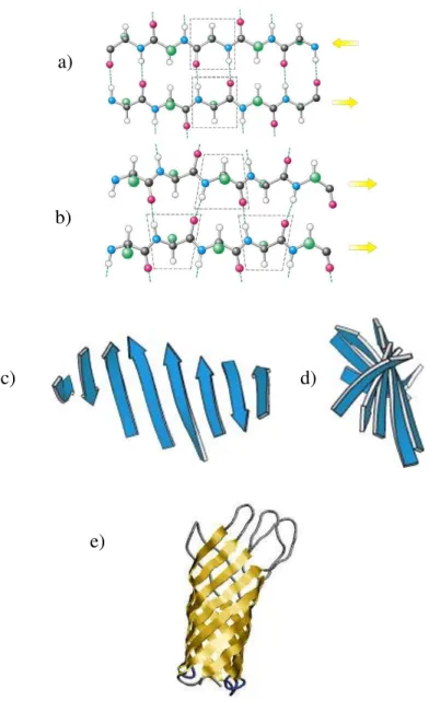
-sheets represent an important motif of secondary structure in proteins and arise when segments of polypeptide chains, called -strands, are connected laterally by at least two or three backbone hydrogen bonds (Figure 4).4
a) b) c) d)
Figure 4: Illustration of -strands and sheets. a) antiparallel b) parallel; c) and d) Twist of
sheets.-strands are illustrated as blue arrows e) -barrel structure.10 a)
b)
c) d)
shape, as beta-barrel (Figure 4e). This latter motif is, for example, characteristic of fatty acid-binding proteins, which are important for lipid metabolism.4,10
I-2.1.3 Polyproline: PPI and PPII
Polyproline motif is one of the major structural elements found in unfolded proteins and since 1993, it has been advocated as secondary structure motif, like the -helix and -sheet. It may adopt two helicoidal secondary structures, defined as polyproline I (PPI) and polyproline II (PPII) helices (Figure 5). PPI and PPII are known since 1951 by Pauling.11 The polyproline II helix (PPII, poly-Pro II) is left-handed and polyproline I helix (PPI, poly-Pro I) is right-handed. PPI contains all cis peptide bonds and is more compact compared to PPII with all trans peptide bonds.12,13,9
Figure 5: Illustration of a) and b) PPI (left) and PPII (right) structure and c) View of N-terminal of PPI structure.9 a) a) b) c) PPI PPII cis trans
The polyproline type II (PPII) helix is a prevalent conformation in unfolded proline-rich proteins. It is found predominantly in collagen and commonly observed in fibrous proteins.12 It is known to play important roles in a wide variety of biological processes.12
I-2.1.4 Turn motives/loops
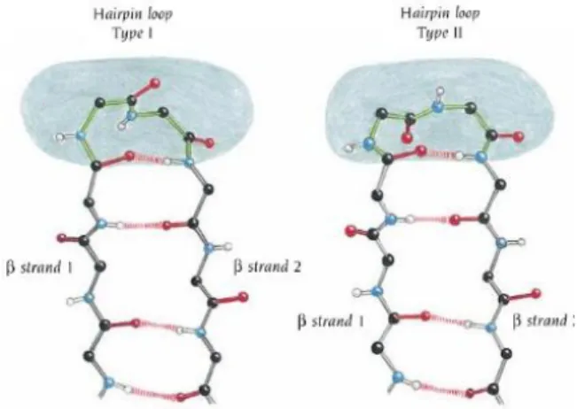
Turns connect consecutive secondary motifs in proteins and reverse the direction of the protein backbone. Globally, turn structures imply two to six amino acids and are classified according to the hydrogen-bond pattern between the carboxyl group of the residue at position “i” and the amide proton at position “i+n”. This forms three families: and-turns, with three, four, five amino acids in length, respectively (n = 2, 3, and 4; Figure 6).14, 10
Figure 7: Illustration of Hairpin loop Type I and Hairpin loop Type II. They differ in the orientation of one amide group.4,10
Moreover, secondary structures that connect the consecutive hydrogen bonded antiparallel-strands with loops/turns represent -hairpins (Figure 8).15
I.-3. Protein-Protein- and
Protein-Nucleic acid
interactions
I.-3.1 Protein-Protein Interactions (PPIs)
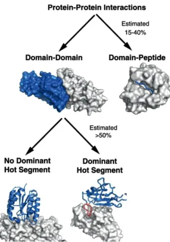
In many biological functions, the communication between proteins is essential, that makes PPIs important molecular targets with strong therapeutic value. Today, the modulation (inhibition or stabilization) of PPIs involved in relevant critical molecular communications is considered as a therapeutic strategy in various diseases and is useful for understanding biological mechanisms. However, PPIs are extremely difficult to target because of their high surface areas, normally flat and featureless. In general, PPIs involve two protein domains (domain–domain) or a short linear sequence of one protein and a domain of the other one (peptide-domain) (Figure 9). In the two cases, many new studies have revealed a high organization level of these interfaces.16,17,18 It is known that diseases can be caused by aberrant PPIs, by loss of essential interactions or through the formation or/and stabilization of protein complexes at an inappropriate time or location.18,19
Figure 9:Two categories of PPIs: domain-mediated and peptide-mediated.20
Normally 40% of interacting peptides adopt a -helix conformation and 30% of proteins contain -strands/sheets motives, which constitute important recognition motives in some protein–protein interactions (PPIs), as in HIV protease-ligands recognition and are implied in neurologic disorders related to in Alzheimer's disease.
The inhibition of PPI constitutes an area of intense research. The most general approaches to inhibit PPIs are primary based on protein topologies (Figure 10). Peptidomimetics have shown their considerable potential in drug design by targeting PPIs.16 One of their main applications is their interaction with an active conformation of a short linear peptide sequence located at the surfaces of a globular protein domain
London et al. defined “hot segments” as continuous epitopes contributing to the majority of the binding energy.17,20
Figure 10: Modulators of PPIs.19
I.-3.2 Protein-Nucleic-acid Interactions
Protein-/Nucleic Acid interactions play an essential role in all mechanisms of genes expression and modulation. Structural, biochemical and molecular-genetic studies have proven that interactions between Proteins and Nucleic Acids are significant and are formed by:21,22,23
1) Electrostatic interactions (salt-bridges): Interaction between negative charged
The networks of specific and non-specific interactions established in DNA-protein- or RNA-protein complexes are intrinsically linked to the 3D-structures of these complexes.
I.-3.2.1 Protein-DNA Interactions
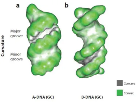
DNA-binding proteins are often used in many major cellular processes to organize and compact the chromosomal DNA and to regulate and effect the processes of transcription, DNA replication, and DNA recombination. The most spacious feature of DNA (A-form and B-form) is the recognition of major and minor grooves by proteins (Figure 11). The structural element employed most frequently is the -helix, -ribbons, -sheets and loops are found less frequently. Considering Figure 11, in the B-DNA helix, the most common DNA form, the major groove is wide enough to accommodate an -helix and the minor groove, on the other hand, is deep and narrow and thus less accessible to secondary structures such as an-helix. In A-DNA helix, the opposite is correct. The minor groove is shallow and broad whereas the major groove is very deep and narrow. In general, however, one might expect proteins to directly decode DNA sequences via interactions in the major groove but discriminate among RNA sequences via interactions in the minor groove. This appears for the most part to be the case.24,25,23
Figure 11: Molecular shape of A- and B-DNA. Convex surface in green and concave surface in dark grey.25
On one side, interactions between proteins and DNA can occur via the protein recognition of a sequence dependent DNA shape (shape readout), but on the other side, it can occur via recognition of the chemical signature of the DNA bases (base readout). That means the contact in the minor and major grooves of DNA is made via the formation of hydrogen bonds between nucleic bases and amino acids, directly or water-mediated, and via hydrophobic contacts.25 In Figure 12, several DNA-binding domains
of proteins are illustrated the zinc finger-eukaryotic transcription factors, the helix-turn/loop-helix characteristic of most prokaryotic regulatory proteins, the leucine zipper, -sheet proteins that facilitate the binding to nucleic acids. In reference 23,24 and 26, more DNA-binding proteins are illustrated and explained.23,24,26
Figure 12: Examples of DNA binding motives in red. The protein binds as a dimer. One monomer is colored in blue and the other in yellow. The DNA is illustrated as space filling model. PDB codes are bracketed.26
I.-3.2.2 Protein-RNA Interactions
Specific interactions between RNA and proteins take part in many aspects of RNA biology. These RNA-binding Proteins (RBP) are involved in important mechanisms like transcription, translation, RNA trafficking, and packaging processes, which have important values for the development of potential therapeutic drugs. If we have a look to the major groove of the dsRNA, which correspond to the A-form of DNA (Figure 11), we realize that it is too narrow and deep to allow penetration by an-helix or -strand without extensive distortion.27,28 The RNA recognition motif (RRM),
-zinc finger (1aay)
Cro and Repressor (1lmb)
Loop-sheet-helix (1tsr)
protein binding domain.29 An example is presented in Figure 13. RRM has a mixed -
and -architecture and recognizes single strand RNAs via its -sheets domain.30
Figure 13: Protein are shown as ribbons (red = helices, yellow = sheets, green = loops). The structural figures are presented by PyMOL. The RNA is presented in blue (cyan stick model). a) RRM in complex with ssRNAs. 3. Crystal structure of the U1A spliceosomal protein RNA hairpin complex (PDB:1URN). Solution structure of RBD of Fox-1 in complex with UGCAUGU (PDB:2ERR). b) RNA binding by ZF proteins. Solution structure of the RBD of Fox-1 in complex with UGCAUGU (PDB entry 2ERR). Crystal structure of three-finger polypeptide from TIFIIIA in complex with truncated 5S RNA (61 nt) (PDB:1UN6). Protein side chain forming base specific contacts with GGU are shown in green. Zn2+ are shown in magenta.30
Mostly zinc finger domains bind to DNA, but some of them are also known to recognize RNA. In general, they are constructed from 30 amino acids and contain two cysteine and two histidine residues, following the sequence X2-Cys-X2,3-Cys-X12-His-X3,4,5.
This sequence can fold in stable -hairpin and -helix structures because of the
The minor or shallow groove of dsRNA should be accessible to -helices and -strands (Figure 11). But the functional groups of nucleic acid bases are not diverse enough among different nucleotides to allow effective discrimination.
Figure 14: RNA binding proteins do not target dsRNA in sequence-specific manner. These recognize single-stranded regions or sites of local distortions induced in double-helical regions by RNA hairpins (U1A, U1 70K, EIAV Tat, N,…), bulges (HIV Tat), or internal loops (U1A, HIV Rev, TFIIIA, …).27
That is the reason why, all known sequence-specific proteins only recognize ssRNAs and hairpin loops, because the functional groups of nucleic bases are free. However, dsRNA is only recognized if there is structural distortion in the double helix, like internal loops or bulges, allowing access to the major groove (Figure 14).27 There are specific proteins, that are known to bind with high affinity and specificity to these disordered RNA sites. Arginine-rich motives (ARM), which are sequences of 10-15
The most common ARM/ARN complexes are the BIV Tat-TAR RNA, HIV Rev/-RRE and N-peptide/-box B-complexes shown in Figure 15.28,30
Figure 15: RNA-peptide complexes.:a) The bovine immunodeficiency virus (BIV) Tat-TAR peptide-RNA complex b) The bacteriophage N peptide-box B RNA complex c) HIV-1 Rev peptide RRE RNA complex.28
Today, inhibitors of protein-protein or protein/nucleic acids interactions have a strong therapeutic value. Mimicking the conformation of the binding domain of proteins is considered as a promising strategy. In the next section, we will present - non-exhaustively - some constrained structures mimicking the interacting sequences (or domains) of proteins. Their therapeutic potential is illustrated by several examples in
I.-4. Designed molecules to
mimicking protein secondary
structures
The defined peptide sequences of proteins involved in PPI seem ideal candidates as inhibitors of these PPI. However, these short sequences, which are highly structured inside the protein rarely adopt defined conformations when they are isolated, and they are often flexible (Figure 16).
Figure 16: Inhibition of PPIs with secondary structure mimetics.31
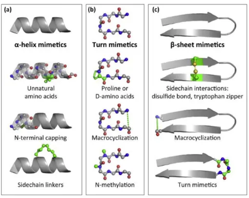
Many different techniques have been developed to stabilize or pre-organize them into their bioactive conformation (Figure 17). Generally, -helices are stabilized by the introduction of unnatural amino acids, N-terminal capping or side-chain (i, i+4 or
i+7/i+11) cyclization. Turn motives are stabilized by the introduction of D-amino acids
or proline, by N-methylation or macrocyclization. -sheets are stabilized by side-chain interactions (disulfide bonds, tryptophan zipper), macrocyclization or turn
Figure 17: Stabilization-techniques of peptide secondary structures a) -helices b) turns c) -sheets.32
Peptide-based drugs are significantly prevented by their degradation by proteases, negligible membrane permeability or oral bioavailability, high clearance and metabolic instability. Numerous constrained secondary structures have been developed to overcome these drawbacks. Special examples are presented in the following chapters.
proteins and nucleic acids are defined as “biotic” foldamers.33,34“Biotic” foldamers are biopolymers, which are constituted by non-natural amino acid units or/and with unusual functions that replace the amide moiety. On the contrary, “abiotic” foldamers are constructed mostly by aromatic rich sequences, which gain new importance, because of their remarkable properties, high stability of folding and relative ease of synthesis. Their robust structures are built by repulsive and attractive interactions. Specific examples of “biotic” and “abiotic” foldamers are shown in Figure 18.1,2,3
Figure 18: Examples of “biotic” and “abiotic” foldamers. Biotic foldamers: a) Peptoids c) ß-Peptides. Abiotic foldamers: b)Aromatic oligoamides d) Aza-aromatic oligomers e)Tertiary aromatic ureas. Arrows represent intramolecular repulsive and attactive interactions.2,3
Moreover, two kind of foldamers exist: homogenous foldamers are built by the same monomer unit, in contrast to heterogonous foldamers, which contain different monomer units (Figure 19). 34
Figure 19:Examples for a) homogeneous b) heterogonous foldamer.34
In the following part, we will focus on biotic foldamers. Most of them have been designed for the inhibition of relevant PPIs, involved in various diseases. Nevertheless, some of them have been elaborated for their application as cell penetrating peptides (CPPs), for drug delivery, and as AMP (antimicrobial peptides), to disrupt microbial cell membranes. The structural requirements for peptides targeting cellular or microbial membranes are usually lower than those interacting with specific targets such as proteins.35
I.-4.1.1
“Biotic” Foldamers
In 1996, Seebach et al. and Gellman et al. reported the first peptide foldamer constituted by -amino acids (10/12-helix).36,37 These kind of foldamers are described in the literature as -peptides. They can adopt a variety of helical secondary structures, depending on the nature of side-chains. Since then, many different kinds of aliphatic peptide foldamers ( and peptide foldamers) have been described (Figure 20).38,39,40
Figure 20: Structure of aliphatic peptide foldamers.34
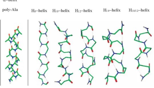
-peptide helices are named according to the number of atoms contained in the ring closed by the helix specific hydrogen bond and include 8-helix, 10-helix, 10/12-helix, 12-helix and 14-helix (Figure 21).38,41
Figure 21.: Helical secondary structures of -peptides compared to -helix poly-Ala.39 helix
are more attractive than -amino peptides counterparts, because of their enhanced conformational and proteolytic stability. These -peptides 1 are used as inhibitors of
the p-53-hDM2 interaction (Figure 22).42,41
Figure 22: 3-peptide foldamers as inhibitors of p53-hDM2 interaction. Side-chains orientation in
helices.31
Homo-oligomers of -peptides have received less attention, but they display a versatility comparable to that of -peptides. In 1998, Seebach et al. and Hanessian et
al. developed -amino acid oligomers, which formed defined 14-helical structures in solutions.43,44 Afterwards, Hofmann et al. confirmed in 2003 with ab initio calculations that 14-helical and 9-helical are the most stable conformations. With further
Figure 23: H-bondings in 14-helix or 9-helix (top) and in mixed helices: 14/12 helix or 24/22 helix (bottom).45
The Hanessian and Seebach groups studied the folding behavior of γ-‐peptides substituted on positions γ4-, γ3-, γ2-, γ2,4- and γ2,3,4. Surprisingly, γ4-, γ2,4- and γ2,3,4
--peptides all fold into the same 14-helix while γ3-‐and γ2-peptides appear to adopt a
flexible structure that is therefore hard to determine. From these results, it clearly appears that a single substituent at the γ4-position is sufficient to promote a robust
14--helix in γ-peptides.46,43,44 Since then, many mimics of γ4-amino acids have been elaborated with the goal to form foldameric structures (Figure 24).
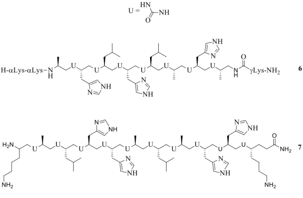
One of these mimics is based on the urea motif. It is one of the strongest H-bond pillars and this leads to a strong restriction of the backbone flexibility. Céline Douat et al. have developed amphiphilic helical oligourea foldamers, mimicking the histidine-rich peptide LAH4 for the transport of DNA into cells (Figure 25).47
Figure 25: Structure of pH-responsive amphiphilic oligoureas, mimics of CPP LAH4, for NA cell delivery 47
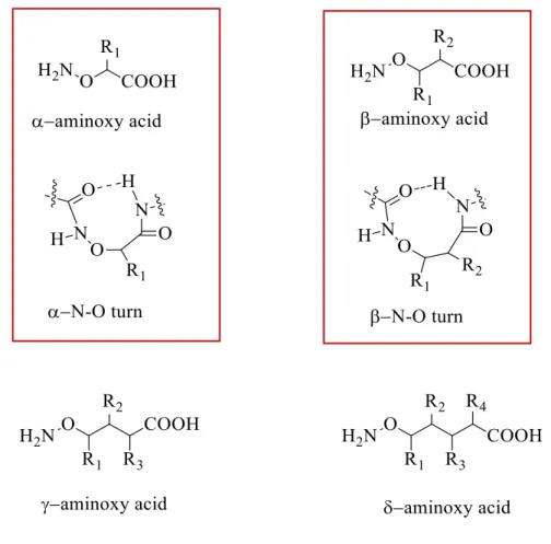
New families of foldamers based on -, -, -aminoxy acids have also been developed (Figure 26). Because of the presence of N-O bonds, - and aminoxy acids shows
heterochiral N-O turns) and sheet-like structures. -aminoxy acids, which are -amino acid analogs in which the carbon is replaced by a oxygen atom, have been successfully used for the design of anion receptors and channels (Figure 26).48,49
Figure 26: and -aminoxy oligomer structures and illustration of and N-O turns.49
Foldamers based on mixed , amino acid, such as and -peptides, also exist that broadens the potential in medicinal chemistry (Figure 29). 50,39 Indeed, the number of foldamers is vastly larger if we include heterogeneous backbones than if we are limited to homogeneous backbones.
Figure 27: Crystal structure of helical heterogeneous peptide backbones ( and -amino acid residues = orange, cyan, green, respectively).2
-peptide foldamers known since 2004, have led to the discovery of new helical secondary structures. In 2007, Choi et al. developed -peptide foldamers displaying 11-helical and 14/15-helical secondary structures, starting from amino acids ACPC (2-aminocyclopentanecarboxylic acid) and -amino acids (Ala or Aib) (Figure 30). Additionally, their torsion angels were determined.48,51
-residue -residue
11-helix -55(3) -40(6) -96(4) 94(6) -88(7)
14/15-helix -62(7) -38(3) -126(11) 83(6) -119(15)
Figure 28: Intramolecular hydrogen bonding pattern: 14/15 helical (green), 11-helical (blue). dotted arrows: possible H-bonding interactions. Torsion angles (deg) from -peptide 1-3 (unfolded c-terminal residues excluded) are given in the table.51
In 2007, Gellman et al. applied and peptide foldamers for the inhibition of PPI involving the Bcl-2 family. They designed a 14/15 helical foldamer to mimic the N-terminal region of the Bak segment and published its crystal structure (Figure 29).52,53,54
a)
Figure 29: Foldamers for inhibition of PPIs. a) X-ray structure of the pro-apoptotic ()-protein BIM interacting with Bcl-XL (PDB:3FDL). This interaction triggers cytochrome C release, leading
to apoptosis b) Chimeric Mimic of BIM-peptide binds to BH3 domain, already illustrated in a)
(PDB:3FDM) c) Top view of -foldamer mimicking BIM (PDB:3FDM). d) Top view and e) lateral view of six-helix bundle, which is formed by N-terminal heptad repeat domain (NHR) -peptides (green) and -foldamer (blue). The-residues are illustrated in purple. f) Close view of X-ray crystal structure of -inhibitor interacting with NHR core helices (PDB:3G7A). The interfacial residues are in purple, the -residues in light purple.53
Concerning mixed - and -foldamers, Baldauf et al. carried out in 2006 ab initio calculations proving, that mixed -peptides form especially 12-helices and mixed 12/10 or 18/20-helices (Figure 30).55
a) b) c)
Figure 30: H-bonding in 12-helix (top) and in mixed helices (12/10 or 18/20-botton).55
-peptide foldamers form especially 11- and 13-helices and mixed 11/13 or 20/22 helices (Figure 31). 55
Figure 31:H-bonding in 11-helix and 13-helix (top) and in mixed helices (11/13 helix or 20/22 helix-bottom). 55
Sharma and Kunwar et al. reported several interesting /and-peptide foldamers with sugar side chains that adopt 12/20 and 11/13 helices in solution. But recently, they reported as well -peptide foldamers, which form 14/12-helices (Figure 32).56,57
Figure 32: Heterogeneous peptide foldamers from Sharma and Kunwar at al. -peptide foldamer-11/13-helix (top) and -peptide foldamer-14/12-helix(below).57,56
On way to construct foldamers is to introduce constrained cyclic amino acids building blocks. Some are presented in Figure 33.
Figure 33: Examples of cyclic amino acid building blocks.
In 2017, Vezenkov et al. foldamers alterning cyclic α-amino-γ-lactam motives and -aminoacids residues having cationic (Arg) and amphipathic (Trp) side-chains, distributed on different faces of the foldamer backbone. They demonstrated that these foldamers adopted a ribbon-like structure (Figure 34) and were able to efficiently deliver a biologically relevant cargo inside the cell.58 Moreover, they showed a dramatically improved protease resistance.
Figure 34: Structure of Agl-AA foldamers and graphical representation of compound 2c. R1 and R2 = Arg or Trp side-chains.58
Manish Nepal et al. published in 2015 a new type of polyproline (PPII) helical scaffold (Figure 35), designed as CPP displaying an intrinsic antibacterial activity. Their goal was to combine the properties of CPPs and antimicrobial peptides (AMPs) in one agent. As CPP, most AMPs have both cationic hydrophilic and hydrophobic side chains, distributed on different faces on the overall folded conformation and actually, AMPs often display their antimicrobial activity through a membrane-lytic mechanism of action. Their good selectivity allows them to cross the bacterial or mammalian cell membranes.59 Among the various polyproline foldamers developed by Nepal, one of them ((PRPRPL)5 (Figure 36) displayed one of the best properties for cell-penetration
Figure 35: Polyproline foldamers (CAPHs) developed by Manish Nepal et al. a) Top view of the helix model b) Structure of labeled CAPHs incorporating the fluorescein fluorophore (in green).60
I.-4.2 Cyclic Peptides secondary structure
mimetics
Today, cyclic peptides generate an ongoing interest, notably in the field of PPI inhibition, since they may adopt thermodynamically stable protein-like conformations (helices, strands and turns), mimicking efficiently the stabilized native states of folded proteins.14,6 As well as foldamers, their conformational rigidity helps to minimize the entropic penalty associated with the target binding. Moreover, these cyclic structures display an increased resistance to degradation by proteases, as compared to their linear counterpart.
I.-4.2.1Helix mimetics
40% of secondary structure motifs are-helix and more than 60% of all proteins in PDB have an -helix sequence at their surface. That’s the reason for the interest of mimicking helical regions to influence protein-protein interfaces.61 Many methods for stabilizing a peptide sequence in an -helix conformation have been published and presented in the review of Pelay-Gimeno et al.6 and Hill et al.14. The most common method consists in “stapling” -covalently or not - two side-chains of two residues that will be located on the same face of the a-helix. A distinction is made between two techniques of stapling: one- vs two-component stapling. The one-component stapling is the stapling of two side chains while, the two-component stapling uses a bifunctional linker to connect two side chains (Figure 36).14,62,63
Figure 36: One- vs. two-component cyclization.61
Figure 37: Various stapling techniques.64
In the next section, we will focus particularly on lactam bridges peptides and hydrocarbon stapled peptides, which have given the best results as biomedical research tools and prototype therapeutics.16,61
a) Lactam bridged peptides
Fujimoto et al. followed the two-component cyclization approach using different kinds of cross-linking agents, to stabilize an helical structure conformation in short peptides containing two Lysine residues in positions (i/i+4), (i/i+7) or (i/i+11) (Figure 38).65
Lactam-bridged peptides following the two-component approach have also been used to inhibit the HIV entry into host cells. The glycoprotein 41 (gp41) is an HIV transmembrane protein responsible for the fusion of the viral envelope and host cell membrane, making gp41 a valuable therapeutic target (Figure 39).66 The insertion of the N-terminal coiled coil of gp41 into the host cell is followed by a structural rearrangement, leading to a six -helix bundle. C-terminal heptad repeat (CHR)-derived peptides, stabilized by the introduction of one (HIV C14 Linkmid)67 or two
lactam-linkers (HIV 31) were developed as antiviral therapeutics because of their interaction with the assembly of the helix bundle (Figure 39).6,66,68
Lys(i) Asp(i+4) to be superior to other lactam bridged macromolecules for the stabilization of -helices (Figure 40).14,62
Figure 40: Lactam-bridged peptides (i --> i+4) to stabilize -helix structures.14,62
b) Hydrocarbon stapled peptides
Hydrocarbon-stapled peptides are peptides in which two alkenyl side-chains of two unnatural -amino acids joined to form a macromolecule, using ruthenium-catalyzed RCM (ring closing metathesis) as the key step in the synthesis. The first stapled peptides were introduced in 2000 by Verdine et al. and expanded by Blackwell and Grubbs (Figure 41).69,70,71,14
Figure 41: Ruthenium olefin metathesis for crosslinking of macropeptides. Metathesis of Blackwell and Grubbs on O-allylserine residues (top). Metathesis of Schafmeister and Verdine on -disubstituted non-natural amino acids (bottom).71
Hydrocarbon linkers are introduced into a polypeptide by incorporating two -methyl, -alkenylglycine residues with defined stereochemistry, in chosen position. As illustrated in Figure 42, these two residues are separated by two (i, i+3), three (i, i+4) or six (i, i+7) amino acids.
inhibit intra- and extra-cellular protein-protein and protein/nucleic acids interactions. Moreover, they are readily accessible through standard solid-phase peptide synthesis.72,71 Most stapled peptides display dramatically improved pharmacologic parameters, such as resistance to proteolytic degradation, cell-penetration, and in vivo half-life as compared to disulfide and lactam bridges.72,71
Today, stapled peptides are largely used to disrupt PPIs involved in human diseases, particularly in cancer. In many types of cancer, proteins MDM2 and MDMX (also called MDM4 and HDM4/HDMX) downregulate the tumor suppressor p53 protein, via their binding to the -helical N-terminal transactivation domain of p53. The crystal structure of this complex is illustrated in Figure 43. The known structural information of p53 hot-spot residues (Phe19, Trp23 and Leu26) given par the crystal structure made it possible to develop new PPI inhibitors for anticancer strategies. One of them, targeting the oncogenic interaction p53-MDM2 / MDMX and another, agonist of the hormone GHRH, are currently under clinical trial trials.6,73
Beak et al. developed a series of stapled -helical p53-derived peptides cross-linked at positions i, i+7 (Figure 43). As compared with the wild-type p53 peptide, they displayed an increased -helicity, improved the binding affinity for MDM2 and enhanced proteolytic stability. Crystallographic data showed, that the hydrocarbon linker is involved in the binding to MDM2, explaining the affinity increase "Hydrocarbon" stapling has been also used in the context of HIV, to obtain inhibitors of viral replication acting on fusion, viral DNA integration and capsid steps. These molecules proved efficiency in animal models of human disease.6,74
I.-4.2.2 Cyclic -strands/sheets mimics
-sheets are formed by several strands that are stabilized by hydrogen bonding. Peptide-sheets are usually antiparallel, parallel or barrel-sheets (Figure 2). These structures constitute important recognition motifs in protein–protein interactions (PPIs), in the case of proteases, amyloids major histocompatibility complex (MHC) proteins, transferases, SH2, and PDZ domain proteins.14,5 They are also involved in many
neurological disorders, characterized by the formation of abnormal aggregates in
which proteins tend to adopt a β-sheet structure. Often, tri-and tetrapeptides cyclized
via side chains (i, i+2) form -sheet mimetics. In Figure 44, potent and selective HIV proteases inhibitors are presented. They bind to the viral enzyme under a suitable preorganized -strand conformation.5
62
Figure 44: Cyclic peptides forming -strands (PDB:1d41)(PDB:1d4k).5
In Figure 45, the binding conformation, in the HIV-1 protease active site, of the cyclic inhibitor 17 (PDB entry: 1d4l, orange) and of the cognate linear peptide substrate (PDB entry: 1mt7, yellow) are compared.75
Figure 45: Comparison of the HIV-1 protease active site binding conformation of the cyclic inhibitor 17 (PDB:1d41) in orange and the linear peptidic substrate (PDB:1mt7) in yellow. Illustration by Insight II.75
Many cyclic -strands mimetics have been developed to inhibit proteases, like aspartic proteases and serine proteases, as well as -secretase and metalloproteases. Many examples are presented in the review of Hill et al..14
I.-4.2.3 Turn mimetics
Mimicking the conformation of PPI-relevant turn structures is considered as a promising strategy towards PPI inhibitors. Most examples involve inhibitors of enzymes (e.g. proteases) or of interactions between peptide ligands and proteins (e.g. ligand-activated G-protein-coupled receptors).14 Introducing Proline, Glycine, D-amino acids or N-methyl D-amino acids into linear peptide structures has a turn-inducing effect, which can be enhanced or locked in place by subsequent cyclization. In Figure 46, examples of such structures are presented. In nature, Proline has the highest tendency of all amino acids to form reverse turns. Glycine has the smallest side chain and, because of that, the most conformational freedom.14
Figure 46: Cyclic peptides inducing turn motives as D-amino acids (14), D-Proline (15), L-Proline (16), N-methylated amino acids(19), aromatic linker (18) or combination of several of them (17).14
Somatostatin is a 14-amino acid cyclic peptide expressed in the central nervous system, gastrointestinal tract, and endocrine tissues. It is used in clinical treatment against cancer and acromegaly. However, Somatostatin is limited in its in vivo application because its rapid proteolytic degradation. The octapeptide Sandostatin, also used in therapy, represent a potent analogue of somatostatin showing higher metabolic stability. The Somatostatins structure is characterized by a type II´--turn structure, while structural studies from Melacini et al. and Pohl et al. have shown that Sandostatin adopts a type II -turn or a type II´ -turn (Figure 7). This structure has been stabilized in hexapeptide and bicyclic compound (Figure 47). These two compounds show similar biological properties as somatostatin and a 10-fold potency enhancement in the inhibition of insulin, glucagon and growth hormone release.76,77,78
Figure 47: Structures of Somatostatin, Sandostatin, bicyclic compound and hexapeptide.14
Several examples of - and-turn structures for PPIs inhibition are published and are
14 Turns and loops specially exist in
I.-4.2.4 -Hairpin structure mimetics
Many -hairpin loops are present on protein surface as recognition motifs. -hairpin structures mimics have been mostly synthesized for the modulation of protein-/protein and protein/nucleic acids interactions. To introduce stability in these -hairpin mimetics, several strategies have been developed, mostly head-to-tail cyclization, disulfide-linkage and D-Pro-L-Pro turns.15
In the literature, one special example of -hairpin mimetic was applied to inhibit the HIV replication. The interaction between the basic region of the HIV Tat protein and the TAR RNA element of the HIV genome play an essential role in the transcription step of the later, constituting thus an attractive target. Athanassion et al. published the design of a-hairpin structure mimicking the basic region of the viral Tat, in view of inhibiting the natural Tat/TAR complex (Figure 48). The design was based on the known structure of the complex established in solution between TAR RNA and the linear basic region of BIV-Tat. This sequence adopts a -hairpin conformation when it bounds to TAR. 79,80 In BIV-2 the linear sequence is cyclized via a D-Pro-L-Pro
dipeptide template, inducing a stable -hairpin conformation, as confirmed by NMR studies.79
Figure 48: a) 3D Solution structure of BIV Tat-TAR RNA complex (PDB: 1mNB) b) BIV-2-TAR RNA complex (PDB: 2A9X). N- and C-terminal of BIV-Tat are illustrated in green... c) NMR structure of BIV2 as compared to the bound form of BIV Tat peptide. 80,79
On the other hand, β-hairpin stabilized peptides have shown potential to modulate PPIs. Thus, one of them, mimicking an α-helical epitope in the N-terminal segment of the p53 protein, binds with a nanomolar affinity to the cognate p53 partner, i.e. the MDM2 protein (Figure 49).16,81
Figure 49: -hairpins as mimics of helices. a) crystal structure of the complex formed between MDM2 and the -helical epitope located in the N-terminal segment of p53 [PDB 3DAC] b) interaction between MDM2 and the p53 derived -hairpin structure [PDB 2AXI].16
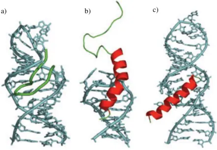
Most -hairpin mimics have been developed as part of the famous interaction between the Rev peptide and the HIV-1 mRNA region called Rev Response Element (RRE). The RRE/Rev interaction is important in the temporal control of HIV-1 mRNA splicing. The Rev sequence interacting with RRE has an helical conformation. In Figure 51b, the X-ray structure shows the binding of Rev in the major groove of the RNA. It is visible that the RNA-interacting side-chains are displayed around almost the entire circumference of the Rev -helix. Further informations concerning the HIV-1 Rev/RRE complex are in the publication of Battiste et al..82 In 2007, Robinson et al. made calculations showing that the interacting domain of Rev could be mimicked by 12-residues model 2:2-hairpin structures (Figure 50c). They designed several -hairpin mimetics stabilized by the D-Pro-L-Pro template, in view of inhibiting the Rev/RRE interaction. NMR studies of these mimics revealed that their structures were disordered in solution, since stable hairpin conformations could not be detected. Just one structure, R-27, was stable enough because of the introduction of a disulfide bridge (Figure 50e). This -hairpin structure was presented as a relative successful Rev peptide mimic for
a) b)
c)
b)
II.
Aims of our work
The aim of the first part of this thesis was to elaborate and to study new families of peptide foldamers, based on unnatural enantiomerically pure cyclic -amino acids, i.e. 6-substituted 4-oxopipecolic acids (Figure 51), whose the stereoselective synthesis and structures were published by Daly et al. in 2012.84
Figure 51: 6-substitued 4-oxo-pipecolic acid derivatives. -amino acid function=blue, side chain=green, ketone-group=red. R = Phenyl, Phenylethyl.
Various characteristics of these 6-substituted 4-oxo-pipecolic acid derivatives confer them interesting properties in the area of foldamers:
- The cyclic structure of the 6-substituted 4-oxo-pipecolic acid should introduce rigidity and it has been shown in the previous chapter that various cyclic amino acids containing five and six atoms (e.g. S-proline chimeras in Chapter 2.3.1) can lead to foldamers.
- The introduction of different side chains in the cyclic monomer at position 6 could be a possibility to modulate the biological properties of the foldamer. - On the other hand, the foldameric structures should have an overall dipole
We planned to develop two strategies to elaborate homogenous and heterogenous peptide foldamers (Scheme 52). In strategy 1, homogenous foldamers should be obtained via repetitive couplings of substituted 6-substituted 4-oxopipecolic acid units. In strategy 2, cyclic monomers should be alternatively coupled with natural -amino acids, in order to obtain heterogeneous foldamers. Heterogeneity in foldameric structures has been already published.One of the newest publications are from Amblard
et al. in 2017.
Figure 52: Illustration of both synthetic strategies for peptide foldamers.
Our first goal was to set up synthetic methodologies in liquid-phase to obtain di- and trimeric sequences of homo/hetero foldamers, then to apply them for the solid-phase
III.
Synthesis of
constrained
peptidomimetics
In this work, we decided to use the major diastereomer (2S, 6R) of 4-oxo-pipecolic acid incorporating a phenyl substituent at position 6. Two synthons should be first synthetized: the phenyl-4-oxo-pipecolic acid methyl ester and a N-protected 6-phenyl-4-oxopipecolic acid residue (Figure 53).
III.-1 Synthesis of protected (2S,
6R)-6-phenyl-4-oxo-pipecolic acid derivatives
III.-1.1 6-phenyl-4-oxo-pipecolic methyl ester
The synthesis of the phenyl-4-oxo-pipecolic acid methyl ester was performed according to the thesis and publication of Marc Daly et al. in 2012.84,86 This monomer was prepared in five steps in 13 % overall yield (Scheme 1 and 2).
Scheme 1: Synthesis of ,-unsaturated ketone. A) AcCl, MeOH, 65°C; b) TrtCl, TEA, DCM; c) DMMP, n-BuLi, THF (dry), -78°C d) Benzaldehyde, K2CO3, MeCN, reflux.
In the first step, L-aspartic acid was esterified with acetyl chloride in MeOH, giving the dimethyl diester hydrochloride salt 1 in 85% yield. In the following step, the amino
without isolation of intermediates (Scheme 2). First, the cleavage of the trityl-protecting group was performed with a 2M HCl solution in MeOH, followed by the cyclization in H2O with DIPEA at pH = 8. These last steps were poorly reproducible and led to
diastereomers 69 and 70 in relatively low yields (47% and 12%, respectively), after Flash Chromatography purification.
Applying another protocol, published by Harkiss et al. in 2018, allowed us to improve yields in compounds 6 and 7.87 First, the cleavage of the trityl group was realized using
TFA in DCM and the TFA salt 5 was isolated in quasi-quantitative yield. In a second step, the cyclization of 5 was carried out by means of DIPEA in MeOH. In this way, diastereomers 6 and 7 were isolated in respectively 60% and 30% yields (Scheme 2).
III.-1.2
N-protected
phenyl-4-oxo-pipecolic
acid
derivatives
We first focused on the synthesis of N-protected derivatives 8, presented in Scheme 3.
Scheme 3: Synthetic scheme of the synthesis of N-protected 4-oxo-pipecolic acid derivatives.
First, we tried to introduce three protecting-groups on the free secondary amine: the Fmoc, Boc and Alloc ones, via Fmoc-Cl, Boc2O and Alloc-Cl reagents, respectively
(Scheme 4). Only the Alloc protection could be performed in 95% yield. In the case of Boc, no reaction occurred while in the case of Fmoc, the only product formed was the dibenzofulvene (DBF), due to the elimination, on Fmoc-Cl reagent, of HCl by the secondary amine of 6.
This likely signifies that the strong steric hindrance due to the presence of substituents on 2- and 6-positions of the 4-oxo-pipecolic acid moiety did not allow the protection by the spacious Fmoc- and Boc-protecting-groups.
The second step was the saponification of the methyl ester 8 (Scheme 5). It was carried out under different conditions that are presented in Table 2.
Scheme 5: Saponification of cyclic monomer 8.
The saponification of 8 was assayed in basic and acid media. In acidic conditions, the degradation of the molecule was observed. In basic medium, the best results could be obtained (50% yield of 9) using the conditions published by Nicolaou et al., i.e Me3SnOH in 1,2-dichlorethane at 80°C (Table 2).88
Table 2: Conditions of saponification of cyclic monomer 8.
Conditions Yield
LiOH/Dioxane/H2O 20%
6M HCl84 /
The two synthons 6 and 9 required for the peptide synthesis were then obtained. However, the synthesis of 6 according to Daly et al. is very time-consuming, and the yields are neither satisfying nor really reproducible.84 Consequently, we tried to synthesize the monomer 6 following a new strategy.
III.-2 Development of a new methodology for
the synthesis of 6-phenyl-4-oxo pipecolic acid
derivatives via the imino-Diels-Alder-reaction
III.-2.1 Imino Diels Alder reaction
To synthesize a six-membered ring, the Diels-Alder reaction is the most known approach. The publication of Aznar et al.90 in 2006 showed us a new opportunity to synthesize diastereospecifically the protected monomer 9, via a L-proline catalyzed imino Diels-Alder reaction (Scheme 6). Starting from the -unsaturated ketone and from N-allyl phenylmethanimine, they obtained the product with a high diastereomeric excess.
Scheme 7: Synthesis of N-protected 4-oxo-pipecolic acid methyl esters 12 and 14.
Compound 10 was synthesized according to the publication of Zari et al. in 2014.91
Methyl levulinate was first brominated with Br2 in CHCl3, then in situ debrominated in
presence of NEt3, giving the desired product 10 in 34% yield (Scheme 8).
Scheme 8: Synthesis of methyl (2E) 4-oxo pent-2-enoate 10.91
Imines 11 and 13 were synthesized in one step, via the protocol described by Trost et
al. in 2015 (Scheme 9). Benzaldehyde reacted with allyl or benzyl amine in DCM, in presence of MgSO4, affording the desired imines 11 and 13 in 95% yield.92
Scheme 10 shows the Diels-Alder-reaction between 10 and imines 11 and 13. 1 eq of the imine 11 or 13 and 4 eq of compound 10 in presence of 20 mol% L-proline as catalyst in MeOH formed the desired compounds 12 or 14 in 40% yield. Using only 1 eq of compound 10 decreased the yield from 40 to about 25% in the two cases.
Scheme 10: Imino-Diels-Alder reactions. PG = Allyl; 12 and PG = Benzyl; 14.
Encouraged by this experiment, we investigated different reaction conditions to optimize the yield of the Diels-Alder reaction, taking compound 12 as target. First, we studied the solvent effect. Neither anhydrous solvents nor the use of inert atmosphere conditions were found to be necessary or beneficial. Different solvents were used for the reaction (DCM, DMF, ACN, CH, H2O, EtOAc, Et2O, Toluene, DMSO, IPA), but
none of them led to higher yields.
As expected, 12 and 14 were obtained as racemic mixtures. Indeed, the optical optical rotation angles of these products, measured by polarimetry, were found to be 0. Moreover, in the 1H-NMR-spectra, adding Eu(hfc)3 induced the doubling of the signal
Figure 54: 1H-NMR-spectrum of 12 in presence of Eu(hfc)3 (blue) compared to 1H-NMR of compound
12 alone (orange). Doubling of the signal of the methyl group was observed and is shown.
We tried other catalysts (Quinidine, (R)-(+)-N-Amino-2-methoxymethyl-pyrrolidine and L-ProPro) in view of both improving the yield and obtaining some enantioselectivity, but all of them failed to obtain the desired product (LC-MS and TLC).
Even if monomers 12 and 14 were obtained as racemic mixtures, this strategy based on the Diels-Alder reaction remained interesting since coupling with a chiral amino acid should lead to two possibly separable diastereomers. In view of peptide synthesis, we then focused on i) deprotection of the N-allyl and benzyl group on respectively compounds 12 and 14 ii) saponification of compounds 12 and 14.
III.-2.2 Synthesis of racemic phenyl-4-oxo-pipecolic methyl
ester
The allyl cleavage was attempted on 12 using several conditions, illustrated in Table 3 (Scheme 11).
Scheme 11: Illustration of allyl-deprotection.
Under conditions 1, the conversion to the desired product occurred. However, the reaction was only partial, even after one week of reaction time (10 %). Using rhodium (conditions 2) or palladium (conditions 3) based-catalysts did not give the deprotected monomer.
Table 3: Conditions of allyl-deprotection.
Conditions
1 5%mol Grubbs, toluene, argon, 80°C93,90
On the other hand, several conditions for the N-benzyl deprotection were carried out (Table 4).
Table 4: Conditions of benzyl-deprotection.
Conditions
1 Pd/C, H2, MeOH
2 Pd(OH)/C, MeOH, acetic acid96
3 H-Cube, 10 bar, 0.5mL/min
4 H-Cube, 10 bar, 0.1mL/min
All conditions allowed conversion into the product. However, as in the case of allyl cleavage, deprotection reactions were incomplete, even after one week. Due to lack of time, further assays could not be achieved.