Thermal restraint of a bacterial exopolysaccharide of shallow vent
origin
Maria
Teresa
Caccamo,
Vincenzo
Zammuto,
Concetta
Gugliandolo, Claire Madeleine-Perdrillat, Antonio Spanò,
Salvatore Magazù
PII:
S0141-8130(18)30825-0
DOI:
doi:
10.1016/j.ijbiomac.2018.03.160
Reference:
BIOMAC 9382
To appear in:
Received date:
22 February 2018
Revised date:
23 March 2018
Accepted date:
26 March 2018
Please cite this article as: Maria Teresa Caccamo, Vincenzo Zammuto, Concetta
Gugliandolo, Claire Madeleine-Perdrillat, Antonio Spanò, Salvatore Magazù , Thermal
restraint of a bacterial exopolysaccharide of shallow vent origin. The address for the
corresponding author was captured as affiliation for all authors. Please check if
appropriate. Biomac(2017), doi:
10.1016/j.ijbiomac.2018.03.160
This is a PDF file of an unedited manuscript that has been accepted for publication. As
a service to our customers we are providing this early version of the manuscript. The
manuscript will undergo copyediting, typesetting, and review of the resulting proof before
it is published in its final form. Please note that during the production process errors may
be discovered which could affect the content, and all legal disclaimers that apply to the
journal pertain.
ACCEPTED MANUSCRIPT
1
Thermal Restraint of a bacterial exopolysaccharide of shallow vent origin
Maria Teresa Caccamo
1,4, Vincenzo Zammuto*
5,6, Concetta Gugliandolo
5, Claire
Madeleine-Perdrillat
3, Antonio Spanò
5, Salvatore Magazù
1,2,3,4vzammuto@unime.it Department of Earth and Marine Sciences, University of Palermo, Via
Archirafi, 22, 90123 Palermo, Italy
1Dept of Mathematical and Informatics Sciences, Physical Sciences and Earth Sciences of Messina University, Viale
Ferdinando Stagno D' Alcontres 31, 98166 Messina, Italy.
2Le Studium Loire Valley Institute for Advanced Studies, 1 rue Dupanloup 45000, Orléans, France.
3Centre de Biophysique Moleculaire (CBM)-CNRS UPR 4301 du CNRS, rue Charles Sadron, 45071 Orleans CEDEX 2
France; Laboratoire Interfaces, Confinement, Matériaux et Nanostructures (ICMN) - UMR 7374 CNRS - Université d’Orléans, 1b rue de la Férollerie, CS 40059, 45071 Orléans cedex 2, France.
4Istituto Nazionale di Alta Matematica "F. Severi" – INDAM - Gruppo Nazionale per la Fisica Matematica – GNFMV.
Piazzale Aldo Moro, 5, 00185 Roma, Italy
5Dept. of Chemical, Biological, Pharmaceutical and Environmental Sciences, Messina University, Viale Ferdinando
Stagno D' Alcontres 31, 98166 Messina, Italy.
6Dept. of Earth and Marine Sciences, University of Palermo, Via Archirafi, 22, 90123 Palermo, Italy
Abstract
To dynamically characterize the thermal properties of the fructose-rich exopolysaccharide (EPS1-T14), produced by the marine thermophilic Bacillus licheniformis T14, the Attenuated Total Reflectance Fourier Transform Infra-Red spectroscopy was coupled to variable temperature ranging from ambient to 80 °C.
The spectra were analyzed by the following innovative mathematical tools: i) non-ideal spectral deviation, ii) OH-stretching band frequency center shift, iii) spectral distance, and iv) wavelet cross-correlation analysis.
The thermal restraint analysis revealed that the whole EPS1-T14 system possessed high stability until 80 °C, and suggested that fucose was mainly involved in the EPS1-T14 thermal stability, whereas glucose was responsible for its molecular flexibility.
Our results provide novel insights into the thermal stability properties of the whole EPS1-T14 and into the role of its main monosaccharidic units. As a new biopolymer, the thermostable EPS1-T14 could be used in traditional biotechnology fields and in new biomedical areas, as nanocarriers, requiring high temperature processes.
Keywords: Extremophiles, Thermal analysis, Attenuated-Total-Reflectance Infra-Red spectroscopy, Spectral distance, Wavelet cross-correlation analysis
ACCEPTED MANUSCRIPT
2
1.
Introduction
Biomolecules, such as complex polysaccharides and exopolysaccharides (EPSs), from extremophiles inhabiting hot environments are expected to possess unique characteristics, mainly the thermostability, useful in responding to the increasing demand of new attractive bio-products in medical and pharmaceutical applications. Thermophilic bacteria, with optimal growth at temperature ranging from 45°C to 70°C, offer interesting features for biotechnological applications, as they are able to produce their biomolecules faster than those from mesophilic and psychrophilic counterparts [1]. New thermophilic and thermotolerant bacteria, isolated from the shallow hydrothermal vents of Eolian Islands (Italy), were able to produce novel EPSs potentially useful in various applicative fields [1-4]. More recently, a novel alkalophilic and thermophilic Bacillus licheniformis strain T14, isolated from a submarine vent off Panarea Island (Italy), has been reported as producer of a new exopolysaccharide (EPS1-T14), by using relatively simple purification process [5]. In details, EPS1-T14, containing fructose, fucose and glucose as major monosaccharides, displayed structural characteristics (high carbohydrate content, 99% and high molecular weight, 1,000 KDa), and interesting rheological properties (i.e. viscoelasticity) (Tab. 1) for which it could be used as a convenient thickener in industrial products [5]. Moreover, EPS1-T14 is soluble in water, non-cytotoxic up to 400g ml-1, and it was also able to inhibit
the cytotoxic effects of Avarol in an brine shrimp assay, and therefore, applications of the biopolymer in the development of anti-cytotoxic drugs could be envisaged [5]. Furthermore, it represents a promising exopolymer in pharmaceutical applications, as natural antiviral, immunostimulant and immunomodulator [6] and with anti-adhesive properties able to prevent biofilm formation by pathogenic bacteria [7]. Many EPSs from thermophilic bacteria have been reported to be thermostable, as determined using the thermo-gravimetrical analysis (TGA) technique, with the highest thermostability registered for the exopolymer from Geobacillus tepidamans V246 (280 °C) [8], followed by those from G. thermodenitrificans B3-72 (240 °C) [9] and by EPS1-T14 from B. licheniformis T14, that was very stable up to 210 °C and started to decompose at 240 °C, yielding a weight loss of the half at 260 °C [5].
However, further information about the influence of the temperature on the molecular structure of EPS1-T14 are needed to address its potential use in biotechnological areas requiring high-temperature processes, such as food and cosmetic industries, and also for novel prospectives in medical and pharmaceutical applications, especially for drug delivery [10].
The changing of molecular structure is quantitatively measurable by InfraRed (IR) spectroscopy, providing a unique label-free tool for studying molecular composition and dynamics [11-14]. Among the IR techniques, the Fourier transform IR (FTIR) in the Attenuated Total Reflectance-FTIR (ATR-FTIR) mode represents a fast analytical method that permits to determine changes in molecular structure, using simultaneous analyses of multiple components from the same sample in a single instrumental measurement [15,16]. ATR-FTIR technique is widely employed to characterize the elemental structures and the molecular interactions among components in polymers [17]. Furthermore, FTIR technique is complementary to Raman scattering, inelastic neutron scattering and density function simulations, these techniques furnishing valuable information with different probe-system coupling [18-21]. Although FTIR analysis if used alone has been reported to be unable to ascertain the composition of polymorphic forms, the empowering of vibrational spectroscopy, by association with mathematical tools for the extraction of valuable information from complex evolving data, has demonstrated to be highly successful [22]. Only a few recent studies reported ATR-FTIR technique as a useful tool for monitoring the thermal behavior of pharmaceutical products [23-25].
In this work, the thermal restrain of the whole biopolymer EPS1-T14 and of its main monosaccharidic components (i.e. fructose, fucose and glucose) was investigated using a biophysical approach able to provide a measure of the
ACCEPTED MANUSCRIPT
3
system sensitivity to temperature changes [26]. The Attenuated Total Reflectance Fourier Transform Infra-Red (ATR-FTIR) spectroscopy was applied over a temperature range from 20 °C to 80 °C, and each spectrum was analyzed by using innovative mathematical tools: i) non-ideal spectral deviation; ii) OH-stretching band frequency center shift; iii) spectral distance; and iv) wavelet cross-correlation analysis.
2.
Material and Methods
2.1. The EPS-T14 production and characterization
The isolation of the halophilic and thermophilic Bacillus licheniformis T14 from a thermal fluid sample, emitted at a shallow hydrothermal vent of Panarea Island (Eolian Islands, Italy), and the production of a new exopolysaccharide (EPS1-T14) have been previously reported [5]. Briefly, characteristics of the emitted fluid were: temperature 50 °C, pH 5.42, and conductivity 42.90 mS m-1. Strain T14 grew aerobically from 25 to 60 °C and its optimal temperature
occurred at 50 °C. The pH range for growth was 4–10, with optimum at pH 8. Strain T14 grew in a range 2–10 % NaCl with optimal growth at 5 %.
The exopolysaccharide was produced (366 mg l-1 yield) at the beginning of the stationary growth phase of strain T14
in the minimal medium SWY (yeast extract, 0.1 %, sucrose 5 %, in seawater) after 48 h of incubation in bioreactor, under optimal growth condition (temperature 50 °C, pH 8, and NaCl 5 %) and air flow at 1 vvm. After purification, EPS1-T14 fraction displayed the highest carbohydrates content (99%), with a molecular weight estimated about 1,000 kDa (Table 1). Monosaccharide analysis revealed the presence of fructose/fucose/glucose composing the saccharide unit. NMR suggested a β-manno-pyranosidic configuration (Fig 1). Thermogravimetrical analysis (TGA) was performed using a Mettler-Toledo Star System apparatus, and a sample (5 mg) was heated rpm 30 to 400 °C at a heating rate of 20 °C min-1 under nitrogen. Measurements of viscosity () were performed with different EPS concentrations in
aqueous solution at 30 °C using a Cannon-Ubbelohde 75 suspended-level viscosimeter.
2.2. Experimental setup
ATR-FTIRVertex 70V spectrometer (Bruker Optics) using Platinum diamond ATR was employed to collect spectra on EPS1-T14, fructose, fucose and glucose, in the temperatures range from 20° C to 80 °C. All the spectra were obtained by collecting 48 scans with a resolution of 4 cm-1 in the 4000 to 400 cm−1 wavenumber range. The following
data pre-processing procedures were applied: i) a baseline correction, in order to diminish the dissimilarities between spectra due to baseline shift; ii) a smoothing treatment, to reduce the instrumental noise; iii) a first derivative treatment, to correct baseline shift together, and a second derivative treatment, to better discriminate features associated with spectra; iv) a spectral normalization, to correct the path-length variation and to reduce the differences among single measurements [27-29].
The intramolecular OH-stretching band frequency shift evaluation and the wavelet cross-correlation analysis were performed by MATLAB 2016a software (Mathworks, Natick, USA). The spectral distance calculation was performed by Origin 9 software package (OriginLab Co., Northampton, USA).
ACCEPTED MANUSCRIPT
4
2.3. Non ideal spectral deviation
In order to characterize the interaction mechanisms among the three major components of EPS1-T14, the difference between the experimental spectrum and the ideal spectrum of EPS1-T14, obtained as a weighed sum of the three component spectra, according to Eq. (1), was calculated using Eq. (2):
𝐼𝑖𝑑𝑒𝑎𝑙 (𝜔) = 𝛼𝐼𝑓𝑢𝑐𝑜𝑠𝑒 (𝜔) + 𝛽𝐼𝑓𝑟𝑢𝑐𝑡𝑜𝑠𝑒 (𝜔) + 𝛾𝐼𝑔𝑙𝑢𝑐𝑜𝑠𝑒 (𝜔) (1)
where 𝛼, 𝛽 and 𝛾 represent the weight factors of the three different components, i.e. fructose, fucose and glucose, respectively.
∆𝐼(𝜔) = 𝐼𝑒𝑥𝑝𝑒𝑟𝑖𝑚𝑒𝑛𝑡𝑎𝑙 (𝜔) − 𝐼𝑖𝑑𝑒𝑎𝑙 (𝜔) (2)
2.4. Hypsochromic frequency shift
A first and the second-order derivative spectra treatments were employed to calculate the band center frequency [30,31]. The hypsochromic shift of the band maximum to higher frequencies was used to highlight the change of H-bond crosslinks among the structural components of EPS1-T14 at increasing temperature. The thermal properties of EPS1-T14 were evaluated by analyzing the OH stretching contribution shift.
2.5. Spectral Distance (SD)
To investigate the temperature-induced structural rearrangements of EPS1-T14 H-bond network, the Spectral Distance (SD) of the intramolecular OH stretching band contribution was calculated in the temperature interval (from 20 °C to 80 °C) using Eq. (3):
𝑆𝐷 = {∑ [𝐼(𝜔, 𝑇) − 𝐼(𝜔, 𝑇 = 20°𝐶)]2Δ𝜔
𝑣 }1⁄2 (3)
where 𝐼(𝜔) is the absorbance at frequency 𝜔 and Δ𝜔 is the instrumental frequency resolution.
In order to extract further quantitative information on the whole EPS1-T14 thermal stability, for the SD versus temperature data fit, the following Eq. (4) has been applied:
𝑆𝐷(𝑇) = 𝐴 (1 −1+𝑒−𝐵(𝑇−𝑇0)1 ) + (𝐶 − 𝐷𝑇) (4)
Where A represents the sigmoid amplitude, whose inverse A -1 is connected to the thermal restraint. B is the sigmoid
steepness; T0 represents the temperature value of the sigmoid inflection point and, finally, C-DT takes into account the
low temperature vibrational contribution.
2.6. Wavelet Cross-Correlation Analysis
In order to quantitatively evaluate the correlation degree among the registered spectra at different scales, a Wavelet Cross-Correlation (XWT) analysis between the different couples of spectra was performed [26,32-36].
ACCEPTED MANUSCRIPT
5
3.
Results and discussion
3.1. Attenuated Total Reflectance Fourier Transform Infra-Red (ATR-FTIR) spectroscopy
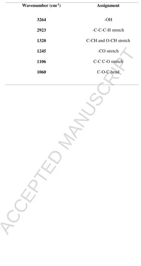
The spectra obtained by means of ATR-FTIR technique on EPS1-T14 and on its main components (i.e. fructose, fucose and glucose) (Fig. 2), according to the peak wavenumber assignment (Table 2), showed the following main features: i) an intense band in the region between 950 cm−1 and 1200 cm−1, corresponding to the –C–O, C–CH stretching and to the –CO and C–O–C bending of the glycoside ring [37]; ii) bands related to the C–CH, O–CH stretching and –CO stretching in the 1300 cm−1 - 1450 cm−1 spectral region, [38]; iii) a broad and intense band in the region between 2500 cm−1 and 3600 cm−1, which is composed by several bands ascribed to the OH stretching of the polymer glycoside ring, and to the OH stretching of free water and of water involved in hydrogen bonds [39,40].
The only detectable change in the reported spectra (Fig 2) is relative to the temperature effects in the region around 3000-3500 cm-1 of EPS1-T14 (Fig 2a), and in the region around 1700-2000 cm-1 of fructose, fucose, glucose (Fig 2
b-d). In detail, the spectrum of EPS1-T14 (fig.2 a) in the region 1700-2000 cm-1 shows a lower thermal variation in
respect with that of glucose (fig.2 d), but similar to that of fructose (fig.2 b) and fucose (fig. 2c). These observations demonstrate that the thermal restrain of EPS1-T14 mainly depends from fructose than glucose.
3.2. Non ideal spectral deviation
The EPS1-T14 ideal spectrum, reconstructed starting from the spectra of its main constituents, is shown in Fig. 3. The spectral deviation, i.e. the difference between the experimental spectrum and the ideal spectrum is reported in Fig. 4, where marked deviations from the ideal behavior are registered in the whole investigated spectral range. Such a circumstance suggests that remarkable interactions take place among the constituents. In particular, although in the 1800cm-1 to 2800cm-1 spectral range the evaluated ideal spectrum is almost coincident to the experimental one,
remarkable differences occur in the OH-stretching region, which is connected with the system H-bond network.
The non-ideal spectral deviation revealed that the real spectrum of EPS1-T14 is not the simple sum of its components, but it seems to acquire new properties and different responses at increasing temperature. This different thermal behavior could be ascribed to the interaction of its three main monosaccharidic components (i.e. fructose, fucose and glucose), that contribute differently to create the complex unique matrix of EPS1-T14.
3.3. Hypsochromic frequency shift
The 3D-FTIR spectra of EPS1-T14 in the 3000 < < 3600 cm-1 range, as a function of temperature are reported in
Fig. 5a. The EPS1-T14 OH stretching band frequency shift, in the temperature range, changes from 3271 cm-1 to 3313
cm-1. Furthermore, this registered hypsochromic frequency shift changes linearly (5) with the increasing of the
temperature, as shown in Fig. 5b. In order to fit the data the following Eq. (5) was employed:
𝜔(𝑇) = 𝐴 + 𝐵𝑇 (5)
The thermal restraint determined by hypsochromic frequency center shift was equal to 1.49.
The spectral analyses (hypsochromic frequency center shift and spectral distance) convey coherent information on the thermal evolution of EPS1-T14, both showing that the whole biopolymer was stable at the maximum value of the tested temperature (80 °C). The high resistance of the biopolymer at the increasing temperature could be explained by
ACCEPTED MANUSCRIPT
6
the increasing H-bounded connectivity. Since the frequency center linearly shifted with the increasing temperature, we may suppose that all the rheological properties (i.e. viscosity, density, etc) of EPS1-T14 could be maintained at 80 °C.
3.4. Spectral Distance (SD)
Spectral distance analysis of the spectra of EPS1-T14 at increasing temperatures showed a sigmoid trend, with an inflection point at T=50 °C (Fig. 6). The SD versus temperature behavior followed a logistic function trend allowing to extract quantitative information on the relaxation amplitude, as well as on its temperature localization.
3.5. Wavelet Cross-Correlation
The analysis by wavelet tools revealed different pathways of EPS1-T14 and its components (Fig. 7), where the shaded region indicates the Cone Of Influence (COI). This advanced investigation indicated that the spectrum of fucose is more closely correlated to that of the whole EPS1-T14, whereas glucose is the less correlated. This finding suggests that fucose plays a key role in the EPS1-T14 structure, and it may be also involved in the biological properties of the exopolysaccharide. The fucose-containing polysaccharides have an increased market value, because fucose is one of the rare sugars difficult to be obtained, and it is also involved in numerous biological activities, such as antioxidant, immune-stimulator and anti-aging agent [41,42]. Fucoidan is the most well-known fucose-rich polysaccharide produced by brown seaweeds, and used in many human activities [43]. Based on results previously obtained using TGA analysis, the decomposition temperature of EPS1-T14 (240 °C) appears higher than that of fucoidan (210 °C) [44]. For its thermal stability and thermal restraint, EPS1-T14 could be more cooking stable than other fucose polysaccharides, such as fucoidan, actually commercially available as nutritional supplements [45]. However, to our knowledge, no data on the thermal behavior of fucoidan have been until now reported for further comparison.
The thermal characterization is also required in the drug deliveries system, a novel pharmaceutical field that allows to use lower doses of drugs and to develop drug or food carriers. Indeed, polymeric nanoparticles can be fabricated from polysaccharides, due to their low bio-toxicity and high adaptability [20]. Sodium carboxymethyl cellulose [46], pectin [47], chitosan [48] and xanthan gum [10] are among polysaccharides until now studied for delivering drugs, nutrients and bioactive substances. Nowadays, chitosan is the most characterized polysaccharide in the field of drug and food deliveries, however its uses are still limited, due to its relatively lower thermal stability (below 40 °C), as spectroscopically determined [49]. For its physicochemical properties and thermo-stability EPS1-T14 could be also considered as useful biopolymer for nanoparticle composites.
4. Conclusion
The fructose-rich exopolysaccharide EPS1-T14, produced by the marine thermophilic Bacillus licheniformis T14, has been previously reported to possess interesting physicochemical properties and biological activities, as anti-cytotoxic, antiviral, immunostimulant, and antibiofilm agent, that make it potentially useful in different biotechnological applications.
Since thermostability represents a prerequisite in applicative areas that require high temperature processes, in this study the structural changes of EPS1-T14 at increasing temperatures have been monitored by using the ATR-FTIR spectroscopic technique, coupled with innovative tools (non-ideal spectral deviation, hypsocromic frequency center shift, spectral distance and wavelet cross correlation analysis). The thermal restraint analysis revealed that the whole EPS1-T14 system possessed high stability until 80 °C, and suggested that fucose was mainly involved in the EPS1-T14 thermal stability, whereas glucose was responsible for its molecular flexibility.
ACCEPTED MANUSCRIPT
7
Our findings provide new insights on the thermal stability of EPS1-T14, enabling us to consider it as a promising biopolymer useful for traditional biotechnological areas, including food, cosmetic and pharmaceutical applications, and novel biomedicine areas that require high-temperature processes, such as drug delivery system and nanoparticle composites.
Funding
This research did not receive any specific grant from funding agencies in the public, commercial, or not-for-profit sectors
Conflict of interest
ACCEPTED MANUSCRIPT
8
References
[1] C. Gugliandolo, V. Lentini, A. Spanò, T.L. Maugeri, New bacilli from shallow hydrothermal vents of Panarea Island (Italy) and their biotechnological potential, J. Appl. Microbiol. 112 (2012) 1102–1112.
[2] T.L. Maugeri, C. Gugliandolo, D. Caccamo, A. Panico, L. Lama, A. Gambacorta, B. Nicolaus, A halophilic thermotolerant Bacillus isolated from a marine hot spring able to produce a new exopolysaccharide, Biotechnol. Lett. 24 (2002) 515–519.
[3] T.L. Maugeri, C. Gugliandolo, D. Caccamo, E. Stackebrandt, Three novel halotolerant and thermophilic Geobacillus strains from shallow marine vents, Syst. Appl. Microbiol. 25 (2002) 450–455.
[4] B. Nicolaus, A. Panico, M.C. Manca, L. Lama, A. Gambacorta, T.L. Maugeri, A thermophilic Bacillus isolated from an Eolian shallow hydrothermal vent, able to produce exopolysaccharides, Syst. Appl. Microbiol. 23 (2000) 426– 432.
[5] A. Spanò, C. Gugliandolo, V. Lentini, T.L. Maugeri, G. Anzelmo, A. Poli, B. Nicolaus, A novel EPS-producing strain of Bacillus licheniformis isolated from a shallow vent off Panarea Island (Italy), Curr. Microbiol. 67 (2013) 21–29.
[6] C. Gugliandolo, A. Spanò, V. Lentini, A. Arena, T.L. Maugeri, Antiviral and immunomodulatory effects of a novel bacterial exopolysaccharide of shallow marine vent origin, J. Appl. Microbiol. 116 (2014)1028–1034.
[7] A. Spanò, P. Laganà, G. Visalli, T.L. Maugeri, C. Gugliandolo, In vitro antibiofilm activity of an exopolysaccharide from the marine thermophilic Bacillus licheniformis T14, Curr. Microbiol. 72 (2016) 518–528.
[8] M. Kambourova, R. Mandeva, D. Dimova, A. Poli, B.Nicolaus, G.Tommonaro, Production and characterization of a microbial glucan, synthesized by Geobacillus tepidamans V264 isolated from Bulgarian hot spring, Carbohydr. Polym. 77 (2009) 338–343.
[9] A. Arena, C. Gugliandolo, G. Stassi, B. Pavone, D. Iannello, G. Bisignano, T.L. Maugeri, An exopolysaccharide produced by Geobacillus thermodenitrificans strain B3-72: antiviral activity on immunocompetent cells, Immunol.
Lett. 123 (2009) 132–137.
[10] W. Xu, W. Jin, Z. Li, H. Liang, Y. Wang, B.R. Shah, B. Li, Synthesis and characterization of nanoparticles based on negatively charged xanthan gum and lysozyme, Food. Res. Int. 71 (2015) 83–90.
[11] C. Branca, S.Magazù, F. Migliardo, P. Migliardo, Destructuring effect of trehalose on the tetrahedral network of water: A Raman and neutron diffraction comparison, Phys. A 304 (2002) 314–318.
[12] C. Branca, S. Maccarrone, S. Magazù, G. Maisano, S.M. Bennington J. Taylor, Tetrahedral order in homologous disaccharide-water mixtures, J. Chem. Phys. 122 (2005) 174513.
[13] P. Ballone, M. Marchi, C. Branca, S. Magazù, Structural and vibrational properties of trehalose: A density functional study, J. Phys. Chem. B 104 (2000) 6313–6317.
[14] S. Magazù, F. Migliardo, M.T.F. Telling, Structural and dynamical properties of water in sugar mixture, Food Chem, 106 (2008)1460–1466.
[15] M. Deepa, N. Sharma, S.A. Agnihotry, R. Chandra, FTIR Investigation on ion—ion interaction in liquid and gel polymeric electrolytes- LiCF3SO3-PC-PMMA, J. Mater. Sci. 37 (2002)1759-1765.
[16] S. Magazù, E. Calabrò, S. Campo, FTIR spectroscopy studies on the bioprotective effectiveness of trehalose on human hemoglobin aqueous solutions under 50 Hz electromagnetic field exposure, J. Phys. Chem. B 114 (2010) 12144–12149.
[17] R.H. Barker, R. Dagher, D.M. Davidson, J.K. Marquis, Review article: tolevamer, a novel toxin-binding polymer: overview of preclinical pharmacology and physicochemical properties. Aliment. Pharmacol. Ther. 24 (2006) 1525– 1534.
[18] L. Hennet, V. Cristiglio, J. Kozaily, I. Pozdnyakova, H.E. Fischer, Bytchkov A, J.W.E. Drewitt, M. Leydier, D. Thiaudière, S. Gruner, S. Brassamin, D. Zanghi, G.J. Cuello, M. Koza, S. Magazù, G.N. Greaves, D.L. Price, Aerodynamic levitation and laser heating: Applications at synchrotron and neutron sources, Eur. Phys. J. Spec. Top. 196 (2011) 151–165.
[19] S. Magazù, F. Migliardo, M.T. Caccamo, Innovative wavelet protocols in analyzing elastic incoherent neutron scattering, J. Phys. Chem. B 116 (2012) 9417–9423.
[20] S. Magazù, F. Migliardo, B.G. Vertessy, M.T. Caccamo, Investigations of homologous disaccharides by Elastic Incoherent Neutron Scattering and Wavelet Multiresolution Analysis, Chem. Phys. 424 (2013) 56–61.
[21] N. Marchese, A. Cannuli, M.T. Caccamo, C. Pace, New generation non-stationary portable neutron generators for biophysical applications of Neutron Activation Analysis. BBA - General Subjects 1861 (2017) 3661–3670.
[22] F. Migliardo, S. Magazù, M.T. Caccamo, Infrared, Raman and INS studies of poly-ethylene oxide oligomers. J. Mol. Struct. 1048 (2013) 261–266.
[23] N.L. Calvo, R.M. Maggio, T.S. Kaufman, A dynamic thermal ATR-FTIR/chemometric approach to the analysis of polymorphic interconversions: Cimetidine as a model drug. J. Pharm. Biomed. Anal. 92 (2014) 90–97.
[24] F. Migliardo, M.T. Caccamo, S. Magazù, Elastic incoherent neutron scatterings wavevector and thermal analysis on glass-forming homologous disaccharides, J. Non Cryst. Solids 378 (2013) 144–151.
[25] F. Migliardo, M.T. Caccamo, S. Magazù, Thermal analysis on bioprotectant disaccharides by elastic incoherent neutron scattering, Food Biophys. 9 (2014) 99–104.
ACCEPTED MANUSCRIPT
9
[26] M.T. Caccamo, S. Magazù, Multiscaling Wavelet analysis of Infrared and Raman data on polyethylene glycol 1000 aqueous solutions, Appl. Spectrosc. 50 (2017) 130–136.
[27] H.M.Al-Qadiri, N.I. Al-Alami, M Lin, M Al-Holy, AG Cavinato, BA. Rasco, Studying of the bacterial growth phases using fourier transform infrared spectroscopy and multivariate analysis, J. Rapid Methods Autom. Microbiol. 6 (2008) 73–89.
[28] R. Davis, L.J. Mauer, Fourier Transform Infrared (FT-IR) Spectroscopy: a rapid tool for detection and analysis of foodborne pathogenic bacteria. In A. Méndez-Vilas (Eds.), Current research, technology and education topics in Applied Microbiology and Microbial Biotechnology, Formatex Research Center Publisher 2010, pp.1582–1594. [29] R. Gautam, S. Vanga, F. Ariese, S. Umapathy, Review of multidimensional data processing approaches for Raman
and infrared spectroscopy, EPJ Techn. Instrum. 2 (2015) 8.
[30] M. Gallignani, R.A. Rondóna, J.F. Ovalles, M.R. Brunetto, Transmission FTIR derivative spectroscopy for estimation of furosemide in raw material and tablet dosage form, Acta Chim. Sinica 4 (2014) 376–383.
[31] Z. Lee, K. Carder, R. Arnone, M. He, Determination of primary spectral bands for remote sensing of aquatic environments, Sensors (Basel) 7 (2007) 3428–3441.
[32] M.T. Caccamo, S. Magazù, Tagging the oligomer-to-polymer crossover on EG and PEGs by infrared and Raman spectroscopies and by wavelet cross-correlation spectral analysis. Vib. Spectrosc. 85 (2016) 222–227.
[33] A. Grinsted, J.C. Moore, S. Jevrejeva, Application of the cross wavelet transform and wavelet coherence to geophysical time series, Nonlinear. Process. Geophys. 11 (2004) 561–566.
[34] H. Li, T. Nozaki, Wavelet cross-correlation analysis and its application to a plane turbulent jet, JSME Int. J. Ser B- 40 (1997) 58–66.
[35] S. Magazù, F. Migliardo, B.G.Vertessy, M.T. Caccamo, Investigations of homologous disaccharides by Elastic Incoherent Neutron Scattering and Wavelet Multiresolution Analysis. Chem. Phys. 424 (2013) 56–61.
[36] D.Veleda, R. Montagne, M. Araujo, Cross-Wavelet bias corrected by normalizing scales. J. Atmos. Oceanic Technol. 29 (2012) 1401–1408.
[37] J.P. Coates, The interpretation of Infrared Spectra: published reference sources. Appl. Spectrosc. Rev. 31 (1996) 179–192.
[38] H.A Szymanski., R.E. Erickson. Infrared Handbook, revised ed., Plenum Publishing Corp., New York, N.Y. 10011, 1970.
[39] S. Magazù, G. Maisano, P. Migliardo, H.D. Middendorf, V. Villari, Hydration and transport properties of aqueous solutions of α-α-trehalose. J. Chem. Phys. 109 (1998) 1170–1174.
[40] S.E. Pagnotta, M.A. Ricci, F. Bruni, S. McLain, S. Magazù, Water structure around trehalose. Chem. Phys. 345 (2008) 159–163.
[41] F. Freitas, V.D. Alves, M.A.M. Reis, Advances in bacterial exopolysaccharides: from production to biotechnological applications. Trends. Biotechnol. 29 (2011) 388–398.
[42] P. Cescutti, A. Kallioinen, G. Impallomeni, R. Toffanin, P. Pollesello, M. Leisola, T. Eerikäinen, Structure of the exopolysaccharide produced by Enterobacter amnigenus. Carbohydr. Res. 340 (2005) 439–447.
[43] B. Li, F. Lu, X. Wei, R. Zhao, Fucoidan: structure and bioactivity, Molecules 13 (2008) 1671–1695.
[44] P.S. Saravana, Y.J. Cho, Y.B. Park, H.C. Woo, B.S. Chun, Structural, antioxidant, and emulsifying activities of fucoidan from Saccharina japonica using pressurized liquid extraction, Carbohydr. Polym. 153 (2016) 518–525. [45] N.C. Moroney, M.N. O’Grady, S. Lordan, C. Stanton, J.P. Kerry, Seaweed polysaccharides (laminarin and
fucoidan) as functional ingredients in pork meat: an evaluation of anti-oxidative potential, thermal stability and bioaccessibility, Mar. Drugs 13 (2015) 2447–2464.
[46] C. Chang, L. Zhang, Cellulose-based hydrogels: Present status and application prospects. Carbohydr. Polym. 84 (2011) 40–45.
[47] C. Chittasupho, M. Jaturanpinyo, S. Mangmool, Pectin nanoparticle enhances cytotoxicity of methotrexate against hepG2 cells. Drug Delivery 20 (2013) 1–9
[48] A. Pramanik, D. Laha, P. Pramanik, P. Karmakar, A novel drug “copper acetylacetonate” loaded in folic acid-tagged chitosan nanoparticle for efficient cancer cell targeting, J. Drug Target22 (2014) 23–33.
[49] E. Szymańsk, K. Winnicka, Stability of chitosan a challenge for pharmaceutical and biomedical applications, Mar. Drugs 13 (2015) 1819–1846.
ACCEPTED MANUSCRIPT
10
Table 1 Characteristics of EPS1-T14 from B. licheniformis strain T14 [5].
EPS1-T14
Carbohydrate (%/v) 99
Protein (%/v) 1.2
Uronic acid (%/v) 0.5
Molecular weight 1000 kDa
Monosaccharide composition (ratio of relative proportion)
Fructose/fucose /glucose/galactosamine/mannose (1.0:0.75:0.28:tr:tr)
Saccharide repeating unit Trisaccharide
Anomeric configuration β-manno pyranoside
Specific viscosity 0.58η
Thermogravimetrical analysis 240 °C
ACCEPTED MANUSCRIPT
11
Table 2 Peak wavenumbers (cm-1) assigned
Wavenumber (cm-1) Assignment
3264 -OH
2923 -C-C-C-H stretch
1320 C-CH and O-CH stretch
1245 -CO stretch
1106 C-C C-O stretch
ACCEPTED MANUSCRIPT
12
Figure Legends
Fig. 1. 1H-NMR (a) and 13C-NMR (b) spectra of EPS1-T14 from Bacillus licheniformis strain T14 [5].
Fig. 2. ATR-FTIR spectra of whole EPS1-T14 a) and its main monosaccharidic components: b) fructose, c) fucose and d) glucose in the 400<∆<4000 cm-1spectral range for positive thermal scans from 20 to 80 °C.
Fig. 3. Ideal spectrum of EPS1-T14 reconstructed from the spectra of its main constituents, obtained using ATR-FTIR in the 400<∆<4000 cm-1spectral range at 25 °C.
Fig. 4. Difference spectrum obtained by subtracting ideal spectrum to experimental spectrum of EPS1-T14. Fig. 5. Hypsochromic frequency shift: a) 3D FTIR spectra of EPS1-T14 in the 3000 < < 3600 cm-1 range, as a
function of temperature and intensity, and b) the center frequency of OH stretching band of EPS1-T14, as a function of temperature. Black dots: experimental data.
Fig. 6. Spectral Distance of the intramolecular OH stretching bands of the EPS1-T14 at the increasing temperature. Fig. 7. Wavelet Cross Correlation between the whole EPS1-T14 and a) fructose, b) fucose and c) glucose at 25 °C.
ACCEPTED MANUSCRIPT
13
ACCEPTED MANUSCRIPT
14
Fig. 2. a) c) b) d)ACCEPTED MANUSCRIPT
15
ACCEPTED MANUSCRIPT
16
ACCEPTED MANUSCRIPT
17
ACCEPTED MANUSCRIPT
18
Fig. 6.Sp
ec
tral
D
is
tan
ce
Temperature ( C)
ACCEPTED MANUSCRIPT
19
Fig. 7.
![Table 1 Characteristics of EPS1-T14 from B. licheniformis strain T14 [5].](https://thumb-eu.123doks.com/thumbv2/123doknet/14550453.536852/11.892.92.647.157.1001/table-characteristics-eps-t-b-licheniformis-strain-t.webp)