SCIENTIFIC ARTICLE
Relationship between Wiberg
's lateral center edge angle,
Lequesne
's acetabular index, and medial acetabular
bone stock
Clément M.L. Werner&Carol E. Copeland&
Thomas Ruckstuhl&Jeff Stromberg&
Clifford H. Turen&Samy Bouaicha
Received: 12 January 2011 / Revised: 15 February 2011 / Accepted: 23 February 2011 / Published online: 15 March 2011 # ISS 2011
Abstract
Objective Knowledge of acetabular anatomy is crucial for cup positioning in total hip replacement. Medial wall thickness of the acetabulum is known to correlate with the degree of developmental dysplasia of the hip (DDH). No data exist about the relationship of routinely used radio-graphic parameters such as Wiberg's lateral center edge angle (LCE-angle) or Lequesne's acetabular index (AI) with thickness of the medial acetabular wall in the general population. The aim of our study was to clarify the relationship between LCE, AI, and thickness of the medial acetabular wall.
Materials and methods Measurements on plain radiographs (LCE and AI) and axial CT scans (quadrilateral plate
acetabular distance QPAD) of 1,201 individuals (2,402 hips) were obtained using a PACS imaging program and statistical analyses were performed.
Results The mean thickness of the medial acetabulum bone stock (QPAD) was 1.08 mm (95% CI: 1.05-1.10) with a range of 0.1 to 8.8 mm. For pathological values of either the LCE (<20°) or the AI (>12°) the medial acetabular wall showed to be thicker than in radiological normal hips. The overall correlation between coxometric indices and medial acetabular was weak for LCE (r =−0.21. 95% CI [−0.25, -0.17]) and moderate for AI (r = 0.37, [0.33, 0.41]).
Conclusions We did not find a linear relationship between Wiberg's lateral center edge angle, Lequesne's acetabular index and medial acetabular bone stock in radiological normal hips but medial acetabular wall thickness increases with dysplastic indices.
Keywords Acetabulum . Bone stock . Acetabular index . Lateral center edge angle . Pelvic radiographs
Introduction
To obtain sufficient coverage and good primary stability for the prosthetic cup in total hip replacement (THR), reaming of the medial acetabular wall is a common procedure. Cup positioning close to the teardrop figure is mandatory to achieve an anatomical offset and to avoid antero-lateral cup overhang causing chronic irritation of the iliopsoas tendon [1–3]. Therefore, knowledge about the individual acetabu-lar anatomy and the bone stock of the medial wall in particular is essential for preventing accidental penetration into the pelvis. Not only for THR but also in acetabular reconstruction surgery the distance between the acetabular
C. M. Werner (*)
:
T. Ruckstuhl:
S. BouaichaDepartment of Orthopaedics, Balgrist University Hospital Zurich, Forchstrasse, 340 8008, Zurich, Switzerland e-mail: [email protected] T. Ruckstuhl e-mail: [email protected] S. Bouaicha e-mail: [email protected]
C. M. Werner
:
C. E. Copeland:
J. Stromberg:
C. H. TurenR. Adams Cowley Shock Trauma Center, University of Maryland Medical Systems, 22 South Greene Street,
Baltimore, MD 21201, USA C. E. Copeland e-mail: [email protected] J. Stromberg e-mail: [email protected] C. H. Turen e-mail: [email protected] DOI 10.1007/s00256-011-1141-3
fossa and the quadrilateral plate is crucial for screw positioning (e.g., screws in the anterior column that may be inserted within the joint but should not interfere with joint movement) [4, 5]. On radiographs, in some cases, it remains difficult to estimate medial wall thickness and lack of information cannot always be substituted by the surgeon's experience. Perforation of the quadrilateral plate with over-reaming may result in insufficient cup fixation and secondary cup migration [6,7].
It is known that medial wall thickness of the acetabulum correlates with the degree of developmental dysplasia of the hip (DDH) regarding Crowe's classification (high-degree DDH with thicker medial bone stock than lower-degree DDH) [8].
No data exists about relationship of coxometric indices such as Wiberg's lateral center edge angle (LCE-angle) [9] or Lequesne's acetabular index (AI) [10] and thickness of the medial acetabular wall in the general population. Knowledge of such a correlation could help orthopedic surgeons in planning total hip replacements preoperatively. Our hypothesis therefore was that commonly used radiographic parameters such as LCE angle and AI indicate the amount of medial acetabular bone stock in the general population. This study was designed to clarify this issue.
Materials and methods Patients
Conventional anterior-posterior pelvic radiographs and pelvic CT scans of patients admitted to the R. Adams Cowley Trauma Center, Baltimore, USA, were extracted from the electronic radiological database. From these patients, 1,201 individuals (2,402 hips), whereof 873 male and 328 female (73/27%) age 14–97 years (median 38.9 years) met the inclusion criteria having plain conventional radiographs of the pelvis obtained in a correct standardized radiographic technique [11] and corresponding CT scans of the pelvis. IRB approval for was obtained for this study.
Validation of radiographs
Rotation in the axial and sagittal plane of the pelvis can significantly change outcome measurements of coxometric indices [12] and therefore to reduce the potential errors, only AP pelvic radiographs, which reveals alignment of the tip of the coccyges with the middle of the symphysis and a distance between the sacrococcygeal joint and the symphysis less than 32 mm in men and 47 in women according to the criteria of Siebenrock et al. [11], were further assessed.
False rotation in the frontal plane could simply be corrected electronically with the picture-archiving
commu-nication system (PACS) imaging program, and therefore such radiographs were not excluded.
Measurements on radiographs
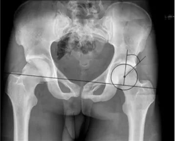
The LCE was measured as follows: First, the horizontal plane of the pelvis was determined by drawing a horizontal line along the inferior boundaries of the two teardrops. A second perpendicular line, crossing the center of the femoral head, which first was defined by the midpoint of a congruent circle, was drawn up to the acetabular roof. The intersection of this vertical line with the acetabular roof represented the starting point of further measuring. At least, a line from the center of the femoral head was drawn out to the most lateral extent of the acetabulum. The angle between the perpendicular line and the oblique line, touching the lateral extent of the acetabulum showed Wiberg's angle (Fig.1).
Lequesne's acetabular index (AI) was measured by first drawing a parallel line to the horizontal plane of the pelvis as described above (teardrop to teardrop), touching the medial border of the sclerotic weight-bearing portion of the acetabular roof, the so-called“sourcil”. Furthermore, a line originating from this medial extent of the “sourcil” was drawn out to the lateral extent of the weight-bearing portion of the acetabulum neglecting any osteophyte formation. The angle between these two lines crossing at the medial border of the“sourcil” showed Lequesne's AI (Fig.2).
All values of conventional radiographs were obtained using digitally measurement tools of the PACS imaging program. Measurement on CT scans
Thickness of the smallest bony wall diameter between the most medial aspect of the acetabular fossa and the
Fig. 1 Measurement method of the Wiberg's lateral center edge angle
quadrilateral plate on axial CT was measured digitally with the PCAS imaging program. Therefore a rectangular line to the quadrilateral plate was drawn through the center of the femoral head pointing the area of medial apex of a prosthetic cup (Fig.3). An analog measurement technique was previously described by Stein et al. who first measured different acetabular distances with CT [13].
Statistical analysis
Gaussianity of angle data was graphically illustrated using frequency polygons. For quantities that involved measure-ments of both sides within a patient (QPAD), that means dependent measurements, we computed confidence inter-vals using standard errors received from a generalized least-squares (GLS) model assuming a compound symmetry correlation structure. Pearson's correlation coefficient is provided to assess the extent of linear association between continuous variables. To compute confidence intervals for correlation coefficients for clustered observations, we use
the fact that Pearson's correlation coefficient between two variables x and y is the slope of a regression model of the standardized y on the standardized x. We compute standard errors for this slope (and thus the correlation coefficient) in a GLS model as above. No correction for multiple testing is done in this analysis, all confidence intervals are computed using a confidence level of 95%, and all tests are computed at a significance level of alpha=0.05. All analyses were performed using R. The add-on package 'nlme' was used to compute the GLS models [14,15].
Results
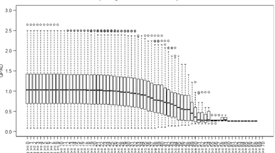
The mean thickness of the quadrilateral plate bone stock (QPAD) was 1.08 mm (95% CI: 1.05-1.10) with a range from 0.1 to 8.8 mm. For correlation between coxometric indices and amount of medial acetabular bone stock, we found only weak to moderate overall correlation for LCE (r =−0.21, 95% CI [−0.25, -0.17]) and AI (r=0.37, 95% CI [0.33, 0.41]). For pathological values of either the LCE (<20°) or the AI (>12°), the medial acetabular wall was shown to be thicker than in radiological normal hips. Figures 4 and 5 show increasing QPAD of both LCE and AI measurements with increasing “dysplastic” angles. The mean wall thickness of individuals with AI above 12° was 1.54 mm compared to 1.04 mm below the pathological cut-off. Less discrepancy showed LCE with a mean thickness of 1.35 mm below 20° and 1.07 mm above.
Discussion
Correct prosthetic cup positioning in total hip replacement is crucial for clinical outcome and longevity of all prostheses [16]. Therefore, knowledge about individual acetabular anatomy is a key issue in the preoperative planning process with direct influence on reaming capac-ities of the medial acetabular wall. Unfortunately, there is no reliable method to estimate medial acetabular bone stock on routinely performed conventional radiographs for THR such as the AP view of the pelvis. CT Scans for this particular question are not routinely performed and are usually not requested by the surgeon planning THR surgery. Thickness of the medial acetabular wall based on CT scans varies with different measurement techniques and locations [8, 13, 17] and therefore comparison of values remains controversial. Stein et al. first published different acetabular distances using computer tomography with different anatomical questions with regard to prosthetic hip surgery. We measured the width of the quadrilateral plate, representing the area of closest and most medial
Fig. 3 Measurement method of the quadrilateral plate acetabulum distance (QPAD) on axial CT scan with PACS
Fig. 2 Measurement method of the Lequesne's acetabular index on
contact to a virtual prosthetic cup accordingly [13]. Because this area is critical for perforation, we opted for the smallest diameter for measurement. With a 1.1-mm mean value for the QPAD, our results were shown to be comparable with historical data [13].
It is known that in developmental dysplastic hips (DDH) the amount of the medial acetabular bone stock correlates positively with increasing dysplasia regarding Crowe's classification. It describes the percentage of dislocation of the femoral head, beginning with less than 50% dislocation in Crowe's class I, to 50–75% in class II, up to 75–100% in class III and over 100% in the most dysplastic class IV [18]. This classification does not address the acetabular morphology quantitatively and usefulness in planning of a THR is not given, especially because in the vast majority of
cases subluxation or dislocation of the femoral head in osteoarthritic hips is absent.
Coxometric parameters such as Wiberg's LCE or Lequesne's AI are routinely used to quantify acetabular roof morphology and to determine the presence or absence of dysplasia (which again can cause difficulties in cup fixation with consecutive loss of primary stability) [16,19]. Since routinely used coxometric indices like LCE and AI already provide important information about acetabular roof orientation, it would be beneficial if the same parameters could indicate the amount of medial acetabular bone stock. Our hypothesis was that (in analogy to the correlation of increasing dysplasia according to the Crowe's classifica-tion) the LCE and AI angles could also indicate the thickness of the medial acetabular wall.
Fig. 4 Boxplots of QPAD in relation to LCE angles
Fig. 5 Boxplots of QPAD in relation to AI angles
Our results confirmed that the coxometric indices correlate with the medial acetabular bone stock in radio-graphic dysplastic hips. This is in concordance with the findings of Liu showing a significant correlation between the degree of dysplasia and medial acetabular wall thickness [8]. Unfortunately, we also could clearly demonstrate the limited applicability to only dysplastic hips as this was not the case in so called "normal" hips. This means that no reliable estimation of medial wall thickness can be made in hips without dysplasia. In routine THR surgery, careful intra-operative estimation of the medial bone stock remains crucial and for difficult cases, CT scan for planning should be considered. While costs for a CT scan of the pelvis are well known, over-reaming of the medial acetabular wall is likely to be underreported, and related follow-up costs due to revision surgery are difficult to take in account. Therefore calculation of the cost-effectiveness for routine CT scans prior to THR remains difficult.
Conclusions
Wiberg's lateral center edge angle and Lequesne's acetab-ular index should not be used to estimate medial acetabacetab-ular bone stock in radiological normal hips.
Conflict of interest None.
References
1. Lachiewicz PF, Kauk JR. Anterior iliopsoas impingement and tendinitis after total hip arthroplasty. J Am Acad Orthop Surg.
2009;17(6):337–44.
2. Dora C, Houweling M, Koch P, Sierra RJ. Iliopsoas impingement after total hip replacement: the results of non-operative manage-ment, tenotomy or acetabular revision. J Bone Joint Surg Br.
2007;89(8):1031–5.
3. Asayama I, Chamnongkich S, Simpson KJ, Kinsey TL, Mahoney OM. Reconstructed hip joint position and abductor muscle strength after total hip arthroplasty. J Arthroplasty. 2005;20
(4):414–20.
4. Starr AJ, Walter JC, Harris RW, Reinert CM, Jones AL. Percutaneous screw fixation of fractures of the iliac wing and fracture-dislocations of the sacro-iliac joint (OTA Types 61-B2.2
and 61-B2.3, or Young-Burgess "lateral compression type II"
pelvic fractures). J Orthop Trauma. 2002;16(2):116–23.
5. Giannoudis PV, Tzioupis CC, Pape HC, Roberts CS. Percutaneous fixation of the pelvic ring: an update. J Bone Joint Surg Br. 2007;89(2):145–54.
6. Eftekhar NS, Nercessian O. Intrapelvic migration of total hip prostheses. Operative treatment. J Bone Joint Surg Am. 1989;71
(10):1480–6.
7. Sharkey PF, Hozack WJ, Callaghan JJ, Kim YS, Berry DJ, Hanssen AD, et al. Acetabular fracture associated with cementless acetabular component insertion: a report of 13 cases. J
Arthro-plasty. 1999;14(4):426–31.
8. Liu RY, Wang KZ, Wang CS, Dang XQ, Tong ZQ. Evaluation of medial acetabular wall bone stock in patients with developmental dysplasia of the hip using a helical computed tomography multiplanar reconstruction technique. Acta Radiol. 2009;50
(7):791–7.
9. Wiberg G. Studies on dysplastic acetabula and congenital subluxation of the hip joint. Acta Chir Scand. 1939;58:5–135. 10. Lequesne M. Coxometry. Measurement of the basic angles of the
adult radiographic hip by a combined protractor. Rev Rhum Mal Osteo-artic. 1963;30:479–85.
11. Siebenrock KA, Kalbermatten DF, Ganz R. Effect of pelvic tilt on acetabular retroversion: a study of pelves from cadavers. Clin
Orthop Relat Res. 2003;407:241–8.
12. Tannast M, Zheng G, Anderegg C, Burckhardt K, Langlotz F, Ganz R, et al. Tilt and rotation correction of acetabular version on
pelvic radiographs. Clin Orthop Relat Res. 2005;438:182–90.
13. Stein MG, Barmeir E, Levin J, Dubowitz B, Roffman M. The medial acetabular wall: normal measurements in different
popu-lation groups. Invest Radiol. 1982;17(5):476–8.
14. Pinhero J, Bates, D., DebRoy, S., Sarkar, D. and the R Core Team. nlme: Linear and nonlinear mixed-effects models. (R package
version 3):1–90.
15. R Development Core Team. R: Language and environment for statistical computing. 2010.
16. Callanan MC, Jarrett B, Bragdon CR, Zurakowski D, Rubash HE, Freiberg AA, et al. The John Charnley Award: risk factors for cup malpositioning: quality improvement through a joint registry at a tertiary hospital. Clin Orthop Relat Res. 2010.
17. Varodompun N, Thinley T, Visutipol B, Ketmalasiri B, Pattarabunjerd N. Correlation between the acetabular diameter and thickness in Thais. J Orthop Surg (Hong Kong). 2002;10
(1):41–4.
18. Crowe JF, Mani VJ, Ranawat CS. Total hip replacement in congenital dislocation and dysplasia of the hip. J Bone Joint Surg
Am. 1979;61(1):15–23.
19. Jasty M, Anderson MJ. Total hip replacement for developmental