ORIGINAL ARTICLE
Micromorphology and adhesive performance of Er:YAG
laser-treated dentin of primary teeth
Simon Flury&Tamara Koch&Anne Peutzfeldt&
Adrian Lussi
Received: 9 August 2010 / Accepted: 30 December 2010 / Published online: 5 February 2011 # Springer-Verlag London Ltd 2012
Abstract This study evaluated (1) the micromorphology by scanning electron microscopy (SEM) and (2) the adhesive performance by microtensile bond strength (μTBS) of diamond bur-treated dentin compared to Er: YAG laser-treated dentin of human primary teeth. (1) For qualitative SEM evaluation, dentin of 18 second primary molars (n=3/method) was treated with either diamond bur as a control (group 1a: 40μm diamond bur only (clinical situation); group 1b: grinding + 40 μm diamond bur) or with Er:YAG laser (group 2a (clinical situation, manu-facturer’s settings): 200 mJ/25 Hz (5 W) + 100 mJ/35 Hz (3.5 W) laser only; group 2b (experimental setting "high"): grinding + 400 mJ/20 Hz (8 W); group 2c (manufacturer’s setting "finishing"): grinding+100 mJ/ 35 Hz (3.5 W); group 2d (experimental setting "low"): grinding + 50 mJ/35 Hz (1.75 W)). (2) For evaluation of adhesive performance, 64 second primary molars were divided into four groups and treated as described for
group 1b and groups 2b/c/d (n=16/method), and μTBS of Clearfil SE/Clearfil Majesty Esthetic to dentin was measured. The SEM micrographs were qualitatively analyzed. The μTBS values were compared with a Kruskal–Wallis test. The significance level was set at α= 0.05. SEM micrographs showed the typical micromor-phologies with a smear layer for the diamond bur groups and open dentin tubules for all laser-treated groups. However, in group 2d, the laser beam had insufficiently irradiated the dentin area, rendering the underlying ground surface partly visible. There were no statistically signifi-cant differences betweenμTBS values of the four groups (p=0.394). This suggests that Er:YAG laser treatment of dentin of primary molars provides bond strengths similar to those obtained following diamond bur treatment. Keywords Erbium:yttrium-aluminum-garnet . Scanning electron microscopy (SEM) . Microtensile bond strength
Introduction
The erbium-doped yttrium aluminum garnet laser (Er:YAG laser) is an alternative used in pediatric dentistry to prepare dental hard tissue. The Er:YAG laser can replace the conventional dental drill for caries removal and cavity preparation especially in anxious children due to a less strident noise and diminished vibrations compared to bur drilling [1–3]. Reportedly, the need for anesthesia is also reduced [1,4].
The Er:YAG laser promotes minimal and conservative preparation. This is attributed to the Er:YAG laser’s property to emit waves in the mid-infrared (λ = 2.94 μm) region that falls in an area of spectrum with high absorption peaks in both water and hydroxyapatite [1, Electronic supplementary material The online version of this article
(doi:10.1007/s10103-011-0882-5) contains supplementary material, which is available to authorized users.
S. Flury (*)
:
T. Koch:
A. Peutzfeldt:
A. LussiDepartment of Preventive, Restorative and Pediatric Dentistry, School of Dental Medicine, University of Bern,
Freiburgstrasse 7, 3010 Bern, Switzerland e-mail: [email protected] T. Koch e-mail: [email protected] A. Peutzfeldt e-mail: [email protected] A. Lussi e-mail: [email protected]
5]. It is a safe and effective method for removing caries-affected hard tissue [2, 6] and in contrast to other laser types, Er:YAG laser irradiation does not seem to lead to irreversible alterations of the pulp or to significant thermal and structural damage in dental tissues [1, 6–8]. Never-theless, the Er:YAG laser generates a surface micromor-phology that differs strongly from that created by tooth preparation with burs: Laser-treated dentin is not covered by the smear layer normally produced by rotating instru-ments [2, 5, 7, 9]. Micromorphology of dentin is of importance when restoring a cavity with adhesive filling materials. As Er:YAG laser-irradiated dentin differs strongly from dentin prepared with a bur, it is likely that conditioning and pretreatment steps have to be adjusted for sufficient dentin adhesion.
The adhesive potential of laser-treated dentin is con-troversially discussed in literature [10]. Some studies claim that laser-treated dentin is favorable for adhesive proce-dures [5, 11]. Other studies have reported no difference between laser and bur [12–14] treatment, while others have found laser treatment to negatively influence the adhesive dentin interface [3,7,8,15].
Most of these studies were carried out on permanent teeth and only a few studies have been done on dentin of primary teeth. However, some differences exist between the dentin of permanent and primary teeth, reportedly related to the number of dentinal tubules as well as to the degree of mineralization and inorganic component [16– 18]. Therefore, the aim of this in vitro study was (1) to qualitatively evaluate the dentin micromorphology of primary teeth after diamond bur and different Er:YAG laser treatments by use of scanning electron microscopy (SEM) and (2) to evaluate the adhesive performance of primary tooth dentin after diamond bur and different Er:
YAG laser treatments by use of the microtensile bond strength (μTBS) method.
Materials and methods
Qualitative SEM evaluation of dentin micromorphology Eighteen non-carious, extracted or exfoliated second human primary molars without restorations (gathered at the Department of Preventive, Restorative and Pediatric Den-tistry, University of Bern with consent of patient and parent) were selected. The molars were cleaned under tap water, stored in 0.5% chloramine solution at 4°C until use, and embedded in circular molds with self-curing acrylic resin (Paladur, Heraeus Kulzer GmbH, Hanau, Germany). The molars were divided into six groups (n=3/group) and treated as described in Table 1. As a clinical reference, molars of group 1a were treated with diamond bur only and molars of group 2a were treated with the Er:YAG laser only. Molars of group 1b and of groups 2b/c/d were previously ground with a grinding machine and silicon carbide (SiC) papers grit #320 (Struers LaboPol-21/SiC #320, Struers, Ballerup, Denmark).
Groups 1a/b were treated with a cylindrical 40 μm grit diamond bur (FG 4305 L, Intensiv, Grancia, Switzerland) in a 1:5 high-speed contra-angle handpiece (GENTLEpower LUX 25 LP, KaVo, Biberach, Germany) with water cooling. The surfaces were diamond bur-treated in three directions at 0°, 45°, and 90°.
Groups 2a/b/c/d were treated with an Er:YAG laser (Er: YAG laser chamber within the handpiece; LiteTouch, Syneron Dental, Yokneam Ilit, Israel) and a laser tip of 14 mm in length and 1 mm in diameter. The laser beam was
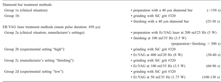
Table 1 Treatment methods of the different groups Diamond bur treatment methods
Group 1a (clinical situation): ▪ preparation with a 40 μm diamond bur (∼150 s)
Group 1b: ▪ grinding with SiC grit #320
▪ finishing with a 40 μm diamond bur (25-30 s) ER:YAG laser treatment methods (mean pulse duration: 450μs)
Group 2a (clinical situation, manufacturer’s settings): ▪ preparation with Er:YAG laser at 200 mJ/25 Hz (5 W) ▪ finishing at 100 mJ/35 Hz (3.5 W)
(preparation+finishing: > 300 s) Group 2b (experimental setting“high”): ▪ grinding with SiC grit #320
▪ Er:YAG at 400 mJ/20 Hz (8 W) (30-40 s) Group 2c (manufacturer’s setting “finishing”): ▪ grinding with SiC grit #320
▪ Er:YAG at 100 mJ/35 Hz (3.5 W) (60-90 s) Group 2d (experimental setting“low”): ▪ grinding with SiC grit #320
applied perpendicular to the ground dentin surface at a focal distance of 2 mm with water cooling. The ground surfaces were irradiated in three directions at 0°, 45°, and 90°. The molars of all six groups were treated by the same operator. The range of time needed for surface treatment per molar is listed in Table1for all six groups.
After surface treatment, the molars were dehydrated in ethanol solutions of ascending concentrations (40%, 60%, 80%, 2×100%) for 24 h each and stored in a desiccator until subsequent sputtering: The molars were mounted on aluminum stubs and gold/palladium sputter-coated (100 s, 50 mA) using a sputtering device (Balzers SCD 050, Balzers, Liechtenstein). SEM was performed with a Stereoscan S360 scanning electron microscope at 20 kV (Cambridge Instru-ments, Cambridge, UK). Digital SEM micrographs of 100× and 4000× magnification were made of every surface (Digital Image Processing System, Version 2.3.1.0, point electronic GmbH, Halle, Germany).
Microtensile bond strength (μTBS) measurements
A final amount of 64 non-carious, extracted or exfoliated second human primary molars without restorations (gath-ered at the Department of Preventive, Restorative and Pediatric Dentistry, University of Bern with consent of patient and parent) were used in this study. The molars were cleaned under tap water and stored in 0.5% chloramine solution at 4°C until use.
For visualization of the dental pulp/residual coronal dentin during further processing, the pulp chamber was retrogradely opened and filled with a colored light-curing compomer restorative material (Twinky Star "pink", VOCO, Cuxhaven, Germany; Lot Nr: 1011453). The molars were then embedded in circular molds with self-curing acrylic resin (Paladur, Heraeus Kulzer GmbH) and ground with a grinding machine and SiC papers grit #320 (Struers LaboPol-21/SiC #320, Struers) until the entire surface was in coronal dentin. Dentin surfaces were checked for absence of residual enamel. Molars exhibiting compomer material through coronal dentin were discarded. The molars were then treated with the diamond bur or the Er:YAG laser as listed in Table1for group 1b and groups 2b/c/d (n=16/treatment method). The molars of all six groups were treated by the same operator. The range of time needed for surface treatment per molar is listed in Table1.
Subsequently, the self-etch adhesive Clearfil SE (Kuraray, Okayama, Japan; Primer Lot Nr: 00965AA, Bond Lot Nr: 01387AA) was applied according to the manufacturer’s instructions. After adhesive treatment, the molars were built up in two layers of 2 mm each with the resin composite Clearfil Majesty Esthetic (Kuraray; shade A4, Lot Nr: 00004GA). Each layer of resin
composite was light-cured for 20 s and all light-curing steps were performed with a LED light-curing unit (Bluephase Polywave, Ivoclar Vivadent AG, Schaan, Liechtenstein) in the "High"-power mode. Light power density was verified to be at least 1,200 mW/cm2at the beginning and end of each day of sample preparation.
The primary molars with the resin composite build-up were then stored for 36 h in 100% humidity at 37°C.
After storage, the molars were sectioned with an electronically programmable diamond saw under water cooling (Struers Accutom-5 with Struers Diamond Cut-Off Wheel 330CA, Struers) perpendicular to the adhesive interface in both x and y directions to obtain nine sticks of the centric dentin surface part. In order to monitor the size and to calculate the bonding surface (BSU (mm2)) of each stick, length and width of the sticks were measured with a digital caliper (Mitutoyo IP 65, Kawasaki, Japan; mean length (standard deviation): 1.05 (0.03) mm, mean width (standard deviation): 1.05 (0.04) mm). The sticks were then fixed by their ends to notched Ciucchi’s jigs mounted in a universal testing machine (Syndicad TC-550, Syndicad Dental Research, Munich, Germany) with Helio-bond (Ivoclar Vivadent AG). The sticks were stressed in tension at a crosshead speed of 1.0 mm/min and the maximum force (Fmax (N)) was recorded. The μTBS
values (MPa) were calculated according to the formula μTBS=Fmax / BSU.
Additionally, the failure mode of each stick was stereomicroscopically determined at 45× magnification (Leica ZOOM 2000, Leica, Buffalo, NY, USA) and classified into one of the five following categories: (1) adhesive failure between adhesive system and dentin, (2) adhesive failure between adhesive system and resin composite, (3) mixed adhesive failure (failure modes 1 and 2), (4) cohesive failure in dentin, and (5) cohesive failure in resin composite.
Four sticks per tooth were measured and stereomicro-scopically analyzed. In the rare cases of pretesting failure (nine pretesting failures/256 final sticks), one of the remaining five sticks of the same tooth was used as a replacement.
Statistical analysis ofμTBS values
Out of the fourμTBS values obtained per primary molar, a mean μTBS value was calculated. Therefore, 16 μTBS values per treatment method were used for statistical analysis. The treatment methods were compared with a Kruskal–Wallis test. The main statistical analysis was performed with R version 2.9.1 (The R Foundation for Statistical Computing, Vienna, Austria;www.r-project.org). TheμTBS data of preliminary tests had been statistically analyzed with NCSS/PASS 2005 (NCSS, Kaysville, UT,
USA) for sample size determination after the level of significance had been set atα=0.05.
Results
Qualitative SEM evaluation of dentin micromorphology Qualitative SEM evaluation revealed the same dentin micromorphology for all three specimens in each of the six groups. Representative micrographs of the qualitative SEM evaluation are shown in Fig.1.
When observed at 100× magnification, the dentin surfaces of groups 1a and 1b showed a relatively smooth surface with intersecting undulations parallel to the three instrumentation directions. At 4000× magnification, both surfaces were covered with a uniform smear layer and no dentinal tubules were visible (Figs.1a/b, small pictures).
Previous grinding with SiC paper did not affect the micromorphological appearance of surfaces treated with the 40-μm diamond bur (group 1b compared to group 1a).
Laser treatment resulted in substantially different surface micromorphology compared to diamond bur treatment. When observed at 100× magnification, Er:YAG
laser-Fig. 1 Scanning electron micro-graphs for qualitative evaluation of dentin micromorphology
treated surfaces showed a multiple overlapping circular pattern in groups 2b and 2c. However, in group 2d, the laser beam had insufficiently irradiated the area, so that the underlying SiC-ground surface was still partly visible (Fig.1f).
Previous grinding with SiC paper did not affect the micromorphology after laser irradiation in groups 2b and 2c and thus, the irradiated surface resulted in a micromorphology very similar to the one obtained after the clinical situation with only Er:YAG laser preparation (group 2a). At 4000× magnification, all Er:YAG laser-treated surfaces (groups 2a/b/c/d) had a similarly distinc-tive, irregular, and scaly appearance exhibiting open tubules and absence of smear layer (Figs. 1c-f, small pictures). Intertubular dentin was selectively more ablated than peritubular dentin. However, the micromorphology varied occasionally according to orientation of the dentin tubules. There was no evidence of thermal damages such as cracking or carbonization.
Microtensile bond strength (μTBS) measurements
TheμTBS values of the four groups are shown in Table2
(median, lower/1st and upper/3rd quartile, and minima/ maxima; mean value and standard deviation). The Kruskal– Wallis test showed no significant differences between the treatment methods (p=0.394).
The distribution of failure modes of the four groups is shown in Table3. Whereas the diamond bur-treated group 1b presented a majority of adhesive failures between adhesive system and dentin, the predominant failure mode of the Er:YAG laser-treated groups 2b/c/d was adhesive failure between adhesive system and resin composite. Similar or equal amounts of mixed adhesive failure or cohesive failure in dentin were observed in all groups except for group 2d, which presented a higher number of mixed adhesive failures and no case of cohesive failure in dentin.
Discussion
The micromorphologies of dentin of primary teeth treated with diamond bur or Er:YAG laser were similar to the corresponding ones reported for dentin of permanent teeth, with a uniform smear layer and no visible dentinal tubules for the diamond bur-treated groups and an absence of smear layer and open tubules for the Er:YAG laser-treated groups [7,8,12,19]. For all four Er:YAG groups (groups 2a/b/c/ d), clinically no evidence of thermal damage such as carbonization was visible on the dentin surfaces, which was confirmed by the SEM micrographs. However, possible pulpal thermal changes may have occurred, especially for the experimental setting "high".
For preparation of μTBS-samples, a standardized flat surface was needed. Thus, abrasive papers were used to achieve flat surfaces before diamond bur or Er:YAG laser treatment. A possible influence of previous SiC grinding on the dentin micromorphology was examined by inclusion of groups 1a and 2a, which served as controls with no SiC grinding. In general, previous grinding had no influence on the dentin micromorphology. However, in group 2d, the SEM evaluation showed that the low laser energy setting (50 mJ/35 Hz (1.75 W); experimental setting "low") was insufficient for broad ablation and for erasure of all traces of the grinding procedure: Although clinically all dentin surfaces of group 2d had the same chalky appearance as the dentin surfaces of the three other Er:YAG groups (groups 2a/b/c), the SEM micrographs of group 2d showed SiC paper ground dentin surfaces to be clearly visible between traces of the laser irradiation (Fig.1f). Due to the difficulty in clinically controlling for sufficient ablation, the adhesive performance of group 2d was likely to depend on both the SiC paper and the Er:YAG laser micromorphologies. On dentin of permanent teeth, other studies which used SiC papers to produce a uniform smear layer and standardized surfaces reported equal or significantly higher bond strength values than those obtained on Er:YAG laser-treated dentin [20–22]. However in the present study, this micromorphology with partly non-ablated areas did not Table 2 Microtensile bond strength values of the different groups (n=
16/group)
Group (lq) median (uq)
(Treatment method) (min-max) Mean value (SD) Group 1b (20.6) 23.8 (30.0) (grinding+40μm grit diamond bur) (14.2–39.4)
24.8 (6.6) Group 2b (grinding+Er:YAG at 400 mJ/20 Hz) (23.2) 25.6 (29.6) (20.2–34.0) 26.1 (4.3) Group 2c (grinding+Er:YAG at 100 mJ/35 Hz) (15.3) 21.4 (28.6) (9.6–37.1) 22.2 (8.6) Group 2d (grinding+Er:YAG at 50 mJ/35 Hz) (20.2) 23.7 (27.8) (12.4–36.1) 23.9 (6.1) lq=lower/1st quartile; uq=upper/3rd quartile; min=minimum; max= maximum; SD=standard deviation
enhance the μTBS: No statistically significant differences among the four treatment methods were found and group 2d did not reach higher μTBS values than the other laser groups. It may be that the three laser energy settings of groups 2b/c/d did not lead to different μTBS values, but they did vary in the speed of ablation. Generally, Er:YAG laser treatment was more time-consuming than diamond bur treatment. Among the groups with different laser energy settings, there was a faster ablation with higher laser energy settings. Correlations between Er:YAG laser energy set-tings, ablation time, and pulpal thermal changes during laser preparation of primary teeth should be investigated in further studies.
Information about bond strength on Er:YAG laser-treated dentin of primary teeth is sparse. On dentin of permanent teeth, however, there are several studies that confirm the findings of this study and in which bond strength values did not differ significantly among the treatment methods. In a study of Ramos and coworkers, equal tensile bond strength values of Clearfil SE were reached on SiC paper-ground dentin and on dentin irradiated with two laser settings [22]. Gurgan and coworkers showed no significant differences in bond strength of a two-step, self-etch adhesive system when used on diamond bur-treated or on Er:YAG laser-treated dentin [23]. Carrieri and coworkers reported no significant differences in tensile bond strength on dentin treated with three different laser settings [13]. However, the dentin had been etched and a resin cement, and not a resin composite, was used. The studies available with bond strength measurements on Er:YAG laser-treated dentin of primary teeth reported shear bond strength values from 10.4 to 12.3 MPa and from 5.1 to 9.7 MPa, depending on the adhesive system and the method used [24,25]. In literature, shear bond strength values on dentin of permanent teeth vary widely from 4.0 to 41.9 MPa [11,15,20,23,26] and can therefore hardly be compared to those obtained on dentin of primary teeth. Also, studies investigating the tensile or microtensile bond strength between Clearfil SE
and dentin of permanent teeth treated with Er:YAG laser have shown a wide range of values from 12.7 MPa to 40.9 MPa [7,12,22,27,28].
In the present study, there was a difference between the diamond bur and the Er:YAG laser groups in their distribution of failure modes. The diamond bur group 1b presented adhesive failure between adhesive system and dentin in 54% of the samples and adhesive failure between adhesive system and resin composite in 16%. The three Er: YAG laser groups presented the opposite distribution with adhesive failure between adhesive system and dentin in 9– 23% of the samples and adhesive failure between adhesive system and resin composite in 53–54%. As previously shown, the self-etch adhesive Clearfil SE interacts super-ficially with bur-treated dentin, forming a relatively thin hybrid layer and preserving the smear layer in the tubules [29, 30]. The fact that there were fewer adhesive failures between adhesive system and dentin in the Er:YAG laser groups compared to the diamond bur group may be due to increased chemical bonding and micromechanical inter-locking to a dentin surface exhibiting no smear layer and open tubules, making the interface between adhesive system and resin composite the weakest link.
The failure mode pattern suggests that Clearfil SE has an acceptable bonding potential to Er:YAG laser-treated dentin of primary molars. This could be in accordance with the findings comparing Clearfil SE to other adhesive systems on dentin of permanent teeth: Clearfil SE reached equal tensile bond strength values to an etch-and-rinse adhesive system [22], and in a second study, Clearfil SE reached higher shear bond strength values than two other self-etch adhesive systems [15]. Both studies were conducted on Er: YAG laser-treated dentin of permanent teeth. Future studies should compare different adhesive systems on laser-treated dentin of primary molars.
In conclusion, the μTBS values of the present study did not differ significantly between the treatment meth-ods, including different laser energy settings. Thus, Table 3 Failure mode analysis
Treatment method Group 1b Group 2b Group 2c Group 2d 64 sticks 64 sticks 64 sticks 64 sticks (4 per molar;
n=16 molars) (4 per molar;n=16 molars) (4 per molar;n=16 molars) (4 per molar;n=16 molars) Failure mode
Adhesive between adhesive system and dentin
54% 23% 22% 9%
Adhesive between adhesive system and composite
16% 53% 54% 53%
Mixed adhesive failure 28% 22% 22% 38%
Cohesive in Dentin 2% 2% 2% 0%
dentin of primary molars treated with Er:YAG laser provided bond strengths to resin composite similar to those provided with diamond bur-treated dentin. Never-theless, it is of importance to further investigate combinations of laser-treated primary teeth and different adhesive systems to establish the Er:YAG laser applica-tion also in pediatric dentistry.
Acknowledgments The authors would like to thank Kuraray Europe, Germany and VOCO, Germany, for providing the materials needed. Furthermore, we thank S. Hayoz and Prof. Dr. J. Hüsler, Institute of Mathematical Statistics and Actuarial Science, University of Bern for statistical advice.
Conflicts of interest The authors declare no conflicts of interest, real or perceived, financial or nonfinancial.
References
1. Keller U, Hibst R (1997) Effects of Er:YAG laser in caries treatment: a clinical pilot study. Lasers Surg Med 20(1):32–38 2. Trajtenberg CP, Pereira PN, Powers JM (2004) Resin bond
strength and micromorphology of human teeth prepared with an erbium:YAG laser. Am J Dent 17(5):331–336
3. Amaral FL, Colucci V, de Souza-Gabriel AE, Chinelatti MA, Palma-Dibb RG, Corona SA (2008) Adhesion to Er:YAG laser-prepared dentin after long-term water storage and thermocycling. Oper Dent 33(1):51–58
4. Keller U, Hibst R, Geurtsen W, Schilke R, Heidemann D, Klaiber B, Raab WH (1998) Erbium:YAG laser application in caries therapy. Evaluation of patient perception and acceptance. J Dent 26(8):649–656
5. Armengol V, Jean A, Rohanizadeh R, Hamel H (1999) Scanning electron microscopic analysis of diseased and healthy dental hard tissues after Er:YAG laser irradiation: in vitro study. J Endod 25 (8):543–546
6. Freitas PM, Navarro RS, Barros JA, de Paula EC (2007) The use of Er:YAG laser for cavity preparation: an SEM evaluation. Microsc Res Tech 70(9):803–808
7. Cardoso MV, Coutinho E, Ermis RB, Poitevin A, Van Landuyt K, De Munck J, Carvalho RC, Van Meerbeek B (2008) Influence of dentin cavity surface finishing on micro-tensile bond strength of adhesives. Dent Mater 24(4):492–501
8. Cardoso MV, Coutinho E, Ermis RB, Poitevin A, Van Landuyt K, De Munck J, Carvalho RC, Lambrechts P, Van Meerbeek B (2008) Influence of Er,Cr:YSGG laser treatment on the micro-tensile bond strength of adhesives to dentin. J Adhes Dent 10 (1):25–33
9. Curti M, Rocca JP, Bertrand MF, Nammour S (2004) Morpho-structural aspects of Er:YAG-prepared class V cavities. J Clin Laser Med Surg 22(2):119–123
10. Bader C, Krejci I (2006) Indications and limitations of Er:YAG laser applications in dentistry. Am J Dent 19(3):178–186 11. Bahrami B, Askari N, Tielemans M, Heysselaer D, Lamard L,
Peremans A, Nyssen-Behets C, Nammour S (2010) Effect of low fluency dentin conditioning on tensile bond strength of composite bonded to Er:YAG laser-prepared dentin: a preliminary study. Lasers Med Sci [Epub ahead of print]
12. Manhães L, Oliveira DC, Marques MM, Matos AB (2005) Influence of Er:YAG laser surface treatment and primer applica-tion methods on microtensile bond strength self-etching systems. Photomed Laser Surg 23(3):304–312
13. Carrieri TC, de Freitas PM, Navarro RS, Eduardo Cde P, Mori M (2007) Adhesion of composite luting cement to Er:YAG-laser-treated dentin. Lasers Med Sci 22(3):165–170
14. Amaral FL, Colucci V, Souza-Gabriel AE, Chinelatti MA, Palma-Dibb RG, Corona SA (2010) Bond durability in erbium:yttrium-aluminum-garnet laser-irradiated enamel. Lasers Med Sci 25 (2):155–163
15. Brulat N, Rocca JP, Leforestier E, Fiorucci G, Nammour S, Bertrand MF (2009) Shear bond strength of self-etching adhesive systems to Er:YAG-laser-prepared dentin. Lasers Med Sci 24 (1):53–57
16. Sumikawa DA, Marshall GW, Gee L, Marshall SJ (1999) Microstructure of primary tooth dentin. Pediatr Dent 21(7):439– 444
17. Schilke R, Lisson JA, Bauss O, Geurtsen W (2000) Comparison of the number and diameter of dentinal tubules in human and bovine dentine by scanning electron microscopic investigation. Arch Oral Biol 45(5):355–361
18. Kornblit R, Bossù M, Mari D, Rocca JP, Polimeni A (2009) Enamel and dentine of deciduous teeth Er:YAG laser prepared. A SEM study. Eur J Paediatr Dent 10(2):75–82
19. Tachibana A, Marques MM, Soler JM, Matos AB (2008) Erbium, chromium:yttrium scandium gallium garnet laser for caries removal: influence on bonding of a self-etching adhesive system. Lasers Med Sci 23(4):435–441
20. de Carvalho RC, de Freitas PM, Otsuki M, de Eduardo CP, Tagami J (2008) Micro-shear bond strength of Er:YAG-laser-treated dentin. Lasers Med Sci 23(2):117–124
21. Ferreira LS, Apel C, Francci C, Simoes A, Eduardo CP, Gutknecht N (2010) Influence of etching time on bond strength in dentin irradiated with erbium lasers. Lasers Med Sci 25(6):849–854
22. Ramos AC, Esteves-Oliveira M, Arana-Chavez VE, de Paula EC (2010) Adhesives bonded to erbium:yttrium-aluminum-garnet laser-irradiated dentin: transmission electron microscopy, scanning electron microscopy and tensile bond strength analyses. Lasers Med Sci 25(2):181–189
23. Gurgan S, Kiremitci A, Cakir FY, Yazici E, Gorucu J, Gutknecht N (2009) Shear bond strength of composite bonded to erbium: yttrium-aluminum-garnet laser-prepared dentin. Lasers Med Sci 24(1):117–122
24. Monghini EM, Wanderley RL, Pécora JD, Palma Dibb RG, Corona SA, Borsatto MC (2004) Bond strength to dentin of primary teeth irradiated with varying Er:YAG laser energies and SEM examination of the surface morphology. Lasers Surg Med 34 (3):254–259
25. Scatena C, Torres CP, Gomes-Silva JM, Contente MM, Pécora JD, Palma-Dibb RG, Borsatto MC (2010) Shear strength of the bond to primary dentin: influence of Er:YAG laser irradiation distance. Lasers Med Sci [Epub ahead of print]
26. Ceballo L, Toledano M, Osorio R, Tay FR, Marshall GW (2002) Bonding to Er-YAG-laser-treated dentin. J Dent Res 81(2):119–122 27. Van Meerbeek B, De Munck J, Mattar D, Van Landuyt K, Lambrechts P (2003) Microtensile bond strengths of an etch&rinse and self-etch adhesive to enamel and dentin as a function of surface treatment. Oper Dent 28(5):647–660 28. Bakry AS, Nakajima M, Otsuki M, Tagami J (2009) Effect of Er:
YAG laser on dentin bonding durability under simulated pulpal pressure. J Adhes Dent 11(5):361–368
29. De Munck J, Vargas M, Iracki J, Van Landuyt K, Poitevin A, Lambrechts P, Van Meerbeek B (2005) One-day bonding effectiveness of new self-etch adhesives to bur-cut enamel and dentin. Oper Dent 30(1):39–49
30. Van Meerbeek B, De Munck J, Yoshida Y, Inoue S, Vargas M, Vijay P, Van Landuyt K, Lambrechts P, Vanherle G (2003) Buonocore memorial lecture. Adhesion to enamel and dentin: current status and future challenges. Oper Dent 28(3):215–235