Full Review
Parvalbumin: calcium and magnesium buffering in the distal
nephron
Eric Olinger, Beat Schwaller, Johannes Loffing, Philippe Gailly and Olivier Devuyst
Division of Nephrology and Laboratory of Cell Physiology, UCL Medical School, Brussels, Belgium, Institutes of Physiology and Anatomy, Zurich Center for Integrative Human Physiology, University of Zurich, Zurich, Switzerland and Unit of Anatomy, Department of Medicine, University of Fribourg, Fribourg, Switzerland
Correspondence and offprint requests to: Olivier Devuyst; E-mail: [email protected]
Abstract
Parvalbumin (PV) is a classical member of the EF-hand protein superfamily that has been described as a Ca2+ buffer and Ca2+ transporter/shuttle protein and may also play an additional role in Mg2+ handling. PV is exclu-sively expressed in the early part of the distal convoluted tubule in the human and mouse kidneys. Recent studies in Pvalb knockout mice revealed a role of PV in the distal handling of electrolytes: the lack of PV was associated with a mild salt-losing phenotype with secondary aldos-teronism, salt craving and stronger bones compared with controls. A link between the Ca2+-buffering capacity of PV and the expression of the thiazide-sensitive Na+–Cl− cotransporter was established, which could be relevant to the regulation of sodium transport in the distal nephron. Variants in the PVALB gene that encodes PV have been described, but their relevance to kidney function has not been established. PV is also considered a reliable marker of chromophobe carcinoma and oncocytoma, two neo-plasms deriving from the distal nephron. The putative role of PV in tumour genesis remains to be investigated. Keywords: DCT; Gitelman syndrome; NCC; purinergic signalling
Properties and distribution of parvalbumin Parvalbumin (PV) is a small protein (109 amino acids in most species; molecular mass ∼12 kDa) first isolated from carp muscle in 1973 [1] and belonging to the sub-family of cytosolic Ca2+buffers in the superfamily of EF-hand proteins [2]. Proteins of the EF-hand family are characterized by a conserved, helix–loop–helix structural unit, which consists of two α-helices bridged by a Ca2+ -chelation loop. These proteins are involved in the regulation of many critical cellular processes including gene transcription, protein phosphorylation, nucleotide metab-olism and ion transport [3–5]. Over 200 members of the
Ca2+-binding EF-hand superfamily have been identified so far in the human genome [2].
PV is characterized by a high affinity for Ca2+ (dis-sociation constant KD,Ca: ∼5–10 nM) and an intermediate
affinity for Mg2+ (KD,Mg: ∼30 µM). Thus, the two
func-tional metal-binding sites are so-called mixed (Ca2+/Mg2+ )-binding sites. These properties and the fact that the intra-cellular concentration of Mg2+ [(Mg2+)cyt, 0.5–1 mM]
exceeds largely that of Ca2+ [(Ca2+)cyt, 50–100 nM] in
basal conditions explain why PV-binding sites are (>80%) occupied mainly by Mg2+in a resting cell. The remaining sites are either Ca2+-bound or metal-free. When a stimulus induces a rise in [Ca2+]cyt levels, Mg
2+
slowly dissociates from the binding sites and is replaced by Ca2+. Because of the slow Ca2+-binding kinetics under physiological con-ditions, which mainly results from this prior Mg2+ dis-sociation, PV is referred to as a slow-onset Ca2+buffer [2]. Binding of two Ca2+ions to the Ca2+-binding sites induces a rather insignificant conformational change [4]. Hence, PV is mostly considered a pure Ca2+ buffer, with little or no Ca2+sensing and direct regulatory properties [6].
The expression of PV is remarkably restricted to a few cell types in the brain, skeletal and heart muscles, para-thyroid glands and kidney [7–9]. In the central nervous system, PV is highly expressed in inhibitory GABAergic interneurons, e.g. in the cortex in chandelier (axo-axonic) and basket cells. These cortical neurons play an important role in controlling pyramidal cell excitability. The absence of PV in these cells is linked to increased drug-induced seizure susceptibility [10]. PV is also expressed in similar types of interneurons in the hippocampus (axo-axonic and basket cells) and cerebellum (stellate and basket cells) [7]. In the cerebellum, PV is additionally expressed in Pur-kinje cells.
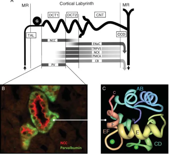
In the kidney, PV is expressed in the epithelial cells lining a subset of tubules in the distal nephron (Figure 1). In the mouse and human kidneys, PV appears to be exclu-sively expressed in the early part of the distal convoluted tubule (early DCT, or DCT1), where it colocalizes with the thiazide-sensitive apical Na+–Cl− cotransporter (NCC).
© The Author 2012. Published by Oxford University Press on behalf of ERA-EDTA. All rights reserved. For Permissions, please e-mail: [email protected]
Nephrol Dial Transplant (2012) 27: 3988–3994 doi: 10.1093/ndt/gfs457
The progression between the early and late part of the DCT is characterized by an abrupt transition from PV (early DCT) to calbindin-D28k (late DCT, or DCT2). Cells
ex-pressing calbindin-D28kalso express NCC and epithelial
Na+channel (ENaC) [11,12]. In the rat kidney, PV has been located in the thick ascending limb (TAL) of the loop of Henle, late DCT, connecting tubule (CNT) and intercalated cells of the collecting duct [13]. Immuno-cytochemical analyses revealed that in most tubular cells, PV shows a diffuse cytosolic pattern, with a signal sometimes enhanced along the basolateral membrane [12,13].
PV is present in fast-contracting and fast-relaxing skel-etal musclefibres (e.g. extensor digitorum longus or tibia-lis anterior) [14]. The protein has also been detected in normal and in hyperplastic and adenomatous human para-thyroid glands, with the strongest expression in chief cells and water clear cells. Of note, PV colocalizes with the parathyroid hormone (PTH) in the same cell types [8].
Role of PV in the distal nephron
The distal nephron plays a major role in the reabsorption of NaCl and the regulation of thefinal excretion of Ca2+ and Mg2+, under the influence of several hormones, in-cluding aldosterone, PTH and 1,25(OH)2-vitamin D3
[15]. Approximately 5% of the filtered load of NaCl is reabsorbed in the DCT, involving the thiazide-sensitive NCC on the apical side and the Na+-K+-ATPase and the ClC-Kb chloride channel on the basolateral side of the cells. The reabsorption of Ca2+and Mg2+in that segment is mediated by a complex interaction of different proteins, involving two specific members of the transient receptor potential (TRP) channel superfamily that have a distinct spatial distribution along the DCT (Figure 2). Approxi-mately 5–10% of filtered Mg2+ is passively reabsorbed through the apical TRPM6 channel and basolateral active transport systems in the DCT1. Specific intracellular Fig. 1. Structure and distribution of PV in the kidney. (A) Segmentation of the distal nephron, with distribution of specific markers in the early (DCT1) and late (DCT2) parts of the distal convoluted tubule, the CNT and the cortical collecting duct (CCD). PV is exclusively expressed in the DCT1, where it colocalizes with the apical NCC. It is of note that the mediators of Ca2+reabsorption are expressed in more distal nephron segments
(TRPV5; NCX, sodium-calcium exchanger; PMCA, plasma membrane calcium ATPase; CB, calbindin-D28K). The epithelial Na +
channel ENac and CB are expressed all along distal nephron segments, starting in DCT2. MR, medullary ray; G, glomerulus. Modified from Loffing et al. [11]. (B) Immunostaining for PV in the human kidney cortex reveals a diffuse cytosolic localization (green signal), sometimes clustered above the basolateral membrane, in cells that express NCC in the apical membrane (red signal). (C) Solution structure of Ca2+-bound human PV. The CD domain (green)
and EF domain (yellow/red) bind one Ca2+ion each (green spheres) in canonical Ca2+-binding loops of 12 amino acids. The helices E and F that gave the name to all EF-hand proteins are marked by letters. Both Ca2+-binding loops in PV are of the Ca2+/Mg2+mixed type. The amino- (N) and
Mg2+ carriers and basolateral extrusion proteins have not yet been identified. Apical TRPV5 channels (previously named epithelial calcium channel 1, ECaC1) mediate the passive entry of Ca2+in the DCT2, before active, basolat-eral transport via the Na+–Ca2+exchanger 1 (NCX1) and plasma membrane Ca2+-ATPase 1b (PMCA 1b). In the DCT, TRPV5 colocalizes with the intracellular Ca2+ buffer/sensor calbindin-D28K [16, 17]. Studies in rabbit
CNTs have shown that calbindin-D28Kcould be important
to lower [Ca2+]cytlevels in order to maintain the necessary
gradient for passive cellular Ca2+ entry and to facilitate the intracellular Ca2+diffusionalflux [18]. Of interest, the Ca2+buffer calbindin-D9k is also expressed in DCT2, but
with a more focused expression than CB-D28k that
extends further into the CNTs and collecting ducts (J. Loffing, personal communication).
In view of the specific expression of PV in DCT1, Belge et al. [12] investigated in detail the renal phenotype of knockout mice (KO) for PV. In comparison with wild-type littermates, Pvalb KO mice had increased diuresis and kaliuresis at baseline, with secondary aldosteronism and salt craving. As expected, an acute administration of the loop diuretic furosemide, aimed to increase sodium
delivery in the DCT, led to increased diuresis and natriur-esis/kaliuresis in both genotypes. However, surprisingly, this treatment did not increase calciuria in Pvalb KO mice. Furthermore, Pvalb KO mice showed no significant diuretic response to hydrochlorothiazide, but rather an ac-centuated hypocalciuria. The PV-deficient mice also showed an increased bone mineral density. These func-tional changes were explained by a strongly decreased expression of NCC at the mRNA and protein levels in the early DCT of Pvalb KO kidneys, in the absence of any ultrastructural changes [12]. The Pvalb KO mice mani-fested a decreased lithium clearance, suggesting that a compensatory increase in sodium transport was taking place in the proximal tubule—as it has been reported in the case of long-term treatment with thiazide diuretics tar-geting NCC [19].
What could be the link between the Ca2+/Mg2+ -buffer-ing capacities of PV and the expression of NCC in the DCT cells? It has been known for a long time that the entire distal nephron, and DCT cells in particular, ex-presses luminal P2Y2 receptors, which trigger rapid intra-cellular Ca2+ transients when stimulated by purinergic agonists such as adenosine triphosphate (ATP) and Fig. 2. Transcellular transport of Ca2+and Mg2+in the distal nephron. The segmentation of the DCT is reflected by a selective transport machinery facilitating the reabsorption of Mg2+in the proximal part of the DCT (DCT1), whereas the Ca2+transport is restricted to the more distal part of the
DCT (DCT2) and the CNT (not represented on thefigure). Magnesium enters the cells lining the DCT1 through apical TRPM6 channels. The nature of the possible transporter and basolateral extruder protein remains unknown. PV is selectively expressed in the cytosol of these cells and plays a role in regulating the Ca2+signalling and the expression of the sodium-chloride cotransporter NCC. The transcellular transport of Ca2+is mediated through apical TRPV5 channels in the DCT2 and in the CNT. The Ca2+buffer calbindin-D
28Kis carrying Ca2+ions to the basolateral membrane,
uridine triphosphate (UTP) [20]. These brief [Ca2+]cyt
transients cause a decrease in several transport systems in-cluding those involved in NaCl reabsorption [21]. Studies in a mouse DCT (mDCT) cell line showed that PV modu-lates the shape and duration of intracellular Ca2+transients induced by ATP [12]. In turn, these changes were re-flected by major modifications in the expression of endogenous NCC expression in these cells. The fact that PV is capable of effectively reducing the amplitude of ATP-evoked elevations in [Ca]cyt in mDCT cells [12]
could be potentially linked to the regulation of NCC expression. Taken together, these studies conducted in Pvalb KO mice and in mDCT cells suggest that PV could regulate the expression of NCC by modulating intracellu-lar Ca2+ signalling in response to extracellular ATP in DCT cells.
Potential roles of PV in Ca2+and Mg2+handling by the kidney
Considering the buffering properties of PV, one may hypothesize that this protein could play a direct role in the transepithelial handling of Ca2+ and Mg2+ in the distal nephron. The major sites for transcellular Ca2+ reabsorp-tion in mouse kidney are the late DCT and the CNT [11]. The early part of the DCT appears to play a minor role in Ca2+transport as apical TRPV5 and basolateral NCX and PMCA are either not detected or only weakly expressed in this segment (Figures 1 and 2). The exclusive distri-bution of PV in the early DCT and the fact that its expression is independent of the vitamin D3 status also argues against a major role in distal calcium handling [13,
18]. Nevertheless, Pvalb KO mice show an increased bone mineral density and a strongly positive calcium balance when stimulated by thiazide diuretics [12]. In fact, such a phenotype is consistent with a reduced NCC expression in the DCT, similar to chronic thiazide admin-istration. Volume contraction in the Pvalb KO mice is probably an important factor to explain the positive calcium balance, as indicated by decreased lithium clear-ance [12, 19]. Nonetheless, other components of the Ca2+-signalling toolkit [3] might be modified in Pvalb KO mice and a study to address this question is underway (Schwaller, unpublished work).
It must be pointed out that even volume-repleted Pvalb KO mice tend to excrete less calcium than their wild-type littermates [12], which could in fact originate from a dys-functional NCC. Gesek and Friedman [22] demonstrated that a reduced Cl−entry in DCT cells leads to decreased intracellular Cl−activity followed by hyperpolarization of the plasma membrane. The hyperpolarization was pro-posed to activate dihydropyridine-sensitive calcium chan-nels in the apical membrane, which enhances transcellular Ca2+ transport [22]. A similar mechanism could operate in DCT cells lacking PV, due to the down-regulation of NCC. However, it is unclear whether a hyperpolarization of the luminal membrane would persist under the chronic conditions of a life-long decrease in NCC activity. Alter-natively, a decreased urinary calcium excretion may also
be explained by a decreased intracellular Na+ concen-tration, resulting from a decreased apical NCC activity. In this scenario, basolateral NCX1 responds by facilitating Na+ entry into the cell and Ca2+ exit out of the cell, en-hancing the net transcellular Ca2+flux [23]. A third mech-anism could be related to a structural hypertrophy of the TRPV5 and NCX/PMCA1b-positive CNT, as observed in NCC-deficient mice [24], which would increase the epi-thelial surface available for transcellular Ca2+ reabsorp-tion. Nevertheless, careful histological examinations have not detected such damages in the kidneys of the Pvalb KO mice. Similarly, no compensatory up-regulation of TRPV5 or calbindin-D28k, as observed after chronic
administrations of thiazide diuretics [25], has been ob-served in the Pvalb KO kidneys.
Elegant studies have shown that calbindin-D28k, which
is expressed in DCT2, may make use its Ca2+-sensing properties to function differently at basal [Ca2+]cytand at
elevated levels caused by a Ca2+ influx. Accordingly, elevated [Ca2+]cyt levels could induce conformational
changes and regulatory processes including a negative feedback on the apical TRPV5 [17]. Nevertheless, such a mechanism seems unlikely to occur for PV, which is more a ‘simple’ Ca2+ buffer protein than a Ca2+ sensing and regulatory protein.
PV is distributed, along with TRPM6 and NCC, in the DCT1 segment that plays a crucial role in Mg2+handling. The affinity of PV for Mg2+(KMg:∼30 µM) makes a
suit-able candidate to feasibly act as an intracellular Mg2+ transporter/shuttle. For instance, inappropriate urinary loss of Mg2+is a hallmark of Gitelman syndrome (GS), an in-herited tubulopathy due to loss-of-function mutations in NCC (see below). Intracellular Mg2+(and also Ca2+) con-centrations tightly regulate their own reuptake through a negative feedback involving apical transporters (TRPM6, TRPV5). Changes in [Mg2+]cyt have been shown to
modify regulatory elements in non-coding mRNA regions, also influencing the transcription of Mg2+ trans-porters in bacteria [26]. Accordingly, a loss of PV could induce such a negative feedback (decreased apical TRPM6), which would decrease the gradient for passive cellular Mg2+entry and impair Mg2+diffusionalflux [17,
27]. A mild Mg2+ wasting phenotype has been observed in Pvalb KO mice on a C57BL/6J background (Olinger, unpublished work), but further investigations have to be done to confirm these findings.
It is important to mention that there are significant differences between Slc12a3 KO mice lacking NCC and those lacking PV. Most importantly, NCC-null mice show a more substantial and constant Mg2+ loss in urine and hypomagnesaemia at baseline, probably consistent with severe DCT damage/loss and a loss of TRPM6-expressing cells in the distal nephron. The morphological alterations in DCT seem to be limited to its early part and are most likely a result from reduced transcellular Na+ transport, raising the question why these ultrastructural changes were not observed in the Pvalb KO mice [12, 24]. Perhaps, the reduction in NCC activity and hence trans-cellular Na+transport in the DCTs of PV-deficient mice is not severe enough to provoke the structural changes as they occur in mice lacking any NCC activity. Experiments
addressing this issue are underway in the laboratory of Loffing.
PVALB as candidate gene in Gitelman syndrome The combination of mild sodium wasting, resistance to thiazide diuretics, hypocalciuria and increased bone density observed in Pvalb KO mice is reminiscent of the manifestations of GS. GS is a recessively inherited salt-losing tubulopathy with hypokalemic alkalosis, hypomag-nesaemia and hypocalciuria. The majority of patients with GS are compound heterozygous for loss-of-function mutations in the SLC12A3 gene that codes for the NCC [28]. A few cases are caused by mutations in the CLCNKB gene that codes for the basolateral ClC-Kb chloride channel in DCT cells [28, 29]. GS is probably the most common tubulopathy, with a prevalence of het-erozygous carrier of an SLC12A3 mutation estimated at 1% in European populations [30]. Most patients with GS are diagnosed in adulthood and are typically normoten-sive, with a mild phenotype. Recent studies have pointed to the possibility of severe complications, sometimes in-volving children and potentially related to male gender and specific allele combinations [31].
Despite advances in mutation detection in SLC12A3, up to 30% of patients with GS carry only a single mutant allele and negative SLC12A3 screening is observed in ∼10% of patients [29, 31]. Mutations in another gene could thus explain the lack of detection of mutant SLC12A3 alleles in GS patients. Considering the pheno-type of the PV KO mouse, it was tempting to hypothesize that mutations in PVALB, the gene coding for PV, could be present in patients with GS heterozygous or in ones negative for SLC12A3 mutations [32]. The PVALB gene, localized on chromosome 22 (22q12-q13.1), has not been linked to any human disorder so far. Direct sequencing of PVALB was performed in 132 GS patients harbouring only one (n = 53) or no (n = 79) mutant SLC12A3 allele. The possible interference of biallelic SNPs (single nucleo-tide polymorphisms) on normal transcription or normal splicing was investigated. No sequence variants resulting in amino acid substitution or a truncated protein within the PVALB gene were found in the 264 chromosomes tested. Ten biallelic SNPs, including six novel polymorph-isms, were identified: five in the 5′ UTR, none of them affecting predicted regulatory elements; three in the coding region, without alteration of the consensus splice sites and two in the 3′ UTR. The observed allelic frequen-cies did not differ significantly between GS patients and controls. These results strongly suggest that mutations in the PVALB gene are not involved in the classical form of GS [32].
Despite these negative results, it may be of interest to screen for PVALB mutations in patients harbouring neuro-logical symptoms, such as epileptic seizures, in particular when these manifestations are linked to hypomagnesae-mia. Central manifestations have been described in Pvalb KO mice, suggesting that a distinct neurological pheno-type could result from the lack of PV (see below). One could also hypothesize that changes in the expression or
function of PV (or in the Ca2+signalling pathway in DCT cells) may participate in the individual response to thia-zide diuretics [12].
PV and renal cell carcinoma
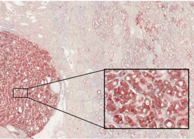
In more than 90% of cases, renal cell carcinoma (RCC) originates from tubular cells. The accurate typing of RCC has important implications for prognosis and therapy. In a normal clinical setting, the histologic diagnosis of RCC is made by routine light microscopy of haematoxylin–eosin stained sections. However, immunohistochemical analysis based on segmental markers is important for the differen-tial diagnosis of non-renal cell neoplasms mimicking RCC, the differentiation of histological subtypes or rare RCC, the analysis of a small biopsy specimen and most importantly, the recognition of RCC metastases in distant organs. The immunohistochemical diagnosis of RCC is based on a set of markers indicative of the nephron segment of origin. As mentioned before, PV is exclu-sively expressed in the first part of the DCT in human kidney [12]. Reactivity for PV is therefore used, along with cadherin, claudins and S100A, as a highly specific and reliable marker of chromophobe RCC and benign on-cocytoma, two neoplasms deriving from the distal nephron (Figure 3). In contrast, PV is not expressed in other renal neoplasms and especially in clear cell and pa-pillary RCC.
Chromophobe carcinoma accounts for 5% of all RCC and is considered to present a rather indolent behaviour with localization restricted to the kidney and nuclear Grade 2 at presentation. It is classically divided into a typical variant and an eosinophilic variant, with a differ-ential diagnosis including clear cell carcinoma, papillary RCC and benign oncocytoma. PV staining is a reliable marker of chromophobe carcinoma, superior to Hale’s colloidal iron and antimitochondrial 113-1 antibodies [33]. Immunostaining for other distal Ca2+-binding
Fig. 3. PV as a marker of chromophobe RCC. Immunohistochemical analysis reveals a strong and ubiquitous expression of PV in a chromophobe renal carcinoma (Inset, illustrating the intense staining in chromophobe cancer cells), and staining of the early DCT of the normal renal parenchyma. (Image courtesy of Dr S. Aydin.)
proteins such as calbindin-D28kis most often negative and
much less specific than PV for oncocytoma and chromo-phobre RCC. Staining for PV is of limited value to differ-entiate the eosinophilic variant of chromophobe RCC from oncocytoma, the most common benign renal neo-plasm. The distinction is particularly challenging for so-called hybrid tumours, revealing features of both onco-cytoma and chromophobe RCC and reflecting probably the common origin of these two tumours. Nevertheless, a negative or a patchy staining for PV is strongly suggestive towards the diagnosis of oncocytoma [34]. PV is also strongly expressed in the (rare) metastases from chromo-phobe RCC [33,35].
Instead of being only a simple marker, could PV be a causative agent in renal tumour development? It is of in-terest thatβ-PV (oncomodulin) has been shown to activate cyclic nucleotide phosphodiesterase and can thereby act as a cellular trigger protein [36]. Oncomodulin is fre-quently expressed in mammalian tumours, for instance in Morris hepatoma [37]. However, PV and oncomodulin differ by their Ca2+-binding sites. Oncomodulin has one Ca2+-specific, non-canonical site (CD domain) and one mixed (Ca2+/Mg2+)-binding site (EF domain). The CD domain shows significant Ca2+-dependent conformation changes, suggestive of additional sensor function for on-comodulin [2]. An active role of PV in tumour genesis cannot be excluded, but would be likely based on altered intracellular Ca2+ signalling. Until now, no interacting partner indicative of a Ca2+ sensor function has been identified.
PV outside the kidney
PV is highly expressed in a subgroup of inhibitory GABAergic interneurons in various brain regions includ-ing cortex, hippocampus, striatum and cerebellum (for more details, see [7]) and in fast-twitch muscle fibres [14]. It has been postulated that the absence of PV in GABAergic interneurons and in particular in chandelier cells and basket cells is correlated with an increased sus-ceptibility to epileptic seizures in Pvalb KO mice [10]. The modulation of [Ca2+]cyt kinetics in neurons lacking
PV leads to increased facilitation of GABAergic trans-mission to postsynaptic pyramidal cells, thus resulting in a shift in the pyramidal cell’s firing properties which could, under certain experimental conditions, lead to epi-leptogenic insults. Indeed, the severity of pentylenetetra-zole-induced generalized tonic–clonic seizures is significantly greater in Pvalb KO mice compared with wild-type littermates [10]. PV KO mice also display a mild impairment in motor coordination and motor learn-ing [38]. There is evidence that PV plays a similar role in the epileptogenic activity of the human neocortex based on results of PV immunoreactivity in epileptic foci [39].
With respect to fast-contracting muscle activity, the contraction–relaxation cycle is prolonged in Pvalb KO musclefibres, with a significantly greater force generated during a single twitch [9]. These changes result from an altered shape of Ca2+transients in the absence of PV.
A possible role of PV as a signal transduction modulator in human parathyroid gland cells by affecting [Ca2+]cytis
also envisaged, potentially related to the control of PTH secretion [8].
Conclusion and perspectives
PV is a classical member of the EF-hand protein super-family that plays a role in regulatory processes operating in very distinct cell types. PV has been described as a Ca2+ buffer and Ca2+ transporter/shuttle protein, but diverse experimental observations hint towards an additional role in magnesium handling. It is particularly puzzling that PV is exclusively expressed in the early part of the DCT of the human and mouse kidneys. A role of PV in the renal handling of electrolytes was demonstrated in Pvalb KO mice, which showed a mild salt-losing phenotype with salt craving, relatively similar to GS. A link between the Ca2+-buffering capacity of PV and the expression of the thiazide-sensitive NCC could be established, with poten-tial relevance for the regulation of sodium transport in the distal nephron. On the basis of these studies, PVALB has been proposed as a candidate gene in patients presenting with GS and displaying none or only a single mutant SLC12A3 allele. However, no link between mutations in PVALB and GS could be established so far. Variants in PVALB have been described, but their relevance to kidney function or response to thiazide diuretics, for instance, has not been investigated. Finally, PV is considered a reliable marker of chromophobe carcinoma and oncocytoma, two neoplasms deriving from the distal nephron. The putative role of PV in tumour genesis has not yet been investi-gated. The role of PV in tissues apart from the kidney is best understood in neurons, where its absence affects short-term modulation of synaptic transmission.
Acknowledgements. We thank Drs Selda Aydin and Sara Terryn for their contributions. Studies mentioned in this review were supported in part by the Fonds National de la Recherche Scientifique; the Fonds de la Re-cherche Scientifique Médicale; an Action de Recherche Concertée; an Inter-University Attraction Pole (IUAP); the NCCR Kidney.CH program and the Swiss National Science Foundation: grant # 130680 (to B.S.). Conflict of interest statement. None declared.
References
1. Kretsinger RH, Nockolds CE. Carp muscle calcium-binding protein. II. Structure determination and general description. J Biol Chem 1973; 248: 3313–3326
2. Schwaller B. Cytosolic Ca2+buffers. Cold Spring Harb Perspect
Biol 2010; 2: a004051
3. Berridge MJ, Bootman MD, Roderick HL. Calcium signalling: dy-namics, homeostasis and remodelling. Nat Rev Mol Cell Biol 2003; 4: 517–529
4. Gifford JL, Walsh MP, Vogel HJ. Structures and metal-ion-binding properties of the Ca2+-binding helix–loop–helix EF-hand motifs. Biochem J 2007; 405: 199–221
5. Grabarek Z. Structural basis for diversity of the EF-hand calcium-binding proteins. J Mol Biol 2006; 359: 509–525
6. Ikura M. Calcium binding and conformational response in EF-hand proteins. Trends Biochem Sci 1996; 21: 14–17
7. Celio MR. Calbindin D-28k and parvalbumin in the rat nervous system. Neuroscience 1990; 35: 375–475
8. Pauls TL, Portis F, Macri E et al. Parvalbumin is expressed in normal and pathological human parathyroid glands. J Histochem Cytochem 2000; 48: 105–111
9. Schwaller B, Dick J, Dhoot G et al. Prolonged contraction-relaxation cycle of fast-twitch muscles in parvalbumin knockout mice. Am J Physiol Cell Physiol 1999; 276: C395–C403
10. Schwaller B, Tetko IV, Tandon P et al. Parvalbumin deficiency affects network properties resulting in increased susceptibility to epi-leptic seizures. Mol Cell Neurosci 2004; 25: 650–663
11. Loffing J, Loffing-Cueni D, Valderrabano V et al. Distribution of transcellular calcium and sodium transport pathways along mouse distal nephron. Am J Physiol Renal Physiol 2001; 281: F1021–F1027 12. Belge H, Gailly P, Schwaller B et al. Renal expression of
parvalbu-min is critical for NaCl handling and response to diuretics. Proc Natl Acad Sci USA 2007; 104: 14849–14854
13. Bindels RJM, Timmermans JAH, Hartog A et al. Calbindin-D9k and parvalbumin are exclusively located along basolateral mem-branes in rat distal nephron. J Am Soc Nephrol 1991; 2: 1122–1129 14. Celio MR, Heizmann CW. Calcium-binding protein parvalbumin is
associated with fast contracting muscle fibres. Nature 1982; 297: 504–506
15. Reilly RF, Ellison DH. Mammalian distal tubule: physiology, patho-physiology, and molecular anatomy. Physiol Rev 2000; 80: 277–306 16. Hoenderop JGJ, Nilius B, Bindels RJM. Calcium absorption across
epithelia. Physiol Rev 2005; 85: 373–422
17. Hoenderop JGJ, Bindels RJM. Calciotropic and magnesiotropic TRP channels. Physiology 2008; 23: 32–40
18. Koster HP, Hartog A, Van Os CH et al. Calbindin-D28K facilitates cytosolic calcium diffusion without interfering with calcium signal-ing. Cell Calcium 1995; 3: 187–196
19. Nijenhuis T, Vallon V, van der Kemp AWCM et al. Enhanced passive Ca2+reabsorption and reduced Mg2+channel abundance ex-plains thiazide-induced hypocalciuria and hypomagnesemia. J Clin Invest 2005; 115: 1651–1658
20. Bidet M, De Renzis G, Martial S et al. Extracellular ATP increases [CA(2+)](i) in distal tubule cells. I. Evidence for a P2Y2 purinocep-tor. Am J Physiol Renal Physiol 2000; 279: F92–F101
21. Leipziger J. Control of epithelial transport via luminal P2 receptors. Am J Physiol Renal Physiol 2003; 84: F419–F432
22. Gesek FA, Friedman PA. Mechanism of calcium transport stimulated by chlorothiazide in mouse distal convoluted tubule cells. J Clin Invest 1992; 90: 429–438
23. Costanzo LS, Windhager EE. Transport functions of the distal con-voluted tubule. In: Andreoli TE, Hoffman JF, Fanestil DD, Schultz SG (eds). Physiology of Membrane Disorders. New York, USA: Plenum Medical Book Company, 1986, pp. 727–750
24. Loffing J, Vallon V, Loffing-Cueni D et al. Altered renal distal tubule structure and renal Na+and Ca2+handling in a mouse model for Gitel-man’s syndrome. J Am Soc Nephrol 2004; 15: 2276–2288
25. Chien-Te L, Shuhua S, Li-Wen L et al. Effect of thiazide on renal gene expression of apical calcium channels and calbindins. Am J Physiol Renal Physiol 2004; 287: F1164–F1170
26. Cromie MJ, Shi Y, Latifi T et al. An RNA sensor for intracellular Mg(2+). Cell 2006; 125: 71–84
27. Hoenderop JGJ, Vennekens R, Müller D et al. Function and expression of the epithelial Ca2+channel family: comparison of mammalian ECaC1 and 2. J Physiol 2001; 537: 747–761
28. Knoers NV, Devuyst O, Kamsteeg EJ. Clinical utility gene card for: Gitelman syndrome. Eur J Hum Genet 2011; 19. doi: 10.1038/ ejhg.2011.14
29. Vargas-Poussou R, Dahan K, Kahila D et al. Spectrum of mutations in Gitelman syndrome. J Am Soc Nephrol 2011; 22: 693–703 30. Devuyst O. Salt wasting and blood pressure. Nat Genet 2008; 40:
495–496
31. Riveira-Munoz E, Chang Q, Godefroid N et al. Transcriptional and functional analyses of SLC12A3 mutations: new clues for the patho-genesis of Gitelman syndrome. J Am Soc Nephrol 2007; 18: 1271–1283
32. Riveira-Munoz E, Devuyst O, Belge H et al. Evaluating PVALB as a candidate gene for SLC12A3-negative cases of Gitelman’s syn-drome. Nephrol Dial Transplant 2008; 23: 3120–3125
33. Martignoni G, Pea M, Chilosi M et al. Parvalbumin is constantly expressed in chromophobe renal carcinoma. Mod Pathol 2001; 14: 760–767
34. Adley BP, Papavero V, Sugimura J et al. Diagnostic value of cyto-keratin 7 and parvalbumin in differentiating chromophobe renal cell carcinoma from renal oncocytoma. Anal Quant Cytol Histol 2006; 28: 228–236
35. Truong LD, Shen SS. Immunohistochemical diagnosis of renal neo-plasms. Arch Pathol Lab Med 2011; 135: 92–109
36. Mutus B, Karuppiah N, Sharma RK et al. The differential stimu-lation of brain and heart cyclic-AMP phosphodiesterase by oncomo-dulin. Biochem Biophys Res Commun 1985; 131: 500–506 37. MacManus JP, Watson DC, Yaguchi M. The complete amino acid
sequence of oncomodulin—a parvalbumin-like calcium-binding protein from Morris hepatoma. Eur J Biochem 1983; 136: 9–17 38. Farré-Castany MA, Schwaller B, Gregory P et al. Differences in
locomotor behavior revealed in mice deficient for the calcium-binding proteins parvalbumin, calbindin D-28k or both. Behav Brain Res 2007; 178: 250–261
39. DeFelipe J, Garcia Sola R, Marco P et al. Selective changes in the microorganization of the human epileptogenic neocortex revealed by Parvalbumin immunoreactivity. Cereb Cortex 1993; 3: 39–48 Received for publication: 25.8.2012; Accepted in revised form: 26.8.2012