Failure of Dexamethasone to Prevent Polymorphonuclear Leukocyte Infiltration
During Experimental Acute Exudative Pyelonephritis and to
Reduce Subsequent Chronic Scarring
Pascal R. Meylan and Michel P. Glauser From the Division of Infectious Diseases, Department of Internal Medicine, Centre Hospitalier Universitaire Vaudois, Lausanne, Switzerland In experimental acute exudative pyelonephritis (AEP), a role for polymorphonuclear
leu-kocyte (PMNL) infiltration in the pathogenesis of kidney scarring has been suggested indirectly. To directly quantitate PMNL infiltration during AEP, we developed an assay for measuring the content in the kidney of myeloperoxidase (MPO), an enzyme present in PMNLs and absent in kidney tissue. This assay was a specific and sensitive marker of the kidney PMNL content. We used this assay to measure in rats with AEP the effect of dexamethasone, administered in an attempt to mitigate the acute inflammatory re-sponse. Compared with saline, dexamethasone given during AEP strikingly reduced kid-ney swelling, measured by the kidkid-ney-weightincrease, but failed to reduce PMNL infiltra-tion, measured by the kidney MPO content. Despite reduced kidney swelling during AEP, dexamethasone treatment failed to prevent subsequent kidney scarring, an observation indicating that PMNLs playa role in the development of permanent kidney damage dur-ing AEP.
We have used a rat model of ascending unilateral ob-structive pyelonephritis to investigate the relation-ship among the presence of bacteria in the kidney parenchyma (infection), the inflammatory processes that occur in response to infection, and the ensuing permanent kidney damage. In this model, acute ex-udative pyelonephyritis (AEP) leads to chronic py-elonephritis (CPN) with scarring and loss of kidney parenchyma [1-3]. We [4] and others [5-7] have pre-viously shown that kidney scars might be prevented if the acute exudative processes were suppressed by early antibiotic therapy. In addition, drug-induced neutropenia aimed at reducing the PMNL infiltra-tion during AEP afforded protecinfiltra-tion against CPN scars despite higher bacterial counts during acute fection [8, 9]. These results were interpreted as in-direct evidence that renal scarring (CPN) resulted
Received for publication 6 May 1987, and in revised form 13 October 1987.
Dr. Meylan was supported by grant 3.836.81 from the Swiss National Foundation for Scientific Research.
We thank Marlies Knaup for technical assistance, Sylvie Glas-son for performing high-pressure liquid chromatography assays for dexamethasone, Dr. Angelika Delaloye-Bischof for help in radioisotopic studies, and Sylviane Bovey for typing the manu-script.
Please address requests for reprints to Dr. Michel P. Glauser, Division of Infectious Diseases, Department of Internal Medi-cine, Centre Hospitalier Universitaire Vaudois, 1011 Lausanne, Switzerland.
480
from tissue damage due to excessive infiltration of PMNLs.
Because the acute inflammatory response appears to playa pivotal role in the pathogenesis of CPN, we tested the effect of dexamethasone administered during AEP on the PMNL infiltration (as measured by the myeloperoxidase [MPO] content of the kid-neys) and on subsequent kidney scarring.
Materials and Methods
Preparation ofrat exudate PMNLs. For prepar-ing solubilized MPO, exudation of peritoneal PMNLs was elicited in rats as described by Baron and Proctor [10].
Extraction and assay of MPO. MPO was ex-tracted and assayed as described by Bradley et al. [11], except that samples were first diluted 1:10in ly-sis buffer.
Experimental design of dexamethasone therapy.
Rats received four ip doses of either 1 mL of 0.90/0 NaCI or 1 mL of 0.9% NaCI containing 0.5 mg of dexamethasone sodium phosphate (Oradexons; Or-ganon, Oss, the Netherlands), corresponding to 1.75 mg/kg of dexamethasone. Injections were admini-stered once a day at 8A.M. over a four-day period. On the afternoon after the first injection, 230 rats were operated on in groups of 35-40 with AEP pro-duced as previously described by using 06:K5:Hl [1,
4]. Rats from each treatment group were randomly selected for killing at 1, 2, 3, 7, or 60 d after opera-tion. Results of separate experiments werereproduc-ible and werethus pooled for statistical analysis. Nine rats died during anesthesia; all surviving rats were included in the results.
Killing of animals and evaluation of effect of dexamethasone treatment. Rats were killed as de-scribed previously [4, 12]. In each experiment the following parameters were evaluated: (1)The inci-dence of macroscopic pyelonephritis was tabulated. Kidneys that do not exhibit pyelonephritic lesions have been shown to harbor only low bacterial counts during the first few days after operation; these find-ings and can be considered to indicate failure to in-duce pyelonephritis [4, 12]. (2) Bacterial counts were enumerated in kidney parenchyma (log cfu/g of kid-ney tissue). (3) The intensity ofthe acute inflamma-tory changes in the animals killed during AEP was assessed by the left/right kidney weight ratio (which provides a quantitative index of the severity of py-elonephritis and increases in proportion to suppu-ration during AEP), and the kidney MPO content (which is calculated from the MPO activity in the kidney homogenates and takes into account the di-lution due to homogenization and to the kidney weight). This parameter was used for estimating the intensity of PMNL infiltration. (4)The severity of kidney scarring in animals killed two months after operation was determined. During CPN the left/right kidney weight ratio decreases due both to destruction of the left kidney tissue and to compen-satory hypertrophy of the right kidney [3].
As well as assessing the left/right kidney weight ratio as a measure of kidney destruction during CPN, we took advantage of the quantitative binding of technetium-99 dimercaptosuccinic acid to cortical tubular cells [13] to measure the remaining functional renal parenchyma. Six hours after iv injection of 125 IlCi of technetium-99 dimercaptosuccinic acid, rats with CPN were killed, and a 1-mL sample of blood was drawn. The kidneys were excised and weighed, and radioactivity was counted by solid scintillation. The amount of circulating label in blood was negligi-ble compared with that bound to kidneys. Thus, in addition to the left/right kidney weight ratio, a simi-lar left/right kidney cpm ratio was computed.
Dexamethasone serum levels. Dexamethasone
levels were determined in sera from five rats after ip injection of 0.5 mg of dexamethasone sodium phosphate. Dexamethasone was assayed by
high-performance liquid chromatography according to the method described by Lambert et al. [14].
Statistical analysis. Comparisons were done by
the Wilcoxon rank-sum test. Relations between parameters were analyzed by the linear regression method. The incidence of pyelonephritis was com-pared by the 'I}test with Yates's correction. Statisti-cal significance was defined asP<.05. All tests were two tailed.
Results
Activity ofMPO and effect ofnormal kidney ho-mogenate on the MPO assay. Normal kidneys con-tained very low levels of peroxidase activity. Further-more, no peroxidase activity was detected in cultures (even when sonicated) ofEscherichiacoli 06:K5:H1.
The MPa activity recovered from peritoneal PMNL suspensions in 0.9070 NaCI (1Q6-1OS cells/mL) was proportional to the number of PMNLs in each sample(r
= .
995): 106PMNLs yielded 1.98± 0.05 (mean ± SE) U of MPa activity.
In testing whether normal kidney homogenate would interfere with the extraction or assay of MPa, samples of PMNL suspensions werecentrifuged and resuspended in either 100 ul, of 0.9% NaCI or 100 ul, of pooled normal kidney homogenate contain-ing a background level of 0.38 U of peroxidase ac-tivity/mL of kidney homogenate. Normal kidney ho-mogenate did interfere to some extent with the MPa assay: The MPO activity recovered from peritoneal PMNL aliquots mixed with normal kidney homog-enate represented only 58.8% ± 3.3% (mean± SE) of that recovered from peritoneal PMNLs suspended in 0.9% NaCl. However, the MPa activity recov-ered from 106-108PMNLs/mL suspended in normal
kidney homogenate was proportional to the num-ber of PMNLs in the specimen (r
=
.992): 106PMNLs yielded 1.11 ± 0.04 (mean± SE) U of MPa activity.
Enzymatic properties of MPO from purified PMNLs and from pyelonephritic kidney homog-enates. In contrast to the low level of peroxidase
found in normal kidney homogenates, the peroxidase activity measured in homogenates of pyelonephritic kidneys during AEP increased considerably (up to 130 U/mL of kidney homogenate). The sensitivity to pH changes, heat, azide, and cyanide of this kid-ney peroxidase activity was similar to that of MPa from PMNLs harvested from peritoneal exudates (data not shown).
Relation between kidney weight increase and kid-ney MPO content during AEP. A strong positive correlation existed between the MPOcontent and the left kidney-weight enlargement, expressed by the left/right kidney weight ratio (r
=
.82), among 25 pyelonephritic rats killed three days after inoculation.Correlation between morphological and radioiso-topic assessment of kidney parenchymal loss (CPN scars). The relation between kidney parenchymal destruction and the reduced kidney weight ratio two months after operation was assessed in experiments using technetium-99 dimercaptosuccinic acid. Nine-teen animals with pyelonephritis of varying severity demonstrated decreased radioactivity binding in the left kidney and corresponding increased binding in the right kidney, a finding showing that the compen-satory hypertrophy of the right kidney was propor-tional to the destruction of the left kidney paren-chyma. There was a very close linear relationship between the left/right kidney cpm ratio and the left/right kidney weight ratio: left/right kidney cpm ratio = [1.04x (left/right kidney weight ratio)]
-0.12(r
=
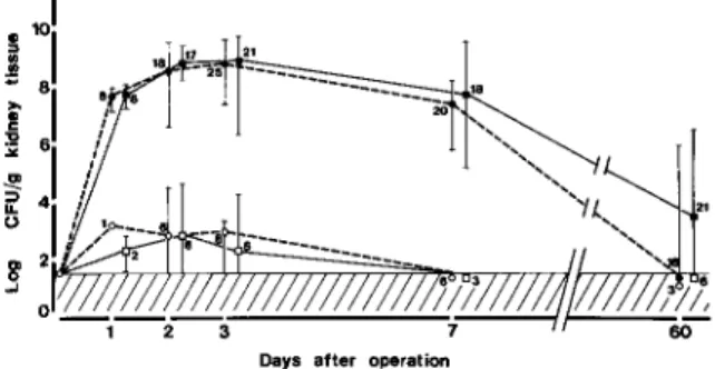
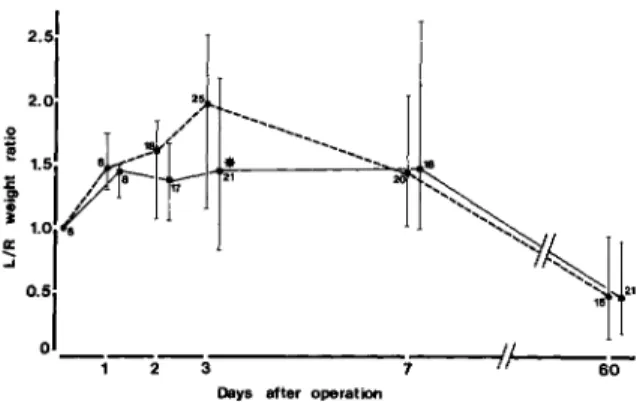
.985; cpm ratio range, 0.20-0.99). These observations demonstrated therefore that the left/ right kidney weight ratio represents an accurate mea-sure of the residual functional mass of the renal pa-renchyma during chronic scarring.Effect ofdexamethasone on bacterial counts and weight increase ofthe left kidney during AEP. Dexa-methasone treatment had no significant effect on bacterial counts in the left kidney at any time at which animals were killed, in both animals with high bacterial counts that developedAEPand animals with low bacterial counts that did not exhibit py-elonephritic lesions (figure 1). Because exudative le-sions developed only in animals with high bacterial counts in the left kidney, the effect of dexametha-sone on the left/right kidney weight ratio was stud-ied in this group of animals only. Compared with saline, dexamethasone treatment reduced by 56070 the median increase in the left/right kidney weight ra-tio in animals killed at three days after bladder in-oculation, the time of peak acute inflammatory re-sponse(P
<
.001; figure 2). This effect disappeared in animals killed later during the course ofAEP.Effect ofdexamethasone on MPO content ofthe left kidney. Figure 3 shows the MPOcontent of the left kidney of animals killed from one to seven days after operation. Rats with low bacterial counts in the left kidney and no macroscopic evidence of
AEPhad aMPO content of the left kidney similar
Days after operation
Figure 1. Effect of dexamethasone (squares) vs. control
(circles)treatment on bacterial counts in the left kidney. The hatched area represents the counts below the thresh-old of bacterial detection. Data are median values; bars indicate ranges for each group. The number of animals in each group is given. Solid symbols represent animals with macroscopic evidence of pyelonephritis, these animals invariablyhad high bacterial counts in the left kidney; open
symbolsrepresent animals with low bacterial counts and without macroscopic evidence of pyelonephritis.
to that of normal kidneys throughout these inter-vals. Dexamethasone treatment did not influence the
MPOcontent in these animals. In rats with high bac-terial counts in the left kidney and macroscopic signs ofAEP,dexamethasone treatment slightly but sig-nificantly reduced theMPOcontent of the left kid-ney in those rats killed early during the course of AEP, i.e.,at 24 h(P
=
.04). However, no such differ-ence was seen at later intervals, when the full devel-opment of the exudative response occurred and theMPO content increased considerably.
Effect of dexamethasone on severity of chronic pyelonephritis. Dexamethasone-treated animals killed two months after bladder inoculation had left/right kidney weight ratios similar to those of con-trols (figure 2). Thus despite the difference in the left/right kidney weight ratio in dexamethasone-treated and control animals duringAEP,the extent of parenchymal loss two months later, measured by kidney weight changes, was similar in both treatment groups.
Dexamethasone serum level. Two hours after ip injection of 2.5 mg of dexamethasone phosphate/kg, the mean ± SD serum level of dexamethasone in five rats was 556 ± 231 ng/mL; values decreased to 119± 60 ng/mL at 6 h and to 22± 11lJg/mL at 12h.
Discussion
Several lines of evidence in the present experiments support the view that theMPOcontent of kidney
Figure 2. Effect of dexamethasone (.) vs. control (.) treatment on kidney swelling during AEP and on severity of scars during CPN, as reflected by the left/right(L/R)
kidney weight ratio. Data are median values; bars indi-cate ranges for each group. The number of animals in each group is given. The star denotes a highly significant 56% reduction(P
<
.001) of the left/right ratio in dexametha-sone-treated animals compared with controls killed three days after operation. Groups of animals that did not de-velop pyelonephritis are not shown here. Their left/right kidney weight ratio increased to 1.4when they were killed 24 h after operation and decreased toward 1.0 in animals killed later.2 3
Days after operation
...
~'~
II 60 10001
000 1~
j
r~
/
!jI-~---
l"
::l 3.0 1 " a" 1.0,' ~+~_________ --~~ 0.31
•
r .
1
0.1 _ 1 2 3 7Days after operation
Figure 3. Effect of dexamethasone (squares) vs, control
(circles)treatment on PMNL exudation, measured by the kidney MPO content. Data are median values; bars indi-cate ranges for each group. The number of animals in each group is given. Solid symbols represent animals with mac-roscopic evidence of pyelonephritis, these animals invari-ably had high bacterial counts in the left kidney; open
sym-bols represent animals with low bacterial counts and without macroscopic evidence of pyelonephritis. The x symbol denotes a slight but significant reduction(P = .04) of the kidney MPO content in dexamethasone-treated rats killed one day after inoculation compared with con-trols. There was no such difference at any later interval.
homogenates can be used for quantitating the PMNL infiltration in kidney tissue.(1)Isolated rat PMNLs contained a large amount of MPO, whereas normal kidney homogenate did not. (2) Despite some interference of kidney homogenate with the MPO assay, the measure of the MPO activity was a quan-titative measure of the number of PMNLs added to normal kidney homogenate samples. (3) The perox-idase activity in kidney homogenates increased in parallel with the macroscopic appearance of sup-purative foci and the microscopic infiltration with PMNLs. (4) The peroxidase activity from pye-lonephritic kidneys had enzymatic properties simi-lar to the MPO activity extracted from purified PMNLs. Thus the kidney MPO content could be used as an index for tissue infiltration by PMNLs, which would allow the study of the effect of ther-apeutic interventions on the PMNL component of the inflammatory response.
The present data show that dexamethasone strik-ingly reduced the weight increase of the left kidney at the time of peak acute inflammatory response, that is, three days after operation. That dexametha-sone had no significant effect on kidney weight at the earlier times of killing (one and two days after operation) is not surprising because the weight in-crease of the left kidney at this time is partly due
to intrarenal hydronephrosis secondary to left ure-teralligation [4, 12]. In contrast to its effect on the weight increase of the left kidney, dexamethasone failed to reduce the kidney infiltration by PMNLs, a finding suggesting that dexamethasone acted on the kidney weight by reducing the plasma exudation, not cellular migration into kidney tissue. This fail-ure to reduce PMNL infiltration during AEP was accompanied two months after operation by a fail-ure to prevent kidney scarring.
The lack of effect of dexamethasone on PMNL infiltration was not due to an inadequate dosage, be-cause the peak serum levelsof dexamethasone in the rats were five to 10times higher than those achieved in humans after standard therapeutic doses of 0.1 mg/kg [15-17]. Furthermore, the schedule of ad-ministration (once a day) seemed appropriate be-cause dexamethasone was still detectable in serum 12 h after injection and because the half-life of the biologic effects of dexamethasone has been shown to be about twice that of the serum levels[17]. In-deed, the dexamethasone treatment schedule used here led to a potent glucocorticoid effect in the rats, as demonstrated by both the shrinkage of the spleen observed in dexamethasone-treated rats (data not shown), a typical glucocorticosteroid effect due to
lymphocytic depletion [18], and the reduction of the weight increase of the left kidney during AEP.
Studies investigating the effects of pharmacolog-ical doses of glucocorticosteroids on the in vivo PMNL migration have led to conflicting results in several experimental systems and demonstrate either a slight reduction or no effect at all[19-26]. These apparently conflicting observations may result from differences with respect to the properties of each an-imal model used in testing the inflammatory re-sponse, to the characteristics and dosages of the glucocorticosteroid used, and to the schedule of ad-ministration. This latter point might be particularly relevant because the onset of the hormonal effect of corticosteroid treatment is usually delayed for sev-eral hours [15].Our experiments differ in three re-spects from most of the observationsmentioned above.
(1)PMNL migration occurred in our experiments in response to the rapid development of high bacte-rial counts rather than in response to chemical or immunologic stimuli. (2) The PMNL infiltrates ac-cumulating in the kidney parenchyma were quanti-fied over several days, in contrast to observation periods of no more than 24 h in all of the above-men-tioned studies. In our experiments, a reduction of PMNL migrationby dexamethasonewasalso observed very early (24 h) during the development of AEP, but this effect vanished with the increasing inflam-matory response, an observation raising questions about the biologic significance of this early reduc-tion of PMNL infiltrareduc-tion. (3) Our model allowed accurate assessment of the permanent deleterious ef-fect of PMNL infiltration on the kidney parenchyma. Here dexamethasone failed to reduce PMNL infiltra-tion during AEP, and the subsequent development of kidney scars was not prevented. This result sug-gests that dexamethasone also failed to prevent the release of cytotoxic inflammatory mediators by stim-ulated PMNLs [27]. Becausewe havepreviously shown that PMNLs play an important role in the control of bacterial multiplication during AEP [8], the pres-ent observation that dexamethasone treatmpres-ent did not influence bacterial counts is additional indirect evidence that dexamethasone did not reduce the ac-tivation and bactericidaleffect of PMNLs on ingested E.coliand confirms that PMNLs are relatively re-fractory to glucocorticosteroid treatment [28].
In conclusion, we showed that measurement of the MPO activity in kidney homogenates can be used for estimating the PMNL infiltration of the kidney parenchyma. When administered during AEP,
dexa-methasone only reduced the swelling of the pyelo-nephritic kidneys, not the PMNL infiltration. There-fore the anti-inflammatory effect of dexamethasone may not necessarily be linked to a reduced migra-tion of phagocytes toward the inflammatory site, in contrast to a widely held belief [28]. Furthermore, dexamethasone failed to protect the rats from py-elonephritic scarring, a finding suggesting that dexa-methasone was not able to reduce the cytotoxic ac-tivity of stimulated PMNLs. These data suggest that even a striking effect of glucocorticosteroids on the classic signs of inflammation(i,e., tumor, in the pres-ent study) is not necessarily linked to reduced tissue damage (functio laesa).
References
1. Brooks SJO, Lyons JM, Braude AI. Immunization against retrograde pyelonephritis. I. Production of an experimental model of severe ascending Escherichia coli pyelonephritis without bacteremia in rats. Am J Pathol1974;74:345-58 2. Brooks SJO, Lyons JM, Braude AI. Immunization against retrograde pyelonephritis. II. Prevention of retrograde Escherichia colipyelonephritis with vaccines.Am J Pathol 1974;74:359-64
3. Brooks SJO, Lyons JM, Braude AI. Immunization against retrograde pyelonephritis. III. Vaccination against chronic pyelonephritis due to Escherichia coli. J Infect Dis 1977;136:633-9
4. Glauser MP, Lyons JM, Braude AI. Prevention of chronic experimental pyelonephritis by suppression of acute sup-puration. J Clin Invest1978;61:403-7
5. Miller T, Phillips S. Pyelonephritis: the relationship between infection, renal scarring, and antimicrobial therapy. Kid-ney Int 1981;19:654-62
6. Slotki IN, AsscherAW.Prevention of scarring in experimental pyelonephritis in the rat by early antibiotic therapy. Nephron 1982;30:262-8
7. Ransley PG, Risdon RA. Reflux nephropathy: effects of an-timicrobial therapy on the evolution of the early pyelo-nephritic scar. Kidney Int 1981;20:733-42
8. BilleJ, Glauser MP. Protection against chronic pyelonephritis in rats by suppression of acute suppuration: effect of col-chicine and neutropenia. J Infect Dis1982;146:220-6 9. Shimamura T. Mechanisms of renal tissue destruction in an
experimental acute pyelonephritis. Exp Mol Pathol 1981;34:34-42
10. Baron EJ, Proctor RA. Elicitation of peritoneal polymorpho-nuclear neutrophils from mice. J Immunol Methods1982; 49:305-13
II. Bradley PP, Priebat OA, Christensen RO, Rothstein G. Mea-surement of cutaneous inflammation: estimation of neu-trophil content with an enzyme marker. J Invest Oermatol 1982;78:206-9
12. Glauser MP, Francioli PB, Bille J, Bonard M, Meylan P. Ef-fect of indomethacin on the incidence of experimental Escherichia colipyelonephritis. Infect Immun 1983;40: 529-33
13. Taylor A Jr. Quantitation of renal function with static imag-ing agents. Semin Nucl Med 1982;12:330-44
14. Lambert WE, De Slypere JPM, Jonckheere JA, Vermeulen A, De Leenheer AP. Improved liquid chromatographic de-termination of serum cortisol with double internal stan-dardization compared to radioimmunoassay and fluorom-etry, and evaluated by isotope dilution/mass spectrometry. Anal Biochem 1983;134:216-23
15. Haynes RC Jr, Murad F.Adrenocorticotropic hormone; adrenocortical steroids and their synthetic analogs; inhib-itors of adrenocortical steroid biosynthesis. In: Gilman Goodman A, Goodman LS, Rail TW, Murad F, eds. Phar-macological basis of therapeutics. New York: Macmillan, 1985:1459-89
16. Hare LE, Yeh KC, Ditzler CA, McMahon FG, Duggan DE. Bioavailability of dexamethasone. II. Dexamethasone phos-phate. Clin Pharmacol Ther 1975;18:330-7
17. Meikle AW, Tyler FH. Potency and duration of action of glucocorticoids. Effects of hydrocortisone, prednisone and dexamethasone on human pituitary adrenal function. Am J Med 1977;63:200-7
18. Claman HN. Corticosteroids and lymphoid cells. N Engl J Med 1972;287:388-97
19. Meier R, EcklinB.Die Wirkung des Hydrocortisons auf die infektionsbedingte lokale Leukozytenansammlung. Ex-perientia 1960;16:204-5
20. Ackerman N, Martinez S, Thieme T, Mirkovich A. Relation-ship between adherence, chemotaxis and the accumulation of rat polymorphonuclear leukocytes at an inflammatory site. J Pharmacol Exp Ther 1982;221:701-7
21. Miyasaka K, Mikami T. Comparison of the anti-inflammatory
effect of dexamethasone, indomethacin and BW 755C on car-rageenin-induced pleurisy in rats. Eur J PharmacoI1982;77: 229-36
22. Tarayre JP, Lauressergues H. Comparison of the effect of phenylbutazone, desonide and cyclophosphamide on four types of experimental pleurisy. J Pharm Pharmacol1980; 32:408-12
23. Perper RJ, Sanda M, Chinea G, Oronsky AL. Leukocyte chemotaxis in vivo. II. Analysis of the selective inhibition of neutrophil or mononuclear cell accumulation. J Lab Clin Med 1974;84:394-406
24. Issekutz AC. Comparison of the effects of glucocorticoid and indomethacin treatment on the acute inflammatory reaction in rabbits. Immunopharmacology 1983;5:183-95 25. Almeida AP, Bayer BM, Horakova Z, Beaven MA. Influence of indomethacin and other anti-inflammatory drugs on mobilization and production of neutrophils: studies with carrageenan-induced inflammation in rats. J Pharmacol Exp Ther 1980;214:74-9
26. Tauber MG, KhayBashi H, Sande MA. Effects of am-picillin and corticosteroids on brain water content, cerebrospinal fluid pressure, and cerebrospinal fluid lac-tate levels in experimental pneumococcal meningitis. J In-fect Dis 1985;151:528-34
27. Weissman G, Smolen JE, Korchak HM. Release of inflam-matory mediators from stimulated neutrophils. N Engl J Med 1980;303:27-34
28. Fauci AS, Dale DC, Balow JE. Glucocorticosteroid therapy: mechanisms of action and clinical considerations. Ann In-tern Med 1976;84:304-15