HAL Id: tel-00974496
https://tel.archives-ouvertes.fr/tel-00974496
Submitted on 7 Apr 2014HAL is a multi-disciplinary open access archive for the deposit and dissemination of sci-entific research documents, whether they are pub-lished or not. The documents may come from teaching and research institutions in France or abroad, or from public or private research centers.
L’archive ouverte pluridisciplinaire HAL, est destinée au dépôt et à la diffusion de documents scientifiques de niveau recherche, publiés ou non, émanant des établissements d’enseignement et de recherche français ou étrangers, des laboratoires publics ou privés.
RÔLE DES MICROPARTICULES MEMBRANAIRES
DANS LA PHYSIOPATOLOGIE DE LA MALADIE
DE CROHN: EFFECTS SUR LA VASOMOTRICITE
ET LES TISSUES CIBLES
Daniela Leonetti
To cite this version:
Daniela Leonetti. RÔLE DES MICROPARTICULES MEMBRANAIRES DANS LA PHYSIOPA-TOLOGIE DE LA MALADIE DE CROHN: EFFECTS SUR LA VASOMOTRICITE ET LES TIS-SUES CIBLES. Pharmacologie. Université d’Angers, 2011. Français. �tel-00974496�
UNIVERSITÉ D’ANGERS
UNIVERSITA’ DEGLI STUDI DI BARI
Année 2011 N° d’ordre 1113
ROLE OF MICROPARTICLES IN THE PATHOPHYSIOLOGY
OF CROHN’S DISEASE: EFFECTS ON VASOMOTRICITY
AND TARGET TISSUES
RÔLE DES MICROPARTICULES MEMBRANAIRES DANS LA PHYSIOPATOLOGIE DE LA MALADIE DE CROHN: EFFECTS SUR LA VASOMOTRICITE ET LES TISSUES
CIBLES
THÈSE DE DOCTORAT EN COTUTELLE Spécialité : Pharmacologie expérimentale et clinique
ÉCOLE DOCTORALE BIOLOGIE SANTÉ Présentée et soutenue publiquement
le 4 Mars 2011 à Bari par Daniela LEONETTI Devant le jury ci-dessous :
Docteur Christian D. MULLER Rapporteur externe
Professeur Maria Antonietta PANARO Rapporteur externe
Docteur Sébastien FAURE Examinateur
Professeur Antonella MUSCELLA Examinateur
Professeur Vincenzo MITOLO Co-directeur de thèse
Docteur Ramaroson ANDRIANTSITOHAINA Directeur de thèse
Mitochondries : Régulations et Pathologie INSERM U694
Acknowledgements 1
Abbreviations 4
Publications 10
Part I 12
Crohn’s disease 13
I. General aspect 13
II. Epidemiology 15
III. Pathogenesis of Crohn’s Disease 16
III.1. Genetic factors 18
III.2. Enteric microflora 20
III.3. Immune Response 23
IV. Therapeutic approaches 27
V. Crohn’s Disease and Vascular Alteration 29
Part II 32
Microparticles 33
I. General aspects 33
II. Definition and generation of Microparticles 34
III. Composition of Microparticles 38
V. Microparticles and inflammation 45
VI. Microparticles and vascular function 47
VII. Microparticles and Crohn’s Disease 52
The aim of study 53
Manuscript I 56
Manuscript II 92
Review 120
Discussion 151
General conclusion and Perspectives 168
References 171
!
"!
!
#!
Ringrazio il Prof. Vincenzo Mitolo che mi ha offerto la possibilità di fare questa esperienza che mi ha arricchito da un punto di vista professionale e umano.
Je remercie profondément le Docteur Ramaroson Andriantsitohaina pour avoir accepté de codiriger cette thèse, ses enseignements, sa disponibilité et le temps qu’il m’a consacré. Je le remercie également de m’avoir transmis l’enthousiasme pour la recherche et de m’avoir permis d’apprécier encore plus ce travail.
Je remercie le Docteur Maria Carmen Martinez à qui je tiens à exprimer ma profonde estime pour ses conseils, sa patience et sa gentillesse, indispensable pour l’achèvement de cette thèse.
Je remercie le Professeur Jean-Marie Reimund pour la précieuse collaboration nécessaire à la réussite de ce travail.
Je remercie les membres de mon jury, le Docteur Christian D. Muller, le Docteur Sébastien Faure, le Professeur Maria Antonietta Panaro et le Professeur Antonella Muscella qui me font l’honneur de juger ce travail.
!
!
%!
!
&!
AA: arachidonic acid Ach: acetylcholine ActD: actinomycin D
CDMPs: MPs isolated from plasma of Crohn’s disease patients CMH: 1-hydroxy-3-methoxycarbonyl-2,2,5,5-tetramethylpyrrolidin COX: cyclooxygenase
COX-1: cyclooxygenase-1 COX-2: cyclooxygenase-2
DETC: Fe2+ diethyldithiocarbamate
EDHF: endothelium-derived hyperpolarizing factor EMPs: endothelial microparticles
EPCs: progenitor cell cultures
EPR: electronic paramagnetic resonance eNOS: endothelial nitric oxide synthase GP: glycoprotein
HSMPs: MPs isolated from plasma of healthy subjects 5-HT: serotonin
!
'!
HUVEC: human umbilical vein endothelial cells IBD: inflammatory bowel disease
ICAM-1: intercellular adhesion molecule-1 Ig: immunoglobulin
iNOS: inducible nitric oxide synthase IL-1!: interleukin-1 ! IL1-": interleukin-1 " IL6: interleukin-6 IL-8: interleukin-8 IL-12: interleukin-12 IL-17: interleukin-17 IL-18: interleukin-18 IL-21: interleukin-21 IL-23: interleukin-23
IL-23R: interleukin-23 receptor IFN-#: interferon-#
!
(!
L-NA: NG-nitro-L-arginine
MAP: Mycobacteriu avium subspecies paratubercolosis M cells: microfold cells
MCP-1: monocyte chemoattractant protein-1
MnTMPyP: manganese(III) tetrakis(1-methyl-4-pyridyl) porphyrin MPs: microparticles
NADPH: nicotinamide adenine dinucleotide phosphate NO: nitric oxide
NS-398: N-2-cyclohexyloxy-4-nitrophenyl OSA: obstructive sleep apnea
PAI-1: plasminogen activator inhibitor-1 PAF: platelet activating factor
PDE4: phosphodiesterase 4 PFP: platelet-free plasma PGI2: prostacyclin
PHA: phytohemagglutinin PKC: protein kinase C
!
)!
PMA: phorbol-myristate-acetate PMPs: platelet-derived MPs
PPAR#: peroxisome proliferator activated receptor # !"#!"#$%"#&'()*+%,-(.,!
!
ROCK: Rho-associated kinase ROS: reactive oxygen species
SC-560: 5-(4-Chlorophenyl)-1-(4-methoxyphenyl)-3-trifluoromethyl pyrazole Shh: sonic hedgehog
SOD: superoxide dismutase SNP: sodium nitroprusside
TAFI: thrombin-activatable fibrinolysis inhibitor TGFbeta1: transforming growth factor beta1 TF: tissue factor
Th1: T helper 1 cells Th2: T helper 2 cells Th9: T helper 9 cells Th17: T helper 17 cells
!
*!
Th22: T helper 22 cells
TNF-!: tumour necrosis factor ! Treg: T regulatory cells
!
"+!
!
""!
Publications in revision: Review:
S. Tual-Chalot, D. Leonetti, R. Andriantsitohaina, MC. Martínez. Microvesicles: intercellular vectors of biological messages. Soumis à Molecular Interventions
Publications under submission:
D. Leonetti, AL. Bretagne, A. Tesse, MC. Martinez, S. Viennot, JM. Reimund, R. Andriantsitohaina. Microparticles are relevant markers of Crohn’s disease activity and cause endothelial and vascular dysfunctions. Soumis à Gastroenterology
D. Leonetti, AL. Bretagne, M. Chalopin, MA. Panaro, MC. Martinez, JM. Reimund, R. Andriantsitohaina. Circulation microparticles from Crohn’s disease patients exert differential effects on nitric oxide and superoxide anion productions, and inflammatory markers. Soumis à The American Journal of Gastroenterology
!
"#!
!
"$!
Crohn’s disease
I. General aspects
Crohn’s disease is, with ulcerative colitis, one of the major forms of inflammatory bowel disease (IBD). The cause of IBD is still unknown but several studies have shown that this pathology is characterised by an abnormal activation of the mucosal immune system in response to bacterial flora or infectious agents which is linked to alteration of barrier function of the intestinal epithelium.
This chronic inflammatory disorder is due to a transmural inflammation that extends through all layers of the bowel wall and can affect the entire gastrointestinal tract, with greater involvement of colon and terminal ileum (Fig 1a). Histopathological features include an aggregation of macrophages that frequently form, with giant cells and epithelioid cells, non-caseating granulomas (Fig 1b).
The clinical presentation is largely dependent on disease location and can include diarrhoea, abdominal pain, fever, clinical signs of bowel obstruction, as well as passage of blood or mucus or both. Crohn and coworkers described the first time the inflammation of the large intestine either localized or diffuse in 1952. Indeed, they designated a disease of the terminal ileum, affecting mainly young adults, characterized by a sub-acute or chronic necrotizing and cicatrizing inflammation (Crohn et al., 1952).
The definition of the disease has been expanded in next ten years as a granulomatous inflammation of the rest of the gastrointestinal tract (including the colon) and distinguishing them from ulcerative colitis on clinical and pathological aspects (Lockhart-Mummery et al., 1960).
!
"%!
Even now, the definition of the disease is not entirely clear because the patients show a clinical heterogeneity. The discovery of genetic and serological markers associated with phenotype in IBD and clinical data have revealed the existence of subtypes of Crohn’s disease based on the location. These subtypes have been defined as terminal ileal (L1), colonic (L2), ileocolonic (L3) and upper GI (L4) during World Congress of Gastroenterology at Montreal (Silverberg et al., 2005).
a b
Fig 1: Crohn’s disease: (a) Crohn’s disease is a transmural inflammation that extends through all layers of the
bowel wall and can affect the entire gastrointestinal tract, with greater involvement of colon and terminal ileum; compared to ulcerative colitis that is a diffuse mucosal inflammation. (b) Histopathological features of Crohn’s disease include granuloma composed of compact macrophages, giant cells and epithelioid cells. Whereas, ulcerative colitis shown crypt abscess composed of transmigrated neutrophils and the surrounding epithelium exhibits features of acute mucosal injury (From Xavier et al., Nature Reviews 2007).
! !
!
"&!
II. Epidemiology
Crohn’s disease affects $ 0.1% of Western population. Epidemiology analysis showed a higher incidence in North America and Northern and Western Europe than Asia, Africa and South America (Logan and Bowlus, 2010).
Since the beginning of the 20th century, there has been a steady increase in reported cases of both Crohn’s disease and ulcerative colitis. This increase in IBD mainly affects the developed countries, especially populations with high living standard and urban areas (Bernstein and Shanahan, 2008). Particularly, Europe has seen a change in the epidemiology of IBD over the past several decades. Recent data showed a stabilization of incidence rates in Northern countries of Europe and a significant increase in some Southern countries tied to an improvement in living standards. In addition, some regions of Europe have shown a particular increase of Crohn’s disease. In Northern France, for example, it has been shown, over a 12 year period, a 23% increase in Crohn’s disease with a 17% decrease in ulcerative colitis over the same period of time (Molinié et al., 2004).
Statistically the frequency of the disease correlates with the introduction of tap water, soap and improvement in the living conditions. The hygiene hypothesis, proposed by Strachan in 1989 (Strachan, 1989), argues therefore, that improved hygiene and a lack of exposure to microorganisms of various types have sensitized our immune system, leading to inadequate reaction to harmless bacteria in our environment (Koloski et al., 2008).
The environmental factors play a major role in the development of disease particularly in teenagers or young adults. There are many factors related to the pathogenesis of IBD such as smoking, prenatal events, breastfeeding, childhood infections, microbial agents, oral
!
"'!
contraceptives, diet, hygiene, occupation, education, tonsillectomy, appendectomy, blood transfusions, contact with animals and physical activities.
Among those one of the most studied factors is the smoking. Recent studies showed that smoking interferes with a shortage of zinc present in subjects with Crohn’s disease, that may facilitate release of pro-inflammatory mediators and their activities and may cause exacerbates symptoms of disease (El-Tawil, 2010). An interesting study reports that emigrates from an area of low prevalence to one of high prevalence have a similar rate of IBD as before, but their children have an increased risk of IBD (Pinsk et al., 2007). This suggests that several factors related to life in a developed country can affect the development of the immune system during infancy.
Finally, Crohn’s disease is a pathology that significantly impairs quality of life, requires expensive drugs, surgery or multidisciplinary care and represents a major burden on public health-care resources.
III. Pathogenesis of Crohn’s Disease
The pathogenesis of Crohn’s disease is still not completely clear but several evidences suggest that the balance between microbes (particularly commensal flora) and host defensive responses at the intestinal mucosal barrier may play an important role.
In healthy individuals, the mucosal immune system represents a complex of interacting mechanisms designed to interpret the environment, distinguish danger from harmless antigenic stimuli, and respond appropriately to maintain internal homeostasis. The maintenance of mucosal homeostasis involves constrained responsiveness to dietary antigens and resident microflora, while retaining the capacity for effective immune responsiveness
!
"(!
against episodic threats from pathogens. Errors in discrimination of danger signals from innocuous stimuli or in regulation of effector immune responses, disrupt mucosal homoeostasis, and predispose the individual to uncontrolled or pathological inflammation (Shanahan, 2000).
Therefore, depending on the genetic susceptibility of the host, the normal intestinal flora may become a liability. The pathogenesis of Crohn’s disease is complex and mainly consists of the three interacting elements: genetic susceptibility factors, enteric microflora and immune-mediated tissue injury (Fig 2).
!
Fig 2: Pathogenesis of Crohn’s disease: Intestinal inflammation in Crohn’s disease results from alteration in
the interaction between resident microbes and the mucosa. This can result from the influence of environmental factors and/ or host factors, which vary depending on genetic inheritance at several susceptibility loci. Genetic factors discovered to date affect barrier function, and innate and adaptive immunity (Modified from Xavier and Podolsky 2007). .
! ! ! !
!
")!
III.1. Genetic factors
Several clinical observation have pointed out that genetic factors contribute to pathogenesis of Crohn’s disease:
- First, the ethnic differences in disease frequency have been shown since the incidence and prevalence of Crohn’s disease varies widely among different populations.
- Second, a familial aggregation of IBD has been demonstrated. A study of familial occurrence of IBD has established that a risk! of IBD in first-degree relatives of an affected patient is 4 to 20 times as high as that among the background population (Orholm et al., 1991).
- Third, in patients with Crohn’s disease, a higher rate of disease concordance in monozygotic (44%) twins than in dizygotic (3-8%) twin has been observed (Tysk et al., 1988).
Also, a manifestation of syndrome resembling IBD in families with rare genetic disorders such as Hermansky-Pudlak syndrome and Turner’s syndrome has been demonstrated (Kouklakis et al., 2007)
;
(Triantafillidis et al., 2010) (Table 1).All these data suggest a genetic complexity of Crohn’s disease, and that the susceptibility of disease is inherited. In recent years, the genome-wide screening for susceptibility genes in Crohn’s disease has identified several genes involved in disease. The first study of screening of DNA has identified a candidate susceptibility gene, in patients with Crohn’s disease, located at the region of chromosome 16 named NOD2 or IBD1 (or also CARD15). This gene encodes a cytoplasmic protein which is expressed in monocytes and is involved in regulation
!
"*!
of macrophage apoptosis and NF-%B activation that is a transcriptional factor directly involved in activation of immunoinflammatory responses (Ogura et al., 2001b).
The patients with Crohn’s disease display the mutations of NOD2 gene and this variant appear to be related of reduced macrophage activation of NF-%B in response to lipopolysaccharide (Hugot et al., 2001).
Over the years have been identified several other loci particularly, their association with genes coding for factors involved in the regulation of immune and inflammatory response or function of mucosal barrier, such as IBD2 (chromosome 12), IBD3 (chromosome6), IBD4 (chromosome14), IBD5 (chromosome 5q31), IL-23R and susceptibility locus identified on the X chromosome (Vermeire et al., 2001; Duerr et al., 2006).
The complexity of the genetic component of Crohn's disease is enhanced by a variable role that the different loci may have in different ethnic population. For example, Silverberg et al. (Silverberg et al., 2007)confirm the importance of IBD5 to Crohn's disease susceptibility and demonstrate that the locus may play a role in non-Jewish individuals only.
The identification of new genes, and understanding about their interaction, is important for the future because it might provide new information about the cause of the disease. At present, it have been identified more than 30 susceptibility genes and loci associated with disease which led to a better understanding of new pathways involved in the pathogenesis of the disease.
!
#+! Table1: Evidence of Genetics contributions to Crohn’s Disease.
II.2. Enteric microflora
The possibility that Crohn's disease is an inflammatory response against microbial agent has been evaluated in several studies. The development of immune reactivity to enteric bacteria is an entirely physiological phenomenon that starts immediately after birth when the immature intestinal immune system begins to be exposed to a variety of microbial antigens and creates a lifelong state of tolerance against them in order to avoid excessive or detrimental responses (Duerkop et al., 2009).
The intestinal mucosa plays a barrier function that physically impedes penetration of macromolecules and intact bacteria. The epithelium is in constant communication with intestinal flora and intestinal epithelial cells express Toll-like receptors, NOD1 and 2 and receptors for different chemokines. In particular, epithelial cells-specific NF-%B seems to play an important role in the suppression or activation of immune response in IBD. Some bacteria seem to be able to modify this process through the reduction of epithelial NF-%B activation by inhibition of I%B! degradation (Neish et al., 2000).
Specialized cells interspersed along the crypt villus axis that enhance protection against microbes and promote repair also form the epithelium. In the base of the crypt, the Paneth cells secrete antimicrobial peptides such as !-defensine. These antimicrobial peptides not only defend against pathogenic bacteria but also control the balance between various bacterial populations and contribute to local homeostasis (Menendez et al., 2010). Some observations suggest that a reduction of !-defensine production might contribute to the pathogenesis of terminal ileal Crohn's disease in patients with mutant NOD2 (Wehkamp et al., 2004; Wehkamp et al., 2005).
!
#"!
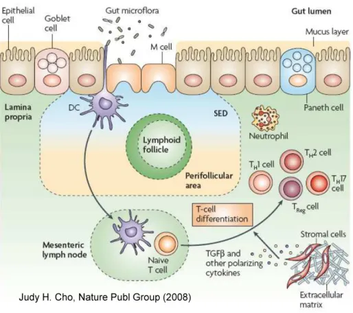
Globlet cells are another important component of epithelium that produces trefoil peptides involved in defence and repair of epithelium and mucosa, whereas Microfold cells (M cells) transport organisms and particles from the gut lumen to immune cells across the epithelial barrier and with dendritic cells sample intestinal contents (Fig 3).
In patients with Crohn's disease, modifications of luminal bacteria concentrations compared to control patients group have been found. For example, a recent study has shown that the neoterminal ileum of CD patients is heavily colonized by a colonic-like bacterial flora, with
E. coli predominating (Barnich et al., 2007).
Evidence of involvement of intestinal microbiota in the pathogenesis of disease is provided by the therapeutic benefits of antibiotics’ treatment in a group of patients with IBD (Gionchetti et
al., 2003). In particular, two microorganisms have been associated with Crohn's disease: Mycobacteriu avium subspecies paratubercolosis (MAP) and Escherichia coli.
MAP has been identified in 1980 but its potential etiological role is cause of debate. Recently, it has been suggested that elimination of MAP in Crohn's disease does not significantly affect the clinical course of patients receiving three anti-mycobacterial antibiotics or placebo (Selby
et al., 2007).
Escherichia coli has been isolated in 1990 from the ileal mucosa of Crohn's disease patients
and its capacity to adhere and penetrate intestinal epithelial cells has been shown. However, it is not yet clear whether this microorganism acts as a pathogens or a commensal in Crohn's disease patients (Darfeuille-Michaud et al., 1998; Darfeuille-Michaud et al., 2004).
The importance of intestinal flora is supported by studies in murine model, in which, colitis is not observed when any of several of these lines are maintained in a gnotobiotic state, but rapidly emerges when they are reconstituted with bacteria that are considered normal constituents of luminal flora (Elson et al., 2005).
!
##!
Based on several reports, it is now well established that essentially all animals raised under germ-free conditions, in the complete absence of commensal flora, do not develop experimental! intestinal inflammation independently from strain and genetic background or method used to induce inflammation.
In patients with IBD, the existence of the immune reactivity against intestinal microbes is provided by series of serum antibodies against a variety of microorganisms such as
Saccharomyces cerevisiae, anti-outer membrane protein C and anti-I2. In patients with
Crohn's disease, these antibodies are currently used as biomarkers and a high level of detected antibodies is often associated with a complicated or rapidly progressive disease course (Mow
et al., 2004; Dubinsky et al., 2008).
A limit on the understanding the microbial-host interrelationship is an incomplete knowledge of the composition of the normal human intestinal microbial flora (Gill et al., 2006). Furthermore, recent studies have shown that some bacteria do not always stimulate an intestinal immune response but can promote an anti-inflammatory status (Mazmanian et al., 2008).
Understanding the distribution, dynamics and responses to microbial flora in this disease could provide additional information on the pathogenesis and regional location of the disease.
!
#$!
III.3. Immune Response
The immune response is essential to functional integrity of the intestinal mucosa and health. Chronic intestinal inflammation appears to result from stimulation of the mucosal immune system by products of commensal bacteria in the lumen or antigens from dietary. The bacterial products may stimulate the surface epithelium, possibly through receptors that are components of the innate immune response system or can penetrate through the mucosal barrier, leading to their direct interaction with immune cells. The innate immunity is carried out mainly trough two types of cells: macrophages and dendritic cells.
In the normal intestine, macrophages are conditioned by the mucosal microenvironment to express a non-inflammatory phenotype that consists of a down-regulated expression of innate immunity receptors and a limited production of pro-inflammatory cytokines. In intestinal tissue affected by IBD, mucosal macrophages show an activated phenotype and are phenotypically heterogeneous. Moreover, macrophages recruited from the peripheral blood, CD14+ pro-inflammatory macrophage activate the production of various pro-inflammatory cytokines such as IL-1!, IL1-" and TNF-! (Rugtveit et al., 1997). In Crohn's disease, the number of CD14+ pro-inflammatory macrophages is increased and these macrophages produce mainly IL-23 and TNF-! and contribute to the production to IFN-# by T cells (Kamada et al., 2008).
The dendritic cells are antigen-presenting cells involved in the initiation and regulation of local innate immune response but also have a role in adaptive immunity. These cells play a role of protection and defence and mediate inflammation. Their function depends on the location, state of maturation and stage of inflammation. In IBD, the dendritic cells are activated and produce elevated levels of pro-inflammatory cytokines such as IL-12 and IL6 (Rescigno and Di Sabatino, 2009; Hart et al., 2005). The innate immune cells are also able to
!
#%!
generate reactive oxygen species (ROS) that are directly involved in inflammation and tissue injury and increase epithelial permeability.
The adaptive immune response is produced by a combination of cell population: B cells and a complex variety of T cells. B cells produce immunoglobulin (Ig) M, IgG and IgA. In IBD patients, the synthesis and secretion of these Igs is altered both! in! peripheral blood! and in mucosal mononuclear cells (MacDermott et al., 1981). In particular, in Crohn's disease patients, the production of IgG1, IgG2 and IgG3 is increased compared to control cells (Scott
et al., 1986).
The two more important types of T cells involved in IBD are CD4+ T helper 1 (Th1) and T helper 2 (Th2). In recent years, it has been identified a series of new types of T cells such as IL-17-producing Th17 cells, Th9 and Th22 cells (Weaver et al., 2007; Annunziato and Romagnani, 2009). In addition to Th cells, T regulatory (Treg) cells are appointed to monitor the immune response and prevent an excessive and potentially harmful immune activation (Izcue et al., 2006) (Fig 3).
!
#&! Fig 3: Intestinal immune system: The intestinal immune system is extensive and unique with respect to its
close apposition to intraluminal bacteria, which are separated from the underlying lamina propria by only a single layer of epithelial cells. The epithelial-cell layer is comprised of absorptive and secretory cells, goblet cells and Paneth cells. Goblet cells contribute to the formation of the protective mucus layer. Microfold cells (M cells) and dendritic cells (DCs) sample intestinal luminal contents.
The presence of either pathogenic bacteria or disruption of the epithelial-cell barrier results in activation and migration of DCs to the mesenteric lymph nodes, where they activate naive T cells, which then undergo differentiation under the influence of factors released by DCs and other stromal elements. A central challenge facing researchers of inflammatory bowel diseases is defining the mechanisms of crosstalk between components of the innate and adaptive immune systems in light of the multiple established genetic variants associated with Crohn’s disease. SED, subepithelial dome; TGF", transforming growth factor-"; TH, T helper; TReg, T regulatory (From Cho JH; Nat Publ Group 2008).
The mucosa of patients with Crohn's disease is dominated by CD4+!lymphocytes with a Th1 phenotype, characterized by the production of IFN-# and IL-12 by lamina propria mononuclear cells (Parronchi et al., 1997). In addition, in Crohn's disease there is also a elevated production of IL-17 by Th17 cells and dual IFN-# and IL-17-producing Th cells, and also IL-21 that regulates IL-17 production (Fujino et al., 2003; Monteleone et al., 2005).
!
#'!
More precisely, in patients with Crohn's disease, the activation of classic antigen-presenting cells, such as dendritic cells, or direct stimulation through pattern-recognition receptors promotes the differentiation of Th1.
In mice, it has been suggested that!the stereotypical Th1 cytokines promote a self-sustaining cycle of activation with macrophages:! Th1 cytokines activate macrophages, which in turn, produce IL-12, IL-18, and macrophage migration inhibitor factor which further stimulate Th1 (Podolsky, 2002). The macrophages produce also a potent mix of active inflammatory cytokines, including TNF-!, IL-1, and IL-6. These factors stimulate a variety of other type of cells such as vascular endothelial cells, which facilitate the recruitment of leukocytes to the mucosa from the vascular space, fibroblasts and epithelium. The recruitment of! additional leukocytes from the vascular space to sites of disease activity is especially important in maintaining inflammation and depends on the expression of cytokines, chemokines and adhesion molecules in the local microvasculature.!
The activation of intestinal immune system is linked to a variety of non nonspecific mediators of inflammation such as many other cytokines, chemokines, and growth factors as well as metabolites of arachidonic acid (prostaglandins and leukotrienes) and reactive nitrogen metabolites such as nitric oxide (NO).
These mediators enhance the inflammatory process and tissue destruction, which lead to the clinical manifestations of disease.
!
#(!
IV. Therapeutic approaches
Crohn's disease is a disease whose anatomical location is fairly stable but the evolution of disease may vary during its course.
A study on population-based cohorts showed that 13–20% of patients with Crohn’s disease
have a chronic active course of disease activity, 67–73% has a chronic intermittent course and
only 10–13% remains in remission for several years. After 20 years old, most patients with
Crohn’s disease will require surgery (Loftus et al., 2002). The ideal therapeutic strategies for
Crohn's disease patients should be able to induce remission and maintain long-term remission, reducing to a minimum the surgery. Furthermore, although surgery might be necessary to treat complications or to induce remission in patients with Crohn's disease, surgery is unable to cure the disease.
Among the different treatment, corticosteroids have been used in the treatment of Crohn's disease for many decades. These drugs are effective in inducing a remission of disease but are unable to secure a long-term remission and are associated to severe and irreversible side effects.
A better understanding of the immunologic mechanism associate with the disease led to the introduction a new concept of therapies, biologic therapies, aimed to correcting the imbalance of the intestinal immune system observed in disease.
Among the molecules that have been shown to be effective in treating the disease, stand the TNF! inhibitors. TNF! is a proinflammatory cytokine that induces cell proliferation and differentiation and promotes the inflammatory response. Three anti-TNF! molecules are currently used to treat IBD: infliximab, adalimumab and cerolizuman pegol. In particular, infliximab is a chimeric monoclonal IgG1 against TNF! that was introduced in 1997 and has
!
#)!
proved highly effective in patients with refractory luminal and fistulising Crohn's disease (Present et al., 1999; Sands et al., 2004). Infliximab treatment reduces hospitalizations and surgery and improves the quality of life in patients with Crohn's disease (Lichtenstein et al., 2005).
A series of promising new molecules for treatment of Crohn's disease were assessed in controlled clinical trial: anti–IL-12/IL-23 p40, anti-IFN-# and anti-IL-6 antibodies. The patients with active Crohn's disease treated with anti-IL-12 display a decrease of production of proinflammatory cytokines such as IL-12, IFN-# and TNF! (Mannon et al., 2004).
Reagents that blocked a receptor complex responsible of T cell activation, such as CD4 and CD3, are also, under development in treatment of Crohn's disease (Plevy et al., 2007).
Recently, a large number of small molecules involved in specific immune pathway disease such as eicosanoides (thromboxane A2, leukotrienes and platelet activating factor (PAF)) and inhibitors of phosphodiesterase 4 (PDE4) are also studied for the treatment of IBD. Particularly, a new promising small molecule is the agonist of activator of peroxisome proliferator activated receptor # (PPAR#) that is associated with an inhibition of signal transduction pathway linked to production of proinflammatory cytokines (van Deventer, 2002).
Furthermore, the alternative approaches therapeutic are being tested especially in patients with severe disease, such as stem cell transplantation (Rutgeerts et al., 2009).
Finally, the progress in the discovery of gene implicated in the pathogenesis of the disease and in understanding of their interaction, could lead in the future, to use of the specific treatments for different disease phenotypes.
!
#*!
Several anatomic and pathological studies have shown, in both IBD patients and in animal models, an altered microvascular anatomy and function in inflamed gut. Then, vascular alterations may be one of the causes of inflammatory disease.
In 1989, Wakefield et al. (1989) have reported vascular lesions in both segments of inflamed and noninflamed intestine in Crohn’s disease patients and a remodelled vascular network in inflamed segments, suggesting that Crohn’s disease is mediated by multifocal gastrointestinal infarction and that vasculitis may be one of the first events leading to mucosal injury.
Recent studies have described an impaired vasodilatation and tissue remodelling in human submucosal arterioles isolated from areas of chronic inflammatory damage of intestine of inflammatory bowel disease patients. This reduced vasodilatory capacity, is due to a functional alteration in endothelium-dependent dilatation and is linked to a reduced endothelial generation of NO and high levels of oxidative stress in blood vessels (Hatoum et
al., 2003).
Some authors have proposed that increased expression of arginase enzymes that convert L-Arg into urea and L-ornithine, and that are important for tissue homeostasis and lesion repair, play an important role in the mechanism involved in microvascular dysfunction present in IBD patients. Effectively, an increase of gene expression and activity of arginase enzymes that was found in inflamed IBD submucosal tissue could decrease microvascular endothelial access to L-Arg that is the fundamental substrate required for NO production (Horowitz et al., 2007).
Microvascular dysfunction is also linked to impaired tissue perfusion and oxygenation (Mori
!
$+!
In contrast, unchanged endothelium-dependent and -independent relaxations in mesenteric arteries from Crohn’s disease patients and a decrease of vascular tone are also reported. The reduction of contractile response, involve an excessive NO production through the overexpression of inducible NOS in vascular smooth muscle. These authors suggest that this hyporeactivity could improve the blood perfusion and could be a possible response of host organism against the injury (Lebuffe et al., 2000).
Tabernero et al. (2003) revealed, despite an increase of release of proinflammatory cytokines in mucosa of Crohn’s disease patients, an unmodified vascular reactivity in small mesenteric arteries from patients with Crohn’s disease. The regulation of vascular contraction involves vasoconstrictor metabolites of cyclooxygenase (COX) and, interestingly; there is a balance between vasoconstrictor products from COX-2 and vasodilator products that maintained the vascular reactivity unchanged.
All these data, suggest a possible implication of vascular alterations in pathogenesis of disease (Fig 4).
!
$"! Fig 4: Vascular alterations in Crohn’s Disease: vascular alterations include both vascular dysfunction and
modification of vascular tone. These alterations involve NO pathway, oxidative stress, COX-2-derived metabolites and impaired perfusion and oxygenation.
!
$#!
!
$$!
Microparticles
I. General aspects
In the multicellular organisms, the communication between cells occurs through direct cell-cell contact or mediator production such as protein that binds to receptors on neighboring cells. Another way of intercellular communication which has recently generated an increasing interest is the release of membrane vesicles.
The consequences of this membrane transfer involved the induction, amplification or modulation of immune response and the acquisition of new functional properties in the recipient cells. Furthermore recent studies suggest a possible transfer of genetic material between cells achieved through the transfer of mRNA present in small vesicles.
In this context, microparticles (MPs) are vesicles shed from the blebbing plasma membrane that have been widely studied. Recent data show that MPs can be considered as biological vectors able to transfer information from one cell to another. In addition, MPs can mediate long-range signalling, acting on cells different from their cells of origin (Martínez et al., 2005).
The MPs can allow intracellular communication trough different pathways. They can interact directly with the ligands present on the surface of target cells transferring proteins, mRNA, miRNA, receptors and bioactive lipids (Hunter et al., 2008). MPs may also enable communication between cells without direct cell contact. MPs can bind to the membrane of target cells which can acquire new surface antigens and then new biologic properties and activities. Finally, MPs can be absorbed by fusion or by internalization.
!
$%!
MPs are present in the blood from healthy subjects and patients, they could than participate in the maintenance of homeostasis under physiological conditions or have a deleterious effect in pathological situations.
II. Definition and generation of MPs
The concept of MPs was introduced in 1967 by Wolf that describes inert “cellular dust” released from activated platelet in human plasma (Wolf, 1967). Based on several data, it is now well established that MPs are small vesicles heterogeneous in size (0.05-1 &m) and composition with procoagulant and proinflammatory properties. Although MPs can potentially be produced by all cell types, in general, MPs derive from circulating cells such as platelets (that represent the highest percentage of MPs), leukocytes, monocytes and erythrocytes, and cells that compose vessel wall such as endothelial cells, macrophages and smooth muscle cells; MPs can derived also from cancer cells (Martínez et al., 2005); (Mostefai et al., 2008).
The mechanism that leads to the formation of MPs is not completely clear but there are two well-known cellular processes that can stimulate the production of MPs: chemical and physical cell activation (by agonist or sheer stress) and apoptosis (/-$0'#!1&2'$-!),"-(3&'($.! $-!&"$"'$'(2!(.)42,-%5!(Benameur et al., 2009).
Following cell activation by different agonists, MPs formation is dependent on an increase of cytosolic calcium that is associated with calpain activation required for proteolysis of cytoskeletal proteins and then cytoskeleton disruption (Pasquet et al., 1996). The cytoskeleton disruption involved also a kinase activation and phosphatase inhibition caused by increase of cytosolic calcium level (Heemskerk et al., 1997
;
Kunzelmann-Marche et al., 2002).!
$&!
MP formation is linked to a loss of membrane asymmetry. The membrane bilayer is formed by two leaflets each of which having a specific lipid composition. 67(.$"#$%"#$+("()%! 8"#$%"#&'()*+%,-(.,! 89:5! &.)! "#$%"#&'()*+,'#&.$+&7(.,5! &-,! %",2(1(2&++*! %,/-,/&',)! (.! '#,! (..,-! +,&1+,';! 0#,-,&%! "#$%"#&'()*+2#$+(.,! &.)! %"#(./$7*,+(.! &-,! "-,%,.'! (.! '#,! $4',-! +,&1+,'<! During MP formation, there is an alteration of bilayer structure that is controlled by three major players: an aminophospholipid translocase which is an inward-directed pump that transport PS and phosphatidylethanolamine from the outer to inner leaflet of plasma membrane against the concentration gradient; an outward-directed pump, an ATP-dependent floppase which acts in conjunction with the translocase and a lipid scamblase, can rapidly move aminophospholipids between the membrane leaflets by a calcium-dependent mechanism and may lead to collapse of membrane asymmetry (Bevers et al., 1999; Hugel et
al., 2005). A sustained rise of intracellular calcium concentration regulates positively
scramblase and floppase activities and inhibiting the translocase. The main effect of redistribution of membrane lipids is the exposure of PS and a consequent release of MPs. The PS exposure is present in the surface of most but not all MPs and PS plays two important functions: promote the blood coagulation and is involved in signal recognition for clearance of senescent cells by the reticuloendothelial system (Hugel et al., 2005) (Fig 5).
MPs production can be also induced by physical stimulation such as changes in blood flow. Holme et al (Holme et al., 1997) have shown that in arteries with several stenosis, high shear stress activates platelets and triggers platelet MP formation. In another study, it has been found that platelet MP formation increases with the duration of shear stress and that activation of protein kinase C (PKC) promotes shear-dependent MP formation. Furthermore, both MPs and platelets showed the exposure of procoagulant activity on their surfaces suggesting that MPs formation could contribute to arterial thrombosis by providing and expanding a catalytic surface for the coagulation cascade (Miyazaki et al., 1996).
!
$'! Fig 5: The plasma membrane response to cell stimulation: The plasma membrane is a well-structured entity
characterized by a controlled transverse distribution of lipids and proteins between the two leaflets but also by a lateral organization in domains termed “rafts.” Following stimulation, a general redistribution occurs, leading to raft structuration, phosphatidylserine externalization, and microparticle release. The private membrane response characterized by controlled inclusion or exclusion of specific constituents into rafts leads to the release of microparticles of a particular composition. (From Hugel et al., 2005).
The second main process that leads to production of MPs is apoptosis. Apoptosis is an important biological process for normal cellular homeostasis in multicellular organisms that can be induced by different extracellular stimuli such as: irradiations, proinflammatory cytokines, chemotherapeutic drugs, hormones or bacterial agents. The cell death by apoptosis
!
$(!
consists of an intracellular cell death program that involved changes of the cell morphology (condensation of the cytoplasm and segregation of chromatin from compact masses on the nuclear membrane inner leaflet), cytoskeleton disruption, cell shrinkage and membrane blebbing (Thompson, 1995). The membrane blebbing is followed by shedding of membrane vesicles that transport nuclear fragments, and release of apoptotic bodies or MPs which expose PS on their surface. The PS exposure characterizes apoptotic bodies and apoptotic cells allowing their recognition and phagocytosis by macrophages and mesenchymal cells. MP formation by apoptosis involved a Rho-associated kinase (ROCK I) activity, due to caspase 3 activation. ROCK I promotes generation of myosin force, couples actin-myosin filaments to the plasma membrane cell and than leads to disruption of membrane skeleton structure and consequently membrane blebbing and MP formation (Coleman et al., 2001).
Another study showed that thrombin plays a role in endothelial MP generation by activating RhoA/ROCK II via caspase-2 pathway in endothelial cells, despite the absence of cell death (Sapet et al., 2006).
III. Composition of MPs
The composition of MPs reflects the organization of plasma membrane of the origin cell at the precise moment of MP generation and this allows their characterization with respect to their cellular source. The membrane and cytoplasmic constituents of MPs influence their biological
!
$)!
activities and their phenotype. Several studies showed that the type of MPs can vary depending on a given stimulus which initiates their production. For example, endothelial cells release qualitatively and quantitatively distinct endothelial MPs (EMPs) during activation compared to apoptosis. In particular, EMPs issued the following inflammatory activation are characterized by their richness in E-selectin. On the contrary, MPs produced by apoptotic endothelium are particularly rich in CD31 (Jimenez et al., 2003).
Furthermore, recently a comparative proteomic analysis indicated that EMPs generated from human umbilical vein endothelial cells by stimulation with plasminogen activator inhibitor type 1 (PAI-1) or tumor necrosis factor-alpha (TNF-alpha) showed a distinct proteins compositions (Peterson et al., 2008).
Martinez et al (Martínez et al., 2006) have reported that MPs generated from activated (by phytohemagglutinin, PHA and phorbol-myristate-acetate, PMA) and apoptotic (actinomycin D, ActD) CEM T lymphocytes (cell line) or lymphocytes from diabetic patients expose on their surface the morphogen Sonic Hedgehog (Shh) (protein implicate in embryonic and adult development). In the presence of PHA alone, PMA alone and ActD alone, these cells generate MPs lacking Shh.
Another study has shown a different protein composition in MPs produced by human monocyte TH1 depending on whether activated with lipopolysaccharide or a soluble P-selectin chimera (Bernimoulin et al., 2009).
The membrane of MPs is formed mainly of lipids and several proteins and results negatively charged because of presence of PS and phosphatidylethanolamine. Several reports have shown that the lipid environment could modify the activity of certain proteins carried by MPs. For example cholesterol enrichment of human monocytes/macrophages induces the generation of highly biologically active PS-positive and TF-positive MPs (Liu et al., 2007).
!
$*!
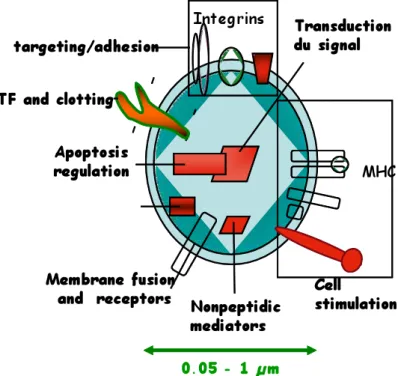
Furthermore, these vesicles express antigens derived from the original cell and carry different membrane, cytoplasmic and nuclear constituents (Fig 6). For example, platelet MPs (PMPs) incorporate plasma membrane glycoproteins (GP), such as GP IIb/IIIa (CD41) and GP Ib/IX complex and the alpha-granulate membrane protein (GMP-140) (Wiedmer et al., 1990). PMPs are also major carrier of platelet-activating factor (PAF) a potent phospholipid involved in the pathogenesis of inflammation (Iwamoto et al., 1997). In addition, they can expose markers of activated cells such as P-selectin (CD62P) (Diamant et al., 2004).
Fig. 6: Cellular MPs: a disseminated storage pool of bioactive effectors.. MPs are shed from the plasma membrane of stimulated cells. They harbor membrane and carry cytoplasmic proteins as well as bioactive lipids implicated in a variety of fundamental processes. This representation does not intend to be exhaustive with respect to the different hijacked components. MHC, major histocompatibility complex. (From Hugel et al., 2005).
Smalley et al. (Smalley et al., 2007) have shown a comparative analysis of MPs isolated from plasma (plasma MPs) versus PMPs in which it was identified 21 proteins that were present in plasma MPs but were essentially absent in PMPs. These proteins include proteins associated with apoptosis (CD5-like antigen, galectin 3 binding protein), iron transport (transferrin, transferrin receptor, haptoglobin), immune response (complement components, immunoglobulin J and kappa chains), and the coagulation process (protein S, coagulation
!
%+!
factor VIII). Only two proteins were present in both types of MPs: von Willebrand Factor and albumin.
Functional and proteomic analysis of PMPs size fractions has shown that PMPs can be separated into different size classes that differ in their contents of plasma membrane receptors and adhesion molecules, chemokines, growth factors and protease inhibitors and functional effects on platelets and endothelial cells (Dean et al., 2009).
A proteomic approach has revealed that the protein content of EMPs consists mainly of metabolic enzymes, proteins involved in adhesion and fusion processes, cytoskeleton-associated proteins and nucleosome (Banfi et al., 2005). There are a limited number of endothelial-specific markers such as CD31, CD146 and E-selectin (CD62E).
MPs released by leukocytes, are normally present in low concentration in blood healthy subjects but their production can be enhanced by inflammatory cell stimulation in vitro or in the presence of various diseases (Sabatier et al., 2002). Leukocyte-derived MPs carried different complex such as CD4, CD3 or CD8 (Martin et al., 2004).
Regarding the overall content of proteins of MPs, another proteomic analysis has identified 390 proteins in MPs among which 34% were localized in the plasma membrane. Half of the detected proteins are intracellular proteins and only a few number of proteins derived from nucleus, mitochondria, Golgi apparatus and endoplasmic reticulum. (Miguet et al., 2006). Recent studies reveal the presence of MPs released by endothelial progenitor cell cultures (EPCs). The analysis of the composition of these MPs obtained by proteomic approach has identified 618 proteins among which there is a large concentration of platelet proteins such as glycoprotein IIb/IIIa (Prokopi et al., 2009).
!
%"!
IV. MPs and disease
The role of MPs has been studied in various diseases states and their number, cellular source and composition are altered compared to healthy controls. It is well established that MPs play an important role in inflammation, coagulation and vascular dysfunction but the mechanisms involved are not yet fully clear.
High levels of circulating MPs have been shown in diseases linked with an increased risk of thromboembolic events such as diabetes, myocardial infarction, preeclampsia, rheumatoid arthritis, hypertension, cancer-associated thrombosis and metabolic syndrome (Sabatier et al. 2002; Mallat et al., 2000; Meziani et al., 2006; Preston et al., 2003; Knijff-Dutmer et al., 2002; Agouni et al., 2008) but the phenotype of circulating MPs may change in different pathological states.
Differences on the phenotype of MPs have been reported by Sabatier et al. (Sabatier et al., 2002) in diabetic patients. Indeed, although there is an elevated level of circulating MPs in type I and type II diabetic patients, the cellular origin and the procoagulant activity of MPs differ according to type of diabetes: in type I diabetic patients, there is an elevated level of EMPs, PMPs and procoagulant activity, instead in type II diabetic patients only the level of annexin-V positive (procoagulant) is increased. The large increase of EMPs in type I diabetic patients suggests that they could be markers in endothelial damage observed in microvascular complications associated to this disease.
Diabetes, particularly type II diabetes, is associated with another disease linked to cardiovascular risk and inflammation, the metabolic syndrome (MS), in which was observed, in the same way, an increase of the rate of circulating MPs. Interestingly, in addition to the increase of MPs from endothelial cells, erythrocytes and platelets in MS patients, there is also
!
%#!
an increased level of procoagulant (annexin V+) MPs that could participate to fibrinolysis impairment and thrombogenesis involved in this disease (Agouni et al., 2008).
An elevated level of MPs with procoagulant potential has been also detected in patients with acute coronary syndrome compared with patients with stable angina and noncoronary heart disease. The cellular origin of these MPs is endothelial and this suggest an important role for endothelial injury in inducing the procoagulant potential and that MPs may contribute to the generation and perpetuation of intracoronary thrombi (Mallat et al., 2000).
The inflammatory process and an enhanced cardiovascular morbidity and mortality are aspects that have also been observed in the rheumatoid arthritis. In the pathology of this disease, has been suggested the involvement of PMPs. Indeed, level of PMPs is increased and is correlated to activity of disease (Knijff-Dutmer et al., 2002).
High concentration of circulating MPs is often associated with high blood pressure. In particular, in patients with several uncontrolled hypertension, the pressure-induced endothelial and platelet activation is linked with a release of EMPs and PMPs (Preston et al., 2003). Therefore, MPs could be markers of organ injury resulting from the combined effects of EMPs and PMPs on coagulation, leukocytes, and endothelium.
Two others pathologies associated with systemic vascular inflammation and risk of thrombosis are preeclampsia and sepsis.
Women affected by preeclampsia showed various disorders characterized by a generalized endothelium dysfunction that lead to hypertension and proteinuria. Van Wijk and co-workers (Vanwijk et al., 2002) have shown an unaltered total number of MPs in preeclamptic patients and normal pregnancy despite en increase level of T-lymphocytes and granulocytes MPs in preeclamptic patients compared to women with normal pregnancy. However, Meziani et al., (Meziani et al., 2006) have found in preeclamptic patients, an increased level of MPs and
!
%$!
phenotypic characterization of their cellular origin showed increased leukocyte-and platelet-derived MPs in the bloodstream of these patients. Furthermore, MPs from preeclamptic women are also involved in vascular alteration and proinflammatory process. Then, preeclamptic MPs could act as vectors of vascular inflammation in this disease.
Sepsis is an acute and systemic immune response mainly to bacterial infection. The production of procoagulant MPs has been shown in patients with meningococcal sepsis. These MPs originating from platelets and granulocytes could contribute to development of disseminated intravascular coagulation that affects these patients (Nieuwland et al., 2000). Conversely, Soriano et al. have demonstrated a positive correlation between high circulating levels of MPs and survival in septic patients (Soriano et al., 2005) and this protective role has been confirmed in a recent study. In this work, Mostefai et al. (Mostefai et al., 2008) have suggest a possible protective role in vascular function of MPs which are present in high amounts in septic patients compared with nonseptic subjects. Particularly, there is an increase of 1.7- and 3-fold of the circulating level of PMPs and EMPs, respectively, in septic patients compared with nonseptic subjects. These MPs are capable to counter the hyporeactivity associated with sepsis, suggesting that, during sepsis, MPs may have a protective role rather than a deleterious role. Indeed, the septic MPs enhanced the contraction of mouse aorta in response to serotonin and this effect was associated with increased thromboxane A2 production, and was sensitive to a selective thromboxane A2 antagonist.
The inflammation is the key pathogenic component of atherosclerosis and is orchestrated by the interaction by inflammatory cells such as smooth muscle. In particular, it has been showed that smooth muscle cells could be able to release tissue factor-bearing MPs in the atherosclerotic plaque that could contribute to thrombus formation during atherosclerotic plaque disruption (Schecter et al., 2000).
!
%%!
Finally, recent data show a possible involvement of MPs on endothelial dysfunction in patients with obstructive sleep apnea (OSA), a disease characterized by recurrent episodes of partial or complete obstruction of the upper airways during sleep, leading to repeated falls in oxygen saturation. Indeed, MPs seem to participate to endothelial dysfunction involved in vascular complications of OSA. Although the level of circulating MPs is unchanged between the two groups considered (desaturators and nondesaturators patients), there is higher levels of granulocytes and activated leukocyte (CD62L+)-derived MPs in desaturators compared to nondesaturators. In addition, MPs from desaturator patients increased expression of endothelial adhesion molecules including E-selectin, “intercellular adhesion molecule-1” (ICAM-1) and integrin-!5, and COX-2 (Priou et al., 2010).
All these data suggest that, in several pathological states, MPs could be used as biomarkers to identify a disease or to highlight the disease-related complications.
!
%&!
V. Microparticles and inflammation
Several reports suggest the involvement of MPs in the inflammatory process. Indeed, MPs can induce the production of potent pro-inflammatory mediators by cells and the up-regulation of several pro-inflammatory enzymes. Furthermore, the production of platelet, endothelial and leukocyte MPs can be increased during inflammatory conditions (Daniel et al., 2006).
MPs are rich in aminophospholipids and are a preferential substrate for phospholipase A2 that is involved in production of lysophosphatidic acid which can triggers platelet aggregation and inflammatory process (Fourcade et al., 1995). The first study about the effect of the PMPs on endothelial cells has shown that they facilitate the transcellular transport of arachidonic acid (AA) which could lead to an increase expression of COX-2 and ICAM-1. Both COX-2 and ICAM-1 regulate the vascular and platelet functional interaction. Thus, the MP-induced modulation of COX-2 expression in human monocytoid cell line induces the translocation of protein kinase C from the cytosol to the membrane and triggers activation of different kinase (Barry et al., 1999; Barry et al., 1997). Furthermore, the unmetabolized AA present in PMPs is involved in platelet aggregation and interactions of platelets and monocytes with endothelial cells (Barry et al., 1998). This suggests a mechanism whereby MPs modulate cell function by the transcellular delivery of bioactive substances.
Also, it has been shown that MPs from platelet and leukocyte origins participate in the production of several endothelial (IL-1", IL-6, IL-8 and monocyte chemoattractant protein-1 (MCP-1)) and monocytes (IL-1", TNF-!, IL-8) cytokines that could facilitate the interaction between leukocytes and endothelium (Mesri and Altieri, 1999). In this respect, Mesri and Altieri showed that after incubation of human umbilical vein endothelial cells (HUVECs) with leukocyte-derived MPs, there is an increased production of IL-6, IL-8 and membrane adhesion molecules implicated in interaction between endothelial cells and leukocytes. In
!
%'!
addition, an up-regulation of IL-6 mRNA was observed suggesting the ability of MPs from leukocytes-to modify gene expression (Mesri and Altieri, 1998).
In turn, cytokines also can be involved in the production of MPs. For example, in patients with arteriosclerosis, it has been shown that increased levels of IL-6 are associated with enhanced expression of P-selectin and generation of PMPs in condition of high shear stress (Nomura et al., 2000). MPs from activated platelets can also mediate in vitro leukocyte-leukocyte interactions via binding of P-selectin to its ligand PSGL-1 on leukocyte-leukocytes. These attachments can then lead to increased accumulation of leukocytes on a P-selectin surface, for example, activated endothelium at sites of vascular injury (Forlow et al., 2000).
Furthermore, MPs derived from platelets could be able to facilitate the recruitment of various immune cells such as monocytes, T and B lymphocytes and NK cells that play a major role in inflammatory process (Ogura et al., 2001a).
A possible role of MPs as pro-inflammatory vectors in disease, has been suggest in preclamptic patients, since, circulating MPs are able to induce an up-regulation of iNOS and COX-2 and activate the NF-%B pathway, suggesting that MPs could be a pro-inflammatory vectors (Meziani et al., 2006).
All these data suggest that MPs may therefore contribute to the increased risk of thrombosis in systemic inflammatory diseases where increased numbers of MPs have been identified, or in localised inflammatory environments, such as atherosclerotic lesions where activated monocytes, endothelial cells and platelets are co-localised.
On the contrary, other authors suggested potentially anti-inflammatory role of MPs. Indeed, it has been reported that MPs released by neutrophils do not possess proinflammatory activity on human macrophages since they increase the release of transforming growth factor beta1 (TGF "1) and then down-modulate cellular activation in macrophages (Gasser et al., 2004).
!
%(!
VI. Microparticles and vascular function
The endothelium is a primary target of cardiovascular risk factors and endothelial dysfunction is the primary event leading to the failure of vasoactive, anticoagulant, and anti-inflammatory effects of healthy endothelium.
The endothelial response can be immediate following the release of several factors, or delayed involving the modulation of genes expression responsible of structural and functional regulation of the vascular wall. In this context, MP-associated effects may represent an adaptive phenomenon or contribute to the aggravation of disease. For this reason, several groups have studied the effects of MPs on vascular function and it was established that MPs are able to affect endothelial (Fig 7) and smooth muscle cells.
!
%)! Fig 7. Microparticle (MP) effects on endothelial cells. Depending on the cell origin and the stimulation used
for their generation, MPs have different effects on endothelial cell function. MPs can have beneficial or deleterious effects by acting on the nitric oxide pathway, pro-inflammatory enzymes, reactive oxidative species production, and angiogenesis. COX-2, inducible cyclo-oxygenase; NO, nitric oxide; ROS, reactive oxygen species; TF, tissue factor (From Benameur et al., 2009).
Concerning the role of MPs in the regulation of vascular function, it has been observed in several studies, that circulating MPs are able to reduce NO production via the reduced activity of endothelial NO-synthase (eNOS) and/or by decreasing its bioavailability. For example, MPs isolated from patients with myocardial infarction, diabetes or preeclampsia induce endothelial dysfunction by impairing endothelial NO transduction pathway (Boulanger et al., 2001; Martin et al., 2004; Vanwijk et al., 2002).
A potentially deleterious effect of MPs on the cardiovascular system and, notably, on the endothelial cells were observed in other disease such as MS, OSA and end-stage renal failure. Indeed, MPs from patents with MS are able to induce in vitro, the reduction of NO and
!
%*!
superoxide anion productions in endothelial cells, resulting in protein tyrosine nitration; in vivo, the same MPs impair the endothelium-dependent relaxation by decreasing eNOS activity (Agouni et al., 2008).
A direct correlation between a specific phenotype of MPs and endothelial dysfunction has been shown in patients with OSA. Indeed, in these patients, there is an increased level of MPs from granulocytes and activated leukocytes (CD62L+) and levels of CD62L+ MPs correlate with endothelial dysfunction that may initiate atherogenic processes in patients with OSA (Priou et al., 2010).
In patients with end-stage renal failure, an increased level of MPs is associated in vitro with impaired endothelium-dependent relaxations and cyclic guanosine monophosphate generation. In addition, endothelial dysfunction correlated with endothelial-derived MPs that induced a decrease of endothelial nitric oxide release (Amabile et al., 2005).
Furthermore, using a model of rat pulmonary arterial hypertension has been showed that circulating MPs from hypoxic rats reduce NO bioavailability by decreasing eNOS activity and by enhancing oxidative stress in pulmonary endothelial cells (Tual-Chalot et al., 2010)
.
MPs issued by smooth muscle cells may also have effects on vascular system. Indeed, MPs from apoptotic smooth muscle cells cause endothelial dysfunction via diminished NO production. In addition, pre-treatment of endothelial cells with two blockers of !3-integrins, abciximab or eptifibatide, restored NO production, suggesting that !3-integrins are implicated in the effects of smooth muscle MPs (Essayagh et al., 2005).
On the other hand, MPs can interact with smooth muscle cells, induce vascular inflammation and modify the vessel contractility. Indeed, in preeclamptic patients, MPs cause ex vivo vascular hyporeactivity in human omental arteries and mouse aortas by increasing NO