i Université du Québec
Institut National de la Recherche Scientifique Institut Armand Frappier
INVESTIGATING THE STRUCTURE-FUNCTION-FLEXIBILITY
RELATIONSHIP OF CHITIN- AND XYLAN-DEGRADING ENZYMES
Par
Nhung Nguyen-Thi
Thèse présentée pour l‘obtention du grade de Philosophiae doctor (Ph.D.) en biologie
Jury d’évaluation
Président du jury et David Chatenet
examinateur interne INRS-Institut Armand-Frappier
Examinateur externe Denis Groleau
Université de Sherbrooke
Examinateur externe Steve Bourgault
Université du Québec à Montréal
Directeur de recherche Nicolas Doucet
INRS-Institut Armand-Frappier
iii
ACKNOWLEDGEMENTS
Completing my PhD has been a long journey that has required a lot of energy and effort. I could not have succeeded without the tremendous help and guidance from my professor, Nicolas Doucet. He has inspired me during the course of the research and has given me a lot of knowledge, particularly of new techniques like NMR - an interesting approach in protein science. I would like to deeply thank you for your great direction and continuous encouragement during more than 4 years of my study here. You have been a great mentor to me. I will remember all the enjoyable moments when working with you, during which I always received priceless advice and knowledge with good-humor. I thank you again for being your student to get the scholarships and chances to participate in national and international conferences.
I would also like to express my gratitude to my previous director, Professor Claude Dupont, who gave me this wonderful chance to study at INRS. You are the first person who trained me in techniques in protein science, which were completely new to me when I started my study at INRS. I wish you good health and all the best of luck during your retirement. I am also very thankful to my professor at VAST (Vietnam Academy of Science and Technology), Tong Kim Thuan, who recommended the program to me and encouraged me throughout my study at INRS. I would also like to thank my committee members, Dr. David Chatenet, Dr. Denis Groleau, and Dr. Steve Bourgault for serving as my committee members. Your corrections, comments and suggestions are of remarkable help for completing my thesis.
I would like to thank my lab-mate, Donald Gagne, for his help, especially in the NMR experiments and analyses. You gave me a lot of useful advice and invaluable constructive criticism during my research. You always took responsibility in the lab and helped me with a lot of enthusiasm and kindness. I will remember all the friendships we have developed during the time I spent at INRS. I would also like to express my gratitude to Anastasia Nikolakakis for her help in correcting my writing. I would especially like to thank Roger Dubuc who supported me a lot. I will remember forever the cheerful and warm times when working with you. I will never forget Raymonde Jette for her help and encouragement during the beginning of my studies at INRS. I would also like to thank Julie Payet for her kindness in helping me with biomolecular techniques. And many thanks to all my lab-mates who worked and shared
iv with me every day in the lab in a friendly atmosphere. I also want to thank Sameer Al-Abdul-Wahid and Tara Sprules from the Québec/Eastern Canada High Field NMR Facility (McGill) and Michael Osborne from IRIC-Institut de Recherche en Immunovirologie et Cancérologie (Université de Montréal) for their excellent assistance in the NMR experiments. Without you guys, I would not have the amazing data for my thesis.
I would like to take this opportunity to thank Mrs. Vu Thi Bich, Professor Sinh Le Quoc, Professor Alain Fournier and others from VAST and INRS. I am sincerely grateful to them for building and operating the Co-operation Program in Doctoral Training between VAST and INRS, which gave me the excellent chance and wonderful time to study at INRS. This was an invaluable help for my career. I would also like to thank the leaders of Institute of New Technology, Institute of Military Science and Technology, Group 871 for facilitating my study process and to sincerely thank the Vietnamese Ministry of Education and Training and INRS for the financial assistance.
I also want to give my thanks to my friends – especially Mai, Nhung, My, and Oanh – who have been always beside me, helped me, and shared with me thousands of happy, glad, funny, sad, and stressful moments not only in life but also in science during my time in Canada. Love you all.
Finally, special thanks and love to my beloved husband and little girls for your support, sacrifice and care for me from the other side of the earth. Words cannot express how grateful I am to you. I also want to thank my parents who always care, encourage and worry for me in all my life. I am also very grateful to my parents-in-law who supported me from abroad during my studies and took care of my little girl with love and protection during my absence. I would like to send my gratitude to my mother-in-law who is now blessing my family and me from the sky. I would also like to underline the support and help from my brother-in-law, sisters-in-law, my brothers, and to most deeply thank Ms Loan for taking care of my daughter during my absence.
v
RÉSUMÉ
La dégradation enzymatique de la biomasse est un procédé biotechnologique de pointe visant la production de petites molécules hautement pertinente en synthèse organique. Ce processus a été largement étudié en raison de ses avantages en chimie verte, en plus de son application potentielle dans de nombreux domaines de la vie. Malgré l‘accumulation des connaissances au niveau des principes qui régissent la dégradation enzymatique de la biomasse, les objectifs fondamentaux et industriels de cette technologie demeurent encore sommaires. La découverte d‘enzymes efficaces et adaptées aux domaines biotechnologiques demeure un défi majeur en raison des problèmes de pureté des produits synthétisés, des faibles rendements de production enzymatique et du coût élevé des processus industriels provenant de la complexité de la biomasse de départ. Conséquemment, nous visons principalement à découvrir et à caractériser des glycoside hydrolases hautement actives et adaptées à de nombreuses applications industrielles.
Une β-N-acétylhexosaminidase (ScHEX) et une chitinase (ChiC) de Streptomyces coelicolor A3(2) possédant des activités élevées envers les chitooligomères et les chitines cristallines ont été exprimées et caractérisées avec succès. En collaboration avec un groupe de l'Université York (York, Royaume-Uni), les structures cristallines de ScHEX native et du mutant ScHEX-E314Q ont été résolues afin de comprendre le mécanisme catalytique d'hydrolyse des chitooligosaccharides. En utilisant un mélange de divers surnageants composés de ScHEX et ChiC dans un essai en présence de chitine cristalline comme substrat, le produit final GlcNAc a été obtenu avec une pureté de 95% après 8 heures d'incubation, promettant un moyen efficace pour la production industrielle de GlcNAc pur.
La flexibilité atomique chez les enzymes se révèle d'une importance cruciale dans la conversion enzymatique de polysaccharides. Il a été démontré que les mouvements moléculaires de protéines à différentes échelles de temps peuvent être impliqués dans la régulation de l'activité enzymatique, en jouant notamment un rôle essentiel dans le contrôle de la relation entre la structure et la fonction chez plusieurs systèmes enzymatiques. Pour étudier la dynamique interne et dans le but de déterminer si cette dynamique doit être considérée dans le génie enzymatique des glycoside hydrolases, des expériences de RMN ont été réalisées
vi avec XlnB2 et XlnB2-E87A de Streptomyces lividans 66 en l'absence et en présence de ligands (xylobiose et xylopentaose). Les résultats de cristallographie révèlent que XlnB2 possède une structure conservée appelée "β-jelly-roll", ressemblant à l'architecture de la main droite. Nos données RMN montrent que, sous forme libre, les mouvements à l'échelle de temps de la catalyse enzymatique sont principalement regroupés sur la face de la cavité catalytique. Lors de la liaison aux ligands, l‘échange conformationnel émerge sur les deux faces de la cavité catalytique et dans le motif de la boucle "thumb loop", ce qui suggère un mouvement global de fermeture. D'autres résultats indiquent que l'implication des résidus de la boucle et de la cavité catalytique demeurent similaires suite à la liaison avec de courtes et longues chaînes de glucides. Ces résultats fournissent une preuve expérimentale directe validant le mécanisme d‘ouverture et de fermeture précédemment prédit par des structures cristallines et des simulations de dynamique moléculaire effectuées sur des xylanases homologues. En bref, cette étude permet de mieux comprendre la dynamique à l'échelle atomique de XlnB2, démontrant une relation directe entre la fonction et la dynamique de l'enzyme.
Les résultats recueillis dans cette thèse permettent de mieux comprendre comment les glycoside hydrolases dégradent la biomasse. D'autres études seront nécessaires pour maximiser leur activité enzymatique et leur efficacité catalytique, ainsi que pour obtenir des informations supplémentaires visant l‘amélioration par ingénierie enzymatique de biocatalyseurs efficaces dans de multiples applications industrielles.
vii
SUMMARY
The enzymatic degradation of biomass is an advanced biotechnological method for the production of small molecules of organic synthesis relevance. This process has been extensively studied because of its benefits in green chemistry and its potential application in many fields of life. Although much knowledge about the principles that govern the enzymatic breakdown of biomass has been gathered, fundamental and industrial goals remain unachieved. Finding efficient enzymes adapted to applied biotechnology disciplines remains a challenge due to product purity issues, yields of enzymatic production, and high costs of degrading processes due to biomass recalcitrance. As a result, we are interested in finding and characterizing highly active glycoside hydrolases aimed at numerous industrial applications.
A β-N-acetylhexosaminidase (ScHEX) and a chitinase (ChiC) from Streptomyces coelicolor A3(2) with high activity toward chitooligomers and crystalline chitins were successfully expressed and characterized. In collaboration with a group at the University of York (York, UK), crystal structures of the native ScHEX and of the mutant ScHEX-E314Q have been resolved in order to understand the catalytic mechanism of chitooligosaccharide hydrolysis. Using a mixture of supernatants from ScHEX and ChiC in an assay with crystalline chitin as substrate, GlcNAc was produced as a final product with yield of 90% after 8h of incubation, promising efficient applicability for the industrial production of pure GlcNAc.
Enzyme flexibility was found to play an important role in enzymatic conversion of polysaccharides. It has been shown that internal protein motions on various timescales can be involved in regulating enzyme activity, playing a critical role in controlling the relationship between structure and function in several enzyme systems. To study internal dynamics and to investigate whether they should be considered in glycoside hydrolase engineering, NMR experiments were carried out with XlnB2 and XlnB2-E87A from Streptomyces lividans 66 in the absence and presence of ligands (xylobiose and xylopentaose). Crystallographic results reveal that XlnB2 conserves a β-jelly-roll scaffold, with a right-hand architecture. Our NMR data show that in the free XlnB2 form, catalytic time-scale motions primarily cluster on one face of the catalytic cleft. Upon ligand binding, conformational exchange emerges on both faces of the catalytic cleft and in the thumb-loop motif, suggesting a global clamping
viii movement. Other results indicate very similar involvement of residues located on the thumb loop and active-site cleft upon binding with short- and long-chain carbohydrates. These results provide direct experimental evidence to validate the previously postulated open-closed mechanism predicted by crystal structures and molecular dynamics simulations performed on xylanase homologues. In short, this study provides further insight into the atomic-scale dynamics of XlnB2, indicating a relationship between function and the dynamics of the enzyme.
The results collected in this thesis provide a better understanding of the glycoside hydrolase enzymes in biomass degradation. Further studies are required to optimize their enzymatic activity and catalytic efficiency, as well as to gain additional information for the proper engineering of improved biocatalysts used in several industrial applications.
ix
TABLE OF CONTENTS
ACKNOWLEDGEMENTS.………..……….………iii
RÉSUMÉ……….……..……….………….…..…………v
SUMMARY.………..………...……..….……….vii
TABLE OF CONTENTS.………..………..ix
LIST OF TABLES.………..………xii
LIST OF FIGURES.………..……….xiii
ABBREVIATIONS..……….……….…..xv
CHAPTER 1. INTRODUCTION ... 1
1.1 Polysaccharides and their degradation ... 7
1.1.1 Biomass, polysaccharides ...7
1.1.2 Chitin, degradation of chitin, and GlcNAc production ...7
1.1.2.1 Chitin, chitooligosaccharides ...7
1.1.2.2 Chitin degradation ...9
1.1.2.3 GlcNAc production ...11
1.1.3 Xylan and degradation of xylan ...14
1.2 Enzymes in enzymatic degradation of polysaccharides ... 18
1.2.1 Carbohydrate-active enzymes ...18
1.2.2 Glycoside hydrolases ...19
1.2.2.1 General properties.………...………19
1.2.2.2 Structure of glycoside hydrolases………..……….…….……20
1.2.2.3 Mode of action.……...……….………..……...21
1.3 Chitinolytic enzymes ... 25
1.3.1 Chitinases GH18 ...27
1.3.1.1 Biochemical properties.……….……….28
1.3.1.2 Substrate specificity.………..……….28
1.3.1.3 Structure and catalytic mechanism.…...……….…….…………29
x
1.3.2.1 Biochemical properties……….30
1.3.2.2 Structure and catalytic mechanism…….……….………31
1.3.2.3 Substrate specificity………..…...……….32
1.3.3 Chitinolytic machinery of Streptomyces coelicolor A3(2)………...34
1.4 Xylanases ... 35
1.4.1 Xylanases ... 35
1.4.2 Xylanases GH11... 35
1.4.2.1 Biochemical properties…….……….……….35
1.4.2.2 Substrate specificity ...37
1.4.2.3 Structure and catalytic mechanism ...37
1.4.2.4 Dynamic property ...39
1.4.2.5 Applications of GH11s ...41
1.4.3 Xylanase B from Streptomyces lividans ...43
1.5 Streptomyces ... 44
1.6 Structure-function-flexibility relationship ... 45
1.7 NMR approach and dynamics at the atomic level ... 48
1.8 Objectives ... 49
CHAPTER 2. CHITINOLYTIC POTENTIAL OF β-N-ACETYLHEXOSAMINIDASE AND CHITINASE C FROM STREPTOMYCES COELICOLOR A3(2) IN N-ACETYLGLUCOSAMINE PRODUCTION ... 51
2.1 Context of chapter2... 52
2.2 Presentation of article 1 – “Structure and activity of the Streptomyces coelicolor A3(2) β-N-acetylhexosaminidase provides further insight into GH20 family catalysis and inhibition……...………...………...53
2.2.1 Contribution of authors ... 53
2.2.2 Résumé ... 54
2.2.3 Article 1 ... 54
2.3 Presentation of article 2 – "Characterization of chitinase C from Streptomyces coelicolor A3(2) and itS application in N-acetylglucosamine production"…………..………70
2.3.1 Contribution of authors ... 70
xi
2.3.3 Article 2 ... 71
2.4 Discussion... 108
2.4.1 Synergistic interactions and importance of the substrate-binding domain...110
2.4.2 Enzyme flexibility plays an important role for efficiently degrading the recalcitrant polymer……..………..……….……..112
CHAPTER 3. THE STRUCTURE-FUNCTION-FLEXIBILITY RELATIONSHIP OF XYLANASE B………...……….………..…….116
3.1 Context of chapter 3 ... 117
3.2 Presentation of article 3 – “Conformational exchange experienced by free and ligand-bound xylanase B2 from Streptomyces lividans 66”....…..………..…….……..………120
3.2.1 Contribution of authors ... 120
3.2.2 Résumé ... 120
3.2.3 Article 3 ... 121
3.3 Discussion... 150
CHAPTER 4. CONCLUSIONS AND PERSPECTIVES ... 154
4.1 Conclusions ... 155
4.2 Perspectives ... 155
xii
LIST OF TABLES
Table 1.1. Biochemical properties of several GH20 enzymes……….….……30 Table 1.2. Biochemical properties of several GH11 representatives.…………..……….……36
xiii
LIST OF FIGURES
Chapter 1Figure 1.1. Repetitive unit of chitin………...……….8
Figure 1.2. The chemical formula of a chitooligosaccharide...….…….….……..………...…..9
Figure 1.3. Enzymatic degradation of chitin ………...……….10
Figure 1.4. Production of GlcNAc using chitin as substrate……….………13
Figure 1.5. Structure of xylan from different sources………..….………16
Figure 1.6. Enzymatic breakdown of xylan………..………16
Figure 1.7. Carbohydrate-active enzymes……….19
Figure 1.8. Active site shapes in GH enzymes....………..….……….…………...….21
Figure 1.9. Endo-, exo-acting cleavage mechanism type.……….………21
Figure 1.10. Retaining, inverting mechanism types with GH enzymes…………..….…...…..22
Figure 1.11. Inverting mechanism with GH enzymes…….……….……...………...22
Figure 1.12. Retaining mechanism with GH enzymes (for a β-glycosidase)....….………..…23
Figure 1.13. Substrate-assissted mechanism utilized by GH enzymes...24
Figure 1.14. Subsite nomenclature in GH enzymes………..24
Figure 1.15. The processive mechanism of GH enzymes……….25
Figure 1.16. A typical (α/β)8 protein fold in GH18 enzymes.……….……..…..26
Figure 1.17. Ribbon diagram of SpHex in complex with NAG-thiazoline (NGT)..…………..31
Figure 1.18. Xylohexaose modeled into six subsites in the active cleft of Tx-Xyl…………..38
Figure 1.19. Residues paving the catalytic cleft of GH11 enzymes……….…...….…………39
Figure 1.20. General mechanism for ligand binding coupled to conformational change…...47
Figure 1.21. Solution NMR techniques cover the complete range of dynamic events in enzymes……….49
Chapter 2 Figure 2.1. Proposed mechanism of synergistic activities of ChiC and ScHEX in chitin degradation...……...110
Figure 2.2. Conformational flexibility in ScHEX……..….…...……..………..………..…...113
Chapter 3 Figure 3.1. Conserved active-site residues mapped on crystal structure of XlnB2...151
xiv
ABBREVIATIONS
Å Ångström (10-10 meter) ANM Anisotrophic network model 13
C Carbon-13
CAZY Carbohydrate-active enzymes CBM Carbohydrate-binding module CBP Chitin-binding protein
CBP21 Chitin-binding protein family 21 CDH Cellobiose dehydrogenase ChtB Chitin-binding domain
CPMG NMR relaxation-compensated Carr-Purcell-Meiboom-Gill DP Degree of polymerization
δ Chemical shift
Δδ Chemical shift variation ε Extinction coefficient EC Enzyme Commission g gravity
GH Glycoside hydrolase
GH3 Glycoside hydrolase family 3 GH11 Glycoside hydrolase family 11 GH20 Glycoside hydrolase family 20 GH84 Glycoside hydrolase family 84 GalNAc N-Acetylgalactosamine
GlcNAc N-Acetyl-β-glucosamine HEX β-N-Acetylhexosaminidases
HSQC Heteronuclear Single Quantum Coherence HPLC High Pressure Liquid Chromatography ITC Isothermal titration calorimetry kcat Catalytic constant
KM Michaelis–Menten Constant kb Kilobases
Kd Dissociation constant kDa Kilo Dalton
xv
MD Molecular dynamics
MeGA 4-O-methyl-α-D-glucuronic acid MW Molecular weight
MWCO Molecular weight cut off
m/z Mass/charge (mass-to-charge ratio) mM Millimolar μs Microsecond (10-6 s) ms Millisecond (10-3 s) 15 N Nitrogen-15 nm Nanometer
NOE Nuclear Overhauser Effect ns Nanosecond (10-9 s)
NA Not available
NMR Nuclear Magnetic Resonance PCR Polymerase chain reaction PDB Protein Data Bank
PM Peritrophic membranes pNP- para-nitrophenyl- ps Picosecond (10-12 s)
R1 Longitudinal relaxation rate R2 Transverse relaxation rate
Rex Conformational exchange parameter (motions μs-ms) RMSD Root Mean Square Deviation
rpm Revolutions per minute
SDS-PAGE Sodium dodecyl sulphate polyacrylamide gel electrophoresis SpHex β-N-Acetylhexosaminidase from Streptomyces plicatus Temp. Temperature
UV Ultraviolet v/v Volume/volume
Vmax Maximum rate of conversion of an enzyme-catalyzed reaction
Xln Xylanase
1
CHAPTER 1. INTRODUCTION
2
Introduction
Polysaccharides, such as chitin and xylan—two of the most abundant biomass polymers of the biosphere—serve as major carbon and energy sources for many bacterial species. In general, growth on these carbohydrate polymers is associated with the secretion of an enzymatic system that breaks down the polymer chains to release monomers, which are in turn easily utilized as carbon and energy sources. It is hoped that biomass can be used as an inexhaustible and renewable energy source via enzymatic hydrolysis in fuel production, in the food industry, and in medical treatments. As a result, these enzymes have become promising biocatalysts for the industrial breakdown of biomass compounds.
Nowadays, using biocatalysts in organic synthesis or for biomass degradation is considered as a strategy for green chemistry — a global target in order to prevent global warming. Benefits of green chemistry could be obtained from the use of biomass and biocatalysts in manufacturing processes and of renewable carbon in final products (1). Indeed, billions of tons of biomass derived from many sources, such as fungi, insects or plants, are accumulated annually in nature. The processes making use of the biomass reduce the cost of production processes and decrease biomass accumulation. The enzymatic processes using biocatalysts are considered as an approach for sustainable development owing to their potential in decreasing waste emissions, pollution, or negative effects on the environment and human health. The products of biomass degradation that can be used as renewable energy sources are incorporated into the nitrogen and carbon cycle. For instance, xylose produced from xylan degradation can be applied to biofuel production, whose products can be used in transportation – a process that generates CO2 which is incorporated into the carbon cycle on earth. Therefore, the processes using biomass and biocatalysts are advantageous in green chemistry and continue to grow in order to be used on a large scale in the future. In particular, enzymatic degradation of chitin and xylan using biocatalysts meets one of the principles of green chemistry.
Chitinolytic enzymes take part in the degradation of chitin, whose abundance renders it an important polymer that should be made use of in the future. In this degradation, the complete conversion of chitin involves the synergistic action of two hydrolyzing enzymes—
3 β-N-acetylhexosaminidase (HEX) and chitinase—generating the end product N-acetyl-D-glucosamine (GlcNAc), a monosaccharide with great value in medical and cosmetic applications (2). Other derivatives of the degradation, which are chitosan and chitooligosaccharides, can be used in many applications such as antimicrobial, antitumor, anti-cholesterol activities, wastewater treatment, drug delivery, or wound healing (3). Due to the widespread and valuable applications of products of the process, attempts have been made to find efficient enzymes as well as to modify the enzymes in order to adapt to such applications. However, to this day, these objectives remain a challenge due to product purity issues, yields of enzymatic production and high costs of degradation processes due to biomass recalcitrance. Due to the above restraints, the processes require many conversion and separation steps, causing higher costs and energy demands and waste emissions. Consequently, improving the efficiency of known enzymes, finding new and more active enzymes, and creating optimized enzyme mixtures can be solutions in order to break the barrier for large-scale production of the target products (4).
With regards to GlcNAc production, despite the strong demand due to its potential use as a non-toxic supplement to protect the bone surfaces of the friction joint (5, 6), to treat inflammatory bowel disease (including Crohn's disease) (7, 8) and to use in dermatological and cosmetic applications (2), large-scale GlcNAc production faces many problems. Typically, GlcNAc is produced by hydrolysis of chitin using hydrochloric acid (15-36% HCl, 40-80oC) (2), which is extremely energy demanding, costly and polluting—an issue that is being avoided in green biochemistry nowadays. The products somehow cannot be considered as a safe supplement due to chemical processes or modifications. In fact, the chemical methods carried out under these extreme conditions have many drawbacks including instability of products. Enzymatic hydrolysis does not show these negative effects on the sugar product. Therefore, enzymatic processes for GlcNAc production using more efficient and environmentally friendly methods are being developed. Indeed, the enzymatic production of GlcNAc by endochitinases (EC 3.2.1.14), exochitinases (EC 3.2.1.52) and N-acetylhexosaminidases (HEX) (EC 3.2.1.96) yielded about 80 g/L/h of GlcNAc with no environmental release (9). However, the final product is contaminated with chitobiose [(GlcNAc)2] and other derivatives whose removal is necessary to obtain a pure product for commercial demand. Other enzymatic process for GlcNAc productions are limited in
4 productivity and time-cost consideration. Hence, finding efficient enzymes that can overcome these issues and which have appropriate properties for large-scale or industrial-scale production is crucial.
Other enzymes seen as promising biocatalysts for the industrial breakdown of biomass are xylanases. They are glycoside hydrolases that cleave internal β-1,4-xylosidic bonds in heteroxylan, a major hemicellulose fraction of plant cell walls. These glycosidases are of widespread importance in biofuel, food and non-food biotechnological applications, bioconversion of hemicellulosic biomass to fermentable sugars and platform molecules. The enzymes are particularly important in bioethanol production. In fact, bioethanol is produced from starch sources or other food plants (the first generation of biofuels) that are considered unsustainable since it affects food security. The production of the first generation of bioethanol is also limited because of inconsistent supply sources due to the dependence on weather and agriculture (10). Therefore, the second generation of bioethanol produced from abundant xylan and cellulose is one of the most popular biofuel technologies nowadays. Moreover, other breakdown products of xylan degradation have been used in many diverse fields, such as pulp and paper production and the textile industry. Because of high industrial demands for environmentally friendly and cost-effective biomass technologies, the principles that govern the enzymatic breakdown of xylan are of particular interest. Because of the highly complex nature of heteroxylans as well as the diversity in biochemical properties of xylanases, efficient use of the enzymes requires a molecular insight into the structure-function relationship of their catalytic mechanisms to improve their efficiency. Moreover, proteins are naturally dynamic and are thought to depend on flexibility to perform their function. Therefore, understanding the mechanism that regulates enzyme activity and the relationship between structure, function and motion is a serious option for engineering improved biocatalysts.
Xylanases form a group of highly homologous biocatalysts displaying a conserved β-jelly-roll structure. A number of three-dimensional structures demonstrated that xylanases adopt different mechanisms and architectures in the substrate-binding cleft, whereby xylanases bind and hydrolyze structurally different heteroxylans and xylo-oligosaccharides. Evidence from kinetics, mutagenesis, crystal structures and molecular dynamic simulations in
5 the presence and absence of ligands have proposed the existence of a ―thumb-loop‖ motion and an ―open-closed‖ movement of the active site which may play a major role in substrate binding, catalysis, inhibition, and stability of the proteins (11). The hypothesis was obtained from inappropriate methods such as crystallography or molecular dynamic simulations – methods which give a rigid image of protein motion or information on a timescale that is not relevant to enzymatic catalysis, respectively. The hypothesis is neither in agreement with other results suggesting the immobility of the active site or the thumb motif. As a result, further information about protein dynamics in xylanases is required for understanding the structure-function-motion relationship in biomass degradation.
In enzymatic degradation of biomass, the ability of enzymes to disrupt crystalline polysaccharides contributes to enzyme efficiency in the process. However, in xylan degradation, the study of this process faces difficulties due to the variety and recalcitrance in the structure of xylan, as well as difficulties in detecting and determining substrate and product molecules in the reactions. Insights on how enzymes access and disrupt structural polysaccharides were obtained from studies on enzymatic conversion of chitin (12). The lesson learned from chitin conversion could be useful for bioethanol production including the importance of accessible accessory proteins and the processive mechanism, which could improve enzyme efficiency and substrate accessibility, respectively. Some hallmarks for the processivity have been identified including protein flexibility (13). These findings suggest new approaches for the design and development of enzyme technology in degradation of recalcitrant biomass. This thesis includes studies on chitinases and xylanases, and could bring more information to the field.
In the biomass structure, both chitin and xylan contain β-1,4-glycosidic bonds, which link GlcNAc and xylose residues, respectively. The chitinases and xylanases that catalyze the cleavage of the linkages belong to different glycoside hydrolase (GH) families. Because of the same ability to cleave the β-1,4-glycosidic bonds of the polymers with long saccharide chains, all of the GH families utilize a general acid/base catalytic mechanism and may share a similar active-site architecture with multiple subsites to fit polysaccharide chains. Therefore, the commonality in the structural, functional and motional properties of these enzymes may provide more understanding of the polymer cleavage mechanism.
6 In general, the principles that govern the enzymatic breakdown of these polymers remain elusive, while great fundamental and industrial goals could be achieved from their understanding. As a result, we are interested in characterizing highly active glycoside hydrolases aimed at numerous industrial applications. Streptomyces are known to degrade cellulose, chitin and xylan polymers. To develop highly efficient biocatalysts and to get a better insight into their structure-function-flexibility relationship, we aim at characterizing β-N-acetylhexosaminidase (ScHEX), and chitinase (ChiC) from Streptomyces coelicolor A3(2) and at studying enzyme flexibility in xylanase B2 (XlnB2) from Streptomyces lividans 66. In addition to providing a better understanding of the glycoside hydrolase enzymes, we expect this project to offer additional information for engineering improved biocatalysts.
7
1.1
Polysaccharides and their degradation
1.1.1 Biomass, polysaccharides
Biomass is a biological material derived from living organisms, mostly plants, and some from other kingdoms such as fungi, yeast, insects or animals (Biomass Energy Center.: Biomassenergycentre.org.uk). Apart from providing food and feed, biomass plays a crucial role for energy supplies that are utilized for cooking, heating, power and transportation. In fact, biomass is the most attractive and the largest source of renewable energy, which accounts for 14% of the total energy demand on the world (14). Every year, more than 130 billion tons of biomass are produced in nature (15), making biomass an inexhaustible source of energy. Moreover, biomass was employed throughout recorded history to extract valuable products such as medicinal drugs, supplements, flavors and fragrances (2, 14).
Biomass is carbon-based and mainly composed of carbohydrate polymers (cellulose, hemicellulose, chitin), and aromatic polymers (lignin). Carbohydrates, with the general formula Cm(H2O)n and known as saccharides, are involved in a variety of biological processes. Monosaccharides are the simplest forms of carbohydrates. They consist of one carbohydrate molecule (e.g., glucose). They serve as the main source of energy for metabolism and are used in biosynthesis or bioconversion. Polysaccharides usually contain more than 20 monosaccharides linked together. They also serve as the main source and storage form of energy or exist as structural components. Polysaccharides vary in the type of monosaccharide unit, type of linkage, degree of branching and degree of polymerization. Some polysaccharides, such as cellulose, hemicellulose, and chitin, are crystalline networks of polymer chains held together by strong hydrogen bonds, thus, forming insoluble and highly rigid structures (2, 14).
1.1.2 Chitin, degradation of chitin, and GlcNAc production
1.1.2.1. Chitin, chitooligosaccharides
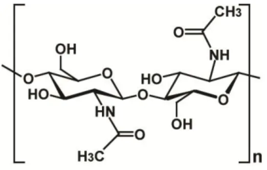
Chitin is a long-chain biopolymer of N-acetylglucosamine (GlcNAc) units linked together by β-(1, 4) bonds. This linear biopolymer is of variable length and the GlcNAc units
8 are rotated 180° relative to each other, making this functional and structural unit a disaccharide (Figure 1.1) (16). It is one of the most abundant natural polymers in the world, and is found in the main component of the cell walls of fungi, the exoskeletons of arthropods such as crustaceans (e.g. crabs, lobsters and shrimps) and insects, the radulas of mollusks and the beaks of cephalopods (17). Chitin may be compared to the polysaccharide cellulose and has also been proven useful in numerous manufacturing processes including food, chemical, pharmaceutical, medical, and industrial (18). Chitin, like cellulose, is insoluble in most solvent systems since its regular hydrogen bonding network requires solvents, which either induce inter-chain repulsions or disturb intermolecular hydrogen bonding for dissolution. The annual accumulation of chitin in nature is estimated to be 100 billion tons, making chitin an unlimited source of material for the production of GlcNAc and its derivatives (17-20).
Figure 1.1. Repetitive unit of chitin. Figure adapted from reference (17).
The molecular configuration of a single chitin chain includes 6 amino-sugar residues per turn. In nature, the orientation of this single crystalline chain defines three allomorphs: α-, β- and γ-chitin (21, 22). The most abundant and stable form is the α-chitin in which one polymer chain is antiparallel to the other, while in the β-chitin form the chains are oriented in a parallel fashion with reducing ends pointing in the same direction (23). The least common form of chitin, γ-chitin, is a mixture of parallel and antiparallel chain packing. α-Chitin is found in insect cuticles, crustacean shells, and cell walls of fungi, while β-chitin is mainly found in squid pen and in some algae. γ-Chitin was described in the PM matrix and insect cocoons. The different packing patterns of the chitin polymorphs confer their different physicochemical properties. α-Chitin, having the most tightly packed crystalline structure, is insoluble in water, whereas β-chitin and γ-chitin being less tightly packed are structurally more flexible and more soluble in water (17, 24).
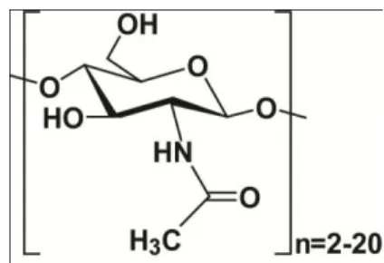
9 Chitooligosaccharides (GlcNAc)n (n=20) are water soluble chitin polymers of 2-acetylamino-2-deoxy-D-glucose monomers with a molecular formula: (C6H11O4N)n.
Figure 1.2. The chemical formula of a chitooligosaccharide.
The chitomonomer (GlcNAc) and chitooligosaccharides are produced from chitin or chitosan by chemical or enzymatic decomposition methods. With their excellent physiological activity and function, they are widely used in medicine, health food, agriculture, and cosmetic chemistry. GlcNAc has been proposed as a treatment for autoimmune diseases. Chitooligosaccharide derivatives have diverse applications in wastewater treatment, drug delivery, and wound healing. Specifically, its chitosan derivatives are known to have antimicrobial, antitumor, and anti-cholesterol activities (3).
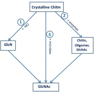
1.1.2.2. Chitin degradation
Chitin degradation is an important process for recycling carbon and nitrogen in the biosphere. Chitin is degraded by enzymes that catalyze the hydrolysis of N-linked acetyl groups from GlcNAc residues or break down 1,4-glycosidic bonds (Figure 1.3). The first type of hydrolysis is deacetylation, which is an effective way to make the polymer soluble (25). Deacetylation is a process that weakens or breaks the inter- or intra-molecular bonds in the compact structure by deacetylases (EC 3.5.1.41, belonging to the carbohydrate esterase family 4). This process can partially deacetylate chitin to 35%, which renders chitin soluble in dilute acids due to protonation of the amino group of GlcNAc. The product, chitosan, is further degraded by chitosanases (EC3.2.1.132, belonging to the glycoside hydrolase families 5, 7, 8, 46, 75 and 80), which cleave the β-1,4-glycosidic bonds to produce oligomers of GlcNAc or GlcN (glucosamine) (12). In the industry, chitosan is made by treating chitin under alkaline conditions at high temperatures (26).
10 Figure 1.3. Enzymatic degradation of chitin. The red (green) arrows correspond to endo-cleavages
(exo-cleavage) catalyzed by endo-chitinase (exo-chitinase).
The second type of chitin hydrolysis is deglycosylation, in which chitin is degraded through the breakdown of 1,4-glycosidic bonds by chitinolytic enzymes (27). This chitinolytic system is comprised of endo- and exo-acting chitinases, working in synergy for the complete degradation of chitin. They include chitinases (EC 3.2.1.14, belonging to the glycoside hydrolase families 18 and 19) which degrade chitin to produce chitooligomers and (GlcNAc)2, which is further hydrolyzed to monomers by N-acetyhexosaminidases (HEX) (EC 3.2.1.29, belonging to the glycoside hydrolase family 20). For commercial GlcNAc or chitooligosaccharides, chitin is hydrolysed using high concentrations of HCl or NaOH.
Due to the recalcitrant nature of the chitin polymer, it is energetically demanding for chitin-degrading enzymes to access the insoluble substrate (28, 29). Recently, lytic polysaccharide monooxygenases (LPMOs), which cleave glycosidic linkages on the crystalline surface of chitin by an oxidative mechanism, have been found. These enzymes create an entry point for hydrolytic chitinases, helping to increase substrate accessibility towards chitinases, thereby ensuring effective hydrolysis of chitin (30, 31).
In nature, chitin microfibrils are normally associated with proteins (arthropods) or glucans (fungi), therefore, chitin hydrolysis often requires accompanying protease or glucanase activities. In some chitin-forming organisms, such as filamentous fungi and yeast, chitin degradation is a fundamental physiological process for growth and development.
11 Fungal chitinases are responsible for the normal growth of chitin-containing fungi, including morphogenesis (hyphal growth and branching, sporulation, spore germination), autolysis, nutrition, and mycoparasitism (32-35). They control cell wall lysis and regulate their reconstitution in hypha expansion and cell bursting prevention (36, 37). They assist nutrient release in the saprophytic and mycoparasitic growth phases and may be involved in insect pathogenesis and phytopathogenesis (38). In Coccidioides spp., the complex morphologic changes during the spherule-endospore phase require the biosynthesis and degradation of cell wall structural components such as chitin, glucan or maman. This process needs the participation of chitin synthase, chitinase, β-glucosidase, and HEX (39-41). In insects, chitin is a structural part of the cuticle and peritropic membranes, where it is degraded by chitinolytic HEX in the turnover of chitin exoskeleton during metamorphosis (38, 42, 43). In some insects, such as mosquitoes, these enzymes are also important in the formation and in the degradation of the peritrophic matrix, which is formed in the midgut of adult females feeding on blood. Chitin is not only a nutritional source for chitinolytic micro-organisms but also for invertebrate and vertebrate organisms. Some insectivorous plants or mycopathogens can use chitinolytic systems to access insect prey or to penetrate fungal cell walls (17). In other plants, induction of chitinase is one of the defense mechanisms that plants use to react to attacks by phytopathogens and arthropod pests.
In general, chitin degradation occurs widely in many organisms. The synergic action of chitinolytic enzymes as well as other proteins such as chitosanases, glucanases or LPMOs is required for complete degradation of chitin in nature. In many industrial applications, in order to produce chitosan, chitooligomers or GlcNAc, chitin is usually hydrolyzed under extreme conditions using chemicals, which is not the most efficient and environmental-friendly method.
1.1.2.3. GlcNAc production
GlcNAc is the monomeric unit that constitutes the chitin polymer. It is a basic component of hyaluronic acid located in the extracellular matrix and of keratia sulfate on the cell surface. In mammals, it plays a role as building block of biomacromolecules such as glycoproteins, proteoglycans, glycosaminoglycans, and other connective tissue building
12 blocks (2). It also exists in human milk in the free form at concentrations of 600-1500 mg/mL. It is also present in growth factors and hormones, such as follicle-stimulating hormone (FSH), luteinizing hormone (LH), thyroid-stimulation hormone (TSH) and human chorionic gonadotropin (hCG) hormone. GlcNAc is also a component of chondroitin, which is abundant in connective tissues, particularly in blood vessels, bone and cartilage. In fact, GlcNAc and sulfated GlcNAc can be isolated from heparin or keratan sulfate, which are distributed in the cornea, cartilage and bone and usually act as an anticoagulant and a cushion in joints to absorb mechanical shock, respectively (44, 45). Besides, GlcNAc plays a role in plant organogenesis and invertebrate embryogenesis. Due to its versatile functions and non-toxic properties, GlcNAc can be used as a potentially therapeutic element in the treatment of many diseases as well as in economic feed-stocks. It has been used among others as a supplement to protect bone surfaces of the friction joint (5, 6) and to treat inflammatory bowel disease (including Crohn's disease) (7, 8). It emerges as a novel candidate for drug development, cosmetic, and other significant applications (17-20).
Every year, billions of tons of chitin are accumulated in nature, making chitin an unlimited source of material for the production of GlcNAc. Because of extreme insolubility, large-scale production of GlcNAc from this resource is rarely applied and is mainly based on chemical hydrolysis methods, which use concentrated HCl at high temperatures (15-36%, at 40-80°C) (46, 47). This process causes many problems, such as acidic wastes, low product yield, high cost of energy and product separation (2). On the other hand, enzymatic processes have gained efficiency, and environmentally-friendly performances but issues of final product purity and low productivity remain (46, 48-53). Biotransformation and a new method using glucose as a substrate can also be applied to produce GlcNAc. However, these two methods show some limitations due to time-cost or high technique issues (2). Production of GlcNAc using chitin as substrate is schemed in Figure 1.4.
13 Figure 1.4. Production of GlcNAc using chitin as substrate. (1) Chemical method. (2) Enzymatic method. (3)
Biotransformation method. Figure modified from reference (2).
Via chemical methods, crude chitin is degraded by a strong acid, such as HCl, at strict exact temperature and concentration to sufficiently degrade chitin but to prevent destroying GlcNAc. In this procedure, GlcNAc can be obtained up to 6.42 g/L per hour. In another procedure, chitin is dissolved in concentrated HCl and heated in boiling water for 3 h to remove the acetyl group of the GlcNAc units. An N-acetylation reaction with acetic anhydride or pyridine must then be executed to produce GlcNAc. After a series of purification procedures, this process can generate an overall yield of 43% or 70% GlcNAc, respectively. Despite acceptable economic yields, these chemical procedures create a product which is not considered a natural material due to its chemical modification (54). Moreover, by-products, such as O-acetylated products or tributylamine, and large quantities of chemical waste resulting from the processes are the main drawbacks for this chemical approach.
The enzymatic hydrolysis of chitin that can produce GlcNAc under mild conditions can overcome the drawbacks of the chemical approach. The collective use of chitinolytic enzymes including endochitinases, exochitinases and N-acetylglucosaminidases can produce GlcNAc from chitin. Many crude enzymes isolated from Penicillium monoverticillium CFR, Aspergillus flavus CFR 10, Fusarium oxysporum CFR, Trichoderma viride, Aspergillus niger,
14 Aeromonas sp. PTCC, and Aeromonium have been found to degrade chitin and to efficiently produce GlcNAc (2, 51, 53, 54). The main problem of enzymatic methods is the production and reuse of the enzymes as well as time-cost of the processes. For industrial applications, a large amount of pure enzymes is required. However, enzymes in organisms are present at low concentrations, thus the production and purification of a large amount of a pure enzyme is expensive. Several inexpensive, commercial enzymes such as those derived from Aeromonas hydrophila H2330 or crude chitinases from Bacillus licheniformis SK-1, or Serratia marcescens QM B1466 (2, 48, 49) can be easily obtained but then require a long process for GlcNAc production.
Using whole microbes in a fermentation system with chitin as substrate is another way to produce GlcNAc. Li et al. (2005) isolated chitinases from Aeromonas caviae DYU-BT4 and after optimizing the fermentation process, about 7.8 g/L of GlcNAc were produced using 2% colloidal chitin as substrate. Another microbe, Chitinibacter tainanensis, isolated from a soil sample in Southern Taiwan, was utilized to produce GlcNAc. This organism produced the sugar with a yield of 75% (98%) when using α-chitin (β-chitin) as substrate. After concentration and crystallization, the purity of the product was determined to be greater than 99%. This process seems to be very effective but unfortunately the microbes are killed and cannot be reused (2, 55).
Finally, GlcNAc can be produced through genetic modification of microorganisms using glucose as a substrate. This method makes use of metabolic pathways for GlcN and GlcNAc synthesis in microorganisms. A genetically engineered E. coli strain has been developed in which some enzymes taking part in GlcNAc synthesis are overexpressed, such as GlcN-6-P acetyltransferase, GlcN-6-P synthase and GlcN-1-P acetyltransferase. In this process, about 120 g/L of GlcNAc was produced after 60 h of fermentation. This process is the most efficient process by far (2).
1.1.3 Xylan and degradation of xylan
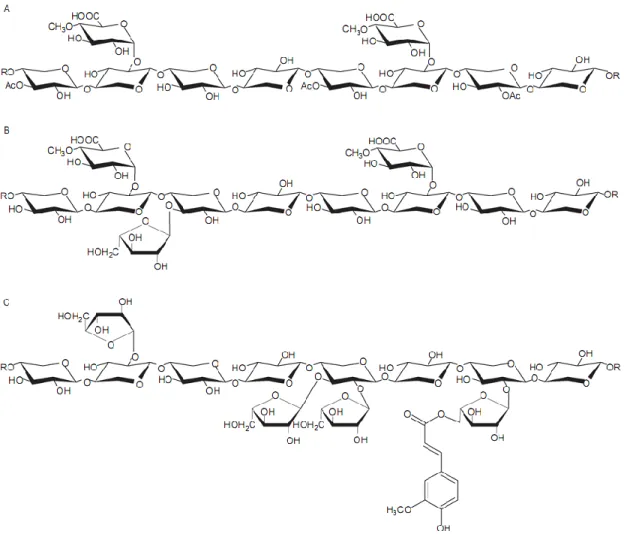
Xylan, a major component of hemicellulose, is a highly complex polysaccharide that is found in plant cell walls and some algae. In some higher plants and agricultural wastes, xylan constitutes from 20-40% of the dry weight of the plant biomass (56, 57). Xylan is a
15 heteropolymer consisting of a repeating β-1,4-linked xylose (a pentose sugar) backbone branched with acetyl, arabinofuranosyl, and 4-O-methyl glucuronyl groups (58). Xylan, derived from different plants, has the same backbone structure but different substitutions in its branches. The proportion of those substitutions is based on the source of the plant tissue. Xylan can be classified into homoxylans, arabinoxylans, glucuronoxylans, and arabinoglucuronoxylans. Homoxylans, which consist of a chain of β-1,4- and β-1,3-linked xylose units, are mainly found in cell walls of red seaweeds. Arabinoxylans, consisting of xyloses with arabinose residues branched at the O-2 or O-3 position, are principal components of plant cell walls, especially in cereal grains. Glucuronoxylans, with 4-O-methyl-α-D-glucuronic acid (MeGA) residues linked at the O-2 xylose backbone, are mainly found in hardwoods, herbs, and woody plants. Finally, arabinoglucuronoxylans are typically found in grass lignocelluloses and have arabinofuranosyl and acetyl side chains linked to the xylose backbone (10).
The chemical formula of xylan and its enzymatic breakdown mechanism are displayed in Figure 1.5 and 1.6.
16 Figure 1.5. Structure of xylan from different sources. A, Glucuronoxylan from hardwoods. B,
Arabino-methylglucuronoxylan from softwoods. C, Arabinoxylan from cereals. Figure taken from reference (59).
17 Xylan, together with cellulose, plays an important role in ruminant animal and insect nutrition, where it can be converted by microorganisms. Heteroxylans and homoxylans in plants also have important functions in many cereal-based food and feed biotechnology processes (11). In plant biomass degradation, complete hydrolysis of the two major components, cellulose and xylan, releases glucose, xylose, and arabinose, which can then be fermented or bioconverted to biofuels (10). Furthermore, other hydrolysis products including acetate, propionate, lactate, or succinate are essential feedstock for the chemical and pharmaceutical industries (61). As a result, xylanases that cleave the internal xylan backbone to modify its physicochemical properties have been used for many applications in the industry. One attractive application is the use of hemicellulase and endoxylanase enzymes to eliminate xylan from wood pulp in the manufacture of dissolved pulp (62). Also, cellulase-free xylanases were applied in pulping and bleaching processes, in which the use of chlorine (Cl2) and chlorine dioxide (ClO2) for biobleaching can be reduced (56). In fact, xylanases hydrolyze reprecipitated xylan to remove it from the surface of the cellulose fibers after pulping (56) due to improvement of permeability of the pulp fibers to bleaching chemicals. Xylanases also help to improve the chemical extraction of lignin from pulp. The use of xylanases instead of chemicals in pulp bleaching reduces the level of toxic chlorine compounds released into the environment. Many xylanases have shown high stability and activity under severe conditions that are applicable and economical in the pulp industry. For instance, xylanases from Actinomadura sp. FC7, Nonomuraea lexuosa (63, 64) and other xylanases from fungi and actinomycetes, which have high temperature and pH stability, have been widely employed in paper and pulp industries.
Despite the rather high level of industrial applications, a larger use of xylanase for different applications in biomass conversion is still difficult to obtain. The major obstacle is the lack of detailed knowledge of the xylan structure, which is diverse due to many linkages of different substitutions within the complex of the heteropolymer. As a result, it is difficult to design enzyme cocktails for efficient hydrolysis of the polymer. It is also unclear as to how individual sugar components in the polymer are chemically linked within the plant cell wall. Also, the complicated structure of the plant cell wall responsible for its recalcitrance is another limitation for bioconversion of plant polymers. Finally, due to different targets, it is appropriate to have suitable, adapted, specific and affordable enzymes for these various biotechnological processes.
18 Therefore, acquiring a better understanding of this type of enzyme helps improving biotechnological methods to hydrolyze polysaccharides, thus providing a fully renewable resource of molecules from biomass.
1.2
Enzymes in enzymatic degradation of polysaccharides
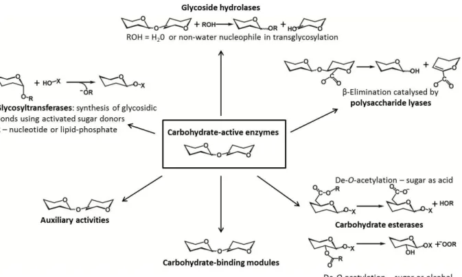
1.2.1 Carbohydrate-active enzymes
Carbohydrates play a role as central metabolites in energy storage and biosynthesis or bioconversion. Carbohydrates are also present in the form of glycoproteins, glycolipids and polysaccharides, which play fundamental roles in cell physiology and development of microbes, plants and animals. The enzymes that catalyze the breakdown, biosynthesis or modification of oligosaccharides, polysaccharides and glycoconjugates, comprise a group of enzymes named ‗carbohydrate-active enzymes‘ (CAZymes) (65). This enzyme group acts on the most structurally diverse substrates in nature. According to the carbohydrate-active enzyme database (CAZy; www.cazy.com), CAZymes are classified into families which reflect homology in amino acid sequence, protein structure and catalytic mechanism. Based on the type of catalysis, they have been grouped into 5 classes: Glycoside Hydrolases (GH), Glycosyl Transferases (GT), Polysaccharide Lyases (PL), Carbohydrate Esterases (CE), and Auxiliary Activities (AA) (66) (Figure 1.7). A sixth class of proteins in CAZymes are the carbohydrate-binding modules (CBM), which are non-catalytic but help the enzyme to bind the substrate (67). Due to the recalcitrance of polysaccharides, many CAZymes face the problem of binding to the substrate, disrupting its crystalline surface, and directing a single polysaccharide-chain into the catalytic site (12). Therefore, synergistic action of several enzymes belonging to different classes is necessary for full degradation. For example, deacetylases (belonging to CE4) can disrupt the crystalline surface of chitin by deacetylating (22, 68), LPMOs (belonging to AA9 or AA10) can make a ‗scratch‘ on the biomass surface to produce an entry point for GHs (30, 31), or CMBs promote binding of GHs to the substrate, disrupting the plant cell wall structure to make it more accessible or sometimes to help by directing the chain into the active site (59, 69).To this date (April 2015), 133 families of GHs,
19 97 families of GTs, 23 families of PLs, 16 families of CEs, 13 families of AAs and 71 families of CBMs are listed in the database.
Figure 1.7. Carbohydrate-active enzymes. Figure modified from reference (66).
1.2.2 Glycoside hydrolases
1.2.2.1. General properties
Among the carbohydrate-active enzymes, glycoside hydrolases (hereafter glycosidases or GHs) are the best-characterized enzymes that are active on disaccharides, oligosaccharides and polysaccharides. The glycosidases are enzymes that enzymatically catalyze the hydrolysis of the glycosidic bond between two or more carbohydrates or between a carbohydrate and a non-carbohydrate moiety (70). The GH enzymes play not only a fundamental role in enzymatic catalysis but also in diverse biological processes. Therefore, glycosidases are widely used in the hydrolysis of biomass as well as in human physiology and disease treatment.
20 For nomenclature, these GH enzymes are classified into a number of families based on amino acid sequence similarity. To this date (April 2015), the Carbohydrate-Active Enzymes database (CAZy) provides a list of 133 glycoside hydrolase families. This classification reflects the homology in structural features and the evolutionary relationships among these enzymes.
1.2.2.2. Structure of glycoside hydrolases
Regarding structure, some of GH families are grouped into ―clans‖, which describe the conserved folding architecture of the proteins. For example, clan GH-A is comprised of 19 families that share the same TIM-barrel fold. Clan GH-B includes the proteins from the GH7 and GH16 families with a β-jelly roll fold in the tertiary structure, etc. (65). Based on substrate specificity and catalytic mechanisms, glycosidases have different active site conformations. Because of the physicochemical resistance of tightly packed linear glycan polysaccharides, such as cellulose and chitin, many GHs that break carbohydrate chains are found to possess a deep cleft or tunnel active site architecture (Figure 1.8) (30). In most cases, this active site shape is effective for enzymes which adopt processive mechanisms in polysaccharide degradation. In processive GHs, the deep cleft or tunnel in the active site is usually paved with aromatic residues that are important and ensure packing, binding and sliding of the polymer through the active site (16, 71-73). These aromatic residues bind to carbohydrate substrates by hydrogen bonding and stacking, which are the dominant interactions in carbohydrate-protein complexes (13, 69). However, non-processive GH enzymes show active sites with a more open cleft or pocket with fewer aromatic residues.
21 Figure 1.8. Active-site shapes in GH enzymes: left, pocket (glucoamylase from Aspergillus awamori, PDB
entry 1GLM); middle, cleft (endoglucanase E2 from Thermobifida fusca, PDB entry 1TML); right, tunnel (cellobiohydrolase II from Trichoderma reesei, PDB entry 3CBH). The catalytic residues are colored in red. Figure adapted from reference (74).
1.2.2.3. Mode of action
GH enzymes can be classified as exo- and endo-enzymes (Figure 1.9), which refers to the ability of a glycoside hydrolase to cleave a substrate in the middle or at the end of a chain (mostly but not always the non-reducing end). For example, ScHEX in this research thesis is an exo-acting enzyme, whereas ChiC and XlnB2 are endo-acting enzymes.
Figure 1.9. Endo-, exo-acting cleavage mechanism type. Figure reproduced from reference (65).
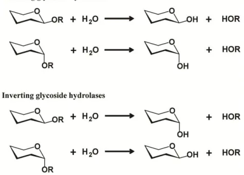
The two standard mechanisms of glycosidic bond cleavage are inversion or retention
of the configuration of the anomeric carbon, as originally outlined by Koshland in 1953 (66) (Figure 1.10). In these mechanisms, the cleavage of the glycosidic bond is catalyzed by two amino acid residues: a general acid/base residue and a nucleophilic one. Depending on the spatial position of these catalytic residues, hydrolysis occurs via overall retention (the initial configuration of the scissile bond is retained by the newly generated reducing end) or overall
22 inversion of the anomeric configuration (65). Moreover, in recent years, several interesting modified mechanisms have been discovered such as substrate assistance, alternative nucleophile, or one fundamentally different mechanism catalyzed by an NADH cofactor. In this thesis, only the two standard and the substrate-assisted mechanisms are described.
Figure 1.10. Retaining, inverting mechanism types with GH enzymes. Figure reproduced from reference (65)
The inverting mechanism is a one step, single-displacement mechanism involving oxocarbenium ion-like transition states (Figure 1.11). The reaction typically occurs with general acid and general base assistance from two amino acid‘s side chains (normally glutamic or aspartic acid) that are typically located 6-11 Å apart. After hydrolysis, the anomeric configuration of the glycoside is inverted (74).
23 The retaining mechanism is a two-step, double-displacement mechanism involving a covalent glycosyl-enzyme intermediate (Figure 1.12). Each step passes through an oxocarbenium ion-like transition state. Similarly to the inversion mechanism, the reaction occurs with acid/base and nucleophilic assistance provided by two amino acid‘s side chains, typically glutamate or aspartate, located 5.5 Å apart. In the first step or glycosylation step, the nucleophilic residue attacks the anomeric centre to displace the aglycon and form a glycosyl-enzyme intermediate, meanwhile the acid catalyst protonates the glycosidic oxygen as the bond is being cleaved. In the second step or deglycosylation step, the base catalyst abstracts protons from the water molecule as it attacks, hydrolyzing the glycosyl intermediate, and finally generating a glycon product with net retention of the configuration.
Figure 1.12. Retaning mechanism with GH enzymes (for a β-glycosidase). Adapted from reference (65).
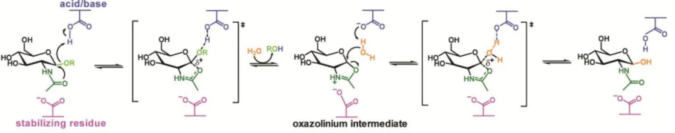
One of the modified mechanisms is the substrate-assisted mechanism with neighboring group participation of the substrate (Figure 1.13). Many polysaccharides contain an N-acetyl (acetamido) or N-glycosyl group at the 2-position of the pyranoside ring. GH families 18, 20, 25, 56, 84, and 85 have no catalytic nucleophile, instead they utilize the acetamido group to act as an intra-molecular nucleophile. The participation of the 2-acetamido group leads to formation of an oxazoline (or more specifically an oxazolinium ion) intermediate, which is stabilized by another conserved residue. In the next step, a general acid/base accepts a proton from the substituent, then the oxazoline ring is attacked by a water molecule at the anomeric center to form a hemiacetal product (38, 75, 76). Not all enzymes that cleave substrates possessing a 2-acetamido group adopt the substrate-assisted mechanism. For instance, GH3 and GH22 enzymes utilize a classical retaining mechanism and hexosaminidases of the GH19 family utilize an inverting mechanism. The research presented in this thesis focuses on three enzymes: chitinase, N-acetylhexosaminidase, and xylanase that are all GH enzymes. Chitinase ChiC from S. coelicolor A3(2) is a GH18 family member,
24 meanwhile N-acetylhexosaminidase ScHEX, also from S. coelicolor A3(2), belongs to the GH20 family, and xylanase XlnB2 from S. lividans belongs to the GH11 family. Among them, ChiC and ScHEX adopt the substrate-assisted mechanism, and XlnB2 shows a retaining mechanism.
Figure 1.13. Substrate-assistedmechanism utilized by GH enzymes. Figure adapted from reference (38).
The subsite theory developed by Hiromi et al. (77, 78) and by Allen, Thoma and co-workers (79, 80) recommends a nomenclature in which the binding surface of the protein is divided into several subsites and represents the point of cleavage. Davies et al. proposed that GHs have five or more subsites, labeling −3, −2, −1, and +1, +2, etc., which accommodate a long substrate inside the active site. This nomenclature indicates that the subsites interact with glycosyl residues from the non-reducing end to the reducing end of the substrate. The cleavage takes place at the point between the -1 and +1 subsites, indicating whether the non-reducing or non-reducing end is being enzymatically attacked (Figure 1.14) (81).
Figure 1.14. Subsites nomenclature in GH enzymes. Subsites -3 to +3 from the non-reducing end to the
reducing end. The arrow indicates the site of cleavage. Figure adapted from reference (81) .
Regarding structure, the cleft or tunnel topology of active sites in GHs allows these enzymes to release the product while remaining firmly bound to the polysaccharide chain by several subsites, thereby, creating the conditions for processivity. Figure 1.15 illustrates the processive mechanism, which is often found in GH enzymes. When a product is released, the enzyme keeps binding to the substrate through several subsites. The empty
25 subsites or other factors, such as loop movement, assist the chain in sliding along the active site for the next hydrolysis to occur (81).
Figure 1.15. The processive mechanism of GH enzymes. Once a dimer product is released (shown as two
linked hexagons), the enzyme remains bound to the polysaccharide chain, the « lid » closing the active site. The chain keeps threading along the active site by two sugar units until the next hydrolysis occurs. Figure reproduced from reference (81).
1.3
Chitinolytic enzymes
Chitinolytic enzymes, which hydrolyze the β(1,4) linkage of chitin, are key enzymes in the natural processing of this material. Based on their hydrolytic functions, chitinolytic enzymes are classified into endochitinases (E.C. 3.2.1.14), exochitinases (E.C. 3.2.1.29), and N-acetylglucosaminidases (HEX) (EC 3.2.1.30) (24). The first group randomly cleaves the chitin polymer to yield soluble, low molecular weight oligomers, such as chitotetraose and chitopentaose. The second group catalyzes the progressive release of chitobiose from the non-reducing or the non-reducing end of the polymer chain. The last category releases N-acetylglucosamine monomers from chito-oligomers and chitobiose, which are produced by the endochitinases and exochitinases, respectively. It was reported that complete degradation of chitin requires chitinase and HEX acting in a concerted fashion (33, 35). In this degradation, chitinase degrades chitin into chitooligomers, which are then hydrolyzed by HEX to release GlcNAc (82, 83). The oligomers of the degradation process seem to be promising candidates for biomedical applications. For example, they can be used in antimicrobial, anticancer, wound-healing, antitumor, or antioxidant effectuations (84).
Based on their primary structure, endo- and exo-chitinases are grouped into two GH families: GH18 and GH19 (85-87). Meanwhile, HEXs that hydrolyze chitooligomers belong