Publisher’s version / Version de l'éditeur:
Journal of the American Chemical Society, 134, 18, pp. 7788-7795, 2012-04-16
READ THESE TERMS AND CONDITIONS CAREFULLY BEFORE USING THIS WEBSITE.
https://nrc-publications.canada.ca/eng/copyright
Vous avez des questions? Nous pouvons vous aider. Pour communiquer directement avec un auteur, consultez la première page de la revue dans laquelle son article a été publié afin de trouver ses coordonnées. Si vous n’arrivez pas à les repérer, communiquez avec nous à PublicationsArchive-ArchivesPublications@nrc-cnrc.gc.ca.
Questions? Contact the NRC Publications Archive team at
PublicationsArchive-ArchivesPublications@nrc-cnrc.gc.ca. If you wish to email the authors directly, please see the first page of the publication for their contact information.
NRC Publications Archive
Archives des publications du CNRC
This publication could be one of several versions: author’s original, accepted manuscript or the publisher’s version. / La version de cette publication peut être l’une des suivantes : la version prépublication de l’auteur, la version acceptée du manuscrit ou la version de l’éditeur.
For the publisher’s version, please access the DOI link below./ Pour consulter la version de l’éditeur, utilisez le lien DOI ci-dessous.
https://doi.org/10.1021/ja211876p
Access and use of this website and the material on it are subject to the Terms and Conditions set forth at
The substrate-bound crystal structure of a Baeyer–Villiger
monooxygenase exhibits a Criegee-like conformation
Yachnin, Brahm J.; Sprules, Tara; McEvoy, Michelle B.; Lau, Peter C. K.;
Berghuis, Albert M.
https://publications-cnrc.canada.ca/fra/droits
L’accès à ce site Web et l’utilisation de son contenu sont assujettis aux conditions présentées dans le site LISEZ CES CONDITIONS ATTENTIVEMENT AVANT D’UTILISER CE SITE WEB.
NRC Publications Record / Notice d'Archives des publications de CNRC:
https://nrc-publications.canada.ca/eng/view/object/?id=548e63dd-952c-4f6a-b68f-da210ae95e9c https://publications-cnrc.canada.ca/fra/voir/objet/?id=548e63dd-952c-4f6a-b68f-da210ae95e9cThe Substrate-Bound Crystal Structure of a Baeyer−Villiger
Monooxygenase Exhibits a Criegee-like Conformation
Brahm J. Yachnin,
†,⊥Tara Sprules,
#Michelle B. McEvoy,
†Peter C. K. Lau,
‡,§,∥and Albert M. Berghuis*
,†,‡,⊥Departments of†
Biochemistry,‡
Microbiology & Immunology, and §
Chemistry, McGill University, 3649 Promenade Sir William Osler, Bellini Pavilion, Room 466, Montreal, QC, Canada H3G 0B1
∥
Biotechnology Research Institute, National Research Council of Canada, 6100 Royalmount Avenue, Montreal, QC, Canada H4P 2R2
#
Québec/Eastern Canada High Field NMR Facility, 3420 University Street, Room 023, Montreal, QC, Canada H3A 2A7
⊥
Groupes de recherche GRASP et PROTEO, Canada
*
S Supporting InformationABSTRACT: The Baeyer−Villiger monooxygenases (BVMOs) are a family of bacterial flavoproteins that catalyze the synthetically useful Baeyer−Villiger oxidation reaction. This involves the conversion of ketones into esters or cyclic ketones into lactones by introducing an oxygen atom adjacent to the carbonyl group. The BVMOs offer exquisite regio- and enantiospecificity while acting on a wide range of substrates. They use only NADPH and oxygen as cosubstrates, and produce only NADP+ and water as byproducts, making them
environmentally attractive for industrial purposes. Here, we
report the first crystal structure of a BVMO, cyclohexanone monooxygenase (CHMO) from Rhodococcus sp. HI-31 in complex with its substrate, cyclohexanone, as well as NADP+and FAD, to 2.4 Å resolution. This structure shows a drastic rotation of the
NADP+ cofactor in comparison to previously reported NADP+-bound structures, as the nicotinamide moiety is no longer
positioned above the flavin ring. Instead, the substrate, cyclohexanone, is found at this location, in an appropriate position for the formation of the Criegee intermediate. The rotation of NADP+ permits the substrate to gain access to the reactive flavin
peroxyanion intermediate while preventing it from diffusing out of the active site. The structure thus reveals the conformation of the enzyme during the key catalytic step. CHMO is proposed to undergo a series of conformational changes to gradually move the substrate from the solvent, via binding in a solvent excluded pocket that dictates the enzyme’s chemospecificity, to a location above the flavin−peroxide adduct where catalysis occurs.
■
INTRODUCTIONThe century-old Baeyer−Villiger (BV) reaction is the oxidative cleavage of a carbon−carbon bond adjacent to a carbonyl; this process transforms ketones to esters and cyclic ketones to lactones using peracids.1,2While this reaction has proven to be
very useful to synthetic chemists, it has a number of shortcomings that include the use of costly and hazardous reagents or chlorinated solvents while producing more waste than product for lack of functional group selectivity and enantioselectivity. Although metal-catalyzed enantioselective BV reactions have also been developed, both cost and contamination of the products with metals may present a serious problem.3 For a variety of reasons, Baeyer−Villiger monooxygenases (BVMOs), a family of bacterial flavoproteins that catalyze the BV reaction,4−9have garnered much attention.
Most BVMOs are involved in the second step of biodegradation of C5 to C15 alicyclic alcohols such as
cyclopentanol, cyclohexanol, and cyclopentadecanol, providing a carbon source for the organisms via the formation of Krebs
cycle intermediates.6,10,11These enzymes can function in water,
and require NADPH as a cofactor and molecular oxygen as the oxidative reactant. While one oxygen atom is introduced adjacent to the carbonyl group of a ketone substrate, the other atom is reduced to water as a byproduct.4,5,12 These green characteristics have made them intriguing targets for use as biocatalysts since the prototypical BVMO, cyclohexanone monooxygenase (CHMO) from Acinetobacter NCIMB 9871 (AcCHMO), was first characterized nearly 40 years ago.10,12 Their frequently exquisite regio- and enantiospecificities, combined with their broad substrate profiles that include substituted and bicyclic ketones, make them of particular interest to the pharmaceutical industry.4−9 In recent years, a
multitude of homologues of these enzymes have been cloned and characterized,4−9thereby expanding the range of potential substrates catalyzed by BVMOs.
Received: January 4, 2012
Published: April 16, 2012
While the BVMOs have been studied extensively, only recently has any structural data been obtained. The first BVMO to have its structure solved was phenylacetone monooxygenase (PAMO) from the thermophile Thermobif ida fisca.13 This structure revealed the overall fold of the enzyme and the location and conformation of the FAD prosthetic group, which remains bound throughout the catalytic cycle. Subsequently, we reported two structures of CHMO from Rhodococcus sp. HI-31 (RmCHMO) in complex with both NADP+and FAD.11This
enzyme is notable for being a close homologue and having a similar substrate profile to the canonical AcCHMO while being substantially more stable. The two crystal structures, designated as CHMOOpenand CHMOClosed, revealed that NADP+can bind
to the enzyme in two distinct conformations. The sliding of the NADP+ cofactor deeper into the protein in the CHMO
Closed
structure was revealed to be coupled with a rotation of the NADPH-binding domain so as to create a well-defined substrate binding pocket. More recently, a series of structures of PAMO with NADP+ and a weak inhibitor,
2-(N-morpholino)ethanesulfonic acid (MES), have corroborated these findings and provided insight into the conformation of the reduced flavoenzyme.14Intriguingly, the PAMO structures identified a funnel-shaped cavity that may provide an alternative entrance for the substrate to gain access to the active site. Another recent paper describing NADP+-bound and NADP+
-free crystal structures of 2-oxo-Δ3-4,5,5-trimethylcyclopentenyl
acetyl-CoenzymeA 1,2-monooxygenase (OTEMO), which is notable for being a dimeric BVMO with a large substrate, also confirmed the dynamic nature of the BVMO family of proteins.15 It should be noted that in addition, a crystal
structure of an atypical BVMO, mithramycin monooxygenase (MtmOIV) has been obtained.16 This enzyme has high sequence and structural similarity to a FAD-dependent hydroxylase of the glucocorticoid receptor (GR2) subfamily.
This makes it a distant relative of the prototypical BVMOs and precludes its detailed comparison with CHMO or PAMO.
Structural studies of BVMOs have provided much insight into these enzymes’ catalytic mechanism; however, they have also raised questions. Notably, the CHMOClosed structure
reveals that for the majority of CHMO’s substrates, which are larger than cyclohexanone, there is actually insufficient room in the active site for the formation of the critical Criegee intermediate, the adduct of the flavin peroxide and the ketone substrate that is required for catalysis. Models of the Criegee intermediate for the cyclohexanone substrate, one of CHMO’s smallest substrates, suggests that this intermediate can be achieved in the Closed conformation.11,17 In contrast, the Criegee intermediates of larger substrates, such as substituted or bicyclic ketones on which CHMO readily acts, would result in severe steric clashes with one or more of NADP+, L146,
F279, and F434 in the Closed conformation. It is clear that in order for a Criegee intermediate to form, a significant reorganization of the active site is required.
Here, we present the first crystal structure of a prototypical BVMO, represented by RmCHMO, bound to its substrate, cyclohexanone, as well as NADP+. This structure reveals a
major rotation in the NADP+ cofactor, allowing CHMO to
adopt a catalytically relevant conformation. The rotation permits the substrate to be positioned above the FAD, thus revealing a Criegee intermediate-like conformation, a snapshot of the most important stage of the catalytic mechanism. It also highlights the role of NADP+ in organizing the active site and
provides a structural explanation for the broad substrate
specificity of the enzyme, which is necessary for a complete understanding of the catalytic mechanism.
■
EXPERIMENTAL SECTIONFor a description of the subcloning of the chnB1 gene from
Rhodococcus sp. HI-31, the construction of various mutants for NMR
and kinetic studies, and the expression and purification of the CHMO enzyme and its variants, please consult the Supporting Information.
Crystal Structure Determination.Crystals were obtained using the hanging drop vapor diffusion method. Wild-type CHMO was prepared at 5 mg/mL in 50 mM Tris pH 8.0 and supplemented with a 5× molar excess of FAD and NADP+. In brief, 2 μL of protein solution
was mixed with 2 μL of reservoir solution on an 18 mm siliconized coverslip (Hampton Research). The reservoir solution consisted of 0.1 M imidazole, pH 8.0, 0.2% TMOS, 20% PEG 3350, and 0.1 M cyclohexanone. This drop was suspended over a 1 mL reservoir in a 24-well ComboPlate (Greiner Bio-One), and the plate was incubated at 4 °C. Crystals suitable for diffraction studies grew in about one week.
Data were collected under standard cryogenic conditions on a Rigaku MicroMax-007HF generator equipped with VariMax HF optics and a Saturn 944+ CCD detector. The data were processed using the
HKL2000 suite of programs (Table 1).18 The structure was
subsequently solved using Phaser,19employing CHMOOpen(PDB ID
3GWF) as the search model.11The model was subjected to multiple rounds of positional and B-factor refinement using Refmac.20Manual model building was performed regularly during refinement using Coot.21The coordinates and structure factors have been deposited in the Protein Data Bank as PDB ID 3UCL. Structural figures were prepared using PyMOL (Schrödinger LLC).
Saturation Transfer Difference NMR Spectroscopy. The buffer used for dialysis of the protein was used to dissolve NADP+
at 5 mM. Samples of the buffer and NADP+ solution were placed
under vacuum until dry, and then reconstituted in an equal volume of
D2O. A 600 μL sample of 75 μM protein and 1.5 mM NADP+was
prepared. Samples of the protein without its ligand were also prepared as a control to confirm that all of the STD signals were derived from protein−ligand interactions.
Table 1. Data Collection and Refinement Statistics for the CHMORotatedCrystal Structure (PDB ID 3UCL)
Data collection statistics
Space group P212121 a, b, c (Å) 55.5, 67.1, 131.4 Resolution range (Å)a 30.6−2.4 (2.5−2.4) Completeness (%)a 97.9 (92.7) Redundancya 11.2 (8.3) Rsyma 8.7 (47.2) Refinement statistics
Total number of reflections (reflections in Rfreeset) 20,214 (2061) Rfactor(%) (Work + Free/Free) 19.8/26.4
Number of atoms 4215
Protein 3986
Water 121
Cofactors and Substrate 108
rmsd
Bond length (Å) 0.017
Bond angle (deg) 1.669
Ramachandran plot 505 (100%)
Residues in favored positions 492 (97.4%) Residues in allowed positions 10 (2.0%) Residues in disallowed positions 3 (0.6%)
aNumbers in parentheses refer to the highest resolution shell.
Journal of the American Chemical Society Article
dx.doi.org/10.1021/ja211876p | J. Am. Chem. Soc. 2012, 134, 7788−7795
All NMR experiments were performed at 15 °C on a Varian INOVA 500 MHz spectrometer equipped with a triple resonance HCN cold probe. A 1D saturation transfer difference pulse sequence with internal subtraction via phase cycling was employed to record difference spectra.22 Residual HDO signal was removed using a double pulsed-field gradient spin echo. On-resonance irradiation of the protein was performed at −0.5 ppm, with off-resonance irradiation at 36.5 ppm. Reference spectra were recorded using the same pulse sequence with saturation pulses applied at 36.5 ppm, and no internal subtraction. Additional details regarding the NMR experiments are provided in the Supporting Information.
Enzyme Kinetics. All enzyme assays were performed at 22 °C
using a Cary 50 Bio UV−Visible Spectrophotometer equipped with a Peltier-thermostatted cell. The enzyme, NADPH, and cyclohexanone were dissolved at the appropriate concentration in 50 mM sodium phosphate, pH 8.0. A 500 μL reaction volume was used. To determine
the KM and kcat for NADPH, a series of assays with 100 μM
cyclohexanone (>40× KM(cyclohexanone)) and between 3 μM and 200 μM
of NADPH were used. These experiments were repeated with varying
concentrations of NADP+ to determine the K
i of NADP+. To
determine the KMand kcatfor cyclohexanone, a series of assays with 50
μM NADPH (>8× KM(NADPH)) and between 0.4 μM and 40 μM
cyclohexanone were used. A higher concentration of NADPH could not be used due to substrate inhibition. In all cases, the NADPH, 10 μL of an appropriate concentration of enzyme, and the substrate were mixed together to start the reaction. The decrease in absorbance at 340 nm was monitored to determine the initial rate of the oxidation reaction. The initial rates were plotted against the substrate (NADPH or cyclohexanone) concentration for each reaction series, and nonlinear regression was performed using SigmaPlot (Systat Software, Inc.) to determine the KMand kcat.
A second set of experiments were performed to determine the uncoupling ratio, defined as the rate of NADPH oxidation in the presence of cyclohexanone (apparent BV activity) divided by the rate of NADPH oxidation in the absence of cyclohexanone (NADPH oxidase activity). Using saturating concentrations of both NADPH (>6× KM(NADPH)) and cyclohexanone (>40× KM(cyclohexanone)), the
decrease in absorbance at 340 nm was assessed both before and after the addition of cyclohexanone. Three different NADPH concen-trations were used (40−80 μM), all of which saturated the enzyme, and the experiments were conducted in quadruplets.
■
RESULTSCrystal Structure of Substrate-Bound CHMO. The crystal structure of RmCHMO in complex with FAD, NADP+, and cyclohexanone was obtained to a resolution of
2.4 Å (Table 1). The polypeptide backbone could be modeled completely from residues 5−534, with the exception of two unstructured loop regions from residues 146−149 and 489−
503. The first loop region corresponds to a long linker between the FAD- and NADPH-binding domains, while the second corresponds to the large loop that is unstructured in the CHMOOpenstructure, but folds in to contact NADP+ in the
CHMOClosedstructure. Density consistent with FAD, NADP+,
and cyclohexanone was also visible (Figure 1A). To differ-entiate it from the CHMOOpenand CHMOClosedstructures that
were previously reported, we will refer to this structure as CHMORotated.
In general, the CHMORotatedstructure most closely resembles
the CHMOOpenstructure (rmsd of 0.56 Å between backbone
atoms present in both CHMOOpenand CHMORotatedstructures
when the FAD-binding domains are aligned, vs 1.31 Å for CHMOClosed). When aligning the two structures based on their
FAD-binding domains, the FAD-binding and helical domains show very little structural divergence. The NADPH-binding domain shows a slight rotation, though this is significantly less pronounced than the substantial domain movements seen when comparing the CHMOOpenand CHMOClosed structures.
This rotation allows for a subtle shift in the position of the adenine portion of NADP+, which may in turn trigger the
rotation of its nicotinamide moiety. Rotation of the NADP+ Cofactor.
One of the most significant features in the crystal structure is the large rotation of the nicotinamide phosphoribose of NADP+when compared
to CHMOOpen and CHMOClosed (Figure 1B). It is for this
reason that we refer to it as the CHMORotatedstructure. This
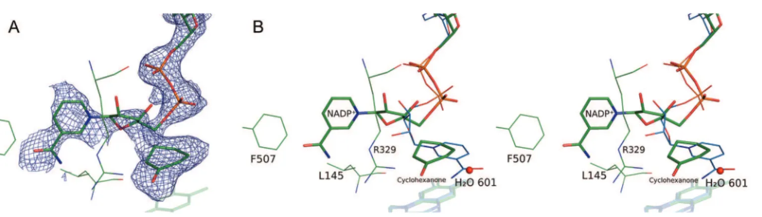
rotation serves to displace the nicotinamide head away from its position above the isoalloxazine ring system of FAD and into the large, primarily hydrophobic cavity that is seen in the CHMOOpenstructure. This movement has two notable effects.
By rotating away from the FAD ring system, space for the substrate to enter into the catalytic position is created. Second, the nicotinamide head serves to partially block the active site region from the bulk solvent, which prevents the substrate from diffusing back into solution while the protein is in the Rotated conformation (Figure 2). It is notable that the nicotinamide moiety forms relatively few strong interactions in this conformation.
The new conformation of NADP+ allows the formation of
intramolecular hydrogen bonds between the 3′ hydroxyl of the nicotinamide ribose and both the nicotinamide and the adenine phosphates. This intramolecular interaction stabilizes this conformation of the NADP+ and may be a driving force in
the adoption of this conformation.
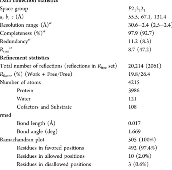
Figure 1.View of the active site in the CHMORotated(green) structure. The positions of FAD, NADP+, and cyclohexanone are shown. (A) The Fo−
Fcelectron density map contoured at 3.0σ around NADP+and cyclohexanone is shown as a blue mesh. The map was calculated with the ligands
omitted from the model. (B) Stereo view with the position of NADP+in CHMO
Closed(blue) overlaid on the CHMORotatedcrystal structure. Key
The Binding of the Substrate, Cyclohexanone. Electron density corresponding in size and shape to what is expected for cyclohexanone was observed above the isoallox-azine ring system of FAD; therefore, we proceeded to model the substrate at this position (Figure 1A). The carbonyl oxygen of cyclohexanone appears to be oriented through interactions with D59 and R329. Both of these residues have been previously implicated in controlling the position of NADP+,11,13,23
as well as in the positioning of a weak inhibitor of PAMO.14A nearby water molecule (H
2O 601) also forms a
weak hydrogen bond with the carbonyl oxygen, linking it to Q192, the backbone atoms of residues L57 and D59, and a larger solvent network.
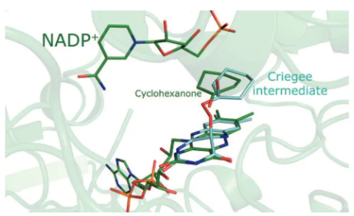
It is known that the formation of the Criegee intermediate, the key catalytic step in the BVMO reaction mechanism, involves a nucleophilic attack on the carbonyl carbon of cyclohexanone by the peroxyanion intermediate, in which the peroxyanion is covalently linked to the C4X carbon of FAD. The distance between C4X and the carbonyl carbon is 3.9 Å in this crystal structure. With the FAD-linked oxygen of the peroxide group expected to occupy an axial position relative to the FAD ring system, if the anionic oxygen were to point toward cyclohexanone, the position occupied by cyclohexanone in this structure would correspond approximately to the expected position for the nucleophilic attack and formation of the Criegee intermediate. A model of the Criegee intermediate that imposes antiperiplanar geometry required for migration of the carbon to form the lactone24 shows that cyclohexanone
nearly coincides with its position in the model of the intermediate (Figure 3). A shift of ∼0.5 Å is required to superimpose the carbonyl carbon of the cyclohexanone molecule and the intermediate, after which a ∼30° rotation is sufficient to make the molecules overlap.
Structure−Function Studies Probing the Rotated Conformation. To probe the relevance of the observed Rotated conformation for catalysis, we designed three mutants that were predicted to specifically perturb nicotinamide cofactor binding in the Rotated conformation, while having no impact on the cofactor in either the Open or Closed conformations. The three mutants, L145N, L145D, and F507Y, are anticipated to stabilize the catalytic conformation by forming a polar contact with the carboxamide group of NADP+ in the Rotated conformation. In contrast, in the
CHMOOpenand CHMOClosedstructures, these residues should
not interact directly with the cofactor, with the nearest distance in either structure being more than 5 Å. Given that many of the residues implicated in NADP+-binding in the CHMO
Rotated
structure are also involved in substrate specificity, the rational design of destabilizing mutations specific for the Rotated conformation was not feasible.
To assess the degree of perturbation in the NADP+binding
mode caused by these mutations, transferred nuclear Over-hauser effect (Tr-NOE) and saturation transfer difference (STD) NMR experiments were performed. No major differ-ences were observed between the Tr-NOESY spectra of NADP+ in the presence of wild-type CHMO or any of the
three mutants, as only some slight changes of NOE intensities and appearance and disappearance of the weakest NOEs were observed. This suggests that the overall set of conformations available to NADP+ in the mutants is not significantly
perturbed. In the STD experiments, the aim was to assess if there was a difference in the relative enhancement of the STD effect of the pyridine protons as compared to the adenine protons when the mutants were compared to the wild-type protein. The differences observed were subtle, and preclude a discussion of the effects of the mutations on any individual proton; however, when the STD effect of the four protons on the pyridine ring (N2, N4, N5, and N6) was compared to the STD effect for the two adenine ring protons (A2 and A8, where A2 is used as the reference for the STD effect), a trend could be observed (Supporting Information Figure S1). The interactions between the protein and the pyridine protons appeared closer than the protein−adenine proton interactions in the mutants as compared to the wild-type CHMO. This is consistent with a minor selective stabilization of the nicotinamide moiety binding mode in the Rotated conformation.
While the structural impact of the mutants was subtle, as assessed by NMR methods, the impact on the kinetic properties of CHMO was substantial (Figure 4, see Supporting Information Table S1 for complete kinetic data). In comparing the KM values for NADPH, the wild-type had the highest KM,
suggesting that all three mutants bind the cofactor more strongly during catalysis. It should be noted that all the KM
values were within an order of magnitude of each other. Furthermore, in terms of kcat, all three mutants had substantially
lower rates than the wild-type, with the kcatdropping by at least
a factor of 6 and by as much as a factor of 100. This effect was seen for the kcat of both NADPH and cyclohexanone. This
Figure 2.NADP+blocks cyclohexanone from diffusing into the bulk
solvent in the Rotated conformation. The protein surface is shown in gray. NADP+ is shown in stick representation and its surface is
outlined in blue. Cyclohexanone is shown using green space-filling representation.
Figure 3. The CHMORotated crystal structure (green) is shown
superimposed on a model of the Criegee intermediate (cyan). The model was produced using molecular dynamics followed by energy minimization in Chem3D, while enforcing the antiperiplanar geometry required for migration of the carbon.
Journal of the American Chemical Society Article
dx.doi.org/10.1021/ja211876p | J. Am. Chem. Soc. 2012, 134, 7788−7795
shows that, although the binding affinity of NADPH had been slightly improved, the mutations appear to have significantly slowed the reaction rate. The Kifor NADP+as a competitive
inhibitor of CHMO is discussed in the Supporting Information, and is largely consistent with the observed trend for NADP+
binding affinities in the mutants.
The uncoupling ratio is an indicator of the efficiency of the complete Baeyer−Villiger reaction (BV activity) relative to the efficiency at which NADPH would be used in the absence of cyclohexanone (NADPH oxidase activity). The wild-type enzyme exhibits the largest uncoupling ratio of 114. For the three mutants, this ratio is significantly reduced, ranging from 4 to 20. To assess how much of this change is caused by the decrease in BV activity and how much is due to changes in the amount of unproductive NADPH oxidase activity, the normalized uncoupling ratio was compared to the normalized kcat/KM(NADPH) and kcat/KM(cyclohexanone) values, using the
wild-type values as 100% (Supporting Information Figure S2). Note that both kcat/KM values were determined using the apparent
BV activity (which includes unproductive NADPH oxidase activity in the presence of cyclohexanone), not the uncoupled reaction (NADPH oxidase activity). For all three mutants, the uncoupling ratio was significantly greater than would be predicted by the normalized kcat/KM(cyclohexanone), and
moder-ately smaller than predicted by the normalized kcat/KM(NADPH).
■
DISCUSSIONA Third NADP+Conformation.Previous structural studies
of BVMOs have raised a critical question: how is the enzyme capable of effecting catalysis with a structure that has its active site blocked by the NADP+ cofactor? The NADP+ has been
shown to be bound throughout the catalytic cycle,23,25,26
presumably to stabilize the peroxyanion intermediate, preclud-ing the possibility of dissociation of NADP+ prior to substrate
binding. At the same time, much of the space required for the substrate to form the expected Criegee intermediate is blocked by NADP+ and other key residues. The rotation of the
nicotinamide away from the FAD provides an explanation for how the substrate can enter the space close to the peroxyanion intermediate while NADP+ remains bound. In this
conforma-tion, the substrate sits in a putative catalytic position that is large enough to accommodate CHMO’s larger substrates. While the structure resembles the Open conformation, the rotation of the NADP+ nicotinamide blocks off much of the
space that is available in the CHMOOpenstructure (Figure 2),
and a shifting of the key residue, R329, narrows the channel further to prevent the substrate from diffusing away from the peroxyanion intermediate. In this way, the substrate is “locked”
into the active site pocket in the Rotated conformation. We propose that in order to avoid the rapid collapse of the peroxyanion intermediate, the substrate is bound in the previously reported Open and Closed conformations. The relatively weak interactions between the nicotinamide and the protein in the Rotated conformation would disfavor this conformation unless the substrate was able to replace some of the lost interactions. In other words, the enzyme would be locked in a conformation that stabilizes the peroxyanion intermediate until the substrate is present in the substrate binding site observed in the CHMOClosed structure. At that
point, the enzyme can switch into the Rotated conformation, allowing catalysis to occur.
The NMR and kinetic data for wild-type and CHMO mutants are in agreement with this proposal. The three mutants were designed such that the altered residues would form favorable interactions specifically with the observed Rotated conformation of the nicotinamide cofactor while having no interactions with this cofactor in either the Open or Closed conformations of the enzyme. The NMR data supports that the design of the mutants was successful as the STD data for the mutants show that the pyridine moiety is bound with higher affinity relative to the adenine moiety. This is consistent with the stabilization of a minor conformational state for the pyridine moiety akin to that observed in the CHMORotated
structure. Kinetic studies reveal that the mutants display a moderate increase in the affinity of NADP+, as seen by the
decrease in KM. This observation strongly suggests that the
Rotated conformation is an enzyme state that must occur sometime during the reaction cycle of CHMO. Moreover, the reaction rate is significantly decreased (6- to 100-fold). This change in kcat for the mutants implies that the enzyme is
spending longer than usual in one or perhaps more catalytically critical steps during the reaction cycle. Finally, the uncoupling ratio data provide insight into which specific step in the reaction mechanism might be delayed. For reasons discussed in the Supporting Information, the uncoupling data are consistent with the interpretation that the mutants are less efficient in stabilizing the peroxyanion intermediate, compared to the wild-type enzyme. This is in agreement with the adoption of the Rotated conformation prior to the binding of the ketone substrate in the mutants, leading to the destabilization of the peroxyanion intermediate and uncoupling of NADPH con-sumption from BV activity. Given that this is the case, it would be expected that the Rotated conformation most likely occurs when the ketone substrate is present, as we propose based on our structural data.
The Rotated structure highlights the need of this enzyme to adopt several arrangements of the substrates and cofactors in order to catalyze the elaborate chemical mechanism. This is a phenomenon seen in other flavoenzymes, including the well-studied para-hydroxybenzoate hydroxylase27−31 and phenol hydroxylase,32,33the former being one of the first flavoenzymes to have its structure solved. Like the BVMOs, these hydroxylases face the problem of needing to bring the substrate in close proximity to a flavin peroxide intermediate that is inherently unstable in protic solvents.34The details and order of substrate and cofactor binding and release are different, as are the solutions employed to overcome this problem. In spite of this, both the hydroxylases and the BVMOs employ changes in the relative arrangements of the substrates and cofactors at different stages of their catalytic mechanisms to protect the peroxide while permitting the reaction of the substrate to
Figure 4.The KMand kcatof NADPH for the wild-type and the three
mutants are shown. Error bars correspond to the standard error as determined by nonlinear regression.
occur.34 The hydroxylases employ three conformations, involving a large rotation of the FAD cofactor, to move from a substrate binding position31to a flavin reduction position27,28 and finally to a substrate hydroxylation position.27,29,30 With CHMO, it is the NADP+ that moves from a flavin reduction
position to a substrate binding position and finally to the Criegee position. In both cases, the peroxyanion intermediate is maintained in a protected environment until the moment that the reaction with the substrate can occur.34
This does not mark the first time a flavin-dependent monooxygenase (FMO) has been observed to have NADP+
present in a rotated conformation. In a recent study, two crystal structures of an FMO from Methylophaga sp. strain SK1 (mFMO) were obtained with a similar conformation of NADP+
(Supporting Information Figure S3).35 These structures were obtained using a mutant enzyme or a NADP+ analogue. As
mFMO has substantial structural differences as compared to CHMO, the minor differences in the conformation are to be expected. As mFMO has a much more open structure than CHMO, there is no need for the enzyme to adopt an alternate conformation to allow catalysis to occur. As such, this conformation is considered to be a product of the high promiscuity of mFMO.35With CHMO, we can see this as being a critical conformation during the catalytic cycle.
The Catalytic Position of Cyclohexanone. The CHMORotated structure unveils the position of CHMO
immediately prior to the reaction with the peroxyanion intermediate to form the Criegee intermediate. Indeed, we see the substrate close to the ideal geometry for catalysis to occur. Assuming that the peroxyanion intermediate were to form prior to adoption of this conformation, a slight shift in the position of the substrate would result in the necessary geometry
for nucleophilic attack on the carbonyl carbon of the ketone. In addition, a fairly minor rotation will result in the antiperiplanar geometry required for the migrating group to form the necessary bond with the peroxide oxygen.
Intriguingly, many of the residues previously implicated in determining the substrate specificity of CHMO and other BVMOs,36−43which generally line the pocket observed in the CHMOClosedstructure, are not in the vicinity of the substrate in
the CHMORotated structure. This suggests that the Rotated
conformation is not the most important conformation for determining substrate specificity, regiospecificity, and enantio-specificity of the enzyme. Rather, the putative substrate binding pocket observed in the CHMOClosed structure is critical in
determining the substrate profile of the enzyme. We can speculate that the key residue R329 may play a role in guiding the substrate from the Closed conformation pocket to the Rotated conformation position while maintaining the chemo-, regio-, and enantiospecificity dictated by the CHMOClosed
structure.
Given the apparent necessity for a substrate-bound form in the Closed conformation, it appears likely that CHMO employs a series of conformational changes to gradually move the substrate from the solvent into the position observed in the CHMORotatedstructure. In contrast to this, a recent study has
reported multiple structures of PAMO with bound NADP+,14
all of which were obtained in a conformation similar to the CHMOClosed structure, and some of which also had bound
MES, a weak inhibitor of PAMO. The MES inhibitor was observed in a funnel-shaped cavity leading to the catalytic site, suggesting that this is a potential route for the substrate to enter the active site. This arrangement has been previously observed in other flavoproteins, like ornithine hydroxylase.44In CHMO,
Figure 5.Schematic of the proposed mechanism for CHMO. The schematics are labeled A to J to correspond to the states referred to in the text. Both the schematics and the chemical mechanism shown in the center are colored using the same scheme. The FAD is denoted by three fused hexagons. The reduced NADPH is shown in blue, with the adenine portion as a rectangle, the nicotinamide as a hexagon, and the phosphate backbone as a thick wavy line. The oxidized NADP+is shown in red. The substrate is shown in green, the lactone in orange, and the Criegee
intermediate in brown. The peroxyanion moiety is shown in blue attached to the FAD.
Journal of the American Chemical Society Article
dx.doi.org/10.1021/ja211876p | J. Am. Chem. Soc. 2012, 134, 7788−7795
this funnel is blocked by a dipeptide insert that is missing in PAMO (residues 278−279, CHMO numbering) (Supporting Information Figure S4), precluding its use for substrate binding in RmCHMO. On the basis of sequence alignments, this dipeptide insert is conserved in almost all CHMOs, including AcCHMO, as well as a number of other closely related BVMOs. The funnel is also blocked in the structures of OTEMO.15It might be that BVMOs like CHMO and OTEMO use different mechanisms for substrate binding as compared to PAMO: the former by adopting a series of conformations that progressively move the substrate toward the catalytic position, and the latter via a tunnel that allows for a “back door” access. The possibility that CHMO and PAMO employ slightly different mechanisms is not new; when the detailed kinetic mechanism for PAMO was elucidated,23it was seen that a spectral change associated with a conformational change during NADP+ release in
CHMO26was not observed. It is also possible that the more
limited substrate profile of PAMO as compared to other BVMOs37,40,42,45 is related to this differing substrate binding mechanisms.
A Structural Mechanism for BVMO Catalysis. The CHMORotatedstructure allows us to propose a mechanism for
BVMO catalysis from a structural perspective that is consistent with the previously elucidated kinetic mechanism of BVMOs.23,25,26 The structural states referred to here will correspond to the states shown in Figure 5. The catalytic mechanism begins in the NADP(H)- and substrate-free form (state A). Following binding of NADPH (state B) and reduction of FAD in an Open-like conformation, the NADP+, R329, and
D59 will occupy positions that stabilize the reduced flavin. Molecular oxygen will react with the reduced flavin, forming the peroxyanion intermediate (state C), which is also stabilized by the same residues. This would also likely take place in a state resembling the Open conformation. The substrate will then bind weakly in the diffuse binding pocket observed in the CHMOOpen structure (state D). This will trigger the
reorganization of the large, unstructured loop, permitting the enzyme to adopt a tight-binding, CHMOClosed-like structure
(state E). It is this structure that will determine whether a substrate will be accepted by the enzyme, as well as what the regio- and enantiospecificity will be. From there, the enzyme will switch into the peroxyanion intermediate version of the Rotated conformation. This involves the rotation of NADP+,
the migration of the substrate into the catalytic position, and the shifting of R329 (state F). We speculate that R329 plays the role of a chaperone guiding the substrate into the catalytic position without allowing it to reorient. This would allow the preservation of the stereochemical requirements imposed in the Closed conformation. At the same time, NADP+obstructs the
exit pathway, preventing the substrate from diffusing away from the reaction site. Formation of the Criegee intermediate will occur (state G), followed by the formation of the lactone product. Once the product is formed, the enzyme will reverse its steps. The R329 will reposition the product, allowing the NADP+ to return to its position above the flavin rings. This
may occur in two stages, with the lactone returning to the tight-binding pocket in the CHMOClosed structure (state H) before
switching to the Open conformation (state I), or it may switch to the Open conformation directly (state I). Either way, the Open conformation causes the product to be bound again in the loose-binding mode. The product can then be released to the solvent (state J), followed by the release of the oxidized
NADP+ cofactor, and the return of the enzyme to the initial
state (state A).
The schematic shown in Figure 5 emphasizes the fact that the protein accommodates a variety of ligand positions, of which three have been observed directly with crystal structures. In spite of this, it appears to do so using only two major global protein conformations: Open-like conformations and Closed-like conformations.
■
CONCLUSIONUp to now, there has been a considerable gap in our understanding of the structural mechanism of the BVMOs. The lack of a crystal structure with the substrate bound to the enzyme has prevented a structural description of the enzyme state allowing for the formation of the Criegee intermediate. The structure presented here shows for the first time a catalytically competent structure of a BVMO. Indeed, it places the ketone substrate in an ideal position for the formation of the Criegee intermediate. At the same time, it provides the necessary space and flexibility for the accommodation of a wide range of substrates of various sizes. Finally, it provides a solution to the problem of how the substrate can reach the active site while the NADP+ cofactor remains bound that is
consistent with the established kinetic mechanism. This fills a critical gap in understanding of the catalytic mechanism of the BVMOs, and has allowed us to propose a structural description of the mechanism of this family of enzymes.
■
ASSOCIATED CONTENT*
S Supporting InformationSubcloning of the chnB1 gene; creation of the L145N, L145D, and F507Y mutants; expression and purification of CHMO; detailed STD procedure; STD results; uncoupling ratio results; comparison of CHMORotatedto mFMO structures; comparison
of the “back” of CHMO and PAMO; complete kinetics data; NADP+ inhibition results; and discussion of the uncoupling
ratio results. This material is available free of charge via the Internet at http://pubs.acs.org.
■
AUTHOR INFORMATIONCorresponding Author
albert.berghuis@mcgill.ca
Notes
The authors declare no competing financial interest.
■
ACKNOWLEDGMENTSThe authors thank past and present members of the Berghuis and Lau laboratories for their assistance and suggestions. We would like to thank Roderick MacCuish in particular for his help in creating the CHMO mutants and preparing protein for NMR. We would also like to thank Joelle Pelletier for helpful discussions regarding the enzyme kinetics. The research was made possible through grants from the Natural Sciences and Engineering Research Council of Canada and Canadian Institute of Health Research awarded to A.M.B. B.J.Y. has held scholarships from the Natural Sciences and Engineering Research Council of Canada, McGill University, and the CIHR Strategic Initiative in Chemical Biology. A.M.B. holds a Canada Research Chair in Structural Biology. NMR experiments were performed at the Québec/Eastern Canada High Field NMR Facility, which is supported by grants from the Natural Sciences
and Engineering Research Council of Canada and the Québec ministère de la recherche en science et technologie.
■
REFERENCES(1) Baeyer, A.; Villiger, V. Ber. Dtsch. Chem. Ges. 1899, 32, 3625− 3633.
(2) Baeyer, A.; Villiger, V. Ber. Dtsch. Chem. Ges. 1900, 33, 858−864. (3) ten Brink, G. J.; Arends, I. W. C. E.; Sheldon, R. A. Chem. Rev. 2004, 104, 4105−4124.
(4) de Gonzalo, G.; Mihovilovic, M. D.; Fraaije, M. W. ChemBioChem 2010, 11, 2208−2231.
(5) Alphand, V.; Wohlgemuth, R. Curr. Org. Chem. 2010, 14, 1928− 1965.
(6) Leisch, H.; Morley, K.; Lau, P. C. K. Chem. Rev. 2011, 111, 4165−4222.
(7) Lau, P. C. K.; Leisch, H.; Yachnin, B. J.; Mirza, I. A.; Berghuis, A. M.; Iwaki, H.; Hasegawa, Y. In Green Polymer Chemistry: Biocatalysis
and Biomaterials; Cheng, H. N., Gross, R. A., Eds.; American Chemical
Society: Washington, DC, 2010; Vol. 1043, pp 343−372. (8) Kayser, M. M. Tetrahedron 2009, 65, 947−974.
(9) Torres Pazmiño, D. E.; Dudek, H. M.; Fraaije, M. W. Curr. Opin.
Chem. Biol. 2010, 14, 138−144.
(10) Donoghue, N. A.; Trudgill, P. W. Eur. J. Biochem. 1975, 60, 1−7. (11) Mirza, I. A.; Yachnin, B. J.; Wang, S.; Grosse, S.; Bergeron, H.; Imura, A.; Iwaki, H.; Hasegawa, Y.; Lau, P. C. K.; Berghuis, A. M. J.
Am. Chem. Soc. 2009, 131, 8848−8854.
(12) Donoghue, N. A.; Norris, D. B.; Trudgill, P. W. Eur. J. Biochem. 1976, 63, 175−192.
(13) Malito, E.; Alfieri, A.; Fraaije, M. W.; Mattevi, A. Proc. Natl.
Acad. Sci. U.S.A. 2004, 101, 13157−13162.
(14) Orru, R.; Dudek, H. M.; Martinoli, C.; Torres Pazmiño, D. E.; Royant, A.; Weik, M.; Fraaije, M. W.; Mattevi, A. J. Biol. Chem. 2011,
286, 29284−29291.
(15) Leisch, H.; Shi, R.; Grosse, S.; Morley, K.; Bergeron, H.; Cygler, M.; Iwaki, H.; Hasegawa, Y.; Lau, P. C. K. Appl. Environ. Microbiol. 2012, 78, 2200−2212.
(16) Beam, M. P.; Bosserman, M. A.; Noinaj, N.; Wehenkel, M.; Rohr, J. Biochemistry 2009, 48, 4476−4487.
(17) Polyak, I.; Reetz, M. T.; Thiel, W. J. Am. Chem. Soc. 2012, 134, 2732−2741.
(18) Otwinowski, Z.; Minor, W. In Methods in Enzymology; Academic Press: 1997; Vol. 276, pp 307−326.
(19) McCoy, A. J.; Grosse-Kunstleve, R. W.; Adams, P. D.; Winn, M. D.; Storoni, L. C.; Read, R. J. J. Appl. Crystallogr. 2007, 40, 658−674. (20) Murshudov, G. N.; Vagin, A. A.; Dodson, E. J. Acta Crystallogr.,
Sect. D: Biol. Crystallogr. 1997, D53, 240−255.
(21) Emsley, P.; Cowtan, K. Acta Crystallogr., Sect. D: Biol. Crystallogr. 2004, D60, 2126−2132.
(22) Mayer, M.; Meyer, B. J. Am. Chem. Soc. 2001, 123, 6108−6117. (23) Torres Pazmiño, D. E.; Baas, B.-J.; Janssen, D. B.; Fraaije, M. W.
Biochemistry 2008, 47, 4082−4093.
(24) Snowden, M.; Bermudez, A.; Kelly, D. R.; Radkiewicz-Poutsma, J. L. J. Org. Chem. 2004, 69, 7148−7156.
(25) Ryerson, C. C.; Ballou, D. P.; Walsh, C. T. Biochemistry 1982,
21, 2644−2655.
(26) Sheng, D.; Ballou, D. P.; Massey, V. Biochemistry 2001, 40, 11156−11167.
(27) Gatti, D. L.; Palfey, B. A.; Lah, M. S.; Entsch, B.; Massey, V.; Ballou, D. P.; Ludwig, M. L. Science 1994, 266, 110−114.
(28) Schreuder, H. A.; Mattevi, A.; Obmolova, G.; Kalk, K. H.; Hol, W. G. J.; van der Bolt, F. J. T.; van Berkel, W. J. H. Biochemistry 1994,
33, 10161−10170.
(29) Schreuder, H. A.; Prick, P. A. J.; Wierenga, R. K.; Vriend, G.; Wilson, K. S.; Hol, W. G. J.; Drenth, J. J. Mol. Biol. 1989, 208, 679− 696.
(30) Schreuder, H. A.; van der Laan, J. M.; Hol, W. G. J.; Drenth, J. J.
Mol. Biol. 1988, 199, 637−648.
(31) Wang, J.; Ortiz-Maldonado, M.; Entsch, B.; Massey, V.; Ballou, D. P.; Gatti, D. L. Proc. Natl. Acad. Sci. U.S.A. 2002, 99, 608−613.
(32) Enroth, C.; Neujahr, H.; Schneider, G.; Lindqvist, Y. Structure 1998, 6, 605−617.
(33) Xu, D.; Ballou, D. P.; Massey, V. Biochemistry 2001, 40, 12369− 12378.
(34) Entsch, B.; Cole, L. J.; Ballou, D. P. Arch. Biochem. Biophys. 2005, 433, 297−311.
(35) Orru, R.; Pazmiño, D. E. T.; Fraaije, M. W.; Mattevi, A. J. Biol.
Chem. 2010, 285, 35021−35028.
(36) Reetz, M. T.; Brunner, B.; Schneider, T.; Schulz, F.; Clouthier, C. M.; Kayser, M. M. Angew. Chem., Int. Ed. 2004, 43, 4075−4078.
(37) Bocola, M.; Schulz, F.; Leca, F.; Vogel, A.; Fraaije, M. W.; Reetz, M. T. Adv. Synth. Catal. 2005, 347, 979−986.
(38) Mihovilovic, M. D.; Rudroff, F.; Winninger, A.; Schneider, T.; Schulz, F.; Reetz, M. T. Org. Lett. 2006, 8, 1221−1224.
(39) Kayser, M. M.; Clouthier, C. M. J. Org. Chem. 2006, 71, 8424− 8430.
(40) Torres Pazmiño, D. E.; Snajdrova, R.; Rial, D. V.; Mihovilovic, M. D.; Fraaije, M. W. Adv. Synth. Catal. 2007, 349, 1361−1368.
(41) Reetz, M. T.; Wu, S. Chem. Commun. (Cambridge, U.K.) 2008, 5499−5501.
(42) Reetz, M. T.; Wu, S. J. Am. Chem. Soc. 2009, 131, 15424−15432. (43) Dudek, H. M.; de Gonzalo, G.; Torres Pazmiño, D. E.; Stepniak, P.; Wyrwicz, L. S.; Rychlewski, L.; Fraaije, M. W. Appl. Environ.
Microbiol. 2011, 77, 5730−5738.
(44) Olucha, J.; Meneely, K. M.; Chilton, A. S.; Lamb, A. L. J. Biol.
Chem. 2011, 286, 31789−31798.
(45) Fraaije, M. W.; Wu, J.; Heuts, D. P. H. M.; van Hellemond, E. W.; Spelberg, J. H. L.; Janssen, D. B. Appl. Microbiol. Biotechnol. 2005,
66, 393−400.
Journal of the American Chemical Society Article
dx.doi.org/10.1021/ja211876p | J. Am. Chem. Soc. 2012, 134, 7788−7795